Embed Size (px)
Citation preview
Oxidative Cleavage of DNA by Ruthenium(II) Complexes Containinga Ferrocene/Non-Ferrocene Conjugated Imidazole Phenol LigandGopal Sathyaraj,† Mathiyalagan Kiruthika,† Thomas Weyhermuller,‡ and Balachandran Unni Nair*,†
†Chemical Laboratory, Central Leather Research Institute, CSIR, Adyar, Chennai 600020, India‡Max-Planck Institut fur Bioanorganische Chemie, D-45470 Mulheim an der Ruhr, Germany
*S Supporting Information
ABSTRACT: Three mixed-ligand ruthenium(II) complexes with general formula[Ru(bpy)2L](PF6) (1−3), where L = 2-{4,5-bis[(E)-2-ferrocenylvinyl]-1H-imidazol-2-yl}phenol (1), 2-{4,5-bis[(E)-2-ferrocenylvinyl]-1H-imidazol-2-yl)-4,6-dichlorophe-nol (2), 2-{4,5-bis[(E)-2-(4-chlorophenyl)ethenyl]-1H-imidazol-2-yl}phenol (3),have been synthesized and characterized. All the three complexes bring about DNAcleavage in the presence of H2O2. Due to the presence of three redox-active metalcenters in complexes 1 and 2 these two complexes show enhanced DNA cleavingactivity in comparison to that exhibited by complex 3, which contains only one redox-active metal center.
■ INTRODUCTION
During the past decade, the interest in the field of metal−nucleic acid interactions has burgeoned. The progress in thisfield is primarily because of the tremendous advances that haveoccurred in nucleic acid technology.1 The modification ofbiological molecules with organometallic and classical coordi-nation compounds has attracted much attention in recent years.A little more than 50 years have elapsed since the discovery offerrocene, and during this period its chemistry has beendeveloped quite extensively.2 The stability of the ferrocenylgroup in aqueous, aerobic media, the accessibility of a largevariety of its derivatives, and its favorable electrochemicalproperties have made ferrocene and its derivatives very popularmolecules for biological applications and for conjugation withbiomolecules. Ferrocene itself exhibits interesting properties asan antianemic or cytotoxic agent. Conjugates of ferrocene withwell-known antibiotics such as penicillins and cephalosporinshave been reported.3 In addition, structural variations ofestablished drugs with the ferrocenyl moiety, such as ferrocenylaspirin, the antimalarial drugs chloroquine (termed ferroquine),quinine, mefloquine, and artemisinin, and the anticancer drugtamoxifen (ferrocifen) have also been reported.4 In addition toferrocene-based compounds, arene−ruthenium complexes havebeen shown to be useful as antimetastasis agents to controltumor malignancy.5
DNA interaction studies on polypyridyl metal complexeshave been the focus of several bioinorganic research groups.Polypyridyl complexes of ruthenium and rhodium exhibitinteresting spectroscopic and luminescence properties onbinding to DNA.6 Ruthenium(II) polypyridyl complexes,because of their excellent redox and photophysical properties,low toxicity, and increased effectiveness toward primary tumorshave the potential to serve as anticancer agents.7 On the otherhand, imidazole is a part of many important biological
molecules and so has become a vital component of a largenumber of pharmacologically active molecules. The imidazolering is coordinated to the transition-metal ions in a number ofbiologically important systems. These facts make imidazole andits derivatives important target analytes. It has been reportedthat the metal coordination compounds of imidazole couldinhibit tumor growth by interacting with DNA.8 Imidazole ispresent in the anticancer medicine mercaptopurine, whichcombats leukemia by interfering with DNA synthesis.9
Phenolate ion, a hard base formed by hydrogen dissociationfrom phenolic molecules, stabilizes the higher oxidation statesof ruthenium upon its coordination.Taking into consideration the medicinal value associated with
both ruthenium-based drugs and iron-based drugs, it is valuableto study their combined effects. A ferrocenyl unit conjugated toa complexed ruthenium(II) center may enhance the com-pound’s ability to induce DNA damage. Furthermore,complexes having two redox-active metal centers may be ableto provide multiple DNA-damaging pathways in photodynamictherapy. This would increase the chances of achievingsuccessful DNA damage and, perhaps more significantly,increase the likelihood of providing a mechanism that isindependent of oxygen. Therefore, by considering all the above,in the present investigation, we have isolated redox-activeheteroleptic Ru(II) complexes of the general formula [Ru-(bpy)2L](PF6) (1−3), where L = 2-{4,5-bis[(E)-2-ferrocenyl-vinyl]-1H-imidazol-2-yl}phenol (1), 2-{4,5-bis[(E)-2-ferroce-nylvinyl]-1H-imidazol-2-yl)-4,6-dichlorophenol (2), 2-{4,5-bis-[(E)-2-(4-chlorophenyl)ethenyl]-1H-imidazol-2-yl}phenol (3),and have analyzed their DNA binding and cleaving properties.
Received: August 15, 2012
Article
pubs.acs.org/Organometallics
© XXXX American Chemical Society A dx.doi.org/10.1021/om3007882 | Organometallics XXXX, XXX, XXX−XXX
■ RESULTS AND DISCUSSION
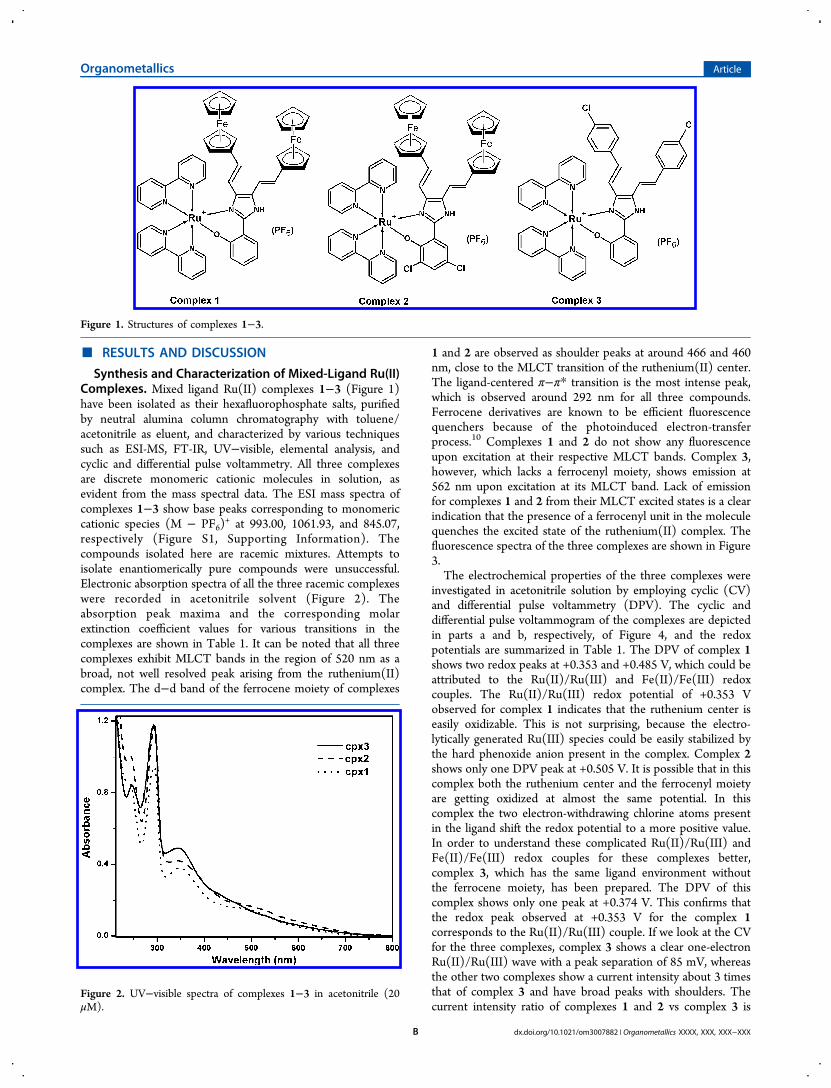
Synthesis and Characterization of Mixed-Ligand Ru(II)Complexes. Mixed ligand Ru(II) complexes 1−3 (Figure 1)have been isolated as their hexafluorophosphate salts, purifiedby neutral alumina column chromatography with toluene/acetonitrile as eluent, and characterized by various techniquessuch as ESI-MS, FT-IR, UV−visible, elemental analysis, andcyclic and differential pulse voltammetry. All three complexesare discrete monomeric cationic molecules in solution, asevident from the mass spectral data. The ESI mass spectra ofcomplexes 1−3 show base peaks corresponding to monomericcationic species (M − PF6)
+ at 993.00, 1061.93, and 845.07,respectively (Figure S1, Supporting Information). Thecompounds isolated here are racemic mixtures. Attempts toisolate enantiomerically pure compounds were unsuccessful.Electronic absorption spectra of all the three racemic complexeswere recorded in acetonitrile solvent (Figure 2). Theabsorption peak maxima and the corresponding molarextinction coefficient values for various transitions in thecomplexes are shown in Table 1. It can be noted that all threecomplexes exhibit MLCT bands in the region of 520 nm as abroad, not well resolved peak arising from the ruthenium(II)complex. The d−d band of the ferrocene moiety of complexes
1 and 2 are observed as shoulder peaks at around 466 and 460nm, close to the MLCT transition of the ruthenium(II) center.The ligand-centered π−π* transition is the most intense peak,which is observed around 292 nm for all three compounds.Ferrocene derivatives are known to be efficient fluorescencequenchers because of the photoinduced electron-transferprocess.10 Complexes 1 and 2 do not show any fluorescenceupon excitation at their respective MLCT bands. Complex 3,however, which lacks a ferrocenyl moiety, shows emission at562 nm upon excitation at its MLCT band. Lack of emissionfor complexes 1 and 2 from their MLCT excited states is a clearindication that the presence of a ferrocenyl unit in the moleculequenches the excited state of the ruthenium(II) complex. Thefluorescence spectra of the three complexes are shown in Figure3.The electrochemical properties of the three complexes were
investigated in acetonitrile solution by employing cyclic (CV)and differential pulse voltammetry (DPV). The cyclic anddifferential pulse voltammogram of the complexes are depictedin parts a and b, respectively, of Figure 4, and the redoxpotentials are summarized in Table 1. The DPV of complex 1shows two redox peaks at +0.353 and +0.485 V, which could beattributed to the Ru(II)/Ru(III) and Fe(II)/Fe(III) redoxcouples. The Ru(II)/Ru(III) redox potential of +0.353 Vobserved for complex 1 indicates that the ruthenium center iseasily oxidizable. This is not surprising, because the electro-lytically generated Ru(III) species could be easily stabilized bythe hard phenoxide anion present in the complex. Complex 2shows only one DPV peak at +0.505 V. It is possible that in thiscomplex both the ruthenium center and the ferrocenyl moietyare getting oxidized at almost the same potential. In thiscomplex the two electron-withdrawing chlorine atoms presentin the ligand shift the redox potential to a more positive value.In order to understand these complicated Ru(II)/Ru(III) andFe(II)/Fe(III) redox couples for these complexes better,complex 3, which has the same ligand environment withoutthe ferrocene moiety, has been prepared. The DPV of thiscomplex shows only one peak at +0.374 V. This confirms thatthe redox peak observed at +0.353 V for the complex 1corresponds to the Ru(II)/Ru(III) couple. If we look at the CVfor the three complexes, complex 3 shows a clear one-electronRu(II)/Ru(III) wave with a peak separation of 85 mV, whereasthe other two complexes show a current intensity about 3 timesthat of complex 3 and have broad peaks with shoulders. Thecurrent intensity ratio of complexes 1 and 2 vs complex 3 is
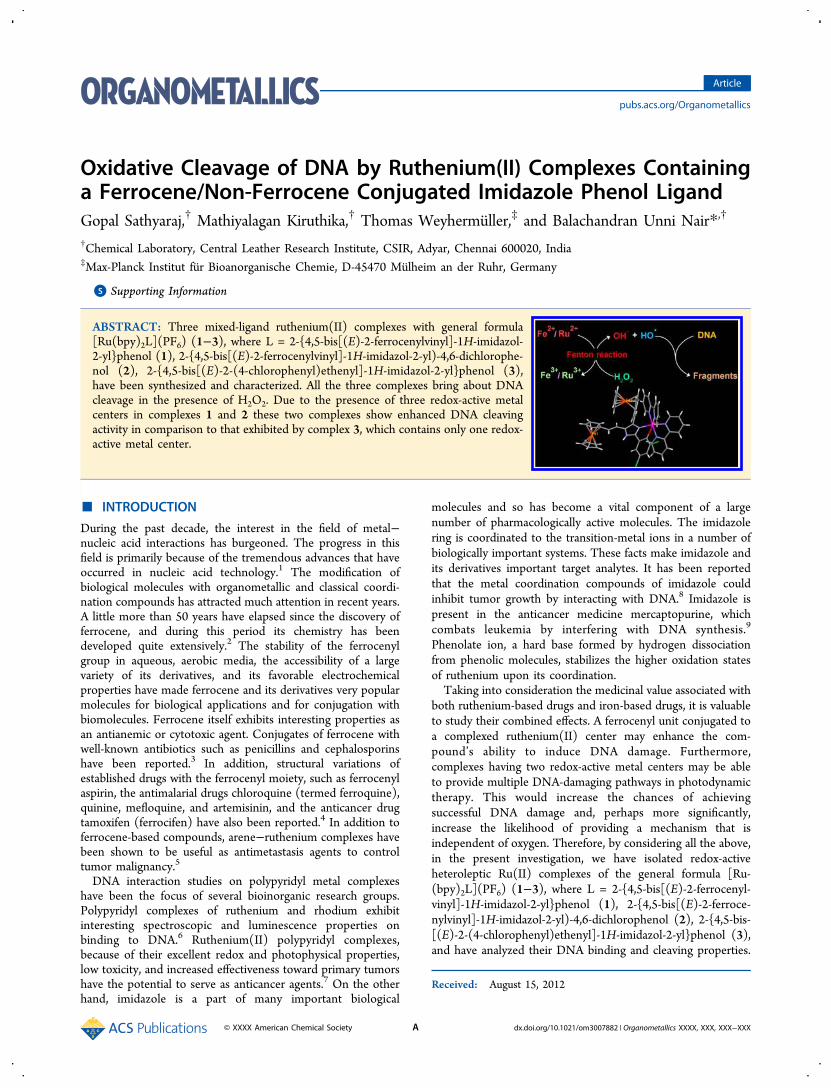
Figure 1. Structures of complexes 1−3.
Figure 2. UV−visible spectra of complexes 1−3 in acetonitrile (20μM).
Organometallics Article
dx.doi.org/10.1021/om3007882 | Organometallics XXXX, XXX, XXX−XXXB

about 3, indicating that a redox process takes place in all threemetal centers in complexes 1 and 2. The redox potentials of theferrocene moieties are shifted to more positive potential incomparison to its free ligand form. Complex 2 has beenstructurally characterized by single-crystal X-ray diffractiontechnique. A single crystal of this compound was obtained bydiffusion of diethyl ether vapor into an acetonitrile solution ofthe complex at room temperature. The crystallographic data forcomplex 2 are given in Table S2 (Supporting Information). An
ORTEP view of complex 2 is shown in Figure 5. It can be seenfrom Figure 5 that the imidazole −NH group of the complex
cation and the perchlorate anion (counterion) are connected bya hydrogen bond through the water of solvation. The phenolmoiety and the imidazole are no longer coplanar in this case,unlike in the case of ligand L2, where they are coplanar.11
In complex 2 both the imidazole nitrogen and the phenolicoxygen are coordinated to the ruthenium(II) ion, and due to
Table 1. Absorption, Fluorescence, and Electrochemical Data of Synthesized Complexes 1−3 in Acetonitrile
complex UV−visible λmax (nm) (ε (M−1 cm−1))λem (λex)(nm)
E1/2(Ru(II)/Ru(III)),E1/2(Fe(II)/Fe(III)) (V vs SCE)
ΔE(mV)
[Ru(bpy)2(L1)]PF6 (1)
∼523.5 (7 400), ∼466 (9 300), 345.5 (21 000), 294.5 (58 600), 243.0 (50 400) +0.353, +0.485 140
[Ru(bpy)2(L2)]PF6 (2)
∼510.5 (6 800) ∼460.5 (8 050) 347.0 (19 000) 293.5 (47 150) 239.0 (43 100) +0.505 217
[Ru(bpy)2(L3)]PF6 (3)
∼525 (5 850) 344.5 (24 600) 292.0 (59 000) 246.0 (42 200) 562 (500) +0.374 85
Figure 3. Fluorescence spectra of 20 μM solutions of complexes 1−3in acetonitrile at 25 °C (excitation wavelength 500 nm).
Figure 4. (a) Cyclic and (b) differential pulse voltammetry of complexes 1 (dashed line), 2 (solid line), 3 (dotted line) in acetonitrile (2 mM). Therecorded potentials are referenced against the SCE at 25 °C at a scan rate of 100 mV s−1.
Figure 5. ORTEP diagram of complex 2.
Organometallics Article
dx.doi.org/10.1021/om3007882 | Organometallics XXXX, XXX, XXX−XXXC

the constraints of the octahedral geometry around theruthenium(II) ion the phenol moiety and the imidazole moietyare not coplanar in this case; the dihedral angle between thephenol and imidazole moieties are 25°02′. The bite anglesgenerated by the phenolate moiety and the imidazole moietyare less than 90°, as can be seen from Figure 6.
The two pyridine rings in the bipyridyl molecule are also notcoplanar; the dihedral angles between the two rings are 4.8 and10°. The ferrocene moieties are in different environments, andas a result the torsion angle between the Cp rings of ferrocene,the Fe−C and C−C bond lengths, and distance between Cprings are not the same as in the case of the ligand. Interestingly,one of the ferrocene moieties shows a maximum torsion angleof 16°34′, which is halfway between staggered and eclipsedconformations (Table 2). The important bond lengths and
bond angles for complex 2 are given in Table 3. Complex 2 hasa one-dimensional hydrogen-bonding network between theimidazole −NH group, the water of solvation, and perchlorateanion, as shown in Figure S3 (Supporting Information). Onecan also see π stacking of bipyridyl moieties from two one-
dimensional networks in the crystal structure of the complex(Figure S3).
Interaction of Substituted Imidazole Phenol Mixed-Ligand Ruthenium(II) Complexes with DNA. AbsorptionSpectral Titration. Electronic absorption spectroscopy is one ofthe most useful techniques in DNA-binding studies. Sincecomplexes 1−3 are coordinatively saturated and have noreplaceable labile monodentate ligand in their coordinationsphere, they are not expected to bind to DNA bases. Thesethree complexes can either bind to DNA groove or bind toDNA intercalatively. Groove binding generally does not lead towell-defined changes in the energy of the absorption band, andin this case the intensity of the absorption band also does notshow any marked change. Intercalative binding, on the otherhand, generally gives rise to hypochromism with a red shift inthe absorption bands of the molecule. No detectable changes inthe energy of spectral bands of these complexes have beenobserved in the presence of DNA, ruling out an intercalativemode of binding of these three complexes.12 The changesobserved in the electronic spectrum of these three complexes inthe presence of DNA can be rationalized in terms of groovebinding.13 However, since all three complexes contain aromaticrings in their ligand structure, partial intercalation of thesemolecules cannot be fully ruled out. The absorption spectra ofthe complexes 1−3 in the presence of increasing amounts ofDNA are shown in Figure 7 and in Figures S4 and S5(Supporting Information), respectively.
Figure 6. Pseudo-octahedral geometry around the central metal atom.
Table 2. Various Parameters Obtained from CrystalStructures of Ligands (L1 and L2) and Complex 2
compdtorsionangle
Fe−C bondlength (Å)
C−C bondlength (Å)
Cpdistance(Å)
ferrocene27 36° 2.05 1.4 3.32L1a OH
side3°38′ 2.043 1.429 3.285
NHside
2°01′ 2.049 1.426 3.302
L2a OHside
6°21′ 2.046 1.425 3.316
NHside
5°17′ 2.047 1.423 3.302
2 OHside
16°30′ 2.042 1.424(5) 3.307
NHside
4°43′ 2.045 1.424(4) 3.296
aReference 11.
Table 3. Important Bond Lengths (Å) and Bond Angles(deg) of Complex 2
Ru1−N61 2.0329(14) Ru1−N72 2.0547(15)Ru1−N41 2.0348(14) Ru1−O1 2.0909(13)Ru1−N52 2.0415(15) Ru1−N12 2.0930(14)N61−Ru1−N41 89.03(6) N52−Ru1−O1 93.02(6)N61−Ru1−N52 96.75(6) N72−Ru1−O1 85.75(6)N41−Ru1−N52 79.36(6) N41−Ru1−N72 101.57(6)N61−Ru1−N72 78.99(6) N41−Ru1−N12 98.39(5)N52−Ru1−N72 175.59(5) N52−Ru1−N12 91.16(6)N61−Ru1−O1 88.25(5) N72−Ru1−N12 92.95(6)N41−Ru1−O1 171.56(5) O1−Ru1−N12 85.30(5)
Figure 7. Absorption spectral titration of complex 1 (20 μM) withDNA (0−175 μM) in Tris buffer, pH 7.2.
Organometallics Article
dx.doi.org/10.1021/om3007882 | Organometallics XXXX, XXX, XXX−XXXD

None of the electronic spectra of three complexes show anyclear isosbestic points during the successive addition of DNA.The intrinsic DNA binding constants, Kb, of the threecomplexes have been obtained by monitoring the changes inabsorbance at 290 nm for complexes 1−3 with increasingconcentrations of DNA. The DNA binding constants forcomplexes 1−3 have been found to be (2.05 ± 0.02) × 105,(1.52 ± 0.03) × 105, and (1.43 ± 0.03) × 105 M−1, respectively.Viscosity Measurements. Hydrodynamic methods are
suitable for detection of small changes in the absence ofcrystallographic structural data and provide essential evidenceto support an intercalation model. Intercalation of a moleculegenerally leads to an increase in the viscosity of DNA. Incontrast, the molecules that bind to DNA either in the groovesor on the external surface give rise to irregularity or no changesin the viscosity of CT DNA. Figure 8 shows the changes in theviscosity of DNA on incremental addition of the rutheniumcomplexes 1−3.
It is clear from the figure that the three complexes bringabout irregular changes in the viscosity of DNA. This clearlyrules out an intercalative mode of binding of these complexes toDNA. Though the complexes contain bipyridyl ligands, in thepresent case, it appears that the ancillary ligand, substitutedimidazole phenol, dictates the mode of binding. Metalcomplexes containing bipyridyl ligands also have been shownto bind DNA non-intercalatively.14 The presence of an −NHgroup in the imidazole would tend to favor hydrogen bondingwith the base pairs of DNA rather than intercalation betweenthe base pairs, thereby leading to groove binding of thesecomplexes. A similar observation has been made in the case ofimidazole-containing ferrocenyl compounds such as 2-ferro-cenyl imidazophenanthroline and 2-ferrocenyl imidazophenan-threne.15 Therefore, the results from the viscosity measure-ments confirm the groove binding of these complexes withDNA.DNA Cleavage Activity. The DNA photocleavage activity of
complexes 1−3 was studied by irradiating SC pUC19 DNA at440 nm for 30 min in the presence of the three complexes (24and 40 μM).
The results of these experiments are depicted in Figure 9.Out of the three complexes, complex 3, which lacks a ferrocene
moiety, showed some photocleavage of plasmid SC DNA toNC DNA in the presence of 40 μM of the complex. At 60 μMconcentration, complex 3 brought about 50% conversion of SCDNA to NC DNA (Figure 10). Conversion of SC DNA to NC
DNA was observed even in the presence of histidine (singletoxygen quencher), as can be seen from lane 7 of Figure 10.Hence, it is clear that cleavage of DNA in the presence ofcomplex 3 under photolytic conditions is due to guanineoxidation by the excited state of ruthenium. It is of interest tonote that complexes 1 and 2, which have ferrocenyl moietiesconjugated to the Ru(II) complex, did not show any significantphotonuclease activity.The oxidative cleavage of SC pUC19 DNA by complexes 1−
3 was studied using hydrogen peroxide as an oxidizing agent.All three compounds showed cleavage in the presence ofhydrogen peroxide (Figure 11).
Iron(II) is known for its Fenton chemistry with hydrogenperoxide and is known to form •OH and •OOH radicals. In thiscase complexes 1 and 2 have Fe(II) and Ru(II) metal ions andcomplex 3 has only the Ru(II) metal ion. The redox potentialsof these metal ions are low enough in complexes 1−3, andhence, the metal ions can be oxidized to their higher valences asin the case of the Fenton reaction. The redox potential of themetal complex is an important parameter for the generation ofhydroxyl and peroxide radicals under the experimentalconditions for the nicking of DNA.16
Figure 8. Relative viscosities of DNA solution in the presence ofvarying amounts of complexes 1−3.
Figure 9. Cleavage of supercoiled pUC19 by complexes 1−3, onincubation for 1 h followed by irradiation at 440 nm for 30 min: (lane1) control DNA; (lanes 2−4) DNA in the presence of 24 μMcomplexes 1−3, respectively; (lanes 5−7) pUC 19 DNA in thepresence of 40 μM complexes 1−3, respectively.
Figure 10. Cleavage of supercoiled pUC19 by complex 3, onincubation for 1 h followed by irradiation at 440 nm for 30 min: (lane1) control DNA; (lanes 2−6) pUC 19 DNA in the presence of 12, 24,36, 48, and 60 μM of complex 3, respectively; (lane 7) DNA in thepresence of 60 μM complex 3 and 300 μM histidine.
Figure 11. Oxidative cleavage of supercoiled pUC19 in the absence(lane 1) and presence of complexes 1−3 (24 μM; lanes 2−4,respectively) and 240 μM H2O2 on incubation for 1 h.
Organometallics Article
dx.doi.org/10.1021/om3007882 | Organometallics XXXX, XXX, XXX−XXXE

The DNA cleavage efficiency of these complexes in thepresence of hydrogen peroxide shows that 24 μM of complexes1 and 2 is able to cleave DNA completely, whereas 24 μM ofcomplex 3 is not able to convert form I to form II completely.This may be due to the fact that complexes 1 and 2 have threepotential active metal ions that can generate 3 times moreradical species in comparison to complex 3, which has only onesuch metal ion in a molecule.
■ CONCLUSIONSFerrocene conjugated ruthenium complexes have attracted theattention of researchers because of their application inchemotherapy. Conjugation of the ferrocene molecule toruthenium(II) complexes is expected to lead to changes inthe photophysical properties of the ruthenium(II) center aswell as the DNA-cleaving properties of the ruthenium(II)complex. Three ruthenium(II) complexes with general formula[Ru(bpy)2L](PF6) (1−3), where L = 2-{4,5-bis[(E)-2-ferrocenylvinyl]-1H-imidazol-2-yl}phenol (1), 2-{4,5-bis[(E)-2-ferrocenylvinyl]-1H-imidazol-2-yl)-4,6-dichlorophenol (2), 2-{4,5-bis[(E)-2-(4-chlorophenyl)ethenyl]-1H-imidazol-2-yl}-phenol (3), have been synthesized and characterizedspectroscopically and electrochemically. Complexes 1 and 2contain two conjugated ferrocene molecules, whereas complex3 lacks a conjugated ferrocene molecule. Complex 2 has alsobeen crystallographically characterized. Electronic spectra of allthree complexes show the MLCT band of the ruthenium(II)center at 540 nm. Complexes 1 and 2 also exhibit ligand fieldtransitions associated with the ferrocenyl moiety. Complex 3upon excitation at its MLCT band shows emission from theMLCT excited state of the ruthenium(II) center. On the otherhand, complexes 1 and 2 do not exhibit emission from theMLCT excited states of their respective ruthenium(II) centers,due to quenching of their excited states by the ferrocenylmoiety present in these two complexes. The DPV of complex 1shows redox peaks at +0.353 V due to the Ru(II)/Ru(III)couple and at +0.485 V due to the Fe(II)/Fe(III) redox couple.Complex 3, which does not contain a conjugated ferrocenemoiety, shows only one DPV peak at +0.353 V. Thisconclusively proves that the redox peak observed in the DPVof complex 1 at +0.353 V is due to the Ru(II)/Ru(III) couple.The Ru(II)/Ru(III) redox potential of +0.353 V observed forcomplex 1 indicates that the ruthenium center is easilyoxidizable. This is not surprising, because the electrolyticallygenerated Ru(III) species could be easily stabilized by the hardphenoxide anion present in the complex. Complex 2 showsonly one DPV peak at +0.505 V. It is possible that in thiscomplex both the ruthenium center and the ferrocenyl moietyare oxidized at almost the same potential. In this complex thetwo electron-withdrawing chlorine atoms present in the ligandshift the redox potential to more positive values. The values ofdiffusion currents in the case of complexes 1 and 2 have beenfound to be almost 3 times that observed for complex 3. This isbecause of the fact that in complexes 1 and 2 the redox processtakes place at three metal centers, whereas in complex 3 there isonly one redox-active metal center. All three complexes havebeen found to exhibit groove binding to CT DNA. Complex 3,which contains only Ru(II) as the redox-active metal ion,exhibits photonuclease activity, whereas complexes 1 and 2,which have conjugated ferrocenyl moieties, do not show anyphotonuclease activity. All three complexes exhibit nucleaseactivity in the presence of H2O2. Complexes 1 and 2 exhibitenhanced nuclease activity in comparison to complex 3 due to
the presence of three redox-active metal centers in complexes 1and 2. Complex 3, which has only one redox-active metalcenter, evidently shows lower nuclease activity. Studies on theantiproliferative activity of these three complexes on cancer celllines are currently in progress, and the results will be reportedsubsequently.
■ EXPERIMENTAL SECTIONMaterials and Methods. Ruthenium chloride trihydrate,
ferrocenecarboxaldehyde, biacetyl, piperidine, salicylaldehyde, 3,5-dichlorosalicylaldehyde, and 4-chlorobenzaldehyde were purchasedfrom Aldrich. Plasmid DNA pUC-19, suitable for gel electrophoresis,was purchased from Genie, Bangalore, India, and used as received. Calfthymus DNA (CT DNA) and agarose were procured from SRL(India). Acetonitrile, dimethyl sulfoxide, dichloromethane, ethanol,and methanol were of chromatographic grade and were used withoutfurther purification. Tris(hydroxymethyl)aminomethane-HCl (Tris-HCl) buffer was prepared using deionized and degassed triple-distilledwater. All the experiments regarding the binding and cleavage of DNAusing complexes 1−3 were carried out in Tris buffer (pH, 7.2). Asolution of CT DNA in the buffer gave a ratio of UV absorbance at260 and 280 nm of about 1.8−1.9, indicating that the DNA wassufficiently free from protein.17 The DNA concentration pernucleotide was determined by absorption spectroscopy using molarabsorption coefficient as 6600 M−1 cm−1 at 260 nm.18
Physical Measurements. Absorption spectra were recorded on aShimadzu UV-160A UV−visible recording spectrophotometer;emission spectra were recorded on a Varian Cary Eclipsespectrofluorometer. ESI mass spectra were obtained from a FinniganLCQ Advantage max ion trap mass spectrometer. Elemental analysiswas carried out using a Euro Vector C,H,N analyzer. Viscosityexperiments were made with an Ostwald viscometer, immersed in awater bath maintained at 25 °C. All cyclic (CV) and differential pulse(DPV) voltammetry experiments were conducted on a CH Instru-ments (USA) Model CH-620 B electrochemical analyzer. Tetrabuty-lammonium perchlorate (TBAP) was used as the supportingelectrolyte. The sample, dissolved in dried acetonitrile, was purgedwith nitrogen prior to each electrochemical measurement. A standardthree-electrode system comprised of glassy carbon as the workingelectrode, platinum electrode as the auxiliary electrode, and saturatedcalomel as the reference electrode (SCE) was used.
Synthesis of 2-(4,5-Bis((E)-2-ferrocenylvinyl)-1H-imidazol-2-yl)phenol (L1) and 2-(4,5-Bis((E)-2-ferrocenylvinyl)-1H-imida-zol-2-yl)-4,6-dichlorophenol (L2). 2-(4,5-Bis((E)-2-ferrocenylvin-yl)-1H-imidazol-2-yl)phenol and 2-(4,5-bis((E)-2-ferrocenylvinyl)-1H-imidazol-2-yl)-4,6-dichlorophenol were prepared according to theprocedure available in the literature.11
Synthesis of 2-{4,5-Bis[(E)-2-(4-chlorophenyl)ethenyl]-1H-imidazol-2-yl}phenol (L3). 1,6-Bis(chlorophenyl)hexa-1,5-diene-3,4-dione19 (0.5 g, 1.51 mmol), salicylaldehyde (0.185 g, 1.51mmol), and ammonium acetate (2 g, 25 mmol) were dissolved in15 mL of acetic acid and heated to reflux for 3 h.20 After cooling, coldwater (10 mL) was added to the solution, during which a yellowprecipitate appeared. The precipitate was filtered, washed using coldwater, and purified by column chromatography on silica gel with ethylacetate/hexane (1/4) as eluent to give the product. Yield: 0.35 g, 54%.ESI-MS: 434 (M + 1)+. Anal. Calcd for C25H18Cl2N2O: C, 69.29; H,4.19; N, 6.46. Found: C, 69.20; H, 4.25; N, 6.59.
Synthesis of [Ru(bpy)2(L1)](PF6) (1). A mixture of [cis-Ru(bpy)2Cl2]·2H2O
21 (0.18 g, 0.34 mmol) and L1 (0.2 g, 0.34mmol) in the presence of Et3N (50 μL) was suspended in an EtOH/H2O solvent mixture (3/1, v/v). The mixture was refluxed under aninert atmosphere for 4 h while vigorous stirring was maintained. Thereaction mixture was cooled to room temperature; the solvent wasreduced under vacuum to one-third of its initial volume. A saturatedaqueous solution of NH4PF6 was added to precipitate [Ru-(bpy)2(L1)]
+ as its hexafluorophosphate salt. The product was filteredand washed with water (3 × 10 mL) and then purified by columnchromatography on neutral alumina using acetonitrile/toluene (1.5/1,
Organometallics Article
dx.doi.org/10.1021/om3007882 | Organometallics XXXX, XXX, XXX−XXXF

v/v) as eluent . Yield: 0.37 g, 95%, Anal . Calcd forC53H43F6Fe2N6OPRu: C, 55.95; H, 3.81; N, 7.39. Found: C, 55.90;H, 3.88; N, 7.27. ESI-MS: m/z 993 (M − PF6)
+. IR (cm−1; KBrpellet): 3402, 3085, 1600, 1477, 1265, 844.Synthesis of [Ru(bpy)2(L2)](PF6) (2). The synthesis and
purification of compound 2 were similar to those of 1 using[Ru(bpy)2Cl2]·2H2O (0.2 g, 0.38 mmol) and L2 (0.25 g, 0.38mmol). Yield: 0.43 g, 93%, Anal. Calcd for C53H41Cl2F6Fe2N6OPRu:C, 52.76; H, 3.42; N, 6.97. Found: C, 52.68; H, 3.49; N, 6.89; ESI-MS:m/z 1061.93 (M − PF6)
+. IR (cm−1; KBr pellet): 3082, 2927, 1600,1461, 1122, 1107, 763. The perchlorate salt of complex 2 was preparedfor single-crystal X-ray diffraction studies by addition of excess sodiumperchlorate instead of ammonium hexafluorophosphate as theprecipitating agent. Single crystals of this molecule have been obtainedby diffusion of diethyl ether into a solution of the compound in anacetonitrile/water mixture (95/5).Synthesis of [Ru(bpy)2(L3)](PF6) (3). This compound was
synthesized and purified by a procedure similar to that described forcompound 1, except that L3 (0.5 g, 1.15 mmol) was used instead ofL1. Yield: 1.09 g, 96%, Anal. Calcd for C45H33Cl2F6N6OPRu: C, 54.55;H, 3.36; N, 8.48. Found: C, 54.48; H, 3.29; N, 8.41; ESI-MS: m/z845.07 (M − PF6)
+. IR (cm−1; KBr pellet): 3398, 3066, 1600, 1485,1091, 1014, 844.X-ray Crystallographic Data Collection and Refinement of
the Structure. A red single crystal of the compound [Ru(bpy)2(L2)]-(ClO4)·H2O was coated with perfluoropolyether, picked up with anylon loop, and immediately mounted in the nitrogen cold stream of aBruker-Nonius KappaCCD diffractometer equipped with a Mo-targetrotating-anode X-ray source. Graphite-monochromated Mo Kαradiation (λ = 0.710 73 Å) was used. Final cell constants wereobtained from least-squares fits for all measured reflections. Intensitydata were corrected for absorption using intensities of redundantreflections with the program SADABS.22 The structure was readilysolved by Patterson methods and subsequent difference Fouriertechniques. The Siemens ShelXTL23 software package was used for thesolution and creation of artwork of the structures; ShelXL9724 wasused for the refinement. All the non-hydrogen atoms wereanisotropically refined, and hydrogen atoms bound to carbon wereplaced at calculated positions and refined as riding atoms withisotropic displacement parameters. Hydrogen atoms bound tonitrogen and oxygen were located from the difference map.Crystallographic data of the compound are given in Table S2(Supporting Information). CCDC reference number: 837444.Absorption Titration. Calf thymus (CT) DNA solutions of
various concentrations (5−175 μM) were added to the metal complex(20 μM dissolved in 10% CH3CN/10 mM tris HCl, pH 7.2), and theMLCT transitions of complexes 1−3 were monitored. The absorptionspectra were recorded after 10 min of incubation. The nature ofequilibrium DNA binding constants, Kb, was calculated from the eq 1,
where εa, εf, and εb correspond to Aobsd/[Ru], the extinction coefficientfor the free ruthenium complex, and the extinction coefficient for theruthenium complexes in the fully bound form, respectively. A plot of[DNA]/(εa − εf) versus [DNA] gives Kb as the ratio of the slope tothe intercept.25
Viscosity Studies. Viscosity experiments were performed using anOstwald viscometer of 3 mL capacity. A viscometer was thermostatedin a water bath to maintain constant temperature (28 °C). Theconcentration of DNA (200 μM) was kept constant, and theconcentrations of metal complexes were varied (0−50 μM). Theflow times for the buffer (10 mM Tris-HCl), DNA alone, and DNA inthe presence of ruthenium complexes were monitored at least threetimes using a manually operated digital stopwatch, and an average flowtime was calculated. The relative specific viscosity was calculated usingthe equation η = (t − t0)/t0, where t0 is the flow time for the buffer andt is the observed flow time for DNA in the absence and presence of
complex. A plot of (η/η0∧(1/3) vs (1/R) (R = [DNA]/[complex]),where η is the viscosity of DNA in the presence of the complex and η0is the viscosity of DNA alone, was constructed from viscositymeasurements.26
DNA Cleavage Studies. Photonuclease activity of the complexeswas monitored using gel electrophoresis of plasmid DNA (pUC19).Two sets of 25 μL solutions were prepared for the photolysisexperiment. One set of solutions contained 3 μL of 100 μg/mL ofplasmid DNA in Tris buffer with varying amounts of metal complex1−3 (0−24 μM). Each solution was incubated for 1 h and thenirradiated at 440 nm for various time intervals varying from 10 to 60min. The samples were then subjected to electrophoresis in 0.8%agarose gel (tris-boric acid-EDTA buffer, pH 8.0) at 50 V for 2 h. Thegel was stained with 0.5 μg/mL of ethidium bromide. The stained gelwas illuminated under an UV lamp and gel documented. In a separateexperiment the DNA was incubated with 24 μM of the metal complexand 240 μM of histidine and irradiated at 440 nm. The photolyzedsolution was subsequently subjected to electrophoresis. Also, oxidativecleavage of DNA with complexes 1−3 in the presence of hydrogenperoxide as an oxidizing agent was studied. A 3 μL portion of 100 μg/mL of plasmid DNA in Tris buffer with metal complex 1−3 (24 μM)and 240 μM of H2O2 was prepared. Each solution was incubated for 1h before being subjected to electrophoresis.
■ ASSOCIATED CONTENT*S Supporting InformationFigures, a table, and a CIF file giving mass spectra ofsynthesized compounds, crystallographic data of complex 2,and absorption spectral titration of complexes 2 and 3. Thismaterial is available free of charge via the Internet at http://pubs.acs.org.
■ AUTHOR INFORMATIONCorresponding Author*E-mail: [email protected] authors declare no competing financial interest.
■ ACKNOWLEDGMENTSWe thank the Council of Scientific and Industrial Research,New Delhi (for a research fellowship to G.S.).
■ REFERENCES(1) Reddy, H. Bioinorganic Chemistry; New Age International: NewDelhi, 2005.(2) Kopf, H.; Kopf-Maier, P. Angew. Chem. 1979, 91, 509−512.(3) (a) Edwards, E. I.; Epton, R.; Marr, G. J. Organomet. Chem. 1976,122, C49−C53. (b) Edwards, E. I.; Epton, R.; Marr, G.; Rogers, G. K.;Thompson, K. J. Spec. Publ. Chem. Soc. 1977, 128, 92−100.(c) Simionescu, C.; Lixandru, T.; Tataru, L.; Mazilu, I.; Vata, M.;Luca, S. J. Organomet. Chem. 1983, 252, C43−C46. (d) Scutaru, D.;Tataru, L.; Mazilu, I.; Diaconu, E.; Lixandru, T.; Simionescu, C. J.Organomet. Chem. 1991, 401, 81−85. (e) Scutaru, D.; Mazilu, I.; Vata,M.; Tataru, L.; Vlase, A.; Lixandru, T.; Simionescu, C. J. Organomet.Chem. 1991, 401, 87−90.(4) (a) Sawamura, M.; Sasaki, H.; Nakata, T.; Ito, Y. Bull. Chem. Soc.Jpn. 1993, 66, 2725−2729. (b) Biot, C.; Delhaes, L.; Maciejewski, L.A.; Mortuaire, M.; Camus, D.; Dive, D.; Brocard, J. S. Eur. J. Med.Chem. 2000, 35, 707−714. (c) Beagley, P.; Blackie, M. A. L.; Chibale,K.; Clarkson, C.; Meijboom, R.; Moss, J. R.; Smith, P. J.; Su, H. DaltonTrans. 2003, 3046−3051. (d) Top, S.; Vessieres, A.; Leclercq, G.;Quivy, J.; Tang, J.; Vaissermann, J.; Huche, M.; Jaouen, G. Chem. Eur.J. 2003, 9, 5223−5236.(5) (a) Ang, W. H.; Daldini, E.; Scolaro, C.; Scopelliti, R.; Juillerat-Jeannerat, L.; Dyson, P. J. Inorg. Chem. 2006, 45, 9006−9013.(b) Suss-Fink, G. Dalton Trans. 2010, 39, 1673−1688. (c) Mishra, A.;
ε ε ε ε ε ε− = − + −K[DNA]/( ) [DNA]/( ) 1/ ( )a f b f b b f(1)
Organometallics Article
dx.doi.org/10.1021/om3007882 | Organometallics XXXX, XXX, XXX−XXXG

Jung, H.; Park, J. W.; Kim, H. K.; Kim, H.; Stang, P. J.; Chi., K.-W.Organometallics 2012, 31, 3519−3526.(6) (a) Erkkila, K. E.; Odom, D. T.; Barton, J. K. Chem. Rev. 1999,99, 2777−2795. (b) Metcalfe, C.; Thomas, J. A. Chem. Soc. Rev. 2003,32, 215−224. (c) Sigman, D. S.; Mazumder, A.; Perrin, M. D. Chem.Rev. 1993, 93, 2295−2316. (d) Dandlier, P. J.; Holmlin, R. E.; Barton,J. K. Science 1997, 274, 1465−1468. (e) Elias, B.; Kirsh DeMesmaeker, A. Coord. Chem. Rev. 2006, 250, 1627−1641. (f) Turro,N. J.; Barton, J. K.; Tomalia, D. A. Acc. Chem. Res. 1991, 24, 332−340.(7) Maurya, M. R.; Woo, L. K. J. Organomet. Chem. 2005, 690, 4978−4981.(8) (a) Liu, H. -K.; Berners-Price, S. J.; Wang, F.; Parkinson, J. A.; Xu,J.; Bella, J.; Sadler, P. J. Angew. Chem., Int. Ed. 2006, 45, 8153−8156.(b) Bergamo, A.; Sava, G. Dalton Trans. 2007, 1267−1272.(9) (a) Lednicer, D.; Mitscher, L. L. The Organic Chemistry of DrugSynthesis; Wiley-Interscience: New York, 2005; Vol. 2, pp 242−260.(b) Laurence, B. L.; John, S. L.; Keith, P. L Goodman and Gilman’s thePharmacological Basis of Therapeutics; McGraw-Hill: New York, 2000;pp 986−1032.(10) Fery-Forgues, S.; Delavaux-Nicot, B. J. Photochem. Photobiol. A:Chem. 2000, 132, 137−159.(11) Sathyaraj, G.; Muthamilselvan, D.; Kiruthika, M.; Weyhermuller,T.; Nair, B. U. J. Organomet. Chem. 2012, 716, 150−158.(12) (a) Barton, J. K.; Dannenberg, J. J.; Raphael, A. L. J. Am. Chem.,Soc. 1984, 106, 2172−2176. (b) Tysoe, S. A.; Morgan, R. J.; Baker, A.D.; Strekas, T. C. J. Phys. Chem. 1993, 97, 1707−1711.(13) Vijayalakshmi, R.; Kanthimathi, M.; Subramanian, V.; Nair, B. U.Biochem. Biophys. Acta 2000, 1475, 157−164.(14) Kumar, C. V.; Barton, J. K.; Turro, J. J. J. Am. Chem. Soc. 1985,107, 5518−5523.(15) (a) Zhao, X. -L.; Han, M. -J.; Zhang, A. -G.; Wang, K. -Z. J.Inorg. Biochem. 2012, 107, 104−110. (b) Maity, B.; Chakravarthi, B. V.S. K.; Roy, M.; Karande, A. A.; Chakravarty, A. R. Eur. J. Inorg. Chem.2011, 1379−1386.(16) Indumathy, R.; Kanthimathi, M.; Weyhermuller, T.; Nair, B. U.Polyhedron 2008, 27, 3443−3450.(17) Marmur, J. J. Mol. Biol. 1961, 3, 208−218.(18) Reichmann, R. E.; Rice, S. A.; Thomas, C. A.; Doty, P. J. J. Am.Chem. Soc. 1954, 76, 3047−3053.(19) Krohnke, F. Synthesis 1976, 1, 1−24.(20) Davidson, D.; Weiss, M.; Jelling, M. J. Org. Chem. 1937, 2, 319−327.(21) Sullivan, B. P.; Salmon, D. J.; Meyer, T. J. Inorg. Chem 1978, 17,3334−3341.(22) Sheldrick, G. M. SADABS, Bruker−Siemens Area DetectorAbsorption and Other Correction, Version 2006/1; University ofGottingen, Gottingen, Germany, 2006.(23) ShelXTL 6.14; Bruker AXS Inc., Madison, WI, 2003.(24) Sheldrick, G. M. ShelXL97; University of Gottingen, Gottingen,Germany, 1997.(25) Wolfe, A.; Shimer, G. H.; Meehan, T. Biochemistry 1987, 26,6392−6396.(26) Cohen, G.; Eisenberg, H. Biopolymers 1969, 8, 45−55.(27) Dunitz, J. D.; Orgel, L. E.; Rich, A. Acta Crystallogr. 1956, 9,373−375.
Organometallics Article
dx.doi.org/10.1021/om3007882 | Organometallics XXXX, XXX, XXX−XXXH