Embed Size (px)
Citation preview

Trans. Nonferrous Met. Soc. China 30(2020) 1928−1942
Oxidation of chalcopyrite in air-equilibrated acidic solution:
Inhibition with phenacyl derivatives
Paul CHIRIȚĂ1, Mădălina I. DUINEA1,2, Laura G. SÂRBU3,
Lucian M. BÎRSĂ3, Mihaela BAIBARAC4, Florinel SAVA4, Elena MATEI4
1. Department of Chemistry, University of Craiova, Calea Bucuresti 107I, 200478, Craiova, Romania;
2. Department of Chemistry, Physics and Environment,
‘‘Dunarea de Jos” University of Galati, 111 Domneasca Street, Galati 800201, Romania;
3. Department of Chemistry, “Al. I. Cuza” University of Iasi, 11 Carol I Blv., Iasi 700506, Romania;
4. Lab. Optical Processes in Nanostructured Materials, National Institute of Materials Physics,
P. O. Box MG-7, Bucharest, R077125, Romania
Received 2 November 2019; accepted 20 May 2020
Abstract: The effects of 4-(2-hydroxyphenyl)-2-(morpholin-4-yl)-1,3-thiazole (Pr02), 1-(3,5-dibromo-2-hydroxyphenyl)- 1-oxoethan-2-yl-N,N-diethyldithiocarbamate (Pr04) and 1-(5-bromo-2-hydroxy-3-methylphenyl)-1-oxoethan-2-yl-O- ethyl xanthate (Pr06) on the aqueous oxidation of chalcopyrite (CuFeS2) in air-equilibrated solution at a temperature of 25 °C and a pH of 2.5 were studied. The effects were investigated by using potentiodynamic polarization, electro- chemical impedance spectroscopy (EIS), scanning electron microscopy coupled with energy dispersive X-ray (SEM/EDX) analysis, aqueous batch experiments, Fourier transform infrared (FTIR) spectroscopy, Raman scattering and quantum chemical calculations. It is found that the anodic current densities decrease in the order of EtOH > Pr02 > Pr04 > Pr06. These results, along with those of the EIS measurements, show that Pr02, Pr04 and Pr06 are effective anodic inhibitors of chalcopyrite aqueous oxidation. Both Raman scattering and FTIR spectroscopy indicate that the elemental sulfur, polysulfide and ferric oxyhydroxides that form on the surface of the mineral are not responsible when it comes to the aqueous oxidation inhibition of chalcopyrite. Quantum chemical calculations show that the adsorption of the tested compounds on the chalcopyrite surface is energetically favorable and so, it can explain the inhibiting effects that were observed. Key words: chalcopyrite oxidation; phenacyl derivatives; inhibition; potentiodynamic polarization; quantum chemical calculation
1 Introduction
Chalcopyrite (CuFeS2) is the most important copper-bearing mineral on the Terra. The aqueous oxidation of chalcopyrite existing in rocks or the waste resulting from the processing of ores causes the release of copper and other toxic elements present in its matrix (Pb, Cd, As, Sb, etc.) and the generation of acid drainage which seriously pollutes the natural waters [1,2]. Therefore, the aqueous
oxidation of chalcopyrite has been extensively investigated [2−7]. In the environment, molecular oxygen (O2) is a common oxidant [2]. The aqueous oxidation of chalcopyrite by O2 can be summarized by the following overall reaction [8]: CuFeS2+4O2=Cu2++Fe2++ 2
42SO (1)
Depending on the experimental conditions, chalcopyrite oxidation may be partial, with the sulfur from CuFeS2 being oxidized to elemental sulfur [5,8,9]:
Corresponding author: Paul CHIRIȚĂ; Tel/Fax: +40-251597048; E-mail: [email protected] DOI: 10.1016/S1003-6326(20)65351-1
Paul CHIRIȚĂ, et al/Trans. Nonferrous Met. Soc. China 30(2020) 1928−1942
1929
CuFeS2+4H++O2=Cu2++Fe2++2S+2H2O (2)
Elemental sulfur (S0) can be found embedded in polysulfide species (−S−S0
n−S−) [10−13]. Together with iron oxyhydroxides (resulted from the hydrolysis of Fe3+, the product of Fe2+ oxidation [9,14]), the elemental sulfur and polysulfide species form a surface layer (SL) which is considered the main factor that inhibits the aqueous oxidation of chalcopyrite [4,7,15,16]. One considers that the inhibition represents an important problem for the optimal copper extraction by atmospheric leaching process [4]. Yet, the low rate of aqueous oxidation of chalcopyrite could be the result of the reaction complexity (it is a multi-step reaction). Those up to 16 electrons lost by chalcopyrite can only be transferred in a sequence of several elementary reactions, the number of electrons transferred in each elementary reaction being no more than two [17,18].
In contrast, the SL formed on chalcopyrite does not sufficiently slow its natural dissolution [5]. Hence, chalcopyrite is seen as an important contributor to acid metalliferous drainage [2], a major environmental problem. Considering the results obtained for other sulfides [19−21], a way to inhibit the chalcopyrite aqueous oxidation could be the treatment of the mineral with various organic compounds. According to the general mechanism of metal corrosion [22−26], the adsorbed organic molecules obstruct the transfer of electrons to the oxidant. As with metals and other metal sulfides, the heteroatoms and multiple bonds in the structure of the organic molecules facilitate their adsorption on the chalcopyrite surface, fact that blocks the active centers of the mineral surface.
The aim of this study is to examine the influence of three phenacyl derivatives (4-(2- hydroxyphenyl)-2-(morpholin-4-yl)-1,3-thiazole(Pr02), 1-(3,5-dibromo-2-hydroxyphenyl)-1- oxoethan-2-yl-N,N-diethyldithiocarbamate (Pr04) and 1-(5-bromo-2-hydroxy-3-methylphenyl)-1- oxoethan-2-yl-O-ethyl xanthate (Pr06)) on the aqueous oxidation of chalcopyrite in air- equilibrated acidic solution. The heteroatoms (O, S, N, Br) and double bonds present in their structures suggest that the three compounds can be adsorbed on the chalcopyrite surface, for example, they are adsorbed on the sphalerite surface [20]. The effects of organic molecules on chalcopyrite reactivity were investigated through potentiodynamic
polarization, electrochemical impedance spectro- scopy (EIS) and aqueous batch experiments. The uninhibited/inhibited surfaces were characterized through scanning electron microscopy coupled with energy dispersive X-ray (SEM/EDX) analysis, Raman scattering and Fourier transform infrared (FTIR) spectroscopy. The relationship between the tested structures and their inhibitory effect was studied by quantum chemical method. 2 Experimental 2.1 Materials
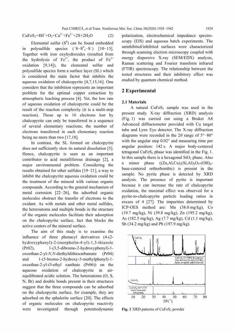
A natural CuFeS2 sample was used in the present study. X-ray diffraction (XRD) analysis (Fig. 1) was carried out using a Bruker A8 Advanced diffractometer provided with Cu target tube and Lynx Eye detector. The X-ray diffraction diagrams were recorded in the 2θ range of 5°−80° with the angular step 0.02° and measuring time per angular position: 142 s. A major body-centered tetragonal CuFeS2 phase was identified in the Fig. 1. In this sample there is a hexagonal SiO2 phase. Also a minor phase ((Zn,Al,Cu)3(Si,Al)2O5-(OH)4- base-centered orthorhombic) is present in the sample. No pyrite phase is detected by XRD analysis. The presence of pyrite is important because it can increase the rate of chalcopyrite oxidation, the maximal effect was observed for a pyrite-to-chalcopyrite particle loading ratios in excess of 4 [27]. The impurities determined by ICP-OES method are: Mn (38.0 mg/kg), Co (19.7 mg/kg), Ni (39.8 mg/kg), Zn (195.2 mg/kg), As (182.5 mg/kg), Ag (7.7 mg/kg), Cd (1.1 mg/kg), Sb (34.2 mg/kg) and Pb (197.9 mg/kg).
Fig. 1 XRD patterns of CuFeS2 powder
Paul CHIRIȚĂ, et al/Trans. Nonferrous Met. Soc. China 30(2020) 1928−1942
1930
The acid solutions were prepared from reagent grade purity HCl. Distilled water was used for the preparation of acid solutions with initial pH 2.50.
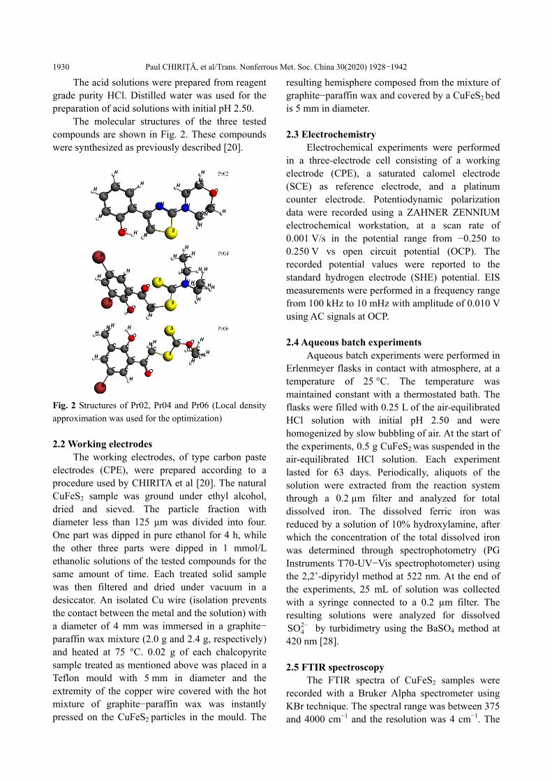
The molecular structures of the three tested compounds are shown in Fig. 2. These compounds were synthesized as previously described [20].
Fig. 2 Structures of Pr02, Pr04 and Pr06 (Local density
approximation was used for the optimization)
2.2 Working electrodes
The working electrodes, of type carbon paste electrodes (CPE), were prepared according to a procedure used by CHIRITA et al [20]. The natural CuFeS2 sample was ground under ethyl alcohol, dried and sieved. The particle fraction with diameter less than 125 µm was divided into four. One part was dipped in pure ethanol for 4 h, while the other three parts were dipped in 1 mmol/L ethanolic solutions of the tested compounds for the same amount of time. Each treated solid sample was then filtered and dried under vacuum in a desiccator. An isolated Cu wire (isolation prevents the contact between the metal and the solution) with a diameter of 4 mm was immersed in a graphite− paraffin wax mixture (2.0 g and 2.4 g, respectively) and heated at 75 °C. 0.02 g of each chalcopyrite sample treated as mentioned above was placed in a Teflon mould with 5 mm in diameter and the extremity of the copper wire covered with the hot mixture of graphite−paraffin wax was instantly pressed on the CuFeS2 particles in the mould. The
resulting hemisphere composed from the mixture of graphite−paraffin wax and covered by a CuFeS2 bed is 5 mm in diameter. 2.3 Electrochemistry
Electrochemical experiments were performed in a three-electrode cell consisting of a working electrode (CPE), a saturated calomel electrode (SCE) as reference electrode, and a platinum counter electrode. Potentiodynamic polarization data were recorded using a ZAHNER ZENNIUM electrochemical workstation, at a scan rate of 0.001 V/s in the potential range from −0.250 to 0.250 V vs open circuit potential (OCP). The recorded potential values were reported to the standard hydrogen electrode (SHE) potential. EIS measurements were performed in a frequency range from 100 kHz to 10 mHz with amplitude of 0.010 V using AC signals at OCP. 2.4 Aqueous batch experiments
Aqueous batch experiments were performed in Erlenmeyer flasks in contact with atmosphere, at a temperature of 25 °C. The temperature was maintained constant with a thermostated bath. The flasks were filled with 0.25 L of the air-equilibrated HCl solution with initial pH 2.50 and were homogenized by slow bubbling of air. At the start of the experiments, 0.5 g CuFeS2 was suspended in the air-equilibrated HCl solution. Each experiment lasted for 63 days. Periodically, aliquots of the solution were extracted from the reaction system through a 0.2 µm filter and analyzed for total dissolved iron. The dissolved ferric iron was reduced by a solution of 10% hydroxylamine, after which the concentration of the total dissolved iron was determined through spectrophotometry (PG Instruments T70-UV−Vis spectrophotometer) using the 2,2’-dipyridyl method at 522 nm. At the end of the experiments, 25 mL of solution was collected with a syringe connected to a 0.2 µm filter. The resulting solutions were analyzed for dissolved
24SO by turbidimetry using the BaSO4 method at
420 nm [28].
2.5 FTIR spectroscopy
The FTIR spectra of CuFeS2 samples were recorded with a Bruker Alpha spectrometer using KBr technique. The spectral range was between 375 and 4000 cm−1 and the resolution was 4 cm−1. The
Paul CHIRIȚĂ, et al/Trans. Nonferrous Met. Soc. China 30(2020) 1928−1942
1931
FTIR measurements were performed soon after pellet preparation, in order to avoid the oxidation of the chalcopyrite particles. 2.6 SEM and EDX analysis
A Zeiss EVO 50 XVP scanning electron microscope (SEM) equipped with an energy dispersive X-ray spectroscopy (EDX) Quantax Bruker 200 system as attachment was used to investigate the morphology and surface composition of the CuFeS2 samples. 2.7 Raman spectroscopy
A T64000 Raman spectrophotometer, acquired from Horiba Jobin Yvon, endowed with Ar laser was used at recording of the Raman spectra of treated CuFeS2. The Raman resolution was 2 cm−1. 2.8 Quantum chemistry analysis
For the estimation of the electronic properties of chalcopyrite surface, phenacyl derivatives (PD) and CuFeS2-PD complexes, the Amsterdam Density Functional (ADF, 2016 program version) was used [29,30]. Scalar relativistic effects were taken into account using the zero order regular approximation (ZORA) [31−33].
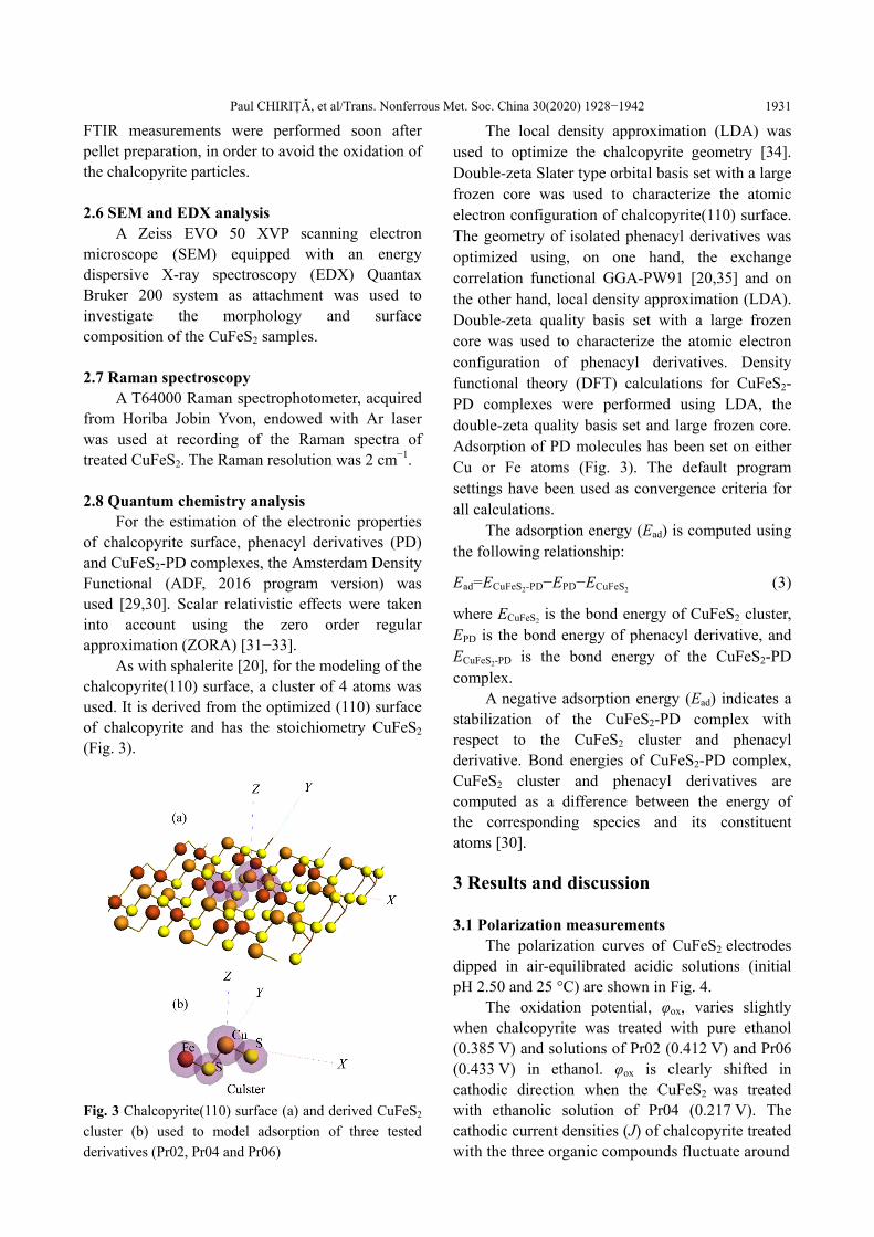
As with sphalerite [20], for the modeling of the chalcopyrite(110) surface, a cluster of 4 atoms was used. It is derived from the optimized (110) surface of chalcopyrite and has the stoichiometry CuFeS2
(Fig. 3).
Fig. 3 Chalcopyrite(110) surface (a) and derived CuFeS2
cluster (b) used to model adsorption of three tested
derivatives (Pr02, Pr04 and Pr06)
The local density approximation (LDA) was used to optimize the chalcopyrite geometry [34]. Double-zeta Slater type orbital basis set with a large frozen core was used to characterize the atomic electron configuration of chalcopyrite(110) surface. The geometry of isolated phenacyl derivatives was optimized using, on one hand, the exchange correlation functional GGA-PW91 [20,35] and on the other hand, local density approximation (LDA). Double-zeta quality basis set with a large frozen core was used to characterize the atomic electron configuration of phenacyl derivatives. Density functional theory (DFT) calculations for CuFeS2- PD complexes were performed using LDA, the double-zeta quality basis set and large frozen core. Adsorption of PD molecules has been set on either Cu or Fe atoms (Fig. 3). The default program settings have been used as convergence criteria for all calculations.
The adsorption energy (Ead) is computed using the following relationship: Ead=ECuFeS2-PD−EPD−ECuFeS2 (3) where ECuFeS2
is the bond energy of CuFeS2 cluster, EPD is the bond energy of phenacyl derivative, and ECuFeS2-PD is the bond energy of the CuFeS2-PD complex.
A negative adsorption energy (Ead) indicates a stabilization of the CuFeS2-PD complex with respect to the CuFeS2 cluster and phenacyl derivative. Bond energies of CuFeS2-PD complex, CuFeS2 cluster and phenacyl derivatives are computed as a difference between the energy of the corresponding species and its constituent atoms [30].
3 Results and discussion 3.1 Polarization measurements
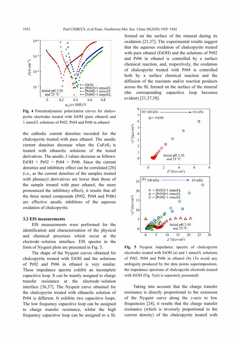
The polarization curves of CuFeS2 electrodes dipped in air-equilibrated acidic solutions (initial pH 2.50 and 25 °C) are shown in Fig. 4.
The oxidation potential, φox, varies slightly when chalcopyrite was treated with pure ethanol (0.385 V) and solutions of Pr02 (0.412 V) and Pr06 (0.433 V) in ethanol. φox is clearly shifted in cathodic direction when the CuFeS2 was treated with ethanolic solution of Pr04 (0.217 V). The cathodic current densities (J) of chalcopyrite treated with the three organic compounds fluctuate around
Paul CHIRIȚĂ, et al/Trans. Nonferrous Met. Soc. China 30(2020) 1928−1942
1932
Fig. 4 Potentiodynamic polarization curves for chalco-
pyrite electrodes treated with EtOH (pure ethanol) and
1 mmol/L solutions of Pr02, Pr04 and Pr06 in ethanol
the cathodic current densities recorded for the chalcopyrite treated with pure ethanol. The anodic current densities decrease when the CuFeS2 is treated with ethanolic solutions of the tested derivatives. The anodic J values decrease as follows: EtOH > Pr02 > Pr04 > Pr06. Since the current densities and inhibitory effect can be correlated [20] (i.e., as the current densities of the samples treated with phenacyl derivatives are lower than those of the sample treated with pure ethanol, the more pronounced the inhibitory effect), it results that all the three tested compounds (Pr02, Pr04 and Pr06) are effective anodic inhibitors of the aqueous oxidation of chalcopyrite. 3.2 EIS measurements
EIS measurements were performed for the identification and characterization of the physical and chemical processes which occur at the electrode−solution interface. EIS spectra in the form of Nyquist plots are presented in Fig. 5.
The shape of the Nyquist curves obtained for chalcopyrite treated with EtOH and the solutions of Pr02 and Pr06 in ethanol is very similar. These impedance spectra exhibit an incomplete capacitive loop. It can be mainly assigned to charge transfer resistance at the electrode−solution interface [36,37]. The Nyquist curve obtained for the chalcopyrite treated with ethanolic solution of Pr04 is different. It exhibits two capacitive loops. The low frequency capacitive loop can be assigned to charge transfer resistance, whilst the high frequency capacitive loop can be assigned to a SL
formed on the surface of the mineral during its oxidation [21,37]. The experimental results suggest that the aqueous oxidation of chalcopyrite treated with pure ethanol (EtOH) and the solutions of Pr02 and Pr06 in ethanol is controlled by a surface chemical reaction, and, respectively, the oxidation of chalcopyrite treated with Pr04 is controlled both by a surface chemical reaction and the diffusion of the reactants and/or reaction products across the SL formed on the surface of the mineral (the corresponding capacitive loop becomes evident) [21,37,38].
Fig. 5 Nyquist impedance spectra of chalcopyrite
electrodes treated with EtOH (a) and 1 mmol/L solutions
of Pr02, Pr04 and Pr06 in ethanol (b) (To avoid any
ambiguity produced by the data points superimposition,
the impedance spectrum of chalcopyrite electrode treated
with EtOH (Fig. 5(a)) is separately presented)
Taking into account that the charge transfer
resistance is directly proportional to the extension of the Nyquist curve along the x-axis to low frequencies [24], it results that the charge transfer resistance (which is inversely proportional to the current density) of the chalcopyrite treated with
Paul CHIRIȚĂ, et al/Trans. Nonferrous Met. Soc. China 30(2020) 1928−1942
1933
EtOH (Fig. 5(a)) is lower than that of the chalcopyrite treated with solutions of Pr02, Pr04 and Pr06 in ethanol (Fig. 5(b)). This finding is in good agreement with the results of polarization measurements, which indicates that the anodic current densities decrease (i.e., charge transfer resistances increase) when the chalcopyrite particles are treated with ethanolic solutions of the three tested compounds. 3.3 Aqueous batch experiments
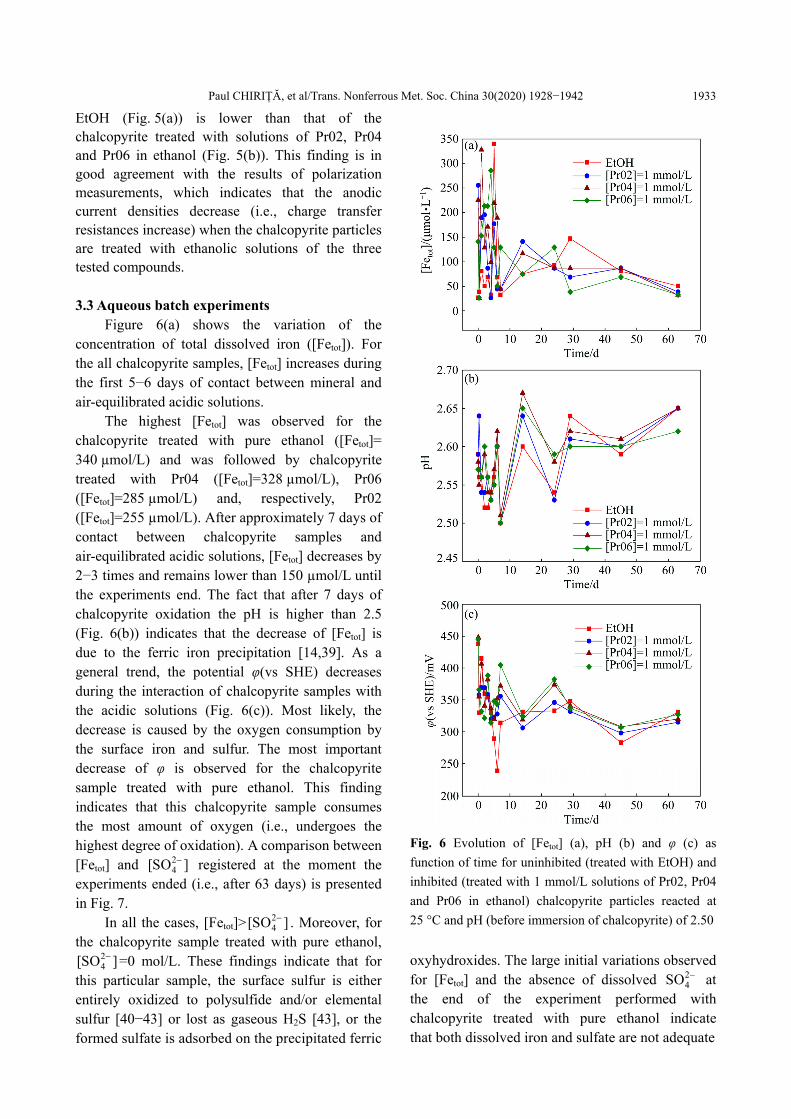
Figure 6(a) shows the variation of the concentration of total dissolved iron ([Fetot]). For the all chalcopyrite samples, [Fetot] increases during the first 5−6 days of contact between mineral and air-equilibrated acidic solutions.
The highest [Fetot] was observed for the chalcopyrite treated with pure ethanol ([Fetot]= 340 µmol/L) and was followed by chalcopyrite treated with Pr04 ([Fetot]=328 µmol/L), Pr06 ([Fetot]=285 µmol/L) and, respectively, Pr02 ([Fetot]=255 µmol/L). After approximately 7 days of contact between chalcopyrite samples and air-equilibrated acidic solutions, [Fetot] decreases by 2−3 times and remains lower than 150 µmol/L until the experiments end. The fact that after 7 days of chalcopyrite oxidation the pH is higher than 2.5 (Fig. 6(b)) indicates that the decrease of [Fetot] is due to the ferric iron precipitation [14,39]. As a general trend, the potential φ(vs SHE) decreases during the interaction of chalcopyrite samples with the acidic solutions (Fig. 6(c)). Most likely, the decrease is caused by the oxygen consumption by the surface iron and sulfur. The most important decrease of φ is observed for the chalcopyrite sample treated with pure ethanol. This finding indicates that this chalcopyrite sample consumes the most amount of oxygen (i.e., undergoes the highest degree of oxidation). A comparison between [Fetot] and 2
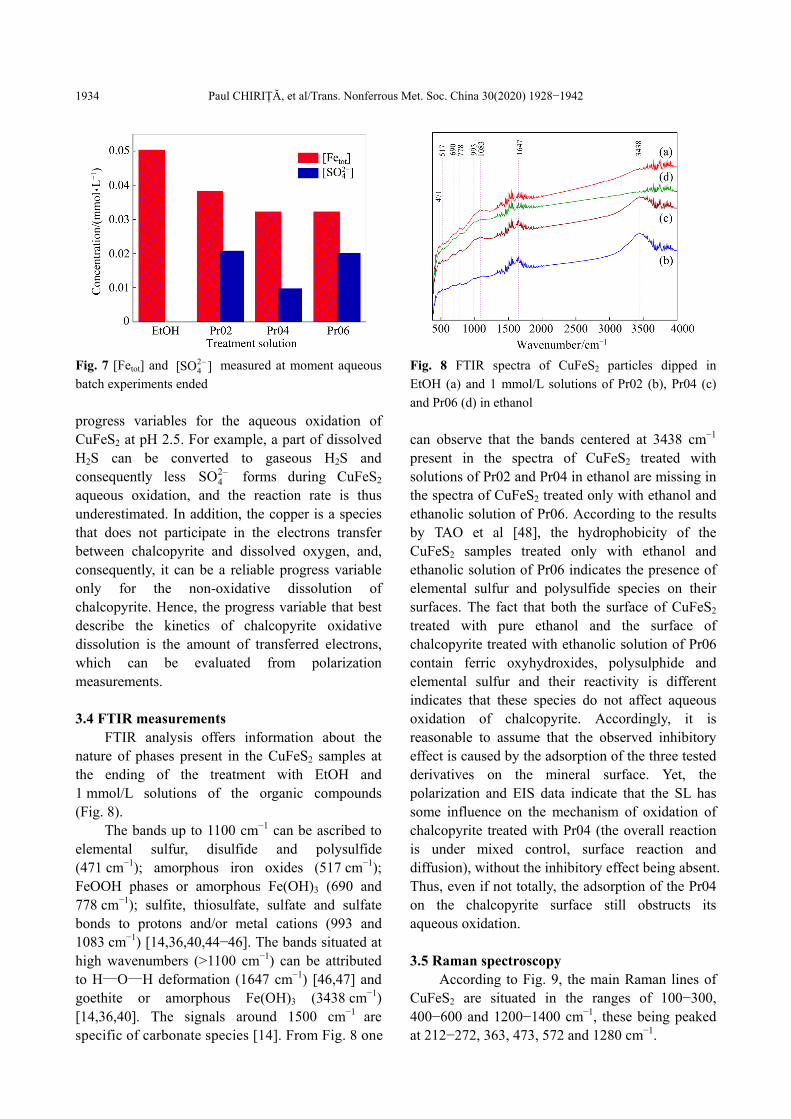
4[SO ] registered at the moment the experiments ended (i.e., after 63 days) is presented in Fig. 7.
In all the cases, [Fetot]>24[SO ] . Moreover, for
the chalcopyrite sample treated with pure ethanol, 24[SO ] =0 mol/L. These findings indicate that for
this particular sample, the surface sulfur is either entirely oxidized to polysulfide and/or elemental sulfur [40−43] or lost as gaseous H2S [43], or the formed sulfate is adsorbed on the precipitated ferric
Fig. 6 Evolution of [Fetot] (a), pH (b) and φ (c) as
function of time for uninhibited (treated with EtOH) and
inhibited (treated with 1 mmol/L solutions of Pr02, Pr04
and Pr06 in ethanol) chalcopyrite particles reacted at
25 °C and pH (before immersion of chalcopyrite) of 2.50
oxyhydroxides. The large initial variations observed for [Fetot] and the absence of dissolved 2
4SO at the end of the experiment performed with chalcopyrite treated with pure ethanol indicate that both dissolved iron and sulfate are not adequate
Paul CHIRIȚĂ, et al/Trans. Nonferrous Met. Soc. China 30(2020) 1928−1942
1934
Fig. 7 [Fetot] and 24[SO ] measured at moment aqueous
batch experiments ended
progress variables for the aqueous oxidation of CuFeS2 at pH 2.5. For example, a part of dissolved H2S can be converted to gaseous H2S and consequently less 2
4SO forms during CuFeS2 aqueous oxidation, and the reaction rate is thus underestimated. In addition, the copper is a species that does not participate in the electrons transfer between chalcopyrite and dissolved oxygen, and, consequently, it can be a reliable progress variable only for the non-oxidative dissolution of chalcopyrite. Hence, the progress variable that best describe the kinetics of chalcopyrite oxidative dissolution is the amount of transferred electrons, which can be evaluated from polarization measurements. 3.4 FTIR measurements
FTIR analysis offers information about the nature of phases present in the CuFeS2 samples at the ending of the treatment with EtOH and 1 mmol/L solutions of the organic compounds (Fig. 8).
The bands up to 1100 cm−1 can be ascribed to elemental sulfur, disulfide and polysulfide (471 cm−1); amorphous iron oxides (517 cm−1); FeOOH phases or amorphous Fe(OH)3 (690 and 778 cm−1); sulfite, thiosulfate, sulfate and sulfate bonds to protons and/or metal cations (993 and 1083 cm−1) [14,36,40,44−46]. The bands situated at high wavenumbers (>1100 cm−1) can be attributed to H—O—H deformation (1647 cm−1) [46,47] and goethite or amorphous Fe(OH)3 (3438 cm−1) [14,36,40]. The signals around 1500 cm−1 are specific of carbonate species [14]. From Fig. 8 one
Fig. 8 FTIR spectra of CuFeS2 particles dipped in
EtOH (a) and 1 mmol/L solutions of Pr02 (b), Pr04 (c)
and Pr06 (d) in ethanol
can observe that the bands centered at 3438 cm−1 present in the spectra of CuFeS2 treated with solutions of Pr02 and Pr04 in ethanol are missing in the spectra of CuFeS2 treated only with ethanol and ethanolic solution of Pr06. According to the results by TAO et al [48], the hydrophobicity of the CuFeS2 samples treated only with ethanol and ethanolic solution of Pr06 indicates the presence of elemental sulfur and polysulfide species on their surfaces. The fact that both the surface of CuFeS2 treated with pure ethanol and the surface of chalcopyrite treated with ethanolic solution of Pr06 contain ferric oxyhydroxides, polysulphide and elemental sulfur and their reactivity is different indicates that these species do not affect aqueous oxidation of chalcopyrite. Accordingly, it is reasonable to assume that the observed inhibitory effect is caused by the adsorption of the three tested derivatives on the mineral surface. Yet, the polarization and EIS data indicate that the SL has some influence on the mechanism of oxidation of chalcopyrite treated with Pr04 (the overall reaction is under mixed control, surface reaction and diffusion), without the inhibitory effect being absent. Thus, even if not totally, the adsorption of the Pr04 on the chalcopyrite surface still obstructs its aqueous oxidation. 3.5 Raman spectroscopy
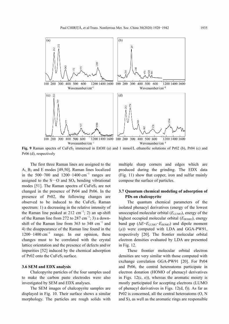
According to Fig. 9, the main Raman lines of CuFeS2 are situated in the ranges of 100−300, 400−600 and 1200−1400 cm−1, these being peaked at 212−272, 363, 473, 572 and 1280 cm−1.
Paul CHIRIȚĂ, et al/Trans. Nonferrous Met. Soc. China 30(2020) 1928−1942
1935
Fig. 9 Raman spectra of CuFeS2 immersed in EtOH (a) and 1 mmol/L ethanolic solutions of Pr02 (b), Pr04 (c) and
Pr06 (d), respectively
The first three Raman lines are assigned to the
A1, B2 and E modes [49,50]. Raman lines localized in the 500−700 and 1200−1400 cm−1 ranges are assigned to the S—O and SO4 bending vibrational modes [51]. The Raman spectra of CuFeS2 are not changed in the presence of Pr04 and Pr06. In the presence of Pr02, the following changes are observed to be induced to the CuFeS2 Raman spectrum: 1) a decreasing in the relative intensity of the Raman line peaked at 212 cm−1; 2) an up-shift of the Raman line from 272 to 287 cm−1; 3) a down- shift of the Raman line from 363 to 348 cm−1 and 4) the disappearance of the Raman line found in the 1200−1400 cm−1 range. In our opinion, these changes must to be correlated with the crystal lattice orientation and the presence of defects and/or impurities [52] induced by the chemical adsorption of Pr02 onto the CuFeS2 surface. 3.6 SEM and EDX analysis
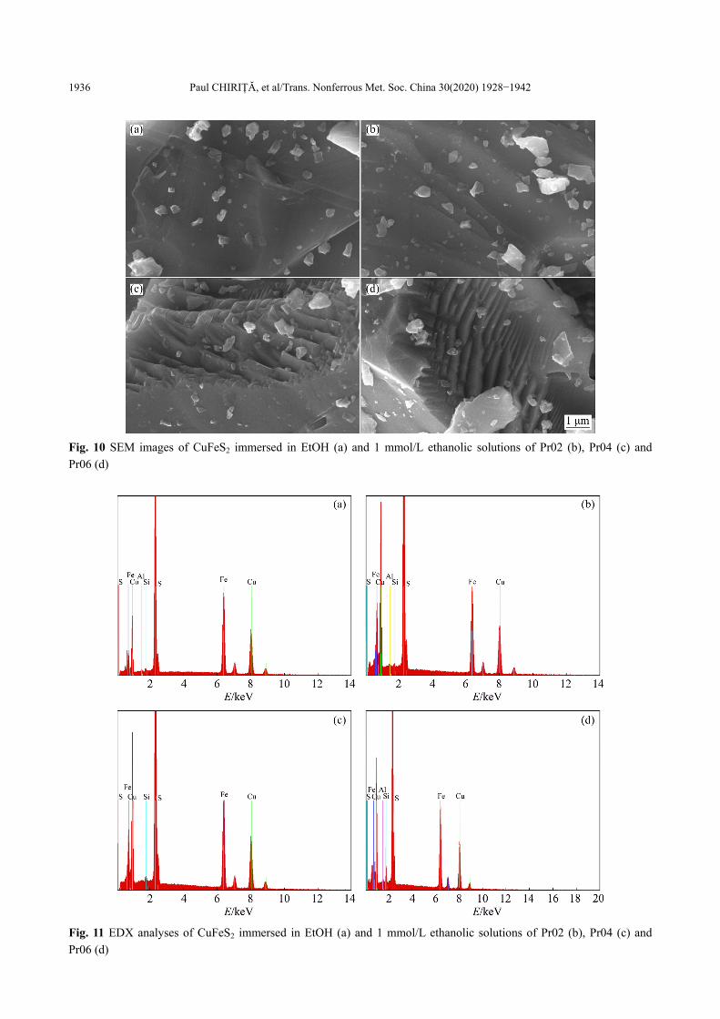
Chalcopyrite particles of the four samples used to make the carbon paste electrodes were also investigated by SEM and EDX analyses.
The SEM images of chalcopyrite samples are displayed in Fig. 10. Their surface shows a similar morphology. The particles are rough solids with
multiple sharp corners and edges which are produced during the grinding. The EDX data (Fig. 11) show that copper, iron and sulfur mainly compose the surface of particles. 3.7 Quantum chemical modeling of adsorption of
PDs on chalcopyrite The quantum chemical parameters of the
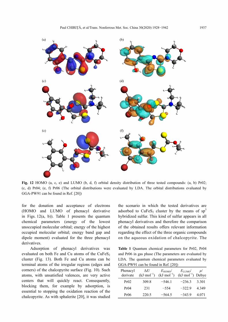
isolated phenacyl derivatives (energy of the lowest unoccupied molecular orbital (ELUMO), energy of the highest occupied molecular orbital (EHOMO), energy band gap (ΔE=ELUMO−EHOMO) and dipole moment (µ)) were computed with LDA and GGA-PW91, respectively [20]. The frontier molecular orbital electron densities evaluated by LDA are presented in Fig. 12.
These frontier molecular orbital electron densities are very similar with those computed with exchange correlation GGA-PW91 [20]. For Pr04 and Pr06, the central heteroatoms participate in electron donation (HOMO of phenacyl derivatives in Figs. 12(c, e)), whereas the aromatic moiety is mostly participated for accepting electrons (LUMO of phenacyl derivatives in Figs. 12(d, f)). As far as Pr02 is concerned, all the central heteroatoms (O, N and S), as well as the aromatic rings are responsible
Paul CHIRIȚĂ, et al/Trans. Nonferrous Met. Soc. China 30(2020) 1928−1942
1936
Fig. 10 SEM images of CuFeS2 immersed in EtOH (a) and 1 mmol/L ethanolic solutions of Pr02 (b), Pr04 (c) and
Pr06 (d)
Fig. 11 EDX analyses of CuFeS2 immersed in EtOH (a) and 1 mmol/L ethanolic solutions of Pr02 (b), Pr04 (c) and
Pr06 (d)
Paul CHIRIȚĂ, et al/Trans. Nonferrous Met. Soc. China 30(2020) 1928−1942
1937
Fig. 12 HOMO (a, c, e) and LUMO (b, d, f) orbital density distribution of three tested compounds: (a, b) Pr02;
(c, d) Pr04; (e, f) Pr06 (The orbital distributions were evaluated by LDA. The orbital distributions evaluated by
GGA-PW91 can be found in Ref. [20])
for the donation and acceptance of electrons (HOMO and LUMO of phenacyl derivative in Figs. 12(a, b)). Table 1 presents the quantum chemical parameters (energy of the lowest unoccupied molecular orbital; energy of the highest occupied molecular orbital; energy band gap and dipole moment) evaluated for the three phenacyl derivatives.
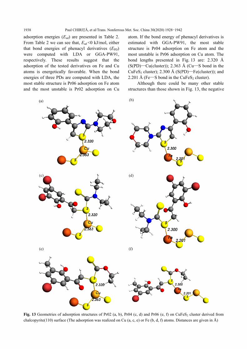
Adsorption of phenacyl derivatives was evaluated on both Fe and Cu atoms of the CuFeS2 cluster (Fig. 13). Both Fe and Cu atoms can be terminal atoms of the irregular regions (edges and corners) of the chalcopyrite surface (Fig. 10). Such atoms, with unsatisfied valences, are very active centers that will quickly react. Consequently, blocking them, for example by adsorption, is essential to stopping the oxidation reaction of the chalcopyrite. As with sphalerite [20], it was studied
the scenario in which the tested derivatives are adsorbed to CuFeS2 cluster by the means of sp3 hybridized sulfur. This kind of sulfur appears in all phenacyl derivatives and therefore the comparison of the obtained results offers relevant information regarding the effect of the three organic compounds on the aqueous oxidation of chalcopyrite. The Table 1 Quantum chemical parameters for Pr02, Pr04
and Pr06 in gas phase (The parameters are evaluated by
LDA. The quantum chemical parameters evaluated by
GGA-PW91 can be found in Ref. [20])
Phenacyl derivate
ΔE/ (kJꞏmol−1)
EHOMO/ (kJꞏmol−1)
ELUMO/ (kJꞏmol−1)
µ/ Debye
Pr02 309.8 −546.1 −236.3 3.301
Pr04 231 −554 −322.9 4.349
Pr06 220.5 −564.5 −343.9 4.071
Paul CHIRIȚĂ, et al/Trans. Nonferrous Met. Soc. China 30(2020) 1928−1942
1938
adsorption energies (Ead) are presented in Table 2. From Table 2 we can see that, Ead <0 kJ/mol, either that bond energies of phenacyl derivatives (EPD) were computed with LDA or GGA-PW91, respectively. These results suggest that the adsorption of the tested derivatives on Fe and Cu atoms is energetically favorable. When the bond energies of three PDs are computed with LDA, the most stable structure is Pr06 adsorption on Fe atom and the most unstable is Pr02 adsorption on Cu
atom. If the bond energy of phenacyl derivatives is estimated with GGA-PW91, the most stable structure is Pr04 adsorption on Fe atom and the most unstable is Pr06 adsorption on Cu atom. The bond lengths presented in Fig. 13 are: 2.320 Å (S(PD)—Cu(cluster)); 2.363 Å (Cu—S bond in the CuFeS2 cluster); 2.300 Å (S(PD)—Fe(cluster)); and 2.201 Å (Fe—S bond in the CuFeS2 cluster).
Although there could be many other stable structures than those shown in Fig. 13, the negative
Fig. 13 Geometries of adsorption structures of Pr02 (a, b), Pr04 (c, d) and Pr06 (e, f) on CuFeS2 cluster derived from
chalcopyrite(110) surface (The adsorption was realized on Cu (a, c, e) or Fe (b, d, f) atoms. Distances are given in Å)
Paul CHIRIȚĂ, et al/Trans. Nonferrous Met. Soc. China 30(2020) 1928−1942
1939
Table 2 Adsorption energies (Ead) for Pr02, Pr04 and Pr06 on Cu and Fe atoms of CuFeS2 cluster derived from
chalcopyrite(110) surface
Chalcopyrite/PD optimizations Center of adsorption Ead/(kJꞏmol−1)
CuFeS2-Pr02 CuFeS2-Pr04 CuFeS2-Pr06
LDA/LDA Fe −89.3 −102.4 −133.9
Cu −65.6 −78.8 −115.5
LDA/GGA Fe −1465.0 −1507.0 −1378.4
Cu −1441.4 −1483.4 −1360.0
values obtained for Ead are enough to reveal the importance of the PDs adsorption on the chalcopyrite surface in the control of its aqueous oxidation. It is considered that the inhibitory effect of organic compounds increases when µ increases, and Ead and ΔE decrease [22−26,53−55]. Starting from these considerations, the anodic current densities are well correlated with Ead values evaluated by LDA/LDA optimization and ΔE values. Instead, the Ead values evaluated by LDA/GGA optimization and µ underestimate the inhibitory effect of Pr06 on the aqueous oxidation of chalcopyrite. 4 Conclusions
(1) Pr02, Pr04 and Pr06 inhibit the aqueous oxidation of chalcopyrite. The three organic derivatives act as anodic inhibitors. The values of the anodic current densities of the chalcopyrite electrodes treated with pure ethyl alcohol and ethanolic solutions of the three phenacyl derivatives decrease in the following order of EtOH > Pr02 > Pr04 > Pr06. These findings are supported by the results of impedance and aqueous batch experiments.
(2) Both polarization and impedance data show that the electrochemical behavior of CuFeS2 sample treated with ethanolic solution of Pr04 is different from that of the other chalcopyrite samples. The aqueous oxidation of chalcopyrite treated with EtOH and the solutions of Pr02 and Pr06 in ethanol is controlled by a surface reaction, while the oxidation of chalcopyrite treated with Pr04 is controlled by a mixed regime of a surface reaction and diffusion.
(3) Raman scattering and FTIR spectroscopy indicate that elemental sulfur, polysulfide and ferric
oxyhydroxides are not responsible for the inhibition of the aqueous oxidation of chalcopyrite.
(4) Theoretical calculations show that the adsorption of the three tested derivatives on chalcopyrite is energetically favorable (Ead< 0 kJ/mol). Hence, it is reasonable to assume that the adsorbed phenacyl derivatives obstruct the aqueous oxidation of CuFeS2.
(5) Our findings show that aqueous oxidation of CuFeS2 is rather controlled by the adsorbed organic matter than the SL formed on its surface. Most likely, the adsorbed organic molecules interpose between chalcopyrite and the dissolved oxygen and do not allow the transfer of electrons from mineral to oxidant. Future investigations should be planned to evaluate the effects of other organic molecules on the aqueous oxidation of chalcopyrite to find the organic structure that most efficiently controls the reaction.
Acknowledgments This work was partly supported by a grant of
the Romanian National Authority for Scientific Research, CNDI−UEFISCDI, project number 51/2012.
References [1] HU J, TIAN G, ZI F, HU X. Leaching of chalcopyrite with
hydrogen peroxide in 1-hexyl-3-methyl-imidazolium
hydrogen sulfate ionic liquid aqueous solution [J].
Hydrometallurgy, 2017, 169: 1−8.
[2] KIMBALL B E, RIMSTIDT J D, BRANTLEY S L.
Chalcopyrite dissolution rate laws [J]. Applied
Geochemistry, 2010, 25: 972−983.
[3] VELASQUEZ P, LEINEN D, PASCUAL J, RAMOS-
BARRADO J R, GREZ P, GOMEZ H, SCHREBLER R,
DEL R, CORDOVA R. A chemical, morphological, and
electrochemical (XPS, SEM/EDX, CV, and EIS) analysis of
Paul CHIRIȚĂ, et al/Trans. Nonferrous Met. Soc. China 30(2020) 1928−1942
1940
electrochemically modified electrode surfaces of natural
chalcopyrite (CuFeS2, and pyrite (FeS2, in alkaline solutions
[J]. The Journal of Physical Chemistry B, 2005, 109:
4977−4988.
[4] GHAHREMANINEZHAD A, DIXON D G, ASSELIN E.
Electrochemical and XPS analysis of chalcopyrite (CuFeS2,
dissolution in sulfuric acid solution [J]. Electrochimica Acta,
2013, 87: 97−112.
[5] LI Y, QIAN G, LI J, GERSON A R. Kinetics and roles of
solution and surface species of chalcopyrite dissolution at
650 mV [J]. Geochimica et Cosmochimica Acta, 2015, 161:
188−202.
[6] RUIZ-SANCHEZ A, LAPIDUS G T. Study of chalcopyrite
leaching from a copper concentrate with hydrogen peroxide
in aqueous ethylene glycol media [J]. Hydrometallurgy, 2017,
169: 192−200.
[7] MARTINEZ-GOMEZ V J, FUENTES-ACEITUNO J C,
PEREZ-GARIBAY R, LEE J. A study of the electro-assisted
reductive leaching of a chalcopyrite concentrate in HCl
solutions. Part I: Kinetic behavior and nature of the
chalcopyrite reduction [J]. Hydrometallurgy, 2018, 181:
195−205.
[8] BARHOUMI N, OLVERA-VARGAS H, OTURAN N,
HUGUENOT D, GADRI A, AMMARE S, BRILLAS E,
OTURAN M A. Kinetics of oxidative degradation/
mineralization pathways of the antibiotic tetracycline by the
novel heterogeneous electro-Fenton process with solid
catalyst chalcopyrite [J]. Applied Catalysis B: Environmental,
2017, 209: 637−647.
[9] THURSTON R S, MANDERNACK K W, SHANKS III W C.
Laboratory chalcopyrite oxidation by Acidithiobacillus
ferrooxidans: Oxygen and sulfur isotope fractionation [J].
Chemical Geology, 2010, 269: 252−261.
[10] ZHAO H, HU M, LI Y, ZHU S, QIN W, QIU G, WANG J.
Comparison of electrochemical dissolution of chalcopyrite
and bornite in acid culture medium [J]. Transactions of
Nonferrous Metals Society of China, 2015, 25: 303−313.
[11] ZHAO H, WANG J, QIN W, ZHENG X, TAO L, GAN X,
QIU G. Surface species of chalcopyrite during bioleaching by
moderately thermophilic bacteria [J]. Transactions of
Nonferrous Metals Society of China, 2015, 25: 2725−2733.
[12] WU S, YANG C, QIN W, JIAO F, WANG J, ZHANG Y.
Sulfur composition on surface of chalcopyrite during its
bioleaching at 50 °C [J]. Transactions of Nonferrous Metals
Society of China, 2015, 25: 4110−4118.
[13] LI Y, QIAN G, BROWN P L, GERSON A R. Chalcopyrite
dissolution: Scanning photoelectron microscopy examination
of the evolution of sulfur species with and without added
iron or pyrite [J]. Geochimica et Cosmochimica Acta, 2017,
212: 33−47.
[14] CHIRITA P, SCHLEGEL M L. Oxidative dissolution of iron
monosulfide (FeS) in acidic conditions: The effect of solid
pretreatment [J]. International Journal of Mineral Processing,
2015, 135: 57−64.
[15] LINGE H G. A study of chalcopyrite dissolution in acidic
ferric nitrate by potentiometric titration [J]. Hydrometallurgy,
1976, 2: 51−64.
[16] DUTRIZAC J E. The kinetics of dissolution of chalcopyrite
in ferric ion media [J]. Metallurgical and Materials
Transactions B, 1978, 9: 431−439.
[17] BASOLO F, PEARSON R G. Mechanisms of Inorganic
Reactions. A study of metal complexes in solution [M]. 2nd
Edition. New York: John Wiley and Sons, Inc, 1967.
[18] SCHOONEN M A A, STRONGIN D R. Catalysis of electron
transfer reactions at mineral surfaces [M]. Environmental
Catalysis. GRASSIAN V H ed.. Boca Raton, Fl: CRC Press,
2005.
[19] SHU X, DANG Z, ZHANG Q, YI X, LU G, GUO C, YANG
C. Passivation of metal-sulfide tailings by covalent coating
[J]. Minerals Engineering, 2013, 42: 36−42.
[20] CHIRITA P, DUINEA M I, SANDU A M, BIRSA L M,
SARBU L G, BAIBARAC M, SAVA F, POPESCU M,
MATEI E. Inhibitory effect of three phenacyl derivatives on
the oxidation of sphalerite (ZnS) in air-equilibrated acidic
solution [J]. Corrosion Science, 2018, 138: 154−162.
[21] BADICA C E, CHIRITA P. An electrochemical study of the
oxidative dissolution of iron monosulfide (FeS) in
air-equilibrated solutions [J]. Electrochimica Acta, 2015, 178:
786−796.
[22] BABIC-SAMARDZIJA K, KHALED K F, HACKERMAN
N. Investigation of the inhibiting action of O−, S− and
N-dithiocarbamato(1,4,8,11-tetraazacyclotetradecane) cobalt
(III) complexes on the corrosion of iron in HClO4 acid [J].
Applied Surface Science, 2005, 240: 327−340.
[23] ABDALLAH Y M. Electrochemical studies of phenyl
sulphonyl ethanone derivatives compounds on corrosion of
aluminum in 0.5 M H2SO4 solutions [J]. Journal of
Molecular Liquids, 2016, 219: 709−719.
[24] EL BELGHITI M, KARZAZI Y, DAFALI A, HAMMOUTI
B, BENTISS F, OBOT I B, BAHADUR I, EBENSO E E.
Experimental, quantum chemical and Monte Carlo
simulation studies of 3,5-disubstituted-4-amino-1,2,4-
triazoles as corrosion inhibitors on mild steel in acidic
medium [J]. Journal of Molecular Liquids, 2016, 218:
281−293.
[25] GECE G, BILGIC S. Quantum chemical study of some
cyclic nitrogen compounds as corrosion inhibitors of steel in
NaCl media [J]. Corrosion Science, 2009, 51: 1876−1878.
[26] QIANG Y, ZHANG S, XU S, LI W. Experimental and
theoretical studies on the corrosion inhibition of copper by
two indazole derivatives in 3.0% NaCl solution [J]. Journal
of Colloid and Interface Science, 2016, 472: 52−59.
[27] KOLEINI S M J, AGHAZADEH V, SANDSTROM A.
Acidic sulphate leaching of chalcopyrite concentrates in
presence of pyrite [J]. Minerals Engineering, 2011, 24:
381−386.
[28] MANESCU S, CUCU M, DIACONESCU M L.
Environmental sanitary chemistry [M]. Bucharest: Medical
Paul CHIRIȚĂ, et al/Trans. Nonferrous Met. Soc. China 30(2020) 1928−1942
1941
Publishing House, 1994. (in Romanian)
[29] TE VELDE G, BAERENDS E J. Precise density-functional
method for periodic structures [J]. Physical Review B, 1991,
44: 7888−7903.
[30] ADF manual, ADF modeling suite 2016 [M]. 2016.
http://www.scm. com.
[31] van LENTHE E, BAERENDS E J, SNIJDERS J G.
Relativistic regular two-component Hamiltonians [J]. The
Journal of Chemical Physics, 1993, 99: 4597−4610.
[32] ROSA A, BAERENDS E J, van GISBERGEN S J, LENTHE
A E, GROENEVELD J A, SNIJDERS J G. Electronic spectra
of M(CO)6 (M=Cr, Mo, W) revisited by a relativistic
TDDFT approach [J]. Journal of the American Chemical
Society, 1999, 121: 10356−10365.
[33] HOU M, MEI Q, HAN B. Solvent effects on geometrical
structures and electronic properties of metal Au, Ag, and Cu
nanoparticles of different sizes [J]. Journal of Colloid
Interface Science, 2015, 449: 488−493.
[34] VOSKO S H, WILK L, NUSAIR M. Accurate spin-
dependent electron liquid correlation energies for local spin
density calculations: A critical analysis [J]. Canadian Journal
of Physics, 1980, 58: 1200−1211.
[35] PERDEW J P, CHEVARY J A, VOSKO S H, JACKSON K.
A, PEDERSON M R, SING D J, FIOLHAIS C. Atoms,
molecules, solids, and surfaces: Applications of the
generalized gradient approximation for exchange and
correlation [J]. Physical Review B, 1992, 46: 6671−6687.
[36] CONSTANTIN C A, CHIRITA P. Oxidative dissolution of
pyrite in acidic media [J]. Journal of Applied
Electrochemistry, 2013, 43: 659−666.
[37] CHIRITA P, SCHLEGEL M L. Pyrite oxidation in air-
equilibrated solutions: An electrochemical study [J].
Chemical Geology, 2017, 470: 67−74.
[38] TUKEN T, YAZICI B, ERBIL M. The corrosion behaviour
of polypyrrole coating synthesized in phenylphosphonic acid
solution [J]. Applied Surface Science, 2006, 252:
2311−2318.
[39] DESCOSTES M, BEAUCAIRE C, MERCIER F, SAVOYE
S, SOW J, ZUDDAS P. Effect of carbonate ions on pyrite
(FeS2) dissolution [J]. Bulletin of the Geological Society of
France, 2002, 173: 265−270. (in French)
[40] MIKHLIN Y L, KUKLINSKIY A V, PAVLENKO N I,
VARNEK V A, ASANOV I P, OKOTRUB A V, SELYUTIN
G E, SOLOVYEV L A. Spectroscopic and XRD studies of
the air degradation of acid-reacted pyrrhotites [J].
Geochimica et Cosmochimica Acta, 2002, 66: 4057−4067.
[41] PENG T, ZHOU D, LIU X, YU R, JIANG T, GU G, CHEN
M, QIU G, ZENG W. Enrichment of ferric iron on mineral
surface during bioleaching of chalcopyrite [J]. Transactions
of Nonferrous Metals Society of China, 2016, 26: 544−550.
[42] CHANG K, ZHANG Y, ZHANG J, LI T, WANG J, QIN W.
Effect of temperature-induced phase transitions on
bioleaching of chalcopyrite[J]. Transactions of Nonferrous
Metals Society of China, 2019, 29: 2183−2191.
[43] CHIRITA P, DESCOSTES M. Troilite oxidation by hydrogen
peroxide [J]. Journal of Colloid and Interface Science, 2006,
299: 260−269.
[44] CHIRITA P. Aqueous oxidation of iron monosulfide (FeS) by
molecular oxygen [J]. Mineral Processing and Extractive
Metallurgy Review, 2016, 37: 305−310.
[45] CHIRITA P. Evaluation and modeling of the surface
characteristics of troilite (FeS) [J]. Applied Surface Science,
2019, 480: 281−287.
[46] YANG B, LUO W, LIAO Q, ZHU J, GAN M, LIU X, QIU G.
Photogenerated-hole scavenger for enhancing photocatalytic
chalcopyrite bioleaching [J]. Transactions of Nonferrous
Metals Society of China, 2020, 30: 200−211.
[47] BAMPOLE D L, LUIS P, MULABA-BAFUBIANDI A F.
Sustainable copper extraction from mixed chalcopyrite-
chalcocite using biomass [J]. Transactions of Nonferrous
Metals Society of China, 2019 29: 2170−2182.
[48] TAO D P, RICHARDSON P E, LUTTRELL G H, YOON R
H. Electrochemical studies of pyrite oxidation and reduction
using freshly-fractured electrodes and rotating ring-disc
electrodes [J]. Electrochimica Acta, 2003, 48: 3615−3623.
[49] XI S, ZHANG X, LUAN Z, DU Z, LI L., LIANG Z, LIAN C,
YAN J. Micro-Raman study of thermal transformations of
sulfide and oxysalt minerals based on the heat induced by
laser [J]. Minerals, 2019, 9(12): 751.
[50] WANG C, XUE S, HU J, TANG K. Raman, far infrared, and
Mossbauer spectroscopy of CuFeS2 nanocrystallites [J].
Japanese Journal of Applied Physics, 2009, 48: 023003.
[51] PRAMEENA B, ANBALAGAN G, GUNASEKARAN S,
RAMKUMAAR G R, GOWTHAM B. Structural, optical,
electron paramagnetic, thermal and dielectric
characterization of chalcopyrite [J]. Spectrochimica Acta
(Part A): Molecular and Biomolecular Spectroscopy, 2014,
122: 348−355.
[52] LIANG C, XIA J, NIE Z, YU S, XU B. Effect of initial pH
on chalcopyrite oxidation dissolution in the presence of
extreme thermophile Acidianus manzaensis [J]. Transactions
of Nonferrous Metals Society of China, 2014, 24:
1890−1897.
[53] FITOZ A, NAZIR H, OZGUR (nee YAKUT) M, EMREGUL
E, EMREGUL K C. An experimental and theoretical
approach towards understanding the inhibitive behavior of a
nitrile substituted coumarin compound as an effective acidic
media inhibitor [J]. Corrosion Science, 2018, 133: 451−464.
[54] HU K, ZHUANG J, DING J, MA Z, WANG F, ZENG X.
Influence of biomacromolecule DNA corrosion inhibitor on
carbon steel [J]. Corrosion Science, 2017, 125: 68−76.
[55] BEDAIR M A, EL-SABBAH M M B, FOUDA A S,
ELARYIAN H M. Synthesis, electrochemical and quantum
chemical studies of some prepared surfactants based on
azodye and Schiff base as corrosion inhibitors for steel in
acid medium [J]. Corrosion Science, 2017, 128: 54−72.
Paul CHIRIȚĂ, et al/Trans. Nonferrous Met. Soc. China 30(2020) 1928−1942
1942
黄铜矿在空气平衡的酸性溶液中的氧化:
苯酰基衍生物的抑制作用
Paul CHIRIȚĂ1, Mădălina I. DUINEA1,2, Laura G. SÂRBU3,
Lucian M. BÎRSĂ3, Mihaela BAIBARAC4, Florinel SAVA4, Elena MATEI4
1. Department of Chemistry, University of Craiova, Calea Bucuresti 107I, 200478, Craiova, Romania;
2. Department of Chemistry, Physics and Environment,
‘‘Dunarea de Jos” University of Galati, 111 Domneasca Street, Galati 800201, Romania;
3. Department of Chemistry, “Al. I. Cuza” University of Iasi, 11 Carol I Blv., Iasi 700506, Romania;
4. Lab. Optical Processes in Nanostructured Materials, National Institute of Materials Physics,
P. O. Box MG-7, Bucharest, R077125, Romania
摘 要:研究 4-(2-羟苯基)-2-(-4-吗啉基)-1,3-噻唑(Pr02)、1-(3,5-二溴-2-羟苯基)-1-(2-氧乙基)-N,N-二乙基二硫代
氨基甲酸酯(Pr04)和 1-(5-溴-2-羟基-3-甲苯基)-1-(2-氧乙基)-O-乙基黄原酸酯(Pr06)对黄铜矿(CuFeS2)在温度
25 °C、pH 2.5 的空气平衡溶液中液相氧化反应的影响。采用动电位极化、电化学阻抗谱(EIS)、能谱扫描电镜
(SEM/EDX)分析、液相间歇实验、傅里叶变换红外光谱(FTIR)、拉曼散射以及量子化学计算等方法研究这些影
响,发现阳极电流密度按 EtOH> Pr02> Pr04> Pr06 的顺序降低。这些结果以及 EIS 测量结果表明,Pr02、Pr04 和
Pr06 是黄铜矿液相氧化的有效阳极抑制剂。拉曼散射和 FTIR 光谱分析表明,矿物表面形成的元素硫、多硫化物
和羟基氧化铁与黄铜矿的液相氧化抑制作用无关。量子化学计算表明,所测试的化合物在黄铜矿表面的吸附在能
量上是有利的,从而可以解释所观察到的这些化合物的抑制作用。
关键词:黄铜矿氧化;苯酰基衍生物;抑制;动电位极化;量子化学计算
(Edited by Xiang-qun LI)