Embed Size (px)
Citation preview

Exp Toxic Pathol 1995; 47: 1-9 Gustav Fischer Verlag Jena
Pharmacia Farmitalia Carlo Erba, R & Offoxicology Opt., Nerviano (Milano), Italy
Overview of toxicological data on Rifabutin
MARCO BRUGHERA, GIOVANNA SCAMPINI, A. JOHN NEWMAN, SILVANA CASTELLINO, UMBERTO SAMMARTINI, and GUY MAZUE
With 3 figures and 3 tables
Received: June 20, 1994; Accepted: July 10, 1994
Address for correspondence: MARCO BRUGHERA, Pharmacia Farmitalia Carlo Erba, R & Offoxicology Opt, Via Giovanni XXIII 24, 20014 Nerviano (Milano) Italy; Tel. 0331.583379, Fax 0331.583377.
Key words: Rifabutin, Toxicological data.
Summary
Rifabutin is a wide spectrum antibiotic particularly active on atypical and rifampicin-resistant mycobacteria. Rifabutin is more potent than rifampicin on Mycobacterium tuberculosis in vitro. Its mode of action is characterized by a high intracellular penetration in treated individuals. Clinical trials have proven the therapeutic value of rifabutin especially in AIDS patients with concomitant MAC.
The preclinical safety evaluation of this compound included single and repeated dose toxicity studies of up to one year in rodents and non-rodents, reproduction and carcinogenicity studies and mutagenicity tests. Ouring toxicological studies the most significant finding after repeated administration of rifabutin was the presence of multinucleated hepatocytes (MNH) in rats. This is a species specific finding which did not affect the life span of the hepatocytes. As shown in carcinogenicity studies, there was no tendency to further proliferative changes.
Another specific histological feature among the species studied was the presence of a lipofuscin-like brown pigment, which was seen in many organs. This is a common finding with amphipilic compounds, such as rifabutin, which bind lipids and proteins, forming membrane-bound complexes. Even in carcinogenicity studies this change did not constitute a stimulus to cell proliferation and did not cause any secondary changes.
In rodents, there was a mild hemolytic anemia at doses higher than 10 mg/kg/day.
At doses ranging from 160-200 mg/kg/day rifabutin inhibited the functions of the male gonads in rats. This effect was reflected in a reduction of implantations observed in the fertility studies. Doses of 40 mglkg/day did not induce any embryotoxic effects or changes in reproductive performance.
The drug was not genotoxic and was devoid of carcinogenic potential.
The exposure levels in the animals used for toxicological tests were higher than those reached in man at therapeutic doses.
Introduction
Rifabutin is a broad spectrum antibiotic particularly active on acid-fast bacilli, including atypical and rifampicin-resistant mycobacteria (1,2,4, 12, 15). In comparison with rifampicin, rifabutin is 4-16 times more potent on Mycobacterium tuberculosis in vitro (12). Moreover, rifabutin is characterized by a high intracellular penetration in treated individuals, by the occurrence of a low frequency of naturally resistant mutants and low emergence of resistant mutants (7,8, 13, 14, 16). Clinical trials have proven the therapeutic value of rifabutin especially in AIDS patients with concomitant MAC (Mycobacterium avium complex) (3, 5, 6, 11).
Rifabutin is rapidly absorbed from the gastrointestinal tract with peak plasma levels being reached within 2 to 4 hours after oral administration. The drug is extensively distributed both in animals and man and reaches much higher levels in tissues than in plasma. The drug is eliminated mainly in the urine. The metabolic pathway in the rat is considered to be that which most closely resembles the situation in man.
The toxicological studies that have been performed are described below and the principle findings are discussed.
Material and methods
Chemical
Rifabutin is a spiro-piperidyl-rifamycin derived from rifamycin-So The chemical name is 4-deoxo-3,4-[2-spiro-
Exp Toxic Pathol 47 (1995) 1
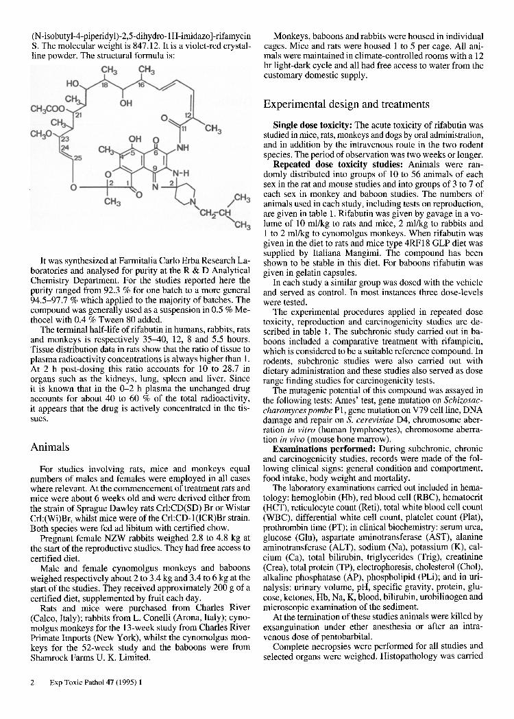
(N -isobuty 1-4-piperid y 1)-2,5-dihydro-l H -imidazo ]-rifamycin S. The molecular weight is 847.12. It is a violet-red crystalline powder. The structural formula is:
CH3 CH3
o
OH 0
It was synthesized at Farmitalia Carlo Erba Research Laboratories and analysed for purity at the R&D Analytical Chemistry Department. For the studies reported here the purity ranged from 92.3 % for one batch to a more general 94.5-97.7 % which applied to the majority of batches. The compound was generally used as a suspension in 0.5 % Methocel with 0.4 % Tween 80 added.
The terminal half-life of rifabutin in humans, rabbits, rats and monkeys is respectively 35-40, 12, 8 and 5.5 hours. Tissue distribution data in rats show that the ratio of tissue to plasma radioactivity concentrations is always higher than I. At 2 h post-dosing this ratio accounts for 10 to 28.7 in organs such as the kidneys, lung, spleen and liver. Since it is known that in the 0-2 h plasma the unchanged drug accounts for about 40 to 60 % of the total radioactivity, it appears that the drug is actively concentrated in the tissues.
Animals
For studies involving rats, mice and monkeys equal numbers of males and females were employed in all cases where relevant. At the commencement of treatment rats and mice were about 6 weeks old and were derived either from the strain of Sprague Dawley rats Cr1:CD(SD) Br or Wistar Cr1:(Wi)Br, whilst mice were of the Cr1:CD-I(ICR)Br strain. Both species were fed ad libitum with certified chow.
Pregnant female NZW rabbits weighed 2.8 to 4.8 kg at the start of the reproductive studies. They had free access to certified diet.
Male and female cynomolgus monkeys and baboons weighed respectively about 2 to 3.4 kg and 3.4 to 6 kg at the start of the studies. They received approximately 200 g of a certified diet, supplemented by fruit each day.
Rats and mice were purchased from Charles River (Calco, Italy); rabbits from L. Conelli (Arona, Italy); cynomolgus monkeys for the 13-week study from Charles River Primate Imports (New York), whilst the cynomolgus monkeys for the 52-week study and the baboons were from Shamrock Farms U. K. Limited.
2 Exp Toxic Pathol 47 (1995) 1
Monkeys, baboons and rabbits were housed in individual cages. Mice and rats were housed 1 to 5 per cage. All animals were maintained in climate-controlled rooms with a 12 hr light-dark cycle and all had free access to water from the customary domestic supply.
Experimental design and treatments
Single dose toxicity: The acute toxicity of rifabutin was studied in mice, rats, monkeys and dogs by oral administration, and in addition by the intravenous route in the two rodent species. The period of observation was two weeks or longer.
Repeated dose toxicity studies: Animals were randomly distributed into groups of 10 to 56 animals of each sex in the rat and mouse studies and into groups of 3 to 7 of each sex in monkey and baboon studies. The numbers of animals used in each study, including tests on reproduction, are given in table I. Rifabutin was given by gavage in a volume of 10 ml/kg to rats and mice, 2 ml/kg to rabbits and 1 to 2 ml/kg to cynomolgus monkeys. When rifabutin was given in the diet to rats and mice type 4RFl8 GLP diet was supplied by Italiana Mangimi. The compound has been shown to be stable in this diet. For baboons rifabutin was given in gelatin capsules.
In each study a similar group was dosed with the vehicle and served as control. In most instances three dose-levels were tested.
The experimental procedures applied in repeated dose toxicity, reproduction and carcinogenicity studies are described in table 1. The subchronic study carried out in baboons included a comparative treatment with rifampicin, which is considered to be a suitable reference compound. In rodents, subchronic studies were also carried out with dietary administration and these studies also served as dose range finding studies for carcinogenicity tests.
The mutagenic potential of this compound was assayed in the following tests: Ames' test, gene mutation on Schizosaccharomyces pombe PI , gene mutation on V79 cell line, DNA damage and repair on S. cerevisiae D4, chromosome aberration in vitro (human lymphocytes), chromosome aberration in vivo (mouse bone marrow).
Examinations performed: During subchronic, chronic and carcinogenicity studies, records were made of the following clinical signs: general condition and comportment, food intake, body weight and mortality .
The laboratory examinations carried out included in hematology: hemoglobin (Hb), red blood cell (RBC), hematocrit (HCT), reticulocyte count (Reti), total white blood cell count (WBC), differential white cell count, platelet count (Plat), prothrombin time (PT); in clinical biochemistry: serum urea, glucose (Glu), aspartate aminotransferase (AST), alanine aminotransferase (ALT), sodium (Na), potassium (K), calcium (Ca), total bilirubin, triglycerides (Trig), creatinine (Crea), total protein (TP), electrophoresis, cholesterol (Cho!), alkaline phosphatase (AP), phospholipid (PLi); and in urinalysis: urinary volume, pH, specific gravity, protein, glucose, ketones, Hb, Na, K, blood, bilirubin, urobilinogen and microscopic examination of the sediment.
At the termination of these studies animals were killed by exsanguination under ether anesthesia or after an intravenous dose of pentobarbital.
Complete necropsies were performed for all studies and selected organs were weighed. Histopathology was carried

Table 1. Experimental design of toxicology studies in animals.
Type of Study Strain No. of Animals/Sex! Group
Species: Rat Subchronic toxicity S.D. 15 Subchronic toxicity S.D. 10 Chronic toxicity Wistar 33 Carcinogenicity S.D. 50 Fertility S.D. 30
Embryotoxicity S.D. 22-25 F Peri- and post-natal S.D. 24F toxicity
Species: Rabbit Embryotoxicity NZW 17-20 F Species: Mouse Subchronic toxicity CD[ 10 Chronic toxicity CD[ 40 Carcinogenicity CD[ 50/56 Species: Monkey Subchronic toxicity Cynomolgus 3 Subchronic toxicity Baboon 4
Chronic toxicity Cynomolgus 7
F = Females; M = Males; d = days.
out on numerous organs in the routine toxicology studies. These included: adrenals, aorta, brain, cecum, colon, duodenum, epididymides, eyes, femur, heart, ileum, jejunum, kidneys, liver, lungs, mammary gland, mesenteric and cervicallymph nodes, optic nerves, ovaries, pancreas, parathyroid, pituitary, prostate, rectum, salivary glands, sciatic nerve, seminal vesicles, skeletal muscle, skin, spinal cord, spleen, sternum, stomach, testes, thymus, thyroid, tongue, trachea, urinary bladder, uterus. Liver sections in the rat studies were sampled from the median and left lobes; two sections, one from each lobe, were processed and examined for each animal. All tissues were fixed in 10 % buffered formalin, sectioned at 5-6 f.l and stained with hematoxylin and eosin.
Electron microscopy was used to investigate the liver changes in the rat, mouse and monkey studies. For this purpose, the liver samples were fixed in unmodified Karnowsky's fluid (2.5 % glutaraldehyde + 2 % paraformaldehyde in 0.1 M phosphate buffer pH 7.3) and embedded in Epon Araldite. Ultrathin sections of approximately 800 A 0
were cut with a Sorvall MT5000 ultramicrotome and were stained with uranyl acetate and lead citrate.
In the teratology studies all treated female rats and rabbits were observed for clinical signs, course of pregnancy, body weight, food consumption in the rat only and pregnancy indices. They were then subjected to a gross pathological examination at caesarean section on day 20 and on day 29 respectively. Corpora lutea were counted and in the rab-
Dosage Route Duration Recovery (mg/kg/day) (oral) (wks) (wks)
25,50, 100,200 gavage 13 50,100,200 diet 13 10,28,80 gavage 26,52 4 15,30,60 diet 104 10,40,160 gavage 84dM
35-54d F 59-78d F
20,40,80 gavage lOdF 12.5,50,200 gavage 27-29d F
20, 40,80 gavage 13dF
50,100,200 diet 13 8,32,128 gavage 52 5 20,60, 180/100 diet 104
10,20,40,80 gavage 13 10,20, 40,80 capsules 13 + rifampicin 8,24,72 gavage 52 6
bit the weight of the pregnant uterus was recorded. At inspection of the uterine contents implantations, resorptions and live and dead fetuses were counted, fetuses were individually weighed and sexed and, in the rat, placentae were also weighed. External and subsequently visceral and skeletal examinations were performed on each fetus to detect abnormalities. Fifty percent of the rat fetuses were used for each examination.
The fertility studies consisted of one with treated males mated with untreated females and the other with treated females mated with untreated males, all F 0 rats were observed for survival, clinical signs, body weight, food consumption and fertility indices. At necropsy of the treated males seminal fluid was inspected fresh and the testes weighed; testes and epididymides from rats that failed to fertilize or presented alterations of the gonads were submitted for histopathology. Approximately one half of Fo mated females, both treated and untreated, were assigned to caesarean section and the remainder to spontaneous delivery. They were observed for survival, clinical signs, body weight, food consumption, estrous cycle, course of pregnancy, day of copulation and pregnancy indices. At necropsy on day 20, dams and their fetuses submitted for caesarean section were examined as for the teratogenicity studies. F 0 dams were allowed to litter and their offspring were observed up to the end of a 21-day lactation period when 2 offspringisexllitter were selected as an F[ generation and the dams and remaining offspring
Exp Toxic Pathol 47 (1995) 1 3

were killed and examined. Offspring were sexed and observed for survival and body weight and examined for abnormalities, external development, functional and psycho-pharmacological development. At maturity one F) ratlsexllitter was observed for reproductive performance and fifty percent of the pregnant females were submitted for caesarean section on day 14 of pregnancy. These were submitted for gross pathology examination and the uterine content inspected as for the F 0 teratology group although the recording of weight and the examination of fetuses were excluded. The remainder of the F 1 pregnant females were allowed to deli ver naturally and were observed with their offspring as for F 0 dams until sacrifice and consequent gross pathology examination on day 21 of lactation.
In the peri- and postnatal study all treated females were allowed to deliver and were observed with their offspring as for Fo dams in the fertility studies until day 21 of lactation when one offspring/sexlliuer was selected as an F) generation and the dams and remaining offspring were killed and examined. Caesarean section was performed on all F) pregnant females on gestation day 20 and uteri and fetuses were examined as for teratology studies.
Results
Single dose toxicity
In rodents the LD 50 (mg/kg) and confidence limits were as follows:
Route Oral Intravenous
MICE males 4801 (3029-7611) 362 (339-388) females 3322 (2946-3747) 266 (255-278)
RATS males >5000 53 (48-58) females >5000 51 (46-56)
When rifabutin was given by the oral route the main signs in both species were a reduction in spontaneous activity and red-violet staining of hairless zones of the body which occurred about two hours after dosing. The staining was interpreted as being caused by the presence of the compound in the blood and tissues. No deaths occurred in rats. In mice, most mortalities occurred during days 2 and 3.
By the intravenous route, mortality occurred a few minutes after dosing. In mice reduced activity, staggering and dyspnea were observed. In rats, convulsions leading to death were noted in addition to the symptoms in mice. No gross changes were recorded in either species at necropsy. The ratio between the LD 50 obtained by the oral and intravenous routes is about 10: 1 for mice and higher in rats.
In dogs and monkeys rifabutin was given orally at doses of 2000 and 4000 mg/kg. This did not induce mortality or signs of toxicity.
4 Exp Toxic Pathol 47 (1995) 1
Repeated dose toxicity
Subchronic toxicity: No drug-related mortality occurred in these studies which involved treatments of up to and including 13 weeks duration.
The most important findings were the occurrence in the rat studies of multinucleated hepatocytes (MNH) in some instances showing as many as 30 nuclei in a single cell (fig. 1), with an incidence and grading which were dose-related and higher in males (9); the presence, again in rats of brown intracytoplasmic pigment in macrophages of many tissues which showed a dose-related increase in incidence and intensity and occurred particularly in the liver, kidneys, lungs, spleen, thymus and G.I. tract. In the mouse dietary study, only liver and kidneys were examined and pigmented macro phages were observed only in the liver at the highest dose. No pigmented macrophages were observed in the monkey studies.
Other less prominent but dose-related changes included effects on the liver in all species. These were characterised functionally by some increases in bilirubin, AST and ALT levels and morphologically by increases in weight of the liver and, only in monkeys by signs of fatty change. There was also a reduction in red blood cell parameters in the rat and monkey studies, with an increased number of reticulocytes in rats only. In rats there was an increase in the weight of the spleen and in those animals treated by gavage at the highest doses hyperplasia was seen in the glandular mucosa of the stomach. There was also evidence of atrophy of the thymus, reduced spermatogenesis in the testis and endometrial hyperplasia in the uterus.
In the study in baboons rifabutin was tested in comparison with rifampicin. The results showed similar effects for the two compounds, but the toxic effects of rifampicin were seen at lower doses than with rifabutin and showed less evidence of recovery after a period without treatment.
A comparison of the results of treatment in rats with rifabutin by gavage or when mixed in the died over a period of 13 weeks gave similar results for both treatments.
Chronic toxicity: The results of studies involving longer periods of dosing reflected those reported for subchronic toxicity. The main target organs and the lowest doses at which toxic changes occurred in subchronic and chronic toxicity and in carcinogenicity studies in mice, rats and monkeys are summarized in table 2.
In repeated dose studies of up to one year's duration no drug-related mortality occurred. MNH were observed only in rats with an incidence and grading which were again dose-related and higher in males. It is noteworthy that after 52 weeks of treatment MNH showed a lower incidence and severity than in the subchronic studies and that, at this stage, a few MNH resembling those induced by treatment were also found in the control group. Pigmented macrophages were observed with dose-related incidence and severity in all species. However, in the rat this finding involved a wider range of organs than in the other species. Liver changes were present in all species

Fig. 1. Liver of rat dosed by gavage with 100 mg/kg/day of rifabutin for 13 weeks. Frequent MNH with varying numbers of nuclei in each cell. Note absence of any additional degenerative or proliferative changes. H & E x 320
Table 2. Main target organs and lowest doses (mg/kg/day) inducing the change during subchronic and chronic toxicity stu-dies in mice, rats and monkeys and during carcinogenicity studies in rodents.
Mice Rats Monkeys
Subchronic Chronic Carcino- Subchronic Chronic Carcino- Subchronic Chronic genicity genicity
MNH(+) NF NF NF 25 10 15 NF NF BP (++) 200 8 20 25 10 15 NF 8 RBC NF NF NF 50 28 NF NF NF Testes NF NF NF 200 80 60 NF NF Stomach NF NF NF 50 80 60 NF NF Liver Enz. 200 32 20 100 80 30 80 72
NF = no drug-related findings; (+) MNH = multinucleated hepatocytes; (++) BP = brown pigment.
and were characterised by increases in bilirubin, AST, ALT, and cholesterol values, by increases in weight and, in monkeys by signs of fatty change. Red blood cell parameters were reduced in rats, and reticulocytes in rats and Heinz bodies in reticulocytes in mice were increased. Spleen weight was increased in both rodent species whilst a moderate degree of hyperplasia in the glandular region of the stomach and atrophy of the testes were seen in rats only at the highest dose.
The above changes were present mainly in animals treated for 52 weeks at higher dose levels but sometimes occurred after 26 weeks of treatment. Regression over recovery periods was seen mainly for some values in hematology and clinical chemistry in the rat and monkey studies. It is noteworthy that the liver changes observed in the rat after 26 and 52 weeks of treatment were much less prominent than those seen in the subchronic studies.
Exp Toxic Patbol 47 (1995) 1 5

Carcinogenicity studies
During the carcinogenicity studies the intake of rifabutin from the medicated diets was confirmed by determination of the plasma levels in both species. At the end of the study these levels were 1.6 to 9.2 mcg/ml in rats and 0.3 to 2.6 mcg/ml in mice. They were higher than those attained at therapeutic doses in humans which range from 0.5 to 1 mcg/ml.
In the mouse study the high dose level was reduced after eighty weeks of treatment from 180 to 100 mg/kg/day in males because the animals were in poor general condition and were showing a high mortality. When, in week 92 deaths reached 76 % in males, all survivors in the high dose group were killed. The cause of death in many of the decedent mice was considered to be heart failure consequent upon vacuolar degeneration of myocardial fibres. Animals receiving lower doses were not affected. In the rat study there was no influence on mortality, but loss of general condition, reduction in body weight gain and other findings already described in repeated treatment studies were again seen with dose-related incidence and severity. These included deposition of brown pigment, liver hypertrophy with concurrent metabolic effects, the presence of MNH, gastric hyperplasia, testicular atrophy and reduced RBC. These results were considered to indicate that the animals of both species had received the maximal practicable challenge.
In both species, the tumors found were recognized as those that occur commonly in laboratory mice and rats. There was no evidence of any dose related effect with regard to type, frequency or time of onset of any tumors. Taking all factors into consideration, it can therefore be concluded that rifabutin, administered to mice and rats at the maximum appropriate exposure, is devoid of carcinogenic potential. This is similar to the situation for rifampicin, a compound belonging to the same family.
Reproduction studies
Fertility and reproductive performance: in rats they were assessed in two separate studies. Treated animals of one sex were mated with untreated partners of the other sex. This experimental scheme was adopted on the basis ofthe results of a 13-week toxicity study, which showed that rifabutin at 200 mg/kg affected the reproductive system of both sexes.
No effects of treatment were seen in males or females up to 40 mg/kg. At the dose of 160 mg/kg the number of implantations was reduced and, when females were treated there was an increase in the weight of the placenta and in the incidence of skeletal changes.
Embryotoxicity: it was investigated in the rat and the rabbit. In the rabbit study rifabutin was compared with equal dose levels of rifampicin. In both species minor effects were seen at 80 mg/kg only. These included slight toxic effects on mothers and, in rabbits an increased re-
6 Exp Toxic Patho147 (1995) 1
sorption rate and lowered fetal weight. At the lower doses of 20 and 40 mg/kg no abnormalities were detected. No teratogenic effects were seen at any dose. In rabbits rifampicin appeared to be slightly more toxic than rifabutin.
Peri- and post-natal toxicity studies: no toxic effects seen at the low and intermediate doses of 12.5 and 50 mg/kg. At 200 mg/kg reduced food consumption and body weight gain were seen in mother rats. The same dose caused reduced litter size and viability and a decrease in early postnatal body weight gain in FI offspring in which eye opening and rota-rod performance were also affected.
Mutagenicity studies
Rifabutin did not cause any bacterial or mammalian point mutations. It demonstrated no clastogenicity in vivo in mouse bone marrow or in vitro with human lymphocytes, and did not posses the property of interfering with DNA repair.
Discussion
During these toxicological studies the most significant finding after repeated administration of rifabutin was the presence of multinucleated hepatocytes (MNH) in both Wistar and Sprague Dawley rats (10). Many studies have confirmed that this finding is only seen in the rat and is not observed in other species, such as mice, monkeys and micro-pigs. MNH were dose-and sex-related, starting from 10 mg/kg and being more evident in males. They began to appear after 5 weeks of treatment and persisted over a long recovery period when the rats received no treatment, however there was no tendency to cell proliferation and in the carcinogenicity study there was no evidence of any carcinogenic effect. Some hints at the mechanism leading to the formation of MNH include the observation that the presence of MNH did not affect the life span of the hepatocytes; that in treated animals they occured with a higher incidence and degree in young rats than in adult rats; that they developed gradually over several weeks of treatment and similarly diminished slowly with age even though treatment was continued. Although these are not critical experiments it does seem possible that the pattern reflects several features of the turnover of liver cells and consequently suggests that the mechanism takes effect at the time of division of hepatocytes.
A specific histological feature among the species studied was the dose-related deposition of brown pigment in many organs and tissues. Studies with both special stains and electron microscopy have confirmed that the pigment is a lipofuscin-like substance. This pigment is considered to arise from a process in which amphiphilic compounds, such as rifabutin, bind lipids and proteins, forming membrane-bound complexes. It has been demonstrated ultra-
2
3
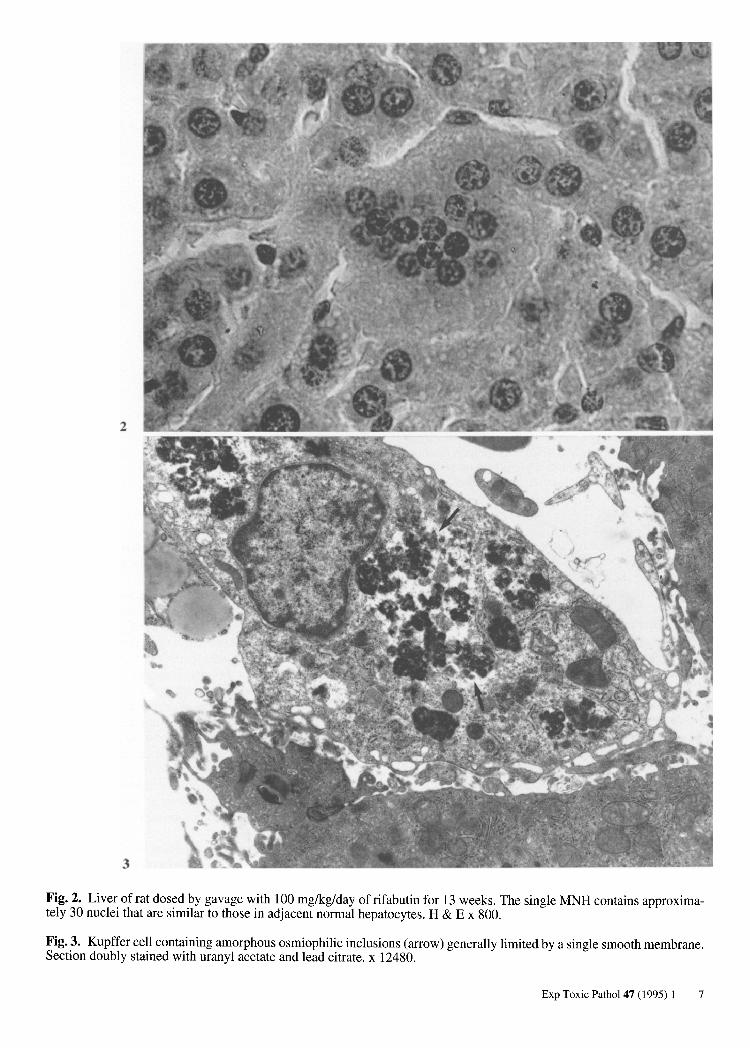
Fig. 2. Liver of rat dosed by gavage with 100 mg/kg/day of rifabutin for 13 weeks. The single MNH contains approximately 30 nuclei that are similar to those in adjacent normal hepatocytes. H & E x 800.
Fig.3. Kupffer cell containing amorphous osmiophilic inclusions (arrow) generally limited by a single smooth membrane. Section doubly stained with uranyl acetate and lead citrate. x 12480.
Exp Toxic Pathol 47 (1995) 1 7

Table 3. Pigment deposition - rifabutin. Rats - 24 months.
Males Females
Dose (mg/kg) C 15 30 60 C 15 30 60
Animal in Group 50 50 50 50 50 50 50 50
Lymph nodes 14 48 46 50 30 48 47 49 Liver 2 18 45 50 9 27 42 50 Lacrimal Glands 25 45 48 2 25 40 45 Lungs 2 5 46 50 1 9 36 50 Heart 1 3 18 48 1 13 9 40 Spleen 36 44 47 50 46 50 49 49 Adrenals 46 45 47 50 46 47 46 50 Salivary Glands 25 47 10 22 40 Stomach 1 22 47 2 32 Jejunum 5 37 45 12 35 Duodenum 18 40 4 26 Ileum 7 29 2 2 Kidneys 20 13 35 47 14 26 33 43 Testes 2 5 27 50 Thymus 11 10 10 3 Pancreas 11 7 10 27 8 13 10 12 Pituitary 27 22 14 26 22 32 36 39
Figures in bold type are considered to include treatment-related change. Figures in small type represent intercurrent pigmentation probably due to hemosiderosis, mela-nin deposits and the typical senile deposition of lipofuscin itself.
structurally that in rat liver these complexes are associated with lysosomes (fig. 2). The accumulation ofthis pigment in tissue macrophages is a continuous process and the pigment is considered to "age" throughout the life of the animals. It does not, however, constitute a stimulus to cell proliferation.
The diffuse presence of brown pigment in many tissues of rodents has also been observed in carcinogenicity studies (table 3). Even in this extreme situation the accumulation of pigmented macrophages caused no secondary changes that were deleterious to the animals.
Effects on red blood cell parameters were seen mainly in rats, with a reduction of RBC, HCT and Hb. Bilirubin values and spleen weight were consistently higher than controls. Moreover, in the 52-week study in mice, Heinz bodies were found in reticulocytes in the high dose group. It is therefore assumed that in rodents rifabutin induces a hemolytic anemia with a regenerative response of the hemopoietic system characterized by an increase in reticulocytes. The anemia was only partially reversible over the recovery periods employed in this series of studies. Depending on the duration of the studies, there was no evidence of anemia developing at doses between 10 and 50 mg/kg.
An in vitro study was performed in comparison with rifampicin and acetylphenylhydrazine as a positive control to detect Heinz bodies in human, monkey, rat and mouse erythrocytes. In this study rifabutin did not cause
8 Exp Toxic PathoI 47 (1995) 1
Heinz body formation up to the highest tested concentration of 20 mcg/mI, that is 20-40 times the human plasma concentration at therapeutic doses.
An effect of rifabutin on male gonads was observed in some rats at the dose of 200 mg/kg given by gavage for 13 weeks, and at 80 mglkg in the 52-week study. This effect was a bilateral, focal or diffuse reduction of spermatogenesis, which showed little tendency to regress. Moreover, signs of testicular atrophy were seen in the carcinogenicity study in the same species in the high dose group of 60 mg/kg. Functionally this change was reflected by a reduction of implantations in the fertility study with males treated at 160 mg/kg. However, in all the reproduction studies no adverse effects were seen in either dams or fetuses at doses of 40 or 50 mg/kg.
The compound was not genotoxic and was devoid of oncogenic potential in mice and rats at doses that can be considered as the maximum appropriate exposure for each species.
When the overall pattern of results is considered it is clear that the animal species tested experienced high cumulative doses of rifabutin. This is particularly well illustrated by the progressive and long lasting persistence of lipofuscin pigment in many tissues in which rifabutin is present as a complex with lipoprotein. The exposure in animals is much higher than that experienced in man at therapeutic doses. For these a dose of 300 mg/day rifabu-
tin is both safe and well tolerated. The rates and types of adverse experiences associated with rifabutin seem to be comparable to those seen with rifampicin.
Considering the increase in disseminated infection caused by MACconcurrent with the AIDS epidemic, effective and relatively safe antimycobacterial drugs like rifabutin are needed to treat these serious infections.
References
1. GROSSET JH: New approaches in antimycobacterial chemotherapy. Drugs of today 1988; 24: 291-301.
2. HEIFETS LB, ISEMAN MD: Determination of in vitro susceptibility of Mycobacteria to ansamycin. Am Rev Respir Dis 1985; 132: 710-711.
3. KOCHERJ, SCAVUZZO D, HORSBURG CR, et al.: Therapeutic response in acquired immune deficiency syndrome patients with disseminated Mycobacterium-avium Mycobacterium-intracellulare. Clin Res 1986; 34 (2): 522A.
4. MITCHISON DA, ELLARD GA, GROSSET J: New antimycobacterial drugs for the treatment of mycobacterial disease in man. Brit Med Bull 1988; 44: 757-774.
5. O'BRIEN RJ, GEITER LJ, LYLE MA: Rifabutin (ansamycin LM 427) for the treatment of pulmonary Mycobacterium avium complex. Am Rev Respir Dis 1990; 141: 821-826.
6. O'BRIEN RJ, LYLE MA, JOHNSON MW, et al.: Preliminary experience with rifabutin (ansamycin LM 427) in the treatment of life-threatening mycobacterial disease. 26th Conference of Tuberculosis and Respiratory Diseases. Singapore, Nov. 4-11, 1986.
7. PERRONE C, GIKAS A, TRUFFOT-PERNOT C, et al.: Activities of cIarithromycin, sulfisoxazole, and rifabutin against Mycobacterium avium complex multiplication within human macrophages. Antimicrob Agents Chemother 1990; 34: 1508-1511.
8. SANFILWPO A: LM 427: Activity on atypical mycobacterial and rifampicin resistant M. tuberculosis. Farmitalia Carlo Erba Internal Report LM 427/210 i, 1983.
9. SCAMPINI G, NAVA A, NEWMAN AJ, et al.: Multinucleated hepatocytes induced by rifabutin in the rat. Toxicology letters. Supplement 1992; 317.
10. SCAMPINI G, NAVA A, NEWMAN AJ, et al. : Multinucleated hepatocytes induced by rifabutin in the rat. Toxicol Patho11993; 21: 369-377.
11. SIEGAL FP, BORESTEIN M, GEHAM K, et al.: Rifabutin may delay the onset of Mycobacterium avium complex infection (MAC) in patients with AIDS. 6th International Conference on AIDS. San Francisco, June 20-21, 1990.
12. TRUFFOT-PERNOT C, GIROIR AM, MAURY L, et al.: A study of the minimal inhibitory concentration of rifabutin (ansamycin LM 427) for Mycobacterium tuberculosis. Mycobacterium xenopy and Mycobacterium aviumintracellulare. Rev Mal Respir 1988; 5: 410-406. (Original text in French).
13. UNGHERI D, SANFILIPPO A: Activity of LM 427 on Legionella SPP.: in vitro study and intracellular killing. 14th International Congress of Chemotherapy, Kyoto, University of Tokyo Press, Tokyo, 1919-1920.
14. VAN DER AWERA T, MATSUMBTO T, HUSSON M: Intraphagocytic penetration of antibiotics. J Antimicrob Chemother 1988; 22: 185-192.
15. WOODLEY CL, KILBURN JO: In vitro susceptibility of Mycobacterium avium complex and Mycobacterium tuberculosis strains to a spiropiperidyl rifamycin. Am Rev Respir Dis 1982; 126: 586-587.
16. YEARSLEY PJ, TREHARNE JL, BALLARD RC: Emergence of resistance to rifampicin and rifabutin in C. trachomatis. 15th Conference of the European Society for Chlamydia Research, Bologna, May 30-June 1, 1988.
Exp Toxic Pathol 47 (1995) 1 9