Embed Size (px)
Citation preview

3968
Abstract. – OBJECTIVE: Imbalance of the ratio of angiotensin converting enzyme (ACE) and ACE2 may lead to pathological conditions in lung. However, its effect on hypoxia-induced pulmonary hypertension (HPH) remains unclear. Therefore, the aim of this study was to investi-gate the effects of ACE2 overexpression on rat primary pulmonary arterial smooth muscle cells (PASMCs) and HPH rat model.
MATERIALS AND METHODS: ACE and ACE2 expression in rat PASMCs under hypoxia condi-tion, as well as in HPH rat model, was detected. The overexpressed ACE2 gene was transfected into PASMCs by Lentiviral. Later, the proliferation and migration of PASMCs were evaluated. Mean-while, the overexpressed ACE2 gene was trans-fected into rats and exposed to hypoxia for four weeks. Finally, the right ventricular systolic pres-sure, the right ventricular hypertrophy, and the percentage of the medial wall thickness were mea-sured to evaluate the development of HPH.
RESULTS: Imbalance of the expression of ACE/ACE2 was indicated in rat PASMCs under hypoxia condition and in the HPH rat model, re-spectively. The overexpression of ACE2 sig-nificantly inhibited PASMCs proliferation and migration. Moreover, the overexpressed ACE2 could significantly attenuate pulmonary hyper-tension, pulmonary vascular remodeling, and right ventricular hypertrophy in HPH rat model.
CONCLUSIONS: ACE2 is related to the forma-tion of pulmonary vascular remodeling and pul-monary hypertension. Furthermore, it may pre-vent hypoxia-induced pulmonary hypertension by inhibiting the proliferation of PASMCs.Key Words:
ACE2, Hypoxia-induced pulmonary hypertension, Pulmonary arterial smooth muscle cells (PASMCs).
Introduction
Progressive elevation of pulmonary arterial pressure is an important feature of pulmonary
hypertension (PH). This may result in the recon-struction of the resistance vessels in the pulmo-nary vascular bed, following right ventricular hy-pertrophy and heart failure1,2. Acute or sustained reduction of alveolar oxygen concentration occurs in many lung diseases. Eventually, this may de-velop into hypoxia-induced pulmonary hyperten-sion (HPH). HPH is an insidious and complex dis-ease with poor prognosis3. A crucial pathological feature of HPH is pulmonary vascular remodel-ing, medial wall thickening of muscular vessels, and pulmonary arterioles muscularization. This implicates the involvement of pulmonary arterial smooth muscle cells (PASMCs)4. The upregula-tion of angiotensin converting enzyme (ACE) in-duced by hypoxia may lead to abnormal prolif-eration and migration of PASMCs. Finally, it can cause pulmonary vascular remodeling associated with HPH5,6.
Angiotensin converting enzyme 2 (ACE2) is an isozyme of ACE. It can counteract the effect of ACE via its degradation of Ang II by promoting Ang-(1-7) synthesis7. The local imbalance of ACE/ACE2 happens in certain tissues. In this process, the expression of ACE is significantly upregulat-ed, while ACE2 level is relatively inadequate. In ACE2 knockout mice, the cardiac development is abnormal and the cardiac contractility is severely damaged, which is gender-related and age-relat-ed8. However, no cardiac development defects are observed in ACE2/ACE double knockout mice9. In addition, myocardial remodeling after hyper-tension or cardiac ischemia and various renal dis-eases are related to the imbalance of ACE/ACE2 ratio10. Previous investigations have also demon-strated that the development of the diseases in-duced by the imbalance of ACE/ACE2 can be re-versed by upregulating the ACE2 expression.
European Review for Medical and Pharmacological Sciences 2020; 24: 3968-3980
H.-H. HU, R.-F. ZHANG, L.-L. DONG, E.-G. CHEN, K.-J. YING
Respiratory and Critical Care Medicine, Sir Run Run Shaw Hospital, Affiliated with Zhejiang University School of Medicine, Hangzhou, China
Corresponding Author: Kejing Ying, MD; e-mail: [email protected]
Overexpression of ACE2 prevents hypoxia-induced pulmonary hypertensionin rats by inhibiting proliferation and immigration of PASMCs

ACE2 prevents hypoxia-induced pulmonary hypertension in rats
3969
It has been reported11 that the long-term hypox-ia increases ACE and decreases ACE2 through hypoxia inducible factor-1α (HIF-1α), resulting in the imbalance of ACE/ACE2. ACE2 inhibits ab-normal proliferation and migration of PASMCs induced by hypoxia, exhibiting an antagonistic effect on ACE12. Therefore, the local upregula-tion of ACE2 may inhibit hypoxia-induced pul-monary vascular remodeling. The overexpression of ACE2 in the heart has been found to protect animals from myocardial injury. Meanwhile, it reduces blood pressure, myocardial fibrosis sur-rounding blood vessel, and left ventricular myo-cardial infarction, as well as preserves the myo-cardial function and contraction13,14. Ferreira et al15 have reported that XNT, an ACE2 receptor agonist, prevents the occurrence of pulmonary hypertension and can eliminate PH-related dam-age. A-779, an Ang(1-7) antagonist, completely weakens those effects. These findings suggest the protective effect of ACE 2-Ang-(1-7)-Mas path-way on pulmonary hypertension16. Furthermore, the overexpression of ACE2 decreases pulmonary artery pressure, corrects right ventricular hyper-trophy, and improves pulmonary vascular remod-eling in rats with pulmonary hypertension17.
In the present work, we investigated the roles of the imbalance of ACE/ACE2 ratio in hypox-ia-induced pulmonary vascular remodeling. The effect of ACE2 overexpression on hypoxic pul-monary vascular remodeling and the process of pulmonary hypertension was evaluated as well. Our findings might provide novel evidence for the function of ACE2 in hypoxia-induced pulmonary hypertension and its clinical treatment.
Materials and Methods
Construction of ACE2-Lentiviral Expression Vector
Lentiviral expression vector pGC-FU was pur-chased from Genechem (Shanghai, China). ACE2 gene was first amplified by PCR using primers (forward containing BamH I site, 5’-CGG GAT CCC GCC ACC ATG TCA AGC TCC TGC TGG CTC-3’; reward containing Age I site, 5’-CGG GTA CCG GTA CGA ATG AAG TTT GAG CAT CAT CACT G-3’) from the plasmid containing ACE2. The entire PCR fragment was digested by BamH I and Age I, which was then cloned into pGC-FU vector. The pGC-FU-containing ACE2 gene was transformed into E.coli strain DH5α. The positive clones were confirmed by PCR us-
ing primers (forward, ACE2-SEQF, 5’-GTT GCC TAT GCC ATG AGA G-3’; reward, EGFP-N-R, 5’-CGT CGC CGT CCA GCT CGA CCA G-3’). Then, they were chosen for DNA sequencing. The plasmids with the correct sequences were trans-fected into 293T cells, and the protein expression of ACE2 was observed under a fluorescence mi-croscope. Western blot was applied to detect the protein expression of ACE2, meanwhile, virus ti-ter was determined by quantitative real-time PCR (qRT-PCR).
Experimental Animals and Animal Facilities
All animal experimental protocols were ap-proved by the Animal Care Committee of Zhe-jiang University School of Medicine18. Male Sprague-Dawley (SD) rats aged 6-8 weeks were obtained from the Animal Center of Zhejiang University School of Medicine. Sixteen rats were randomly assigned into two groups, includ-ing: hypoxia group (N=8) and normoxia group (N=8). The rats in hypoxia group were exposed in a hypobaric hypoxia chamber (10% oxygen) for 4 weeks (8 h/d, 6 d/week). However, the rats in normoxia group (N=8) were housed in room air (21% oxygen) as normal controls. To infect rat air-way with lentivirus, 6 rats received an infusion of 300 µL control lentivirus (Control-Lenti). Mean-while, 5 rats received an infusion of 300 µL len-tivirus carrying ACE2 gene (Lenti-ACE2) (5×10⁸ CFU/ml). After 2 weeks of recovery, the rats in the Control-Lenti group (N=6) or in lenti-ACE2 group (N=5) were treated under hypoxia condi-tion for 4 weeks.
Measurement of Right Ventricular Systolic Pressure (RVSP) and Right Ventricular Hypertrophy
At the end of 4 weeks, the rats were anesthe-tized with sodium thiopental (30 mg/kg). For the measurement of RVSP, a polyethylene catheter was inserted into the right external jugular vein and forwarded to the right ventricle. After track-ing stabilization, the data were recorded. The po-sition of the catheter was confirmed by pressure tracing waveform and validated by postmortem examination, respectively19. The data were ana-lyzed using MedLab Biological Signal Collection System (MedLab-U/501H, Medease Science and Technology, Nanjing, China). After removing the atria, the right ventricle was separated from the left ventricle and the septum. The ratio of the right ventricle to left ventricle plus septum weight
H.-H. Hu, R.-F. Zhang, L.-L. Dong, E.-G. Chen, K.-J. Ying
3970
was calculated as an index of right ventricular hy-pertrophy, namely RVHI = RV/(LV+S). After data collection, the rats were sacrificed, and the heart and lung tissues were collected. Finally, right ven-tricular hypertrophy, lung pathology, and gene expression were determined19.
Culture of Rat Pulmonary Artery Smooth Muscle Cells (PASMCs)
Primary PASMCs were cultured according to previously reported tissue explants adherent methods19,20. Briefly, the male SD rats were anes-thetized and sacrificed. The segments of pulmo-nary arteries were under aseptic conditions. The pulmonary artery was dissected free from con-nective and fat tissues, which were then clipped into blocks in the size of 1 mm3. Subsequently, the tissues were incubated in Dulbecco’s Modified Eagle’s Medium (DMEM) supplemented with 50% fetal bovine serum (FBS) at 37°C for 4 h. After that, the cells were attached to the inner sur-face of the culture dish. The cells were cultured for 14 d, and the medium was changed every 3-5 d. The purity of PASMCs was confirmed by im-munehistochemical staining with anti-α-smooth muscle actin antibody. The cells in 3-10 passages were used for the following experiments.
Hypoxia Exposure of CellsBefore the following experiments, the cells
were subjected to serum starvation for 24 h. Sub-sequently, the cells were induced in a special hy-poxia incubator (1% O2, 94% N2, and 5% CO2) to reach 3% oxygen concentration for 0, 6, 12, 24, and 48 h, respectively. Next, the cells under nor-moxia condition were cultured in an incubator with 21% O2, 74% N2, and 5% CO2.
Quantitative Reverse Transcription-Polymerase Chain Reaction (QRT-PCR)
The total RNA was first extracted from cul-tured PASMCs11. Specific oligonucleotide primers used in this study were shown as follows: ACE2: sense, 5’-CGCTGTCACCAGACAAGAA-3’, an-tisense, 5’-GCCATTATTTCGTCCAATCC-3’, with annealing temperature of 55°C. ACE: sense, 5’-CCAAGACATTTGACGTGAGCA-3’; antisense 5’-ACGTACTTTGGGAAAAAG-GGAAG-3’, with annealing temperature of 56°C; β-actin: sense, 5’-GCCAACACAGT-GCTGTCT-3’; antisense, 5’-AAGATCAAGAT-CATTGCTCCT-3’, with annealing temperature of 56°C. β-actin was used as an internal reference.
Western Blot Analysis The total protein was extracted from PASMCs
and rat lung tissues. The protein expression of ACE was detected by mouse anti-ACE monoclo-nal antibody (ab11734; Abcam, Cambridge, UK), while ACE2 protein was detected by rabbit an-ti-ACE2 monoclonal antibody (ab108252; Abcam, Cambridge, UK). Western blot was performed as previously described11. Western Blot Detection kit and Image J software (NIH) were used to mea-sure blot signal and density.
Immunohistochemical Analysis, Cell Growth, and Migration Assay
To detect ACE and ACE2 proteins, immu-nohistochemical staining was performed ac-cording to the instructions of Strept Avidin Biotin Peroxidase Complex kit. As previously described, the cell growth was evaluated by WST-8 cleavage assay. Meanwhile, cell migra-tion was conducted according to the previously reported method21.
Statistical AnalysisThe Statistical Product and Service Solutions
(SPSS) for Windows (Version 18.0, SPSS Inc., Chicago, IL, USA) was used for all statistical analysis. The experimental data were expressed as means ± standard deviation (SD). The Student’s t-test, Single factor analysis of variance or χ2-test was used when appropriate. Each experiment was repeated for three times. p<0.05 was considered statistically significant.
Results
Effects of Hypoxia Exposure on the MRNA or Protein Expression of ACE or ACE2 in rat PASMCs
The aim of this work was first to investigate the effects of hypoxia treatment on the expressions of ACE or ACE2 at transcriptional and translational levels. Rat primary PASMCs were cultured in vi-tro. Under an optical microscope, the spindle cells were observed climbing out from tissue blocks 2 d after the tissues adhered to the inner surface of the culture dish. At the end of 1 week, the dish was covered with cells with long oval or willow-like morphology. The cellular nuclei were observed by DAPI staining. Fluorescence immunoassay using rabbit anti-α-SM actin antibody showed PASMCs with more than 95% purity (Figure 1A).

ACE2 prevents hypoxia-induced pulmonary hypertension in rats
3971
Figure 1. Culture of rat primary PASMCs and the effects of hypoxia on ACE and ACE2 expression. A, Culture of PASMCs observed under an optical microscope. (a) Two days after tissues adhered to the inner surface of the Petri dish, the spindle cells climbed out from the tissue blocks (magnification × 100). (b) At the end of 1 week, the dish was covered by cells with long oval or willow-like morphology (magnification × 400). (c) Cellular nuclei were observed by DAPI staining (magnification × 100). (d) Fluorescence immunoassay using rabbit anti-α-SM actin antibody showed PASMCs with more than 95% purity (magnification × 400). B, and C, Effects of hypoxia on mRNA or protein expressions of ACE and ACE2 in PASMCs. Rat primary PASMCs were exposed to hypoxia for 0, 6, 12, 24, and 48 h, respectively. The total RNA was extracted for RT-PCR to determine mRNA (B), or total proteins were extracted for Western blot to detect protein levels (C) of ACE and ACE2. The mRNA or protein signals of ACE or ACE2 were normalized to β-actin. Levels of mRNA or protein at 0 h were set as 100%. Finally, changes in percentage of mRNA (A) or protein (B) of ACE, ACE2 or ACE/ACE2 were calculated. *p<0.05, **p<0.01.
H.-H. Hu, R.-F. Zhang, L.-L. Dong, E.-G. Chen, K.-J. Ying
3972
After exposure to hypoxia for 0, 6, 12, 24, and 48 h, respectively, the total RNA was extracted from rat primary PASMCs. RT-PCR was then performed to examine the mRNA expressions of ACE and ACE2, with β-actin as an internal control. As shown in Figure 1B, the expression of ACE in PASMCs gradually increased after ex-posure to hypoxia, which reached the highest lev-el at 48 h. Meanwhile, the mRNA level of ACE2 reached peak at 12 h after exposure to hypoxia, and gradually decreased. No significant change was observed in the mRNA ratio of ACE/ACE2 during 0-24 h after exposure to hypoxia, suggest-ing no imbalance. However, ACE/ACE2 ratio was significantly elevated 48 h after hypoxia treat-ment (p<0.05). This suggested that ACE expres-sion was beyond the compensation of ACE2 and the imbalance of ACE/ACE2 occurred.
Western blots was applied to detect the changes of ACE and ACE2 protein levels, which were sim-ilar to the changes at the transcriptional level after expose to hypoxia (Figure 1C). The protein levels of ACE increased after exposure to hypoxia, and reached the highest level 48 h later. Meanwhile, the protein expression of ACE2 was also elevated, and reached the peak at 24 h. However, it then decreased at 48 h. No significant change was ob-served in the ratio of ACE/ACE2 during the first 24 h of hypoxia exposure. However, it significant-ly increased 48 h after exposure to hypoxia, indi-cating the loss of ACE2 compensation.
Establishment of the Hypoxic Pulmonary Hypertension Model in Rats
Male SD rats in hypoxia group (N=8) were ex-posed to 10% oxygen, while those in normoxia group (N=8) were housed in 21% oxygen for 4 weeks. By compared with normoxia group, the body weight of rats in hypoxia group was signif-icant lower (236 ± 3.6 vs. 293.8 ± 10.5 g, p<0.01). However, RV/LV+S (0.390 ± 0.004 vs. 0.288 ± 0.007, p<0.01) and RVSP (37.66 ± 0.56 vs. 31.39 ± 0.38 mmHg, p<0.01) increased significantly in hypoxia group compared with normoxia group (Figure 2A).
Furthermore, we observed the possible chang-es of the pulmonary vascular morphology. Un-der the light microscopy, H&E staining of lung tissues of rats in hypoxia showed the features of emphysema, including distal lung tissue expan-sion, alveolar wall thinning, as well as alveolar septal narrowing or fracture (Figure 2B, x40). Infiltration of inflammatory cells around small pulmonary vessels was smaller than 100 μm. The
vascular smooth muscle layer was thickened and endothelial cell structure was unclear (Figure 2B, x400). αSM-actin staining showed the structure of the pulmonary vascular smooth muscle layer. In the hypoxia group, the pulmonary muscular arterioles of the medial smooth muscle layer were significantly thickened. Meanwhile, non-muscle small arteries appeared muscularization (Figure 2B). By Image-Pro Plus imaging analysis, we fur-ther calculated the percentage of the wall thick-ness of pulmonary arteries (WT) to endo diameter (ED) (WT% = 2*WT/ED*100) or wall area (WA) [WA% = (TA-LA)/TA*100]. Compared with nor-moxia group, WT% (47.43 ± 4.07% vs. 19.28 ± 2.17%, p<0.05) and WA (59.54 ± 5.83% vs. 19.3 ± 2.01%, p<0.01) (Figure 2C) were both significant higher in hypoxia group. Therefore, the model of hypoxic pulmonary hypertension was successful-ly established in rats.
Changes in Expression Levels of ACE and ACE2 in Lung Tissues ofthe HPH Rat Model
After exposure to normoxia or hypoxia condi-tion for 4 weeks, the total RNA or protein was extracted from lung tissues of rats in the two groups. RT-PCR results showed that, compared with normoxia group, the mRNA expression of ACE significantly increased in hypoxic pulmo-nary hypertension rats. However, there was no evident difference in the mRNA level of ACE2 between the two groups. Accordingly, the ratio of ACE/ACE2 was remarkably higher in the hypoxia group than the control group (p<0.05), suggesting imbalance between ACE and ACE2 (Figure 2D).
Western blots showed that the protein changes of ACE and ACE2 were not similar to the mRNA changes of the two genes after expose to hypoxia. Compared with the normoxia group, the protein level of ACE in lung tissues of rats exposed to hypoxia was not significantly changed (p<0.05). However, ACE/ACE2 ratio was statistically high-er (p<0.05). The above results suggested that there was imbalance between ACE and ACE2 in the lung of hypoxic pulmonary hypertension rats (Figure 2E).
Overexpression of ACE2 by Lentivirus Carrying ACE2 Gene in 293T Cells and Rat PASMCs
The plasmid pGC-FU with the correct se-quence of ACE2 was transfected into 293T cells. Under a fluorescence microscope, the transfect-ed 293T cells appeared green fluorescence, as

ACE2 prevents hypoxia-induced pulmonary hypertension in rats
3973
Figure 2. Establishment of the model of hypoxic pulmonary hypertension in rats and the expressions of ACE and ACE2 in the lung of rats. Male SD rats aged 6-8 weeks were divided into two groups. The rats in hypoxia group (N=8) were exposed to 10% oxygen, while those in the normoxia group (N=8) were housed in 21% oxygen for 4 weeks. A, The body weight, right ventricular hypertrophy index (RV/LV+S), right ventricular systolic pressure (RVSP) were measured. B, H&E and αSM-actin staining for rat pulmonary vascular morphology in the normoxia and hypoxia groups. C, Image-Pro Plus imaging analysis was used to calculate the percentage of wall thickness of pulmonary arteries (WT) and wall area (WA) in normoxia and hypoxia groups. D, and E, Expressions of ACE and ACE2 at the transcriptional and translational levels. The total mRNA or protein was extracted from the lung tissues of rats in the two groups. RT-PCR was performed to determine mRNA (D), or Western blots to detect the protein levels (E) of ACE and ACE2. The mRNA or protein signals of ACE or ACE2 were normalized β-actin. The levels of mRNA or protein at 0 h were set as 100%. Finally, the percentage changes of mRNA (D) or protein (E) of ACE, ACE2, or ACE/ACE2 were calculated. *p<0.05, ** p<0.01.
H.-H. Hu, R.-F. Zhang, L.-L. Dong, E.-G. Chen, K.-J. Ying
3974
the plasmid contained GFP. Western blots with proteins prepared from wild type and transfect-ed 293T cells showed that ACE2 was expressed (Figure 3A). Male SD rats were infected with control lentivirus (Control-lenti) (n=6) or lenti-virus carrying ACE2 gene (Lenti-ACE2) (n=5), respectively. After 2 weeks of recovery, PASMCs were isolated from rats in both groups. PASMCs from Crtl-Lenti rats showed no fluorescence. However, the green fluorescence was observed from Lenti-ACE2 rats. This indicated a success-ful transfection of the ACE2 gene (Figure 3B). The proteins were then extracted from PASMCs, and Western blot was applied to determine the protein level of ACE2. As shown in Figure 3B, significantly higher ACE2 protein level was ob-served in the cells from Lenti-ACE2 rats when compared with that from the Control-Lenti rats (p<0.05) (Figure 3B).
Effects of ACE2 Overexpression on the Proliferation of PASMCs
After exposure to hypoxia for 24 h, the possi-ble effects of ACE2 overexpression on PASMCs were detected using Cell Counting Kit-8 (CCK-8; Sigma-Aldrich, St. Louis, MO, USA). Compared with the normoxia group, OD values of hypoxia group were significantly higher (Table I), sug-gesting that hypoxia promoted the proliferation of primary PASMCs. Compared with the control group, in normoxia environment, OD values of Control-Lentiviral group significantly decreased from 24 hours to 72 h (p<0.01). This suggested that cell proliferation was remarkably inhibited by the toxicity of lentivirus. OD values of Len-ti-ACE2 group were remarkably lower than those of the Control-Lenti group (p<0.01), indicating the inhibiting effect of ACE2 on the proliferation of PASMCs (Figure 3C). Compared with the con-trol group, in the hypoxia environment, OD val-ues of Control-Lenti group decreased (p<0.05). Meanwhile, OD values of Lenti-ACE2 group were significantly lower. However, the extent of OD decreased in Lenti-ACE2 group after expo-sure to hypoxia was smaller than that in normoxia environment. These results indicated that ACE2 exerted protective effect on the proliferation of PASMCs (Figure 3C).
Effect of ACE2 Overexpression on the Migration of the PASMCs
Furthermore, we evaluated the possible effect of ACE2 on the migration of PASMCs using the transwell assay. Both in normoxia and hypoxia
conditions, the number of transmembrane cells in Lenti-ACE2 group was significantly less than that of the Control-Lenti group (p<0.01, Figure 3D). This demonstrated that ACE2 overexpres-sion inhibited the migration of PASMCs under normoxia and hypoxia environments. However, the inhibition effect of ACE2 was more evident under hypoxia (Figure 3D).
Overexpression of ACE2 in Lung Tissues of Rats by Transfection of Lentivirus Carrying ACE2
Two weeks after transfection of the control lentivirus (Control-Lenti) or Lentivirus carrying ACE2 gene (Lenti-ACE2), the sections of lung tissues of rats in both groups were stained with anti-ACE2 antibody for immunohistochemical analysis. The green fluorescence demonstrated the localization of ACE2 (Figure 4A). Compared with the Control-Lenti group, the anti-ACE2 antibody stained pulmonary vascular smooth muscle cells, bronchial epithelial cells, and al-veolar epithelial cells in Lenti-ACE2 group. This suggested that ACE2 gene in lentivirus was effi-ciently expressed in pulmonary vascular smooth muscles and other parts of the lung (Figure 4A). Western blots showed significantly higher ACE2 protein expression level in Leni-ACE2 group when compared with Control-Lenti group (p<0.05) (Figure 4B).
Pulmonary Artery Pressure and Right Ventricular Hypertrophy Index in Different Groups
Two weeks after transfection of Control-Len-ti (n=6) or Lenti-ACE2 (n=5), the rats in both groups were kept in hypoxia for 4 weeks. A group of rats in normoxia (n=8) for 4 weeks were used as normal controls. As shown in Figures 4C and 4D, RVSP of Hypoxia- Lenti-ACE2 (36.45 ± 0.64 mmHg) was significantly higher than that of the Normoxia-Control (34.08± 1.67 mmHg, p<0.05). This suggested that after hypoxia ex-posure, pulmonary artery pressure significantly increased, indicating successful establishment of pulmonary hypertension model in rats. RVSP of Hypoxia-Lenti-ACE2 group was remarkably lower than that of Hypoxia-Control-Lenti group (36.45 ± 0.64 mmHg vs. 41.21 ± 1.26 mmHg, p<0.01). This indicated that ACE2 overexpres-sion reduced the pulmonary artery pressure in PH animal model and ACE2 might improve pul-monary hypertension.

ACE2 prevents hypoxia-induced pulmonary hypertension in rats
3975
Figure 3. Effects of ACE2 overexpression on 293T cells and rat primary PASMCs. A, Plasmid pGC-FU carrying ACE2 and GFP genes was constructed and transfected into 293T cells (magnification × 100). The transfected cells showed green fluorescence of GFP under fluorescence microscope. The protein expression of ACE2 was detected by Western blots. B, PAMCs were isolated from the rats infected with Control-Lenti or Lenti-ACE2 for 2 weeks (magnification × 400). The cells from Lenti-ACE2 rats showed fluorescence of GFP, with significantly higher expression level of ACE2 (p<0.01). C, Effects of ACE2 overexpression on the proliferation of PASMCs. After exposure of PASMCs to hypoxia for 24 h, cell proliferation was measured by the Cell Counting Kit-8 (CCK-8) (Table I). D, Effect of ACE2 overexpression on the migration of PASMCs. PASMCs migration was measured using the transwell assay. Both in the normoxia and hypoxia conditions, the number of transmembrane cells of in Lenti-ACE2 group was significantly less than that of the Control-Lenti group (p<0.01). *p<0.05, **p<0.01.

H.-H. Hu, R.-F. Zhang, L.-L. Dong, E.-G. Chen, K.-J. Ying
3976
RV/LV+S of Hypoxia-Control-Lentivi group was remarkably higher than that of the Nor-moxia-Control group (0.32±0.03 vs. 0.23±0.01, p<0.05, Figure 4E), which was the typical right ventricular change in rats with pulmonary hy-pertension. However, RV/LV+S of Hypoxia-Len-ti-ACE2 group was significantly lower than that of the Hypoxia-Control-Lenti group (0.24±0.01 vs. 0.32±0.03, p<0.05) (Figure 4E). This suggest-ed that right ventricular hypertrophy was im-proved in pulmonary hypertension rats. All these findings indicated the positive effect of ACE2 on right ventricular changes induced by pulmonary hypertension.
Pulmonary Artery Remodeling Caused by ACE2 Overexpression
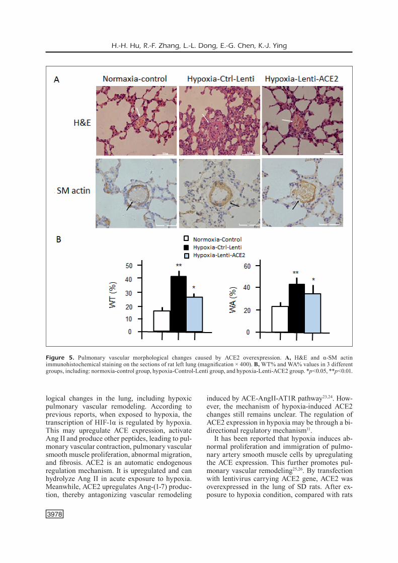
H&E and α-SM actin immunohistochemical staining were performed to observe the possible morphological changes of the pulmonary ves-sels. H&E staining showed that in Hypoxia-Con-trol-Lenti and Hypoxia-Lenti-ACE2 groups, there were manifestations of emphysema, including pulmonary tissue expansion, thinner alveolar wall, as well as narrower alveolar septal or rup-ture. Furthermore, in Hypoxia-Control-Lentivi-rus group, we observed the infiltration of the in-flammatory cells, thickening of vascular smooth muscle layer, narrowed lumen, unclear elastic fiber lay structure and destroyed the endothelial cells of the intimal layer around small vessels of lung tissues. Meanwhile, the diameter was less than 100 mm (Figure 5A). However, in Hypox-ia-Lenti-ACE2 group, the thickening of pulmo-nary artery smooth muscle layer was not evident.
Subsequently, we observed the possible chang-es in the structure of the small pulmonary vascu-lar smooth muscle cells. As shown in Figure 5B,
the smooth muscle layer of pulmonary artery was significantly thicker in the Hypoxia-Control-Len-ti group than that of the Normoxia-Control group (WT%: 41.58±2.81% vs. 16.21±1.12%, p<0.01). This was consistent with vascular remodeling in pulmonary hypertension. Compared with the Hypoxia-Control-Lenti group, the thickening of the pulmonary artery smooth muscle layer sig-nificantly decreased in the Hypoxia-Lenti-ACE2 group (WA%: 32.22±3.24% vs. 44.08±4.96%, p<0.01). However, it was remarkably higher than that of the Normoxia-Control group (24.72±2.94, p<0.05). The results suggested that the pulmonary artery remodeling in pulmonary hypertension rats could be improved by transfection of ACE2.
Discussion
In our investigation, we found that hypoxia in-duced the imbalance of ACE/ACE2 ratio in rat pul-monary arterial smooth muscle cells (PASMCs) and the hypoxia-induced pulmonary hypertension (HPH) rat model. More importantly, our findings provided novel evidence that the overexpression of ACE2 in rats might prevent hypoxia induced pulmonary hypertension by inhibiting the prolif-eration and immigration of PASMCs.
ACE expression gradually increased at both transcriptional and translational levels after expo-sure to hypoxia environment, which was consis-tent with the previous findings22. However, ACE2 expression was not correspondingly upregulated, resulting in the imbalance of ACE/ACE2 ratio. Hypoxia led to increased ACE expression and de-creased ACE2 expression in the lung tissues of male SD rats. This also resulted in the imbalance of ACE/ACE2 ratio, further resulting in the patho-
Table I. CCK2 results (OD) and comparison of Lenti-ACE2 group to control groups.
Tim (1) Control (2) Control-Lenti (3) ACE2-Lenti p (2) vs. (3)/(1) vs. (2)
Normoxia 12 h 0.392 ± 0.035 0.317 ± 0.046 0.291 ± 0.018 0.446/0.093 24 h 0.724 ± 0.013 0.607 ± 0.019 0.465 ± 0.006 0.004/0.002 48 h 0.796 ± 0.037 0.657 ± 0.024 0.488 ± 0.002 0.006/0.009 72 h 0.884 ± 0.041 0.700 ± 0.020 0.534 ± 0.015 0.000/0.007Hypoxia 12 h 0.412 ± 0.059 0.405 ± 0.019 0.335 ± 0.023 0.016/0.848 24 h 0.871 ± 0.011 0.702 ± 0.048 0.607 ± 0.009 0.072/0.022 48 h 1.066 ± 0.056 0.852 ± 0.112 0.709 ± 0.024 0.003/0.019 72 h 1.058 ± 0.068 0.874 ± 0.023 0.743 ± 0.038 0.012/0.032p Normoxia V.S. Hypoxia 12 h 0.635 0.038 0.059 24 h 0.000 0.034 0.000 48 h 0.002 0.000 0.000 72 h 0.019 0.001 0.001

ACE2 prevents hypoxia-induced pulmonary hypertension in rats
3977
Figure 4. Effects of ACE2 overexpression on the lung of rats by infection with lentivirus carrying ACE2 gene. The rats were infected with Control-Lenti (N=6) or Lenti-ACE2 (N=5) for 2 weeks, and were kept under normoxia or hypoxia for 4 weeks. A group of normal rats under normoxia was as normal controls (n=8). A, Immunofluorescence staining with anti-ACE2 antibody and H&E staining showed that ACE2 located on pulmonary vascular smooth muscle cells, bronchial epithelial cells, and alveolar epithelial cells in Lenti-ACE2 group (magnification × 400). B, Western blots showed significantly higher ACE2 protein level in Leni-ACE2 group when compared with the Control-Lenti group (p<0.05). C, and D, RVSP of Hypoxia-Lenti-ACE2 was remarkably higher than that of the Normoxia-Control (p<0.05), and was much lower than that of the Hypoxia-Control-Lenti group (p<0.01). E, RV/LV+S of Hypoxia-Control-Lenti group was remarkably higher than that of the Normoxia-Control group (p<0.05), and was lower than the Hypoxia-Control-Lenti group (p<0.05). *p<0.05, **p<0.01.
H.-H. Hu, R.-F. Zhang, L.-L. Dong, E.-G. Chen, K.-J. Ying
3978
logical changes in the lung, including hypoxic pulmonary vascular remodeling. According to previous reports, when exposed to hypoxia, the transcription of HIF-1α is regulated by hypoxia. This may upregulate ACE expression, activate Ang II and produce other peptides, leading to pul-monary vascular contraction, pulmonary vascular smooth muscle proliferation, abnormal migration, and fibrosis. ACE2 is an automatic endogenous regulation mechanism. It is upregulated and can hydrolyze Ang II in acute exposure to hypoxia. Meanwhile, ACE2 upregulates Ang-(1-7) produc-tion, thereby antagonizing vascular remodeling
induced by ACE-AngII-AT1R pathway23,24. How-ever, the mechanism of hypoxia-induced ACE2 changes still remains unclear. The regulation of ACE2 expression in hypoxia may be through a bi-directional regulatory mechanism11.
It has been reported that hypoxia induces ab-normal proliferation and immigration of pulmo-nary artery smooth muscle cells by upregulating the ACE expression. This further promotes pul-monary vascular remodeling25,26. By transfection with lentivirus carrying ACE2 gene, ACE2 was overexpressed in the lung of SD rats. After ex-posure to hypoxia condition, compared with rats
Figure 5. Pulmonary vascular morphological changes caused by ACE2 overexpression. A, H&E and α-SM actin immunohistochemical staining on the sections of rat left lung (magnification × 400). B, WT% and WA% values in 3 different groups, including: normoxia-control group, hypoxia-Control-Lenti group, and hypoxia-Lenti-ACE2 group. *p<0.05, **p<0.01.

ACE2 prevents hypoxia-induced pulmonary hypertension in rats
3979
injected control lentivirus, ACE2-overexpressed rats showed significantly lower RVSP and RV/LV+S. These results indicated the improvement of pulmonary hypertension. In addition, the overexpression of ACE2 in the lung significant-ly decreased the thickening of pulmonary ar-tery smooth muscle layer, indicating prevention from pulmonary artery remodeling in pulmo-nary hypertension rats. We then cultured prima-ry PASMCs isolated from ACE2-overexpressed rats. Next, the effects of ACE2 on inhibiting the proliferation and migration of PASMCs were con-firmed. The results suggested that ACE2 could be used as a powerful target for gene therapy of hy-poxic pulmonary arterial hypertension.
In the design of transfection approach, we se-lected lentivirus as transfection vector mediated to rat airway. The intravenous dosing lentivirus was unable to efficiently integrate into pulmonary vascular smooth muscle cells and endothelial cells due to short residence time. Airway admin-istration of lentivirus could locate to airway and lung tissues, reducing the purpose of transfection efficiency. The advantage of the lentivirus vector was transfection efficiency and simplicity, how-ever, there were also some shortcomings. A large number of inflammatory cells and macrophages infiltration were observed in lung tissues by HE staining. This showed that lentivirus mediated to the airway might cause local inflammation or immune response. Therefore, how to improve the security and ability of directional gene expression is the key point for future researches.
Conclusions
We provided the evidence that ACE2 might prevent hypoxic pulmonary hypertension by in-hibiting the proliferation and immigration of PASMCs. Therefore, ACE2 could be used as a po-tential target for gene therapy of hypoxia induced pulmonary hypertension.
Conflict of InterestThe Authors declare that they have no conflict of interests.
Funding AcknowledgementsThe targeted treatment of hypoxic pulmonary hypertension by ACE2 transgenic endothelial progenitor cells, a co-fund-ed Project of Zhejiang Province (WKJ2010-2-01).
References
1) Wang Ln, Yu WC, Du CH, Tong L, CHeng ZZ. Hy-poxia is involved in hypoxic pulmonary hyperten-sion through inhibiting the activation of FGF2 by miR-203. Eur Rev Med Pharmacol Sci 2018; 22: 8866-8876.
2) STenmark kr, Fagan ka, FriD mg. Hypoxia-induced pulmonary vascular remodeling: cellular and mo-lecular mechanisms. Circ Res 2006; 99: 675-691.
3) meYriCk B. The pathology of pulmonary artery hy-pertension. Clin Chest Med 2001; 22: 393-404.
4) DonogHue m, HSieH F, BaronaS e, goDBouT k, goS-SeLin m, STagLiano n, Donovan m, WooLF B, roBi-Son k, JeYaSeeLan r, BreiTBarT re, aCTon S. A novel angiotensin-converting enzyme-related carboxy-peptidase (ACE2) converts angiotensin I to angio-tensin 1-9. Circ Res 2000; 87: E1-E9.
5) Yu TZ, ma CT. Effects of angiotensin-converting enzyme and angiotensin II on hypoxia-induced proliferation of cultured intra-pulmonary artery smooth muscle cells. Acta Pharmacol Sin 2000; 21: 381-384.
6) PaSCauD ma, griSCeLLi F, raouL W, marCoS e, oPo-Lon P, raFFeSTin B, PerriCauDeT m, aDnoT S, eDDaHiBi S. Lung overexpression of angiostatin aggravates pulmonary hypertension in chronically hypoxic mice. Am J Respir Cell Mol Biol 2003; 29: 449-457.
7) TiPniS Sr, HooPer nm, HYDe r, karran e, CHriSTie g, Turner aJ. A human homolog of angiotensin-con-verting enzyme. Cloning and functional expres-sion as a captopril-insensitive carboxypeptidase. J Biol Chem 2000; 275: 33238-33243.
8) gurLeY SB, aLLreD a, Le TH, griFFiTHS r, mao L, PHiL-iP n, HaYSTeaD Ta, DonogHue m, BreiTBarT re, aCTon SL, roCkman Ha, CoFFman Tm. Altered blood pres-sure responses and normal cardiac phenotype in ACE2-null mice. J Clin Invest 2006; 116: 2218-2225.
9) BroSniHan kB, neveS La, CHaPPeLL mC. Does the angiotensin-converting enzyme (ACE)/ACE2 bal-ance contribute to the fate of angiotensin pep-tides in programmed hypertension? Hypertension 2005; 46: 1097-1099.
10) re rn. Mechanisms of disease: local renin-angio-tensin-aldosterone systems and the pathogene-sis and treatment of cardiovascular disease. Nat Clin Pract Cardiovasc Med 2004; 1: 42-47.
11) ZHang r, Wu Y, ZHao m, Liu C, ZHou L, SHen S, Liao S, Yang k, Li Q, Wan H. Role of HIF-1alpha in the regulation ACE and ACE2 expression in hypox-ic human pulmonary artery smooth muscle cells. Am J Physiol Lung Cell Mol Physiol 2009; 297: L631-L640.
12) ZHou L, ZHang r, Yao W, Wang J, Qian a, Qiao m, ZHang Y, Yuan Y. Decreased expression of angiotensin-converting enzyme 2 in pancreat-ic ductal adenocarcinoma is associated with tu-mor progression. Tohoku J Exp Med 2009; 217: 123-131.

H.-H. Hu, R.-F. Zhang, L.-L. Dong, E.-G. Chen, K.-J. Ying
3980
13) DíeZ-Freire C, váZQueZ J, Correa De aDJounian mF, Ferrari mF, Yuan L, SiLver X, TorreS r, raiZaDa mk. ACE2 gene transfer attenuates hyperten-sion-linked pathophysiological changes in the SHR. Physiol Genomics 2006; 27: 12-19.
14) CraCkoWer ma, Sarao r, ouDiT gY, YagiL C, koZieraDZ-ki i, SCanga Se, oLiveira-DoS-SanToS aJ, Da CJ, ZHang L, Pei Y, SCHoLeY J, Ferrario Cm, manoukian aS, CHaP-PeLL mC, BaCkX PH, YagiL Y, Penninger JM. Angioten-sin-converting enzyme 2 is an essential regulator of heart function. Nature 2002; 417: 822-828.
15) Ferreira aJ, SHenoY v, YamaZaTo Y, SriramuLa S, Fran-CiS J, Yuan L, CaSTeLLano rk, oSTrov Da, oH SP, kaToviCH mJ, raiZaDa mk. Evidence for angioten-sin-converting enzyme 2 as a therapeutic target for the prevention of pulmonary hypertension. Am J Respir Crit Care Med 2009; 179: 1048-1054.
16) YamaZaTo Y, Ferreira aJ, Hong kH, SriramuLa S, Fran-CiS J, YamaZaTo m, Yuan L, BraDForD Cn, SHenoY v, oH SP, kaToviCH mJ, raiZaDa mk. Prevention of pul-monary hypertension by Angiotensin-converting enzyme 2 gene transfer. Hypertension 2009; 54: 365-371.
17) Haga S, TSuCHiYa H, Hirai T, Hamano T, mimori a, iSHi-Zaka Y. A novel ACE2 activator reduces monocro-taline-induced pulmonary hypertension by sup-pressing the JAK/STAT and TGF-beta cascades with restored caveolin-1 expression. Exp Lung Res 2015; 41: 21-31.
18) Xu X, Hu H, Wang X, Ye W, Su H, Hu Y, Dong L, ZHang r, Ying k. Involvement of CapG in proliferation and apoptosis of pulmonary arterial smooth muscle cells and in hypoxia-induced pulmonary hyperten-sion rat model. Exp Lung Res 2016; 42: 142-153.
19) Xia S, Tai X, Wang Y, an X, Qian g, Dong J, Wang X, SHa B, Wang D, murTHi P, kaLioniS B, Wang X, Bai
C. Involvement of Gax gene in hypoxia-induced pulmonary hypertension, proliferation, and apop-tosis of arterial smooth muscle cells. Am J Respir Cell Mol Biol 2011; 44: 66-73.
20) arJona aa, HSu Ca, Wrenn DS, HiLL nS. Effects of natriuretic peptides on vascular smooth-muscle cells derived from different vascular beds. Gen Pharmacol 1997; 28: 387-392.
21) ZHang r, ZHou L, Li Q, Liu J, Yao W, Wan H. Up-regulation of two actin-associated proteins prompts pulmonary artery smooth muscle cell mi-gration under hypoxia. Am J Respir Cell Mol Biol 2009; 41: 467-475.
22) morreLL nW, morriS kg, STenmark kr. Role of an-giotensin-converting enzyme and angiotensin II in development of hypoxic pulmonary hyperten-sion. Am J Physiol 1995; 269: H1186-H1194.
23) TouYZ rm. Reactive oxygen species and angio-tensin II signaling in vascular cells -- implications in cardiovascular disease. Braz J Med Biol Res 2004; 37: 1263-1273.
24) SamPaio Wo, SouZa DSr, Faria-SiLva r, Da mmL, SCHiFFrin eL, TouYZ rm. Angiotensin-(1-7) through receptor Mas mediates endothelial nitric oxide synthase activation via Akt-dependent pathways. Hypertension 2007; 49: 185-192.
25) Dai H, Jiang L, Xiao Z, guang X. ACE2-angioten-sin-(1-7)-Mas axis might be a promising thera-peutic target for pulmonary arterial hypertension. Nat Rev Cardiol 2015; 12: 374.
26) Yuan Ym, Luo L, guo Z, Yang m, Ye rS, Luo C. Activation of renin-angiotensin-aldosterone sys-tem (RAAS) in the lung of smoking-induced pul-monary arterial hypertension (PAH) rats. J Re-nin Angiotensin Aldosterone Syst 2015; 16: 249-253.

![Overexpression of HIF-2α-Dependent ... - Cell Physiol Biochem · [15-17]. Hypoxia-inducible factor (HIF) is involved in major mechanism mediating oxygen-dependent transcriptional](https://img.pdfslide.us/doc/110x75/60418717ba206b61c053200c/overexpression-of-hif-2-dependent-cell-physiol-biochem-15-17-hypoxia-inducible.jpg)