Embed Size (px)
Citation preview
Overcoming limitations in bioengineering Rubisco
in higher plant chloroplasts
Yi-Leen Lim
A thesis submitted for the degree of
Doctor of Philosophy of The Australian National University
Plant Sciences
Research School of Biological Sciences
The Australian National University
June 2015
iii
ACKNOWLEDGEMENTS
This piece of literature and the research opportunities that may further stem from this
thesis could not have been accomplished without the insight of Associate Professor
Spencer Whitney, whose influence and expertise in the field of Plant Sciences have given
me countless academic exposure and network opportunities over the past few years. To
Spencer – thank you for being an excellent leader, role model, friend and confidante.
I am fortunate to have had wonderful colleagues at the Whitney laboratory, and would
like to especially thank Dr. Robert Sharwood for his vast knowledge and patience in
sharing his academic experiences and technical skills. To Douglas Orr, Laura Gunn,
Robbie Wilson and Sara Milward – I believe I finally made it. Thank you also to Elena
Martin and Maxim Kapralov for your comments on the drafts as well as guidance on
writing this thesis. To Rosemary Birch, Carly & Brendon Conlan and other members of
the laboratory – thank you so much for your words of encouragement and support!
I would also like to address my gratitude to Eliza Middleton, Kevin Tee, Nadia Radzman
and Piyankarie Jayatilaka for being my sparks of love and random joy during the hardest
of times. To Janice Lee, Joyce Khoo, Klein Fernandez, Tam Tran and Timothy Wan –
thank you for all the delicious home-cooked meals, and most importantly – for making
Canberra feel like home.
Lastly, I would like to thank my parents for making it possible for me to embark on this
five-year journey. There is undoubtedly no gift better than the challenging experience of
living independently in a foreign country at a time I have now come to realise as my
transition into adulthood. Mum & Dad – this thesis is yours as much as it is mine.
iv
ABSTRACT
Better structure-function studies of higher plant Rubisco are imperative in improving
catalytic potential of the enzyme Rubisco. In this thesis, a novel system to study Rubisco
using an RNAi tobacco genotype is designed to provide a homogenous environment of
large and small Rubisco subunits for a more genuine assessment of recombinant Rubisco
catalysis, regulation and assembly as well as its photosynthetic capacity in tobacco. The
application of technology and strategies discussed in this thesis will demonstrate a great
leap forward in Rubisco bioengineering and recombinant protein expression in plant
chloroplasts.
The RbcS RNAi of the cmtrLRNAi-S genotype is stable up to three generations,
having selectable resistance against the Basta herbicide while boasting no accumulation
of transcript mRNA from the tobacco RbcS multigene family. Access and ability to
manipulate the Rubisco S-subunit in higher plants have been the final crux in
bioengineering Rubisco and is now possible using the cmtrLRNAi-S master line.
Additionally, application of the intron-containing hairpin loop construct in RNAi
silencing and its effectiveness as shown in this thesis strongly validates the use of this
technology to study other genomic and proteomic components of photosynthesis in higher
plants.
The unperturbed growth of cmtrLRNAi-S to maturity in soil (albeit requiring elevated
CO2 environment) and therefore the development of fertile pollen enhances the prowess
of the cmtrLRNAi-S line to include stable transfer of the RNAi-RbcS system into tobacco
genotypes using cross-pollination. New genotypes generated using pollen from cmtrLRNAi-
S to fertilise genotypes producing S-subunits in the chloroplast mirror similar RbcS
silencing found in cmtrLRNAi-S thus resulting in populations of homogenous, chloroplast-
made S-subunits in the absence of endogenous (cytosolic) S-subunits. In summary, a
more accurate system for determining the innumerable factors and limitations in
recombinant Rubisco expression and biogenesis in higher plants can be achieved using
cmtrLRNAi-S.
v
The recent advent of cmtrLRNAi-S to intrinsically manipulate the S-subunit
encourages further possibilities for comprehensive studies to overcome limitations in
bioengineering higher plant Rubisco. The curious nature of the S-subunit multigene
family and its indispensable role in higher plant photosynthesis once perplexing now
serve as tools to bring fresh perspectives on the S-subunit’s import into the chloroplast,
processing events and interaction with its counterpart subunit. The capacity to experiment
on a single RbcS species in the chloroplast by its expression in an rbcL-rbcS dicistronic
operon presents opportunities for differentiating members of the RbcS multigene family
as well as to study the importance of structure-function differences between intra- and
interspecies variants of RbcS. This thesis details preliminary knowledge gleaned from the
first examples of homogenous hybrid Rubisco populations expressing foreign S-subunit
genes from red Rubisco (G. monilis), C3 (N. tabacum and H. annuus) and C4 (F. bidentis
and S. bicolor) plants as well as various approaches in regulatory elements and sequences
for optimising synthesis of recombinant Rubisco in host surrogate tobacco.
The mention of cmtrLRNAi-S in preceding theses from the Whitney laboratory and
its use in the Whitney laboratory for various other projects in parallel to work done in the
thesis is proof of a pioneering method for stable bioengineering of S-subunit and
subsequently L8S8 Rubisco in higher plants. Ultimately, this thesis showcases new
strategies for improving the transition of transcript mRNA coding for foreign and
recombinant Rubisco as well as other potential proteins of interest to comparable levels
of translated product in higher plant chloroplasts.
1
TABLE OF CONTENTS
Statement of Authorship ................................................................................................... ii
Acknowledgements ......................................................................................................... iii
Abstract ............................................................................................................................ iv
Table of Contents .............................................................................................................. 1
List of figures .................................................................................................................... 9
List of tables .................................................................................................................... 12
List of abbreviations ........................................................................................................ 13
Chapter 1 – General Introduction .................................................................................... 20
1.1 Photosynthesis – Carbohydrate synthesis and the sustenance of life ................... 20
1.1.1 The carbon fixation and light reactions of photosynthesis ............................ 20
1.1.2 Photosynthesis – a target for improvement to increase global crop yields ... 22
1.1.3 Rubisco - an enzyme in need of improvement .............................................. 22
1.1.4 The complexity of Rubisco catalysis – an impediment to speed and
specificity ................................................................................................................. 23
1.1.5 Structural and functional diversity among the varying isoforms of Rubisco 25
1.1.6 Activation and regulation of plant Rubisco by Rubisco activase .................. 29
1.2 Higher plant Rubisco ............................................................................................ 32
1.2.1 The biogenesis of L8S8 Rubisco in the chloroplast stroma ............................ 32
1.2.2 Higher plant Rubisco subunits and their role in catalysis ............................. 33
1.3 CO2 assimilation in C3- and C4 plants .................................................................. 34
1.3.1 C3 versus C4 photosynthesis .......................................................................... 34
1.3.2 Modelling C3 photosynthesis ......................................................................... 36
1.4 Rubisco engineering in leaf chloroplasts ............................................................. 37
1.4.1 Tobacco as a model plant for transgenic studies ........................................... 37
1.4.2 The goals and limitations to bioengineering Rubisco in leaf chloroplasts .... 38
1.4.3 Transgenic manipulation of Rubisco by plastome transformation ................ 39
1.5 Primary objectives of thesis ................................................................................. 41
2
Chapter 2 – Material and Methods .................................................................................. 42
2.1 Molecular techniques ........................................................................................... 42
2.1.1 E. coli growth and transformation ................................................................. 42
2.1.2 Plasmid DNA purification ............................................................................. 42
pDNA mini-preps ...................................................................................... 42
pDNA maxi-preps ..................................................................................... 43
2.1.3 Genomic DNA extraction .............................................................................. 43
Cetyltrimethylammonium bromide (CTAB) gDNA extraction ................ 43
QIAGEN Plant Mini gDNA extraction kit ................................................ 44
2.1.4 Restriction enzyme digests ............................................................................ 44
2.1.5 DNA electrophoresis ..................................................................................... 44
2.1.6 DNA ligations ................................................................................................ 45
2.1.7 Primer design and storage ............................................................................. 45
2.1.8 PCR amplification ......................................................................................... 45
2.1.9 DNA sequencing ........................................................................................... 46
2.1.10 [32P]-labelled DNA probe synthesis and hybridisation ............................... 46
2.1.11 DNA blot analyses ....................................................................................... 47
2.1.12 RNA extraction ............................................................................................ 47
Tri-Reagent® method ................................................................................. 47
QIAGEN RNA extraction kit .................................................................... 48
2.1.13 RNA blot analyses ....................................................................................... 48
RNA slot blot /RNA electrophoresis ......................................................... 48
RNA blot hybridisation and visualisation ................................................. 49
2.2 Protein analysis techniques .................................................................................. 49
2.2.1 Protein extraction and concentration assay ................................................... 49
2.2.2 Non-denaturing (nd)PAGE electrophoresis .................................................. 49
2.2.3 SDS-PAGE electrophoresis and western blot analyses ................................. 50
SDS-PAGE electrophoresis ....................................................................... 50

3
Western blot detection and visualisation ................................................... 50
2.2.4 Immobilized Metal-Affinity Chromatography (IMAC) ................................ 51
2.3 Plant maintenance, tissue culture techniques ....................................................... 52
2.3.1 Seed germination ........................................................................................... 52
2.3.2 Maintenance of plants .................................................................................... 52
2.3.3 Floral pollination processes ........................................................................... 53
2.4 Plant transformation ............................................................................................. 53
2.4.1 Nuclear transformation using Agrobacterium tumefaciens ........................... 53
Growth and transformation of LBA4404 Agrobacterium cells ................. 53
Agrotransformation and selection of putative transformants .................... 54
2.4.2 Biolistic plastome transformation .................................................................. 54
Preparation of tungsten particles ............................................................... 54
DNA coating of tungsten particles ............................................................ 55
Bombardment of DNA into tobacco leaf tissue ........................................ 55
2.5 Biochemical analyses ........................................................................................... 55
2.5.1 [14C]-CABP determination of Rubisco content ............................................. 55
Synthesis of RuBP and [12C]- and [14C]-CPBP ......................................... 55
Rubisco content analysis (per sample) ...................................................... 56
Rubisco content analysis (per lane) for SDS and ndPAGE ....................... 56
2.5.2 Rubisco catalysis measurements ................................................................... 57
2.5.3 14CO2 specific activity determination ............................................................ 57
2.6 Leaf gas exchange ................................................................................................ 58
2.6.1 Optimisation of plant stomatal conditions ..................................................... 58
Chapter 3 – Generating a cmtrLRNAi-S tobacco genotype where S-subunit synthesis is
silenced by RNAi. ........................................................................................................... 59
3.1 Introduction .......................................................................................................... 59
3.1.1 Mutagenic analysis of the RbcS multigene family in leaf chloroplasts ......... 59
3.1.2 Relocating RbcS into the tobacco chloroplast ............................................... 60
4
3.1.3 cmtrL - a tobacco genotype for chloroplast-targeted Rubisco bioengineering61
3.1.4 RNAi silencing- a tool of varying success .................................................... 62
3.1.5 The tobacco RbcS multigene family and its potential silencing by RNAi .... 63
3.1.6 Research objective - Silencing S-subunit synthesis in cmtrL by ihpRNAi-
RbcS .......................................................................................................................... 66
3.2 Results .................................................................................................................. 67
3.2.1 Analysis of the RbcS diversity in tobacco ..................................................... 67
3.2.2 Generation of the cmtrLRNAi-S tobacco genotypes........................................... 69
3.2.3 bar and RbcS mRNA knockout co-segregated in four cmtrLRNAi-S lines ........ 70
3.2.4 Generation and identification of a homozygous cmtrLRNAi-S8 line ................. 72
3.2.5 The T-DNA insertion in cmtrLRNAi-S is stably inherited ................................. 73
3.2.6 The RNAi-RbcS silencing in cmtrLRNAi-S is heritable via cross-pollination ... 74
3.3 Discussion ............................................................................................................ 79
3.3.1 The potency of the RNAi-RbcS transgene in all the transformed genotypes 79
3.3.2 Locating the T-DNA insertion position in cmtrLRNAi-S genomes ................... 80
3.3.3 Exploiting cmtrLRNAi-S for transplastomic study of Rubisco .......................... 82
Chapter 4 – Enabling assembly of Rubisco comprising chloroplast made small subunits
using the cmtrLRNAi-S tobacco genotype. .......................................................................... 83
4.1 Introduction .......................................................................................................... 83
4.1.1 Rubisco assembly in higher plants ................................................................ 83
4.1.2 Synthesis of S-subunits in the cytosol and import into leaf chloroplasts ...... 83
4.1.3 The S- and L-subunits are post-translationally modified for L8S8 assembly 86
4.1.4 Expression of RbcS transformed into the tobacco plastome ......................... 88
4.1.5 Research Objective –producing L8S8 holoenzyme comprising only plastid
made S-subunits ....................................................................................................... 89
4.2 Results .................................................................................................................. 90
4.2.1 Generating the LEVLSSuH transplastomic line ........................................... 90
4.2.2 Phenotype of LEVLSSuH, RVSSuH and RVtpSSuH transplastomic plants 92
5
4.2.3 Inheritance of the RNAi-RbcS T-DNA via pollination with cmtrLRNAi-S pollen
.................................................................................................................................. 93
4.2.4 Growth, maintenance and resulting phenotype of F1 progenies .................... 95
4.2.5 Rubisco content analysis ............................................................................... 95
4.2.6 Rubisco in the transplastome♀ × cmtrLRNAi-S♂ progeny comprises only
plastome made S-subunits ........................................................................................ 98
4.2.7 Rubisco mRNA content ................................................................................. 99
4.2.8 Rubisco catalysis is affected by the S-subunit C-terminal hepta-histidine tag
................................................................................................................................ 101
4.3 Discussion .......................................................................................................... 102
4.3.1 Possible limitations to plastid made S-subunit production and/or assembly
into L8S8 Rubisco in leaf chloroplasts .................................................................... 102
4.3.1.1 Limitations to translational processing of the RbcSH7 mRNA .. 103
4.3.1.2 Are the translated SH7-subunits prone to proteolysis? ............... 106
4.3.2 The RNAi-S genotype is stably inherited .................................................... 108
4.3.3 The RNAi-processing machinery is not present in chloroplasts ................. 108
CHAPTER 5 – Transplastomic production of hybrid Rubisco comprising tobacco L-
subunits and alternative S-subunits. .............................................................................. 110
5.1 Introduction ........................................................................................................ 110
5.1.1 Expression of rbcL-rbcS operons in higher plants chloroplasts .................. 110
5.1.2 Catalytic variability of higher plant hybrid Rubisco ................................... 114
5.1.3 Differential assembly of hybrid Rubisco in tobacco chloroplasts ............... 115
5.1.4 Research Objective – Producing hybrid Rubisco comprising tobacco L-
subunits and heterologous S-subunits .................................................................... 116
5.2 Results ................................................................................................................ 117
5.2.1 Choice of S-subunits for transformation ..................................................... 117
5.2.2 Modifying the codon use of the transplanted RbcS ..................................... 118
5.2.3 Transforming RbcS into the plastome as an rbcL-rbcS operon ................... 118
5.2.4 All four LEVL-SH6 genotypes were unable to grow in soil ........................ 125

6
5.2.5 The LEVL-SH6 genotypes produce very little L8S8 Rubisco ....................... 126
5.2.6 Post-transcriptional limitations also impact Rubisco biogenesis in each
LEVL-SH6 genotype ............................................................................................... 128
5.3 Discussion .......................................................................................................... 130
5.3.1 Lower Rubisco mRNA levels in LEVL-SH6 – cause or effect of low hybrid
enzyme biogenesis? ................................................................................................ 130
5.3.2 Post-transcriptional limitations to hybrid Rubisco biogenesis in LEVL-SH6
................................................................................................................................ 132
5.3.2.1 Problems with initiating chloroplast S-subunit synthesis in the
LEVL-SH6 lines ....................................................................................... 134
5.3.2.2 Synthesis of the S-subunit is slowed by poor codon use ........... 136
5.3.2.3 An increased propensity for recombinant Rubisco subunit
proteolysis? .............................................................................................. 138
5.3.3 Varying structural incompatibilities between heterologous L- and S-subunit
effect on holoenzyme assembly and catalysis ........................................................ 140
5.3.4 Future goals – increasing incorporation of plastid made S-subunits into L8S8
Rubisco ................................................................................................................... 141
CHAPTER 6 – Enhancing hybrid Rubisco production in tobacco chloroplasts ........... 142
6.1 Introduction ........................................................................................................ 142
6.1.1 Chaperone incompatibilities and proteolysis influence Rubisco biogenesis in
E. coli and in chloroplasts ...................................................................................... 142
6.1.2 A popular transplastomic target – hybrid Rubisco of sunflower L-subunits
and tobacco S-subunits ........................................................................................... 143
6.1.3 Considerations for increasing recombinant protein production in chloroplasts
................................................................................................................................ 146
6.1.4 New insights into polycistronic mRNA design ........................................... 147
6.1.5 Research Objective – testing alternative transgene structures to modulate
hybrid Rubisco synthesis in cmtrLRNAi-S .................................................................. 147
6.2 Results ................................................................................................................ 148
6.2.1 Laying the experimental foundation with tobacco and sunflower Rubisco 148
7
6.2.2 Modifying the codon use of the plastome-transformed TobRbcS and SunRbcS
................................................................................................................................ 148
6.2.3 Modifying the translation initiation sequence of TobRbcS and SunRbcS in the
intergenic region ..................................................................................................... 152
6.2.4 Transforming the RbcS into the cmtrLRNAi-S plastome as an rbcL-rbcS operon
................................................................................................................................ 153
6.2.5 Growth, maintenance and resulting phenotype the different tobacco
genotypes ................................................................................................................ 155
6.2.6 Recombinant L8S8 Rubisco production was dependent on the L-subunit
source ...................................................................................................................... 157
6.2.7 Post-transcriptional limitations to Rubisco biogenesis in LEVSLTS and
LEVSLSS ................................................................................................................. 160
6.2.8 Tobacco Rubisco containing tobacco or sunflower SH6-subunits are
catalytically identical .............................................................................................. 161
6.2.9 The altered catalysis of the plastome engineered Rubisco isoforms match that
predicted by leaf gas exchange ............................................................................... 162
6.3 Discussion ............................................................................................................ 165
6.3.1 The varying limitations to L- and S-subunit synthesis in chloroplasts ....... 165
6.3.1.1 Meeting the chaperone requirements of the L –subunit is a core
requirement .............................................................................................. 165
6.3.1.2 The contrasting restraints on L8S8 biogenesis in each tobacco
genotype .................................................................................................. 167
CHAPTER 7 – General Discussion .............................................................................. 172
7.1 Using cmtrLRNAi-S to study chloroplast protein translation and Rubisco assembly
requirements in leaf chloroplasts ................................................................................ 174
7.2 What caused the differing rates of S-subunit synthesis between genotypes? .... 175
7.3 Can chloroplast S-subunit synthesis be enhanced further? ................................ 177
7.3.1 Via a cmtrLRNAi-S cross pollination approach ............................................... 177
7.3.2 Stimulating rbcL-rbcS mRNA cleavage to increase RbcS mRNA production
and translation ........................................................................................................ 178
7.4 Enhancing L-subunit synthesis to increase recombinant L8S8 assembly ........... 181
8
7.5 The effect of the S-subunit on catalysis ............................................................. 182
7.6 Exploiting the RNAi silencing efficiency of cmtrL to study the ancillary protein
requirements of Rubisco ............................................................................................ 184
7.7 What is the minimal Rubisco amount needed to grow tobacco in soil? ............ 187
7.8 Concluding remarks ........................................................................................... 187
References ..................................................................................................................... 188
Appendix A ................................................................................................................... 207
Appendix B ................................................................................................................... 208
9
LIST OF FIGURES
Figure 1.1 Enzymes and metabolites in the CBB and photorespiratory cycles. ............. 20
Figure 1.2 The light reactions of photosynthesis. ........................................................... 21
Figure 1.3 The multi-step reactions of Rubisco carboxylation and oxygenation. .......... 24
Figure 1.4 Rubisco structural diversity in nature. ........................................................... 26
Figure 1.5 Schematic of Rubisco activation. .................................................................. 30
Figure 1.6 Rubisco activation and regulation of its activity by RCA. ............................ 31
Figure 1.7 General overview of Rubisco biogenesis in leaf cells. .................................. 32
Figure 1.8 Architecture and subunit interactions in a L2S4 portion of L8S8 Rubisco...... 33
Figure 1.9 Representative A-Ci response curve for a C3 plant. ....................................... 37
Figure 1.10 Wild-type Nicotiana tabacum cv. Petit Havana. ......................................... 38
Figure 1.11 Genetic variations between the plastome of the cmtrL and wild-type tobacco
genotypes. ................................................................................................... 40
Figure 3.1 Generation and phenotype of the cm
trL tobacco genotype. ............................ 62
Figure 3.2 Schematic of targeted mRNA degradation by RNAi-silencing. .................... 64
Figure 3.3 The T-DNA sequence of psiSS used to suppress RbcS mRNA levels in
tobacco. ...................................................................................................... 65
Figure 3.4 Research objective summary – producing an RNAi-RbcS silenced tobacco
line using the cmtrL genotype. ..................................................................... 66
Figure 3.5 The RBCS multigene family in N. tabacum................................................... 68
Figure 3.6 S-subunit sequence comparisons. .................................................................. 69
Figure 3.7 BastaR segregation analysis of cmtrLRNAi-S T1 progenies and RNA blot
screening. .................................................................................................... 70
Figure 3.8 BastaR segregation and RNA blot analysis of the cmtrLRNAi-S9 to cmtrLRNAi-S11
T1 progeny. ................................................................................................. 72
Figure 3.9 Stable inheritance of the BastaR and RNAi-RbcS silencing genotypes in three
successive generations of the cm
trLRNAi-S
genotype. ................................... 74
Figure 3.10 The conserved phenotype after successive generations of cm
trLRNAi-S
. ....... 75
Figure 3.11 The WT♀ ×
cmtrL
RNAi-S♂ progenies are BastaR ............................................. 76
Figure 3.12 The WT♀× cmtrLRNAi-S♂ progeny are unable to grow autotrophically. ......... 77
Figure 3.13 PAGE analysis of leaf protein from the different tobacco genotypes. ........ 78
10
Figure 3.14 Identifying the T-DNA insertion point in the cmtrLRNAi-S genomes by
“primer walking” ........................................................................................ 81
Figure 4.1 S-subunit precursor import into the chloroplast. ........................................... 85
Figure 4.2 Post-translational modification of the L-subunit prior to L8S8 assembly. ..... 87
Figure 4.3 Summary of L8S8 Rubisco assembly in leaf chloroplasts. ............................. 87
Figure 4.4 RNA blot analyses showing the high abundance of plastome made RbcS
mRNA in the transplastomic RVtpSSuH and RVSSuH tobacco genotypes
relative to wild-type (WT).......................................................................... 88
Figure 4.5 Schematic demonstration of the transgenic crosses tested in this study........ 90
Figure 4.6 Generation of the LEVLSSuH tobacco genotype.......................................... 91
Figure 4.7 Comparative plastomes and phenotypes of the LEVLSSuH, RVSSuH,
RVtpSSuH and wild-type tobacco genotypes. ........................................... 92
Figure 4.8 All progeny of the transplastomic lines pollinated with pollen from cm
trLRNAi-
S show resistance to both Basta and spectinomycin. .................................. 94
Figure 4.9 ndPAGE and [14C]-CABP analysis of leaf Rubisco contents ........................ 96
Figure 4.10 SDS-PAGE and immunoblot analysis Rubisco subunit synthesis. .............. 97
Figure 4.11 SDS-PAGE and immunoblot analysis of Ni-NTA purified Rubisco. .......... 99
Figure 4.12 RNA blot analysis of rbcL, nucRbcS and plastid made cprbcSH7 mRNA levels.
.................................................................................................................. 100
Figure 4.13 Summary of Rubisco subunit expression in cmtrLRNAi-S♂ crossed F1
genotypes and unknown limitations in subunit processing in the chloroplast.
.................................................................................................................. 103
Figure 5.1 Transformation of rbcL-rbcS operons coding heterologous Rubisco in the
tobacco plastome. ..................................................................................... 112
Figure 5.2 Strategy for introducing rbcL-rbcS operons into cmtrLRNAi-S via plastome
transformation. ......................................................................................... 117
Figure 5.3 Comparative alignment and structure of the S-subunits studied. ................ 120
Figure 5.4 Alignment of the RbcS genes studied .......................................................... 122
Figure 5.5 Design and detail of the LEVL-SH6 plastome transforming plasmids. ........ 123
Figure 5.6 Transforming cmtrLRNAi-S and selecting for the transplastomic genotypes. . 124
Figure 5.7 The LEVL-SH6 genotypes could only survive in tissue culture. .................. 125
Figure 5.8 Rubisco content in the LEVL-SH6 genotypes. ............................................. 127
Figure 5.9 Rubisco subunit composition in the LEVL-SH6 genotypes. ........................ 128
Figure 5.10 The LEVL-SH6 genotypes produce plastid Rubisco mRNA...................... 129
11
Figure 6.1 Key features of tobacco genotypes producing sunflower L-subunits. ......... 144
Figure 6.2 Alignment of the sunflower and tobacco Rubisco subunits. ....................... 149
Figure 6.3 Sequence comparison of the RbcS transgenes transformed into the tobacco
plastome. .................................................................................................. 151
Figure 6.4 Design and detail of the plastome transforming plasmids. .......................... 153
Figure 6.5 ndPAGE screening for the transplastomic lines. ......................................... 154
Figure 6.6 Comparative growth phenotypes of the transplastomic genotypes. ............ 156
Figure 6.7 ndPAGE analysis and CABP quantification of Rubisco content. ............... 158
Figure 6.8 SDS-PAGE analysis of Rubisco subunit composition. ............................... 159
Figure 6.9 RNA blot analyses. ...................................................................................... 161
Figure 6.10 CO2 assimilation rates in response to varying intercellular CO2 pressures.
.................................................................................................................. 164
Figure 7.1 Transgenic approaches to enable S-subunit bioengineering in tobacco
chloroplasts. .............................................................................................. 173
Figure 7.2 Schematic summary of the relative Rubisco content and factors influencing
L8S8 biogenesis in the tobacco genotypes detailed in Chapters 4 to 6. .... 174
Figure 7.3 Identifying the elements that increase chloroplast S-subunit synthesis....... 176
Figure 7.4 Sequence and function of the IEE in tobacco chloroplasts.......................... 179
Figure 7.5 Increasing recombinant Rubisco biogenesis in cmtrLRNAi-S. ......................... 180
Figure 7.6 S-subunit sequence diversity amongst various plant species. ..................... 184
Figure 7.7 Strategies for studying Rubisco-associated proteins in chloroplasts ........... 186

12
LIST OF TABLES
Table 1.1 Misfire products and biological molecules that inhibit Rubisco. ................... 24
Table 1.2 Summary of known Rubisco forms and their properties (adapted with
modifications from Tabita et al. [2008]). ................................................... 29
Table 1.3 Characteristic differences between C3 and C4 plants. ..................................... 35
Table 2.1 Blocking reagent, primary and secondary antibodies used in western blot
analyses. ..................................................................................................... 51
Table 3.1 BastaR segrega tion analysis of the cmtrLRNAi-S8 T2 progeny ........................... 73
Table 4.1 Proteins involved in S-subunit import into the chloroplast. ............................ 85
Table 4.2 Rubisco catalysis comparison ....................................................................... 101
Table 4.3 Comparative codon use of the tobacco rbcL and cpRbcS transgene .............. 105
Table 5.1 Summary of Rubisco properties from native and recombinant hybrid or
foreign L8S8 complexes produced in plant leaves. ................................... 112
Table 5.2 Comparative codon use of tobacco rbcL, the tobacco NtS1a/ NtS1b RbcS
mRNAs (*) and synthetic rbcS genes made for this study. ...................... 121
Table 5.3 Variation in the intergenic sequences used in rbcL-rbcS transgene studies in
the tobacco plastome. ............................................................................... 136
Table 5.4 Codon use by the 79 mRNAs produced in tobacco chloroplasts (Nakamura
and Sugiura, 2009) ................................................................................... 138
Table 6.1 Comparative codon use of native tobacco and sunflower rbcL and synthetic
TobRbcS* and SunRbcS. .............................................................................. 150
Table 6.2 Rubisco catalysis comparison. ...................................................................... 162
13
LIST OF ABBREVIATIONS
General terms and processes
ANU Australian National University
BRF Biomolecular Resource Facility, ANU
BS bundle sheath
BSC bundle sheath cells
CBB Calvin-Benson-Bassham cycle
CCM carbon-concentrating mechanism
CER controlled environment rooms
C-terminal carboxyl (COOH)-terminal
EST expressed sequence tags
ETC electron transport chain
Fd/TRX ferrodoxin/thioredoxin
gDNA genomic DNA
GLB gel-loading buffer
GOS global ocean sampling
H6 hexa-histidine
H7 hepta-histidine
ihpRNAi intron containing hairpin RNAi construct
IMAC immobilised metal affinity chromatography
iPCR inverse PCR
LB Luria-Bertani medium
LRE light responsive elements
MC mesophyll cells
MS mineral salts
nd non-denaturing
Nt Nicotiana tabacum
N-terminal amino (NH)-terminal
NUE nitrogen-use efficiency
OAA oxaloacetate
OEC oxygen evolving complex
PAGE polyacrylamide gel electrophoresis
PCR polymerase chain reaction
pDNA plasmid DNA
PSI Photosystem I
PSII Photosystem II
PTGS post transcriptional gene silencing
PTM post-translational modification
RBCS Rubisco S-subunit multigene family
RMOP Murashige and Skoog medium
RNAi RNA interference
RT-PCR reverse transcriptase polymerase chain reaction
RUE radiation-use efficiency
TCR translational control region
TPU trioise phosphate utilisation
UV ultraviolet
WGS whole shotgun contiguous sequences
WT wild-type tobacco

14
WUE water-use efficiency
YM yeast mold medium
Units, formulas and measurements
°C degree Celsius
A absorbance
A CO2 assimilation rate
bar metric unit for pressure
bp base pair
C CO2 concentration in assay
Ca CO2 concentration in Li-COR leaf chamber
Ci intracellular CO2 concentration
cpm counts per minute
Ct total organic carbon
d day(s)
F farad
g gram
g Earth gravitational acceleration
gm stomatal conductance
h hour(s)
Kb kilobase-pair(s)
KC Michaelis constant for CO2
kcat CO2-saturated carboxylase activity
kCcat carboxylation turnover rate
Km Michaelis-Menten constant
Ko Michaelis constant for O2
kOcat oxygenation turnover rate
L litre
M molar
m2 area in metre
min minute(s)
nt nucleotide
opm orbits per minute
pH negative log of the activity of the hydrogen ion in an
aqueous solution
pK logarithmic measure of the acid disassociation constant
psi pound/square inch
q solubility of CO2 in water at 1 atm at 25°C (0.03292
Mol L-1 atm-1)
R universal gas constant
Rs Rubisco content per sample
Rd
Mitochondria respiration not associated with
photorespiration
Rl Rubisco content per lane
rpm revolutions per minute s second(s)
Sc/o, τ, Ω Rubisco relative specificity for CO2 as opposed to O2
V volt(s)
v/v volume per volume
vb volume of gel loading buffer
15
vs volume of sample vf final volume of sample
Vc Rubisco carboxylation rate
Vc max maximal Rubisco carboxylation rate
Vo Rubisco oxygenation rate
Vo max maximal Rubisco oxygenation rate
w/v weight per volume
Γ* CO2 concentration where oxygenation: carboxylation
is 2:1; CO2 compensation point
Chemical compounds
(NH4)NO3 ammonium nitrate
CABP 2-carboxy-D-arabinitol 1,5-bisphosphate
CaCl2 calcium chloride
CaCl2·6H2O calcium chloride hexahydrate
Cl chloride
CoCl2·6H2O cobalt chloride hexahydrate
CPBP carboxypentitiol-1,5-bisphosphate
CRBP carboxyribitol-1,5-bisphosphate
CsCl cesium chloride
CTAB cetyl trimethylammonium bromide
CuSO4·5H2O copper sulfate pentahydrate
DMSO dimethyl sulfoxide
DTT dithiothreitol
EDTA ethylenediamine tetra-acetic disodium salt
FeCl3·6H2O iron chloride hexahydrate
H2O water
H3BO4 boric acid
HCl hydrochloric acid
HCO3- bicarbonate
IPTG isopropyl-C-D-thiogalactoside
K2HPO4·3H2O dipotassium hydrogen phosphate trihydrate
Kan kanamycin
KCN potassium cyanide
KH2PO4 monopotassium phosphate
KI potassium iodide
KNO3 potassium nitrate
MgCl2 magnesium chloride
MgSO4 magnesium sulphate
MgSO4·7H2O magnesium sulfate heptahydrate
MnSO4·2H2O manganese(II) sulphate dihydrate
MOPS 3-(N-morpholino)propansulfonic acid
N2 nitrogen (aqueous)
Na sodium
Na2HPO4 disodium hydrogen phosphate
NAA α – napthaleneacetic acid, auxin
NaCl sodium chloride
NaHPO4 sodium phosphate
NaMoO4·2H2O sodium molybdate dihydrate
NaOH sodium hydroxide
16
NH3 ammonia
Ni-NTA nickel-nitrilotriacetic acid
PMSF phenyl methyl sulfonyl fluoride
PVP-40 polyvinylpyrrolidone
PVPP polyvinylpolypyrrolidone
SDS sodium dodecyl sulphate
Spec spectinomycin
SSC sodium chloride sodium citrate
TAE Tris, acetic acid and EDTA
TBS Tris, boric acid and sodium chloride
TE Tris-EDTA
Tris tris(hydroxymethyl)aminomethane
X-gal 5-bromo-4-chloro-3-indoyl-β- D-galactopyranoside
Zn2SO4·7H2O zinc sulphate heptahydrate
Biochemical molecules and metabolites
1,3-PGA 1,3-bisphosphoglycerate
2-PG 2-phosphoglycolate
3-PGA 3-phosphoglycerate
ADP adenosine diphosphate
ATP adenosine triphosphate
BSA bovine serum albumin
BSD2 BUNDLE SHEATH DEFECTIVE2; DnaJ-like protein
CA1P 2-carboxy-arabinitol 1-phosphate
cDNA complementary DNA
ClpC caseinlytic peptidase
CO2 carbon dioxide
dATP deoxyadenosine triphosphate
dCTP deoxycytidine triphosphate
dGTP deoxyguanosine triphosphate
DHAP dihydroxyacetone phosphate
DNA deoxyribonucleic acid
dNTP deoxynucleoside triphosphate
dsRNA double-stranded RNA
dTTP deoxythymidine triphosphate
E4P erythrose-4-phosphate
F1,6P fructose-1,6-bisphosphate
F6P fructose-6-phosphate
G3P glyceraldehyde-3-phosphate
gDNA genomic DNA
GTP guanosine-5'-triphosphate
H+ proton, cationic form of hydrogen
H2O water
HSP70 heat shock protein 70
KABP 3-ketoarabinitol-1, 5- bisphosphate
miRNA microRNA
mRNA messenger RNA
NAD+ nicotinamide adenine dinucleotide
NADH nicotinamide adenine dinucleotide, reduced form
NADP+ nicotinamide adenine dinucleotide phosphate
NADPH nicotinamide adenine dinucleotide phosphate, reduced
17
form
Nt nucleotide
O2 oxygen
PDBP pentadiulose 1,5-bisphosphate
Pi inorganic phosphate
PKABP 2'-peroxy-3-ketoarabinitol 1,5-bisphosphate
R5P ribulose-5-phosphate
RAF Rubisco accumulation factor; Pterin-4a-carbinolamine
dehydratase-like protein
RbcX Rubisco assembly chaperone
RLP Rubisco-like protein
RNA ribonucleic acid
RuBP D-ribulose-1,5-bisphosphate
RuP ribulose-5-phosphate
S1, 7P sedoheptulose-1,7-bisphosphate
S7P sedoheptulose-7-phosphate
SAM S-adenosyl-methionine
siRNA small interfering RNA
SPP stromal processing peptidase
sRNA small RNA
T-DNA transfer-DNA
TGS transcriptional gene silencing
Tic Translocon of the inner membrane of the chloroplast
Toc Translocon of the outer membrane of the chloroplast
X5P xylulose-5-phosphate
XuBP D-xylulose-1,5-bisphosphate
Genes and coding regions
3’UTR 3’ untranslated region
5’UTR 5’ untranslated region
aadA aminoglycoside-3-adenyltransferase (spectinomycin)
resistance
accD acetyl-CoA carboxylase beta subunit
atpB ATP synthase CF1 beta chain
bar Basta (glufosinate ammonium) resistance
CaMV 35S cauliflower mosaic virus 35S promoter
CDS coding sequence
CHI chalcone synthase intron cmrbcM codon-modified R. rubrum L-subunit gene
IEE intergenic expression element
IS intergenic sequence
LB left border
loxP lox sequence derived from bacteriophage P1
OCS octopine synthase
Prrn plastid rRNA operon promoter
psbA photosystem II Protein D1 gene
RB right border
rbcL Rubisco large subunit
rbcM R. rubrum L-subunit gene
RbcS Rubisco small subunit
18
rca Rubisco activase
rps16 ribosomal protein S16 gene
rrn ribosomal RNA
SD Shine-Dalgarno sequence
T7g10 major capsid protein of phage T7 gene
TATA Goldberg-Hogness box; cis-regulatory DNA sequence
found in the promoter region of genes in archaea and
eukaryotes
tp transit peptide
Proteins and enzymes
AAA+ ATPase-Associated Activity proteins
AGO1 Argonaute-1
CbbY XuBP sugar phosphatase
Cre tyrosine recombinase enzyme from P1 Bacteriophage
Cytbf Cytochrome bf complex
DCL DICER-LIKE1 protein
FBA fructose-1,6-bisphosphate aldolase
FBPase fructose-1,6-bisphosphatase
FNR ferrodoxin NADP+ oxidoreductase
GADPH glyceraldehyde-3-phosphate dehydrogenase
GDC glycine decarboxylase
GOGAT glutamine oxoglutarate aminotransferase
GS glutamine synthase
HEN1 HUA ENHANCER1
HSP70 heat shock protein 70
HYL1 HYPONASTIC LEAVES1
LSu large subunit of Rubisco
NAD-ME NAD malic enzyme
NADP-ME NADP malic enzyme
NAT N-acetyltransferase
P680 pigment 680; Photosystem II chlorophyll, primary
electron donor
P700 pigment 700; Photosystem I chlorophyll, primary
electron donor
PC plastocyanins
PEP phosphoenolpyruvate
PEPC phosphoenolpyruvate carboxylase
PEPCK phosphoenolpyruvate carboxykinase
PGK phosphoglycerate kinase
PGK phosphoglycerate kinase
PGPase phosphoglycolate phosphatase
PQ plastoquinones
PRK phosphoribulokinase
Rca Rubisco activase
RISC RNA induced silencing complex
RISC RNA-induced silencing complex
RNAse ribonuclease
RPE ribulose-5-phosphate 3-epimerase
RPI ribose-5-phosphate isomerase
19
Rubisco ribulose-1,5-bisphosphate carboxylase / oxygenase
SBPase sedoheptulose-1,7-bisphosphatase
SE SERRATE
SOD superoxide dismutase
SPP stromal protein peptidase
SSu small subunit of Rubisco
TK transketolase
TPI triose phosphate isomerase
20
CHAPTER 1 – GENERAL INTRODUCTION
1.1 Photosynthesis – Carbohydrate synthesis and the sustenance of life
Most autotrophic organisms synthesize complex carbohydrates via photosynthesis using
light energy. Ribulose-1,5-bisphosphate [RuBP] carboxylase/deoxygenase; EC 4.1.1.39
(Rubisco) is a fundamentally important enzyme in the carbon-assimilation steps of
photosynthesis wherein it initiates the fixation of inorganic carbon dioxide (CO2) from
the atmosphere into carbohydrates that serve as the source of energy and biomass for all
living species. Within the biosphere, the CO2 and photosynthetic process of fixing CO2
and light energy to produce O2 and organic carbon derivatives is the essence that
sustains life on earth (Blankenship, 2002).
1.1.1 The carbon fixation and light reactions of photosynthesis
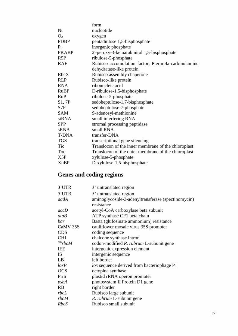
Figure 1.1 Enzymes and metabolites in the CBB and photorespiratory cycles.
Within the chloroplast stroma RuBP (ribulose-1,5-bisphosphate) and carbon dioxide (CO2) are fixed by
Rubisco (Ribulose 1,5-bisphosphate carboxylase/oxygenase) to produce two 3-phosphoglycerate (3-PGA)
molecules that undergoes a series of reducing reactions (enzymes 1 & 2) to produce glyceraldehyde-3-
phosphate (G3P, in bold). The G3P is either used for carbohydrate (CHO) synthesis or used to regenerate
RuBP within the CBB cycle (enzymes 3 – 10). Fixation of O2 to RuBP by Rubisco produces a 3-PGA and
a 2-PG (2-phosphoglycolate) product that is recycled to 3-PGA via the photorespiratory cycle (red arrows)
that spans the peroxisome and mitochondria. This pathway leads to loss of previously fixed CO2 within
the mitochondria. PGK, phosphoglycerate kinase; GAPDH, glyceraldehyde-3-phosphate dehydrogenase;
FBA, fructose-1,6-bisphosphate aldolase; FBPase, fructose-1,6-bisphosphatase; TK, transketolase; RPE,
ribulose-5-phosphate 3-epimerase; PRK, phosphoribulokinase; RPI, ribose-5-phosphate isomerase; TPI,
triose phosphate isomerase; SBPase, sedoheptulose-1,7-bisphosphatase. Metabolites in the cycle include
1,3-PGA, 1,3-bisphosphoglycerate; DHAP, dihydroxyacetone phosphate; E4P, erythrose-4-phosphate;
S1,7P, sedoheptulose-1,7-bisphosphate; S7P, sedoheptulose-7-phosphate; R5P, ribulose-5-phosphate;
X5P, xylulose-5-phosphate; F1,6P, fructose-1,6-bisphosphate; F6P, fructose-6-phosphate; RuP, ribulose-
5-phosphate. Figure modified from Michelet et al. (2013).
21
Of all the known pathways for CO2-fixation, the Calvin-Benson-Bassham (CBB) cycle
is most prevalent in autotrophs (Herrmann et al., 2015). In the cycle Rubisco fixes CO2
onto the substrate RuBP and evenly cleaves the 6-carbon product into 3-
phosphoglycerate (3-PGA) products that form the precursor molecules for carbohydrate
synthesis (Figure 1.1).
The CBB cycle requires the co-ordinated functioning of 11 different enzymes to
catalyse 13 different biochemical reactions. As many of these reactions require ATP and
NADPH, flux through the CBB cycle is regulated by the supply of these energy
equivalents from the photosynthetic light reactions (Michelet et al., 2013). As
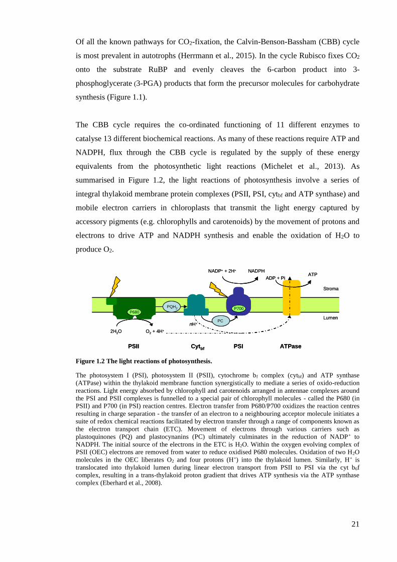
summarised in Figure 1.2, the light reactions of photosynthesis involve a series of
integral thylakoid membrane protein complexes (PSII, PSI, cytbf and ATP synthase) and
mobile electron carriers in chloroplasts that transmit the light energy captured by
accessory pigments (e.g. chlorophylls and carotenoids) by the movement of protons and
electrons to drive ATP and NADPH synthesis and enable the oxidation of H2O to
produce O2.
Figure 1.2 The light reactions of photosynthesis.
The photosystem I (PSI), photosystem II (PSII), cytochrome bf complex (cytbf) and ATP synthase
(ATPase) within the thylakoid membrane function synergistically to mediate a series of oxido-reduction
reactions. Light energy absorbed by chlorophyll and carotenoids arranged in antennae complexes around
the PSI and PSII complexes is funnelled to a special pair of chlorophyll molecules - called the P680 (in
PSII) and P700 (in PSI) reaction centres. Electron transfer from P680/P700 oxidizes the reaction centres
resulting in charge separation - the transfer of an electron to a neighbouring acceptor molecule initiates a
suite of redox chemical reactions facilitated by electron transfer through a range of components known as
the electron transport chain (ETC). Movement of electrons through various carriers such as
plastoquinones (PQ) and plastocynanins (PC) ultimately culminates in the reduction of NADP+ to
NADPH. The initial source of the electrons in the ETC is H2O. Within the oxygen evolving complex of
PSII (OEC) electrons are removed from water to reduce oxidised P680 molecules. Oxidation of two H2O
molecules in the OEC liberates O2 and four protons (H+) into the thylakoid lumen. Similarly, H+ is
translocated into thylakoid lumen during linear electron transport from PSII to PSI via the cyt b6f
complex, resulting in a trans-thylakoid proton gradient that drives ATP synthesis via the ATP synthase
complex (Eberhard et al., 2008).
PC
2H2O O2 + 4H+
nH+
PQH2
NADP+ + 2H+ NADPHATP
ADP + Pi
PSII PSICytbf ATPase
Lumen
Stroma
PCPC
2H2O O2 + 4H+
nH+
PQH2PQH2
NADP+ + 2H+ NADPHATP
ADP + Pi
PSII PSICytbf ATPase
Lumen
Stroma
P680P700
22
1.1.2 Photosynthesis – a target for improvement to increase global crop yields
The significance of addressing the growing concerns on food security is hard to over-
state. Food production needs to rise by >50% within the next 40 years to sustain the
growing global population (Long et al., 2015). The genetic gains in plant productivity
over the last ~50 years have come through plant breeding strategies to improve
resistance to stress – drought, cold, high salinity, pests and diseases – and through more
efficient water and fertilizer use. Adding to the challenge of increasing the global
productivity of natural and cropping ecosystems are the human-induced rises in air
temperature and deficiencies in the availability of additional arable lands. The grim
reality is that further gains at current rates of increase will be insufficient to ensure
future food security (Ainsworth and Ort, 2010). This has led to an increased urgency to
develop new strategies to “supercharge” photosynthesis in C3 crops to improve yield
potential. There are a number of technological remedies being studied for supercharging
photosynthesis. Many of these are targeted at enhancing photosynthetic efficiency by
improving the catalytic properties of the CO2-fixing enzyme Rubisco or by introducing
CO2-concentrating mechanisms that elevate CO2 levels around the enzyme to increase
its activity (von Caemmerer and Evans, 2010; Raines, 2011; Evans, 2013).
1.1.3 Rubisco - an enzyme in need of improvement
As the enzyme linking the inorganic and organic phases of the CBB cycle, the Rubisco
carboxylation reaction is often identified as the initiating step in photosynthetic carbon
assimilation. As highlighted below, Rubisco catalysis is relatively slow and unspecific
compared with other plant enzymes often leading to its catalytic activity limiting flux
through the CBB cycle (Evans, 2013; Parry et al., 2013). This has direct consequence
on the level of glyceraldehyde 3-phosphate (G3P) and other triose phosphates made for
hexose sugar production. These sugars are needed for plant metabolism and growth as
well as supply of other CBB products that are substrates for other pathways essential for
plant development (Raines, 2003; Vriet et al., 2014).
The evolution of oxygenic photosynthesis has seen the predominantly anaerobic,
CO2-rich atmosphere of earth some 3 billion years ago (Kaufman, 2014) increase to the
life preserving levels (~20,600 ppm O2) of today. The increasing abundance of O2
however proved detrimental to the carboxylation chemistry of Rubisco that evolved in
the absence of O2 (Whitney et al., 2011a). As shown in Figure 1.1, O2 is a competitive
inhibitor of CO2 during Rubisco catalysis. The oxygenation of RuBP by Rubisco
produces one molecule of 3-PGA and 2-phosphoglycolate (2-PG) (Bowes et al., 1971;
23
Andrews et al., 1971). Although often considered a waste product, recent studies
suggest 2-PG may play a role in regulating carbon-concentrating mechanism activity in
cyanobacteria (Haimovich-Dayan et al., 2014), be important to the nitrogen flux in
plants (Mallmann et al., 2014) and possibly serve as an electron sink for quenching
excessive free radicals during photosynthesis (Silva et al., 2015). Nevertheless many
photosynthetic organisms (e.g. algae, cyanobacteria, CAM and C4 plants) have evolved
carbon concentrating mechanisms (CCMs) to concentrate CO2 around Rubisco (often at
high expense in metabolic energy) to avoid RuBP oxygenation and evade the resource
costs of recycling 2-PG via photorespiration (Figure 1.1). This process of recycling two
molecules of 2-PG into a 3-PGA molecule by photorespiration spans three different
organelles (chloroplasts, peroxisomes, and mitochondria) (Kisaki and Tolbert, 1969) in
a process that requires energy (ATP and NADH) and through the action of glycine
decarboxylase in the mitochondria can result in up to 50% of the photosynthetic fixed
carbon being released as CO2 (Figure 1.1). This bifunctional activity of Rubisco has
made it a popular target for improving photosynthetic efficiency (Parry et al., 2013).
Such bioengineering efforts in plant leaves are aimed at either directly improving the
capacity of Rubisco itself to better discern CO2 from O2, or indirectly limiting its
oxygenase activity by introducing CCM or photorespiratory bypass systems into leaf
chloroplasts (Maurino and Peterhansel, 2010; Evans, 2013).
1.1.4 The complexity of Rubisco catalysis – an impediment to speed and specificity
It is paradoxical that as such a prominent biological catalyst Rubisco has not evolved
greater catalytic efficiency. Having reportedly originated more than 3 billion years ago
from an ancestral enzyme in an Archaean (Tabita et al., 2008) there has been ample
opportunity for greater catalytic advancement than is currently seen in nature (Whitney
et al., 2011a). While there is significant natural catalytic diversity to demonstrate that
Rubisco catalysis has managed to adapt to the CO2 and O2 pressures around its cellular
location (Tcherkez et al., 2006), Rubisco is still acknowledged as a sluggish, unspecific
enzyme in need of catalytic improvement (Parry et al., 2013). Its slowness to adapt and
improve appears largely attributable to the enzyme’s difficult, multi-step catalytic
chemistry (Figure 1.3) (Pearce, 2006; Kannappan and Gready, 2008) and structural
complementation requirements with varied molecular partners for its biogenesis in
cyanobacteria and chloroplasts (Mueller-Cajar and Whitney, 2008; Durão et al., 2015;
Whitney et al., 2015).
24
In additional to its oxygenase activity, Rubisco catalysis is also impaired by a
slow turnover rate (kcat) and a tendency to bind sugar-phosphate molecules that resemble
its catalytic transition state intermediates that inhibit function (Servaites, 1990; Carmo-
Silva et al., 2014) (see Table 1.1 and Figure 1.3). Compared with superoxide dismutase
(SOD) that catalyses at rates of up to 109 cycles per second (Gleason et al., 2014),
Rubisco is capable of only undergoing 1 to 4 cycles per second in leaf chloroplasts
(Whitney et al., 2011a). In plants like wheat, rice and tobacco this turnover typically
incorporates one oxygenase reaction per three carboxylation reactions.
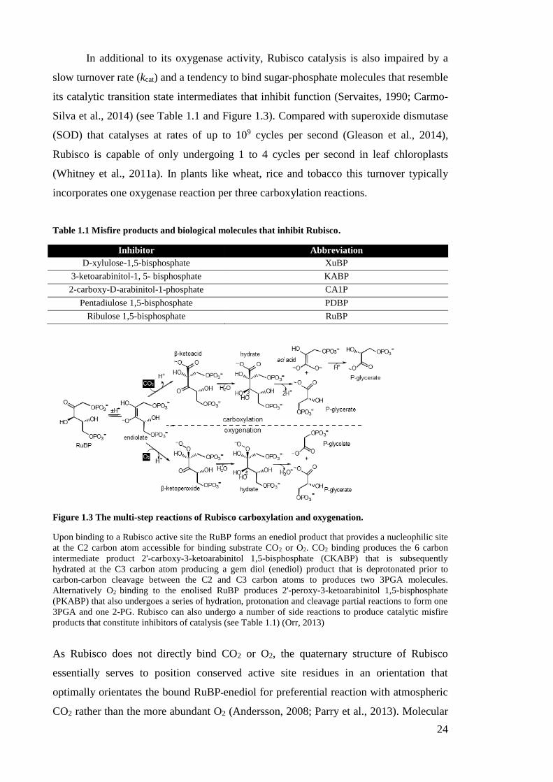
Table 1.1 Misfire products and biological molecules that inhibit Rubisco.
Inhibitor Abbreviation
D-xylulose-1,5-bisphosphate XuBP
3-ketoarabinitol-1, 5- bisphosphate KABP
2-carboxy-D-arabinitol-1-phosphate CA1P
Pentadiulose 1,5-bisphosphate PDBP
Ribulose 1,5-bisphosphate RuBP
Figure 1.3 The multi-step reactions of Rubisco carboxylation and oxygenation.
Upon binding to a Rubisco active site the RuBP forms an enediol product that provides a nucleophilic site
at the C2 carbon atom accessible for binding substrate CO2 or O2. CO2 binding produces the 6 carbon
intermediate product 2'-carboxy-3-ketoarabinitol 1,5-bisphosphate (CKABP) that is subsequently
hydrated at the C3 carbon atom producing a gem diol (enediol) product that is deprotonated prior to
carbon-carbon cleavage between the C2 and C3 carbon atoms to produces two 3PGA molecules.
Alternatively O2 binding to the enolised RuBP produces 2'-peroxy-3-ketoarabinitol 1,5-bisphosphate
(PKABP) that also undergoes a series of hydration, protonation and cleavage partial reactions to form one
3PGA and one 2-PG. Rubisco can also undergo a number of side reactions to produce catalytic misfire
products that constitute inhibitors of catalysis (see Table 1.1) (Orr, 2013)
As Rubisco does not directly bind CO2 or O2, the quaternary structure of Rubisco
essentially serves to position conserved active site residues in an orientation that
optimally orientates the bound RuBP-enediol for preferential reaction with atmospheric
CO2 rather than the more abundant O2 (Andersson, 2008; Parry et al., 2013). Molecular

25
dynamic simulation in silico of the Rubisco catalytic chemistry (Kannappan and Gready,
2008) and understanding the energetic constraints of its highly conserved mechanism
(Walter, 2006; Tcherkez, 2013) have proven of fundamental importance to better
understand Rubisco function at the molecular level. Such studies provide rationales for
the general observations that improvements in the catalytic speed of Rubisco are often
attained at the expense of specificity for CO2 over O2, and vice versa (Tcherkez et al.,
2006) – although greater exploration of Rubisco catalytic diversity and strategic
mutagenic testing is needed to test the accuracy of these hypotheses (Parry et al., 2013;
Galmés et al., 2014; Sharwood and Whitney, 2014).
As a consequence of its poor kinetics most plants invest considerable amounts of
their resources into producing sufficient Rubisco to support adequate levels of carbon
assimilation for plant growth and development. For example Rubisco typically
constitutes 25 to 50% of the leaf protein in C3 plants (such as wheat, cotton, rice,
Arabidopsis and tobacco) (Whitney et al., 2011a; Sharwood and Whitney, 2014) which
accounts for up to 25% of the total leaf nitrogen (Evans, 2013). This high level of
investment bestows Rubisco with the dubious honour of being the most abundant
enzyme on Earth (Ellis, 1979; Raven, 2013).
1.1.5 Structural and functional diversity among the varying isoforms of Rubisco
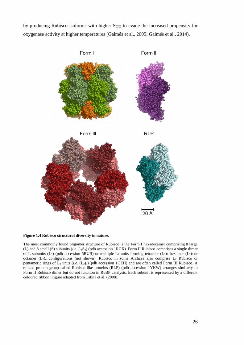
In nature, Rubisco takes on a variety of quaternary structures (Figure 1.4). All structures
comprise oligomers of large (L-) subunits (e.g. Form II Rubisco, Form III Rubisco and
Rubisco like proteins, RLP) or a hexadecameric structure of eight L-subunits and eight
small (S-) subunits (i.e. Form I L8S8 Rubisco) whereby the dimer of two L-subunits (L2)
that are arranged in an anti-parallel fashion (i.e. “head-to-toe”) and contain two active
sites (Andersson and Backlund, 2008; van Lun et al., 2011; Stec, 2012) is the basal
functional unit and is shared by all Rubisco isoforms. While the quaternary structure
and generic conformation of L8S8 Rubisco has remained unperturbed somewhat in
photosynthetic eukaryotes (e.g. plants and algae), there is significant variation in their
catalytic properties (Whitney et al., 2011a) (further detailed in Chapter 4). This
variation often correlates with the level of CO2 in solution around Rubisco (Sharwood
and Whitney, 2014; Galmes et al., 2014). For example Rubisco associated with a CCM
generally has a faster kcat but a lower affinity for CO2 (i.e. a higher Km for CO2) and a
reduced specificity for CO2 over O2 (termed the Rubisco CO2/O2 specificity factor; SC/O)
(Badger et al., 1998; Parry et al., 2013). There is also increasing evidence that catalytic
properties of plant Rubisco have adapted to growth conditions of elevated temperature
26
by producing Rubisco isoforms with higher SC/O to evade the increased propensity for
oxygenase activity at higher temperatures (Galmés et al., 2005; Galmés et al., 2014).
Figure 1.4 Rubisco structural diversity in nature.
The most commonly found oligomer structure of Rubisco is the Form I hexadecamer comprising 8 large
(L) and 8 small (S) subunits (i.e. L8S8) (pdb accession 1RCX). Form II Rubisco comprises a single dimer
of L-subunits (L2) (pdb accession 5RUB) or multiple L2 units forming tetramer (L2)2 hexamer (L2)3 or
octamer (L2)4 configurations (not shown). Rubisco in some Archaea also comprise L2 Rubisco or
pentameric rings of L2 units (i.e. (L2)5) (pdb accession 1GEH) and are often called Form III Rubisco. A
related protein group called Rubisco-like proteins (RLP) (pdb accession 1YKW) arranges similarly to
Form II Rubisco dimer but do not function in RuBP catalysis. Each subunit is represented by a different
coloured ribbon. Figure adapted from Tabita et al. (2008).
27
As shown in Figure 1.4, the L8S8 conformation of Form I Rubisco comprises a
core of eight 50- to 54 kDa L-subunits (L8) with two tetrads of 12 to 18 kDa S-subunits
capping either end (van Lun et al., 2011). S-subunit binding is thought to both stabilise
the holoenzyme structure as well as induce conformational changes within the L8 core
to stimulate catalytic viability (Bracher et al., 2011). In nature, the Form I L8S8 isoform
of Rubisco appears the most prominent as it is the form utilised by plants, algae,
cyanobacteria and various proteobacteria (Andersson and Backlund, 2008; Whitney et
al., 2011a). Based on the phylogeny of L-subunit sequences, Form I Rubisco comprises
of four distinct clades (IA, IB, IC, ID) that generally map to the phylogenetic
distribution of other photosynthetic genes and physiological processes (Badger and Bek,
2008). These phylogenies show the clustering of plant, green algae, cyanobacteria and
certain proteobacteria Rubisco isoforms (IA and IB) in what is often called the “green-
Rubisco” lineage. The Rubisco from non-green algae, other cyanobacteria and different
proteobacteria species cluster in a separate “red-Rubisco” lineage (IC and ID)
(Delwiche, 1999) (see also Table 1.2). Aligning with these different lineages is the
location of rbcL and rbcS coding the L- and S-subunit genes. The Form I red-Rubisco
genes are typically arranged in tandem in a bicistronic operon located in the chloroplast
genome (plastome) or prokaryotic chromosome. In some cases these operons include
other genes that code for proteins related to Rubisco biogenesis (e.g. rbcX) or catalytic
regulation (e.g. cbbX) (Liu et al., 2010; Mueller-Cajar et al., 2011). In contrast, in
vascular (“higher”) plants and green algae rbcL is located in the plastome and multiple
RbcS copies located in the nucleus (Andersson and Taylor, 2003; Andersson, 2008;
Tabita et al., 2008; Whitney et al., 2011a).
Form II Rubisco isoforms exist in proteobacteria and some dinoflagellate algae.
The simplest Form II Rubisco structure is the L2 isoform that is best characterised from
the photosynthetic α-proteobacterium Rhodospirillum rubrum (Figure 1.4) (Tabita and
McFadden, 1974). In dinoflagellates the Form II Rubisco constitutes a more complex
oligomeric structure whose stoichiometry is assumed to be L8, but remains
experimentally unclarified (Whitney and Yellowlees, 1995). Study of R. rubrum
Rubisco has spanned over 40 years by taking advantage of its minimal assembly
requirements that have allowed its expression and mutagenesis in hosts such as E. coli,
cyanobacterium Synechocystis 6803 and leaf chloroplasts (Pierce et al., 1989; Morell et
al., 1990; Whitney and Andrews, 2001b; Mueller-Cajar and Badger, 2007). Other Form
II Rubisco isoforms produced in species of Rhodopseudomonas palustris have been
found to form varying oligomeric structures such as (L2)2 tetramers, (L2)3 hexamers and
28
(L2)4 octamers (Tabita et al., 2008). Common features to Form II Rubisco are the low
sequence identity of their L-subunits to Form I Rubisco (typically sharing only 20 to 30%
identity) and inferior catalytic properties that comprise 4 to 8-fold reductions in SC/O, a
>10-fold higher Km for CO2 that offset the benefits of the ~2-fold increases in kcat for
many Form II Rubisco. Despite these differences however, crystal structure analyses
show the quaternary structure of the conserved active site residues within both Form I
and II Rubisco are highly superimposable making it difficult to discern structural
reasons for their catalytic differences (Tabita et al., 2007; Andersson and Backlund,
2008).
Rubisco isoforms found in Archaea generally show relatively poor sequence
similarity (<50%) to Form I and Form II Rubsico (Andersson and Backlund, 2008;
Badger and Bek, 2008). This Rubisco type is often called Form III Rubisco and
characteristically lacks S-subunits and forms L2 dimers or decameric complexes (i.e.
(L2)5). Unlike the photosynthetic function of Form I and Form II Rubisco, Form III
Rubisco appears to function in metabolising the RuBP generated in Archaea during
nucleotide (i.e. AMP) metabolism (Kitano et al., 2001; Bräsen et al., 2014). Consistent
with this function the affinity of Form III Rubisco for RuBP is extremely high; typically
having a >10-fold lower Km for RuBP than Form I and II Rubisco. However Archaea
Rubisco typically have slow turnover rates (kcat <1 s-1), a high Km for CO2 and SC/O
values that are >40-fold lower than Form I Rubisco making them extremely sensitive to
O2 inhibition (Alonso et al., 2009). Despite their different functional role, replacement
of Form I Rubisco in tobacco chloroplasts with the Form III Rubisco from
Methanococcus burtonii produced plants whose photosynthetic growth could be fully
supported when grown in air containing elevated levels of CO2 (Alonso et al., 2009).
Rubisco-like proteins (RLPs) (sometimes referred to as Form IV Rubisco) show
structural similarities to conventional Rubisco, but do not retain many of the catalytic
residues conserved in all Rubisco forms (Ashida et al., 2005). As a result RLPs are
unable to metabolise RuBP, questioning the validity of classifying them as Form IV
Rubisco (Table 1.2) (Tabita et al., 2007). Like L2 Rubisco, RLPs typically comprise a
homodimer complex that is thought to function in thiosulfate oxidation as part of
sulphur metabolism (Hanson and Tabita, 2001) and methionine salvage pathways such
as in Bacillus subtilis (Ashida et al., 2005).
29
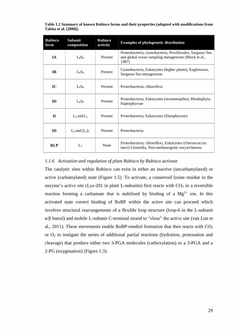
Table 1.2 Summary of known Rubisco forms and their properties (adapted with modifications from
Tabita et al. [2008]).
Rubisco
form
Subunit
composition
Rubisco
activity Examples of phylogenetic distribution
IA L8S8 Present
Proteobacteria, cyanobacteria, Prochlorales, Sargasso Sea
and global ocean sampling metagenome (Block et al.,
1987)
IB L8S8 Present Cyanobacteria, Eukaryotes (higher plants), Euglenozoa,
Sargasso Sea metagenome
IC L8S8 Present Proteobacteria, chloroflexi
ID L8S8 Present Proteobacteria, Eukaryotes (stramenopiles), Rhodophyta,
Haptophyceae
II L2 and Ln Present Proteobacteria, Eukaryotes (Dinophyceae)
III L2 and (L2)5 Present Proteobacteria
RLP L2 None Proteobacteria, chloroflexi, Eukaryotes (Ostreococcus
tauri) Clostridia, Non-methanogenic euryarchaeota
1.1.6 Activation and regulation of plant Rubisco by Rubisco activase
The catalytic sites within Rubisco can exist in either an inactive (uncarbamylated) or
active (carbamylated) state (Figure 1.5). To activate, a conserved lysine residue in the
enzyme’s active site (Lys-201 in plant L-subunits) first reacts with CO2 in a reversible
reaction forming a carbamate that is stabilised by binding of a Mg2+ ion. In this
activated state correct binding of RuBP within the active site can proceed which
involves structural rearrangements of a flexible loop structure (loop-6 in the L-subunit
α/β barrel) and mobile L-subunit C-terminal strand to “close” the active site (van Lun et
al., 2011). These movements enable RuBP-enediol formation that then reacts with CO2
or O2 to instigate the series of additional partial reactions (hydration, protonation and
cleavage) that produce either two 3-PGA molecules (carboxylation) or a 3-PGA and a
2-PG (oxygenation) (Figure 1.3).
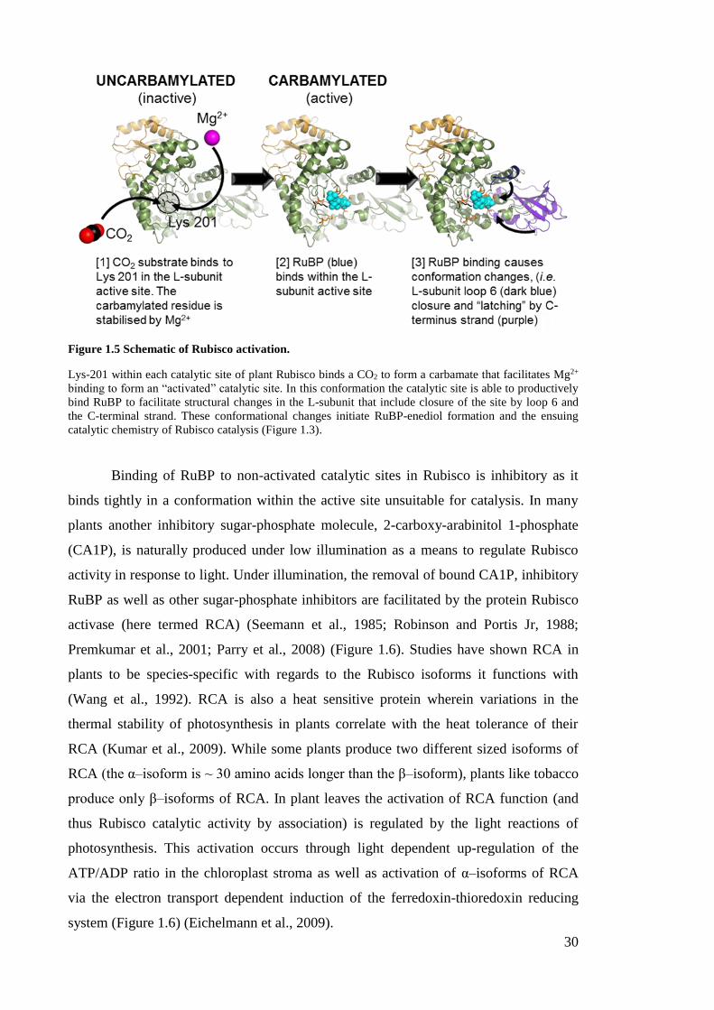
30
Figure 1.5 Schematic of Rubisco activation.
Lys-201 within each catalytic site of plant Rubisco binds a CO2 to form a carbamate that facilitates Mg2+
binding to form an “activated” catalytic site. In this conformation the catalytic site is able to productively
bind RuBP to facilitate structural changes in the L-subunit that include closure of the site by loop 6 and
the C-terminal strand. These conformational changes initiate RuBP-enediol formation and the ensuing
catalytic chemistry of Rubisco catalysis (Figure 1.3).
Binding of RuBP to non-activated catalytic sites in Rubisco is inhibitory as it
binds tightly in a conformation within the active site unsuitable for catalysis. In many
plants another inhibitory sugar-phosphate molecule, 2-carboxy-arabinitol 1-phosphate
(CA1P), is naturally produced under low illumination as a means to regulate Rubisco
activity in response to light. Under illumination, the removal of bound CA1P, inhibitory
RuBP as well as other sugar-phosphate inhibitors are facilitated by the protein Rubisco
activase (here termed RCA) (Seemann et al., 1985; Robinson and Portis Jr, 1988;
Premkumar et al., 2001; Parry et al., 2008) (Figure 1.6). Studies have shown RCA in
plants to be species-specific with regards to the Rubisco isoforms it functions with
(Wang et al., 1992). RCA is also a heat sensitive protein wherein variations in the
thermal stability of photosynthesis in plants correlate with the heat tolerance of their
RCA (Kumar et al., 2009). While some plants produce two different sized isoforms of
RCA (the α–isoform is ~ 30 amino acids longer than the β–isoform), plants like tobacco
produce only β–isoforms of RCA. In plant leaves the activation of RCA function (and
thus Rubisco catalytic activity by association) is regulated by the light reactions of
photosynthesis. This activation occurs through light dependent up-regulation of the
ATP/ADP ratio in the chloroplast stroma as well as activation of α–isoforms of RCA
via the electron transport dependent induction of the ferredoxin-thioredoxin reducing
system (Figure 1.6) (Eichelmann et al., 2009).

31
Figure 1.6 Rubisco activation and regulation of its activity by RCA.
Removal of sugar-phosphate bound inhibitors (e.g. RuBP) from the catalytic sites of non-activated
Rubisco by RCA enable their activation (carbamylation) with CO2 and Mg2+ to enable catalysis (Figure
1.5). Activated Rubisco can also bind sugar-phosphate inhibitors (e.g. CA1P and the catalytic misfire
product XuBP, Table 1.1) whose removal by RCA is critical for enabling/maintaining Rubisco catalysis
during photosynthesis.
Similar to other AAA+ (ATPase-Associated Activity) proteins, RCA removes
bound inhibitors from the catalytic sites in Rubisco using the energy of ATP hydrolysis
(Robinson and Portis Jr, 1989; Portis et al., 2008). Other components involved with
inhibitor removal include CbbY, a sugar phosphatase discovered in Arabidopsis
thaliana that metabolises XuBP to non-inhibitory compounds that can be used for RuBP
regeneration (Bracher et al., 2015). Recent success in deriving structural information for
tobacco RCA by mass spectrometry (Blayney et al., 2011) and protein crystallography
(Stotz et al., 2011) have demonstrated its capacity to form hexameric complexes. These
studies also confirm prior mutagenic studies as to the likelihood of a C-terminal
“recognition” loop in RCA recognise sequences on the holoenzyme surface in the L-
subunit N-domain (between residues 89 and 94) that facilitate conformational changes
to the Rubisco L-subunit loop-6 that facilitate sugar-phosphate inhibitor release (Esau et
al., 1998). Unfortunately the transitory nature of Rubisco-RCA interactions poses a
significant challenge to resolving the influence of the S-subunit and specific
mechanistic detail on how RCA interacts with Rubisco to cause these conformational
changes (Wachter et al., 2013).
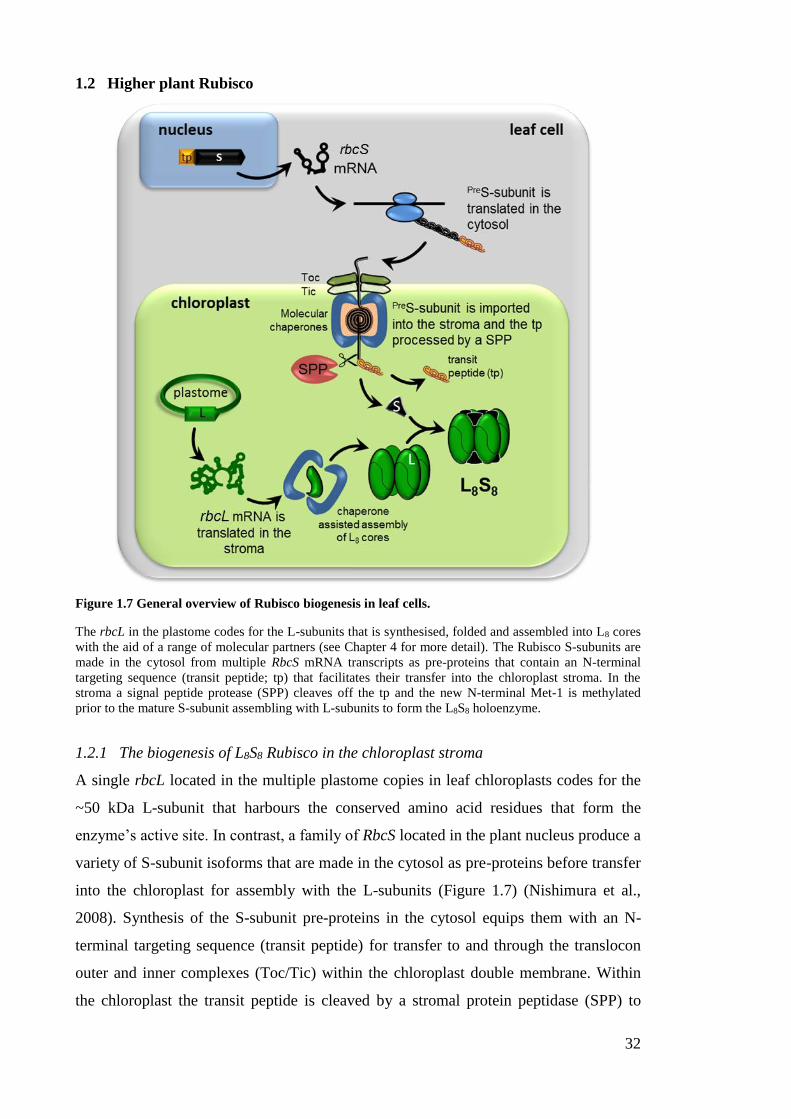
32
1.2 Higher plant Rubisco
Figure 1.7 General overview of Rubisco biogenesis in leaf cells.
The rbcL in the plastome codes for the L-subunits that is synthesised, folded and assembled into L8 cores
with the aid of a range of molecular partners (see Chapter 4 for more detail). The Rubisco S-subunits are
made in the cytosol from multiple RbcS mRNA transcripts as pre-proteins that contain an N-terminal
targeting sequence (transit peptide; tp) that facilitates their transfer into the chloroplast stroma. In the
stroma a signal peptide protease (SPP) cleaves off the tp and the new N-terminal Met-1 is methylated
prior to the mature S-subunit assembling with L-subunits to form the L8S8 holoenzyme.
1.2.1 The biogenesis of L8S8 Rubisco in the chloroplast stroma
A single rbcL located in the multiple plastome copies in leaf chloroplasts codes for the
~50 kDa L-subunit that harbours the conserved amino acid residues that form the
enzyme’s active site. In contrast, a family of RbcS located in the plant nucleus produce a
variety of S-subunit isoforms that are made in the cytosol as pre-proteins before transfer
into the chloroplast for assembly with the L-subunits (Figure 1.7) (Nishimura et al.,
2008). Synthesis of the S-subunit pre-proteins in the cytosol equips them with an N-
terminal targeting sequence (transit peptide) for transfer to and through the translocon
outer and inner complexes (Toc/Tic) within the chloroplast double membrane. Within
the chloroplast the transit peptide is cleaved by a stromal protein peptidase (SPP) to

33
produce a ~14 to 15 kDa mature S-subunit whose new Met-1 N-terminus is N-
methylated by a methyltransferase (Houtz and Mulligan, 1991). Once processed the S-
subunit assembles with the L-subunit with the aid of a range of generic and Rubisco-
specific chaperones via a process that remains poorly understood (Whitney et al., 2011a;
Whitney et al., 2015). Details on the RbcS multi-gene family and Rubisco assembly
process are given in Chapter 3 and Chapter 4 respectively.
Figure 1.8 Architecture and subunit interactions in a L2S4 portion of L8S8 Rubisco.
The catalytic (or active) site of Rubisco (white) and Loop 6 (in red) is formed from conserved residues
coded in the L-subunit (in green). The S-subunits (in yellow) are able to influence Rubisco catalysis
despite their distance from the active site (van Lun et al., 2011). Interactions between the βA/βB loop of
the S-subunit (shown in black) and adjoining L-subunit residues within central solvent channel (CSC) are
considered key contacts involved in influencing the catalytic properties of Rubisco (Spreitzer et al., 2005)
1.2.2 Higher plant Rubisco subunits and their role in catalysis
The location of the higher plant L8S8 Rubisco (Section 1.1.5) catalytic (or ‘active’) site
is positioned at the interface of L-subunits and are dimerised in an antiparallel L2
fashion (Figure 1.8), where the C-terminal β/α barrel domain of each L-subunit and the
N-terminal domain of the other L-subunit contribute conserved His, Gln, Glu, Thr and
Lys residues to each active site (Stec, 2012). As the L-subunit harbours the active sites
of Rubisco, a significant focus has been made on correlating changes in the L-subunit
34
sequence with natural catalytic variability measured for Rubisco from different plant,
algae and prokaryotic sources (Parry et al., 2003; Galmés et al., 2005; Kapralov et al.,
2011; Whitney et al., 2011b; Galmés et al., 2014).
The ability to express and assemble cyanobacteria L8S8 Rubisco in E. coli
proved vital to mutagenic studies of Form I Rubisco in the 80’s and 90’s, in particular
demonstrating the role of the S-subunit on catalysis. Although the S-subunit was found
not to be absolutely essential for the assembly and function of cyanobacteria L8 Rubisco
cores, the addition of S-subunits stimulated activity 100-fold and provided structural
stability (Andrews and Ballment, 1983; Andrews, 1988). Since these pioneering studies,
subsequent mutagenic work using cyanobacteria (Read and Tabita, 1992),
Chlamydomonas (Genkov and Spreitzer, 2009) and plant (Ishikawa et al., 2011)
Rubisco have continued to highlight the pervasive influence of the S-subunit on
catalysis. As a consequence of their tetrameric S-subunit arrangements at both apices of
the L8 core in Form I Rubisco (Figure 1.4), each L-subunit makes contact with three
separate S-subunits (van Lun et al., 2011). Of particular interest are the interactions
between the S-subunit βA/βB loop and residues within its most closely associated L-
subunit. Mutations to residues in either subunit around this region have a significant
influence on catalysis, despite their substantial distance from the active site (Figure 1.8)
(Spreitzer et al., 2001; Spreitzer et al., 2005; Genkov and Spreitzer, 2009). Similar to
cyanobacteria and Chlamydomonas L8S8 enzymes, the S-subunit βA/βB loops of higher
plant Rubisco line the circumference at both ends of the central pore that traverses the
holoenzyme (Spreitzer and Salvucci, 2002; Spreitzer, 2003; Andersson, 2008; van Lun
et al., 2011). Determining the influence of the βA/βB loops and other S-subunit
sequences on higher plant Rubisco catalysis remains elusive due to challenges in
mutating S-subunits in leaf chloroplasts. Further details of these challenges are
considered in Section 1.4.2 below and more thoroughly in Chapter 3.
1.3 CO2 assimilation in C3 and C4 plants
1.3.1 C3 versus C4 photosynthesis
The primary distinguishing factor between C3 and C4 plants is the initial form or
inorganic carbon assimilated into organic carbon. In C3 plants CO2 is the initial carbon
substrate fixed by Rubisco to produce the 3-C product 3-PGA (Figure 1.1) in model
plants species examples include tobacco, Arabidopsis, wheat and rice. In contrast C4
plants are those containing a CCM where carbon assimilation begins with HCO3-
fixation to phosphoenolpyruvate (PEP) by phosphoenolpyruvate carboxylase (PEPC) to
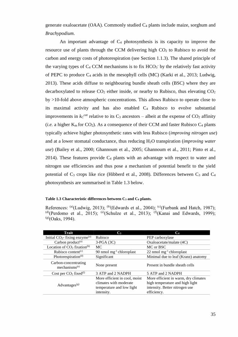
35
generate oxaloacetate (OAA). Commonly studied C4 plants include maize, sorghum and
Brachypodium.
An important advantage of C4 photosynthesis is its capacity to improve the
resource use of plants through the CCM delivering high CO2 to Rubisco to avoid the
carbon and energy costs of photorespiration (see Section 1.1.3). The shared principle of
the varying types of C4 CCM mechanisms is to fix HCO3- by the relatively fast activity
of PEPC to produce C4 acids in the mesophyll cells (MC) (Karki et al., 2013; Ludwig,
2013). These acids diffuse to neighbouring bundle sheath cells (BSC) where they are
decarboxylated to release CO2 either inside, or nearby to Rubisco, thus elevating CO2
by >10-fold above atmospheric concentrations. This allows Rubisco to operate close to
its maximal activity and has also enabled C4 Rubisco to evolve substantial
improvements in kCcat relative to its C3 ancestors – albeit at the expense of CO2 affinity
(i.e. a higher Km for CO2). As a consequence of their CCM and faster Rubisco C4 plants
typically achieve higher photosynthetic rates with less Rubisco (improving nitrogen use)
and at a lower stomatal conductance, thus reducing H2O transpiration (improving water
use) (Bailey et al., 2000; Ghannoum et al., 2005; Ghannoum et al., 2011; Pinto et al.,
2014). These features provide C4 plants with an advantage with respect to water and
nitrogen use efficiencies and thus pose a mechanism of potential benefit to the yield
potential of C3 crops like rice (Hibberd et al., 2008). Differences between C3 and C4
photosynthesis are summarised in Table 1.3 below.
Table 1.3 Characteristic differences between C3 and C4 plants.
References: (a)(Ludwig, 2013); (b)(Edwards et al., 2004); (c)(Furbank and Hatch, 1987); (d)(Perdomo et al., 2015); (e)(Schulze et al., 2013); (f)(Kanai and Edwards, 1999); (g)(Oaks, 1994).
Trait C3 C4
Initial CO2- fixing enzyme(a) Rubisco PEP carboxylase
Carbon product(a) 3-PGA (3C) Oxaloacetate/malate (4C)
Location of CO2 fixation(b) MC MC or BSC
Rubisco content(c) 90 nmol mg-1 chloroplast 22 nmol mg-1 chloroplast
Photorespiration(d) Significant Minimal due to leaf (Kranz) anatomy
Carbon-concentrating
mechanisms(e) None present Present in bundle sheath cells
Cost per CO2 fixed(f) 3 ATP and 2 NADPH 5 ATP and 2 NADPH
Advantages(g)
More efficient in cool, moist
climates with moderate
temperature and low light
intensity.
More efficient in warm, dry climates
high temperature and high light
intensity. Better nitrogen use
efficiency.
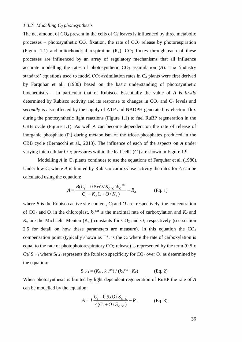
36
1.3.2 Modelling C3 photosynthesis
The net amount of CO2 present in the cells of C3 leaves is influenced by three metabolic
processes – photosynthetic CO2 fixation, the rate of CO2 release by photorespiration
(Figure 1.1) and mitochondrial respiration (Rd). CO2 fluxes through each of these
processes are influenced by an array of regulatory mechanisms that all influence
accurate modelling the rates of photosynthetic CO2 assimilation (A). The ‘industry
standard’ equations used to model CO2 assimilation rates in C3 plants were first derived
by Farquhar et al., (1980) based on the basic understanding of photosynthetic
biochemistry – in particular that of Rubisco. Essentially the value of A is firstly
determined by Rubisco activity and its response to changes in CO2 and O2 levels and
secondly is also affected by the supply of ATP and NADPH generated by electron flux
during the photosynthetic light reactions (Figure 1.1) to fuel RuBP regeneration in the
CBB cycle (Figure 1.1). As well A can become dependent on the rate of release of
inorganic phosphate (Pi) during metabolism of the triose-phosphates produced in the
CBB cycle (Bernacchi et al., 2013). The influence of each of the aspects on A under
varying intercellular CO2 pressures within the leaf cells (Ci) are shown in Figure 1.9.
Modelling A in C3 plants continues to use the equations of Farquhar et al. (1980).
Under low Ci where A is limited by Rubisco carboxylase activity the rates for A can be
calculated using the equation:
d
oci
cat
COCi RKOKC
kSxOCBA
)/1(
)/5.0( / (Eq. 1)
where B is the Rubisco active site content, Ci and O are, respectively, the concentration
of CO2 and O2 in the chloroplast, kCcat is the maximal rate of carboxylation and Kc and
Ko are the Michaelis-Menten (Km) constants for CO2 and O2 respectively (see section
2.5 for detail on how these parameters are measure). In this equation the CO2
compensation point (typically shown as Γ*, is the Ci where the rate of carboxylation is
equal to the rate of photophotorespiratory CO2 release) is represented by the term (0.5 x
O)/ SC/O where SC/O represents the Rubisco specificity for CO2 over O2 as determined by
the equation:
SC/O = (Ko . kCcat) / (kO
cat . Kc) (Eq. 2)
When photosynthesis is limited by light dependent regeneration of RuBP the rate of A
can be modelled by the equation:
d
OCi
OCi RSOC
SxOCJA
)/(4
/5.0
/
/ (Eq. 3)
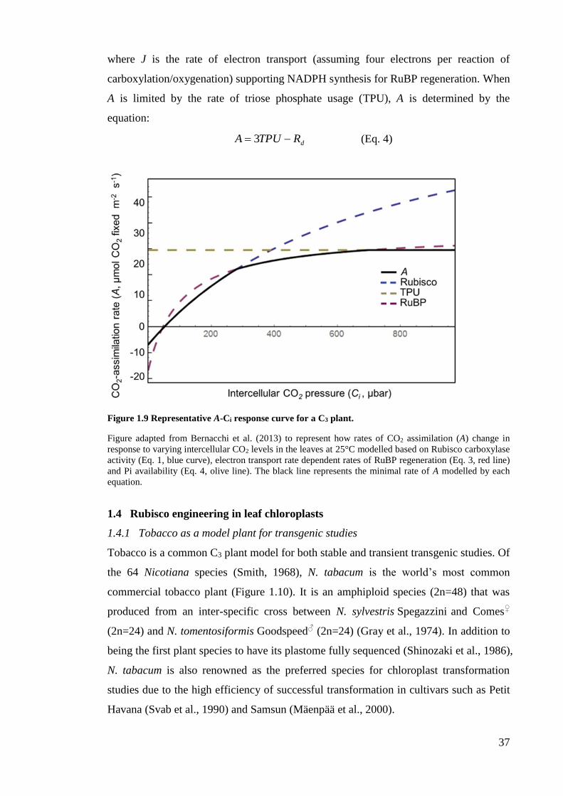
37
where J is the rate of electron transport (assuming four electrons per reaction of
carboxylation/oxygenation) supporting NADPH synthesis for RuBP regeneration. When
A is limited by the rate of triose phosphate usage (TPU), A is determined by the
equation:
dRTPUA 3 (Eq. 4)
Figure 1.9 Representative A-Ci response curve for a C3 plant.
Figure adapted from Bernacchi et al. (2013) to represent how rates of CO2 assimilation (A) change in
response to varying intercellular CO2 levels in the leaves at 25°C modelled based on Rubisco carboxylase
activity (Eq. 1, blue curve), electron transport rate dependent rates of RuBP regeneration (Eq. 3, red line)
and Pi availability (Eq. 4, olive line). The black line represents the minimal rate of A modelled by each
equation.
1.4 Rubisco engineering in leaf chloroplasts
1.4.1 Tobacco as a model plant for transgenic studies
Tobacco is a common C3 plant model for both stable and transient transgenic studies. Of
the 64 Nicotiana species (Smith, 1968), N. tabacum is the world’s most common
commercial tobacco plant (Figure 1.10). It is an amphiploid species (2n=48) that was
produced from an inter-specific cross between N. sylvestris Spegazzini and Comes♀
(2n=24) and N. tomentosiformis Goodspeed♂ (2n=24) (Gray et al., 1974). In addition to
being the first plant species to have its plastome fully sequenced (Shinozaki et al., 1986),
N. tabacum is also renowned as the preferred species for chloroplast transformation
studies due to the high efficiency of successful transformation in cultivars such as Petit
Havana (Svab et al., 1990) and Samsun (Mäenpää et al., 2000).
38
Figure 1.10 Wild-type Nicotiana tabacum cv. Petit Havana.
N. tabacum is commonly used as a model system for transgenic studies in higher plants, in particular the
application of plastome transformation (Maliga, 2004).
1.4.2 The goals and limitations to bioengineering Rubisco in leaf chloroplasts
Transgenic research on Rubisco over the past 30 years in photosynthetic organisms has
highlighted its pivotal influence on photosynthesis. This, along with the finding that
Rubisco from non-green algae have better catalytic properties than plant Rubisco
(Whitney et al., 2001; Andrews and Whitney, 2003) (detailed further in Chapter 4),
have driven Rubisco biologists to identify solutions that improve Rubisco catalysis. On
top of this challenge is identifying transgenic approaches to introducing beneficial
changes to Rubisco function into plant leaves – in particular crops species. Ultimately
by improving Rubisco catalysis it is hoped that photosynthesis can be improved and
crop yield increased. In addition, improving Rubisco performance may potentially
improve resource-use efficiency by reducing a plants’ need for nitrogen (invest less in a
more efficient Rubisco), water (lower the Ci levels needed for photosynthesis thereby
reducing stomata aperture and water transpiration) or/and light (reduced
photorespiration thereby saving energy) (Andrews and Whitney, 2003; Whitney et al.,
2011a; Parry et al., 2013). A key limitation to structure-function studies of L8S8 Rubisco
from higher plants is their inability to assemble in E. coli due the incompatibilities in
their folding and assembly requirements (Parry et al., 2013). For this reason, genetic
39
modification of plant Rubisco has been limited by the slower processes associated with
generating transgenic plants.
Although the S-subunits of higher plant Rubisco are essential to the enzymes
holistic function (see Section 1.2.2), the majority of mutagenic analyses undertaken on
higher plant Rubisco have focused on the L-subunit. Primary reasons for this are the
location of the catalytic sites within the L-subunit (Figure 1.6), the relative ease of
genetically modifying rbcL by chloroplast (plastome) transformation (Section 1.4.3) and
due to limitations in genetically mutating RbcS. The latter limitations stem from there
being multiple allelic copies of RbcS in the nucleus of plant cells – up to 22 copies in
some species (Manzara and Gruissem, 1988; Spreitzer, 2003) – that appear to be
differentially expressed in response to growth and environmental cues and code varying
degrees of amino acid diversity (Sasanuma, 2001; Ogawa et al., 2011). Such factors
complicate mutagenic or replacement strategies targeting RbcS (Whitney et al., 2011a).
More details on prior S-subunit engineering undertakings are provided in Chapter 3.
1.4.3 Transgenic manipulation of Rubisco by plastome transformation
Plastome transformation was originally developed in Chlamydomonas (Boynton et al.,
1988) before being applied to modify the small chloroplast genome (plastome; ~160 kb)
of higher plant chloroplasts (Svab et al., 1990). The technology is available for a limited
range of plant species that includes a few important crop species – tomato, potato
soybean and cotton (Daniell et al., 2005; Day and Goldschmidt-Clermont, 2011; Maliga
and Bock, 2011). The transformation process remains most efficient in N. tabacum (cv
Petit Havana SR1) where flanking plastome sequence (typically ~1 kb) included either
side of the genetic changes guide their introduction into the plastome with pinpoint
accuracy via homologous recombination. Among the genetic modifications introduced
is typically the aadA selectable marker that confers resistance to the antibiotics
spectinomycin and streptomycin. Using this approach a variety of mutations have been
made to the tobacco rbcL in a bid to examine structure-function relationships and better
understand Rubisco biogenesis in plant plastids (Andrews and Whitney, 2003;
Sharwood et al., 2008; Whitney et al., 2011b; Whitney et al., 2015). Initial deletion of
rbcL prevented Rubisco synthesis while the re-introduction of rbcL into the nucleus
only partially recovered leaf Rubisco content (Kanevski and Maliga, 1994). This
confirmed the preferred location of rbcL in the plastome. Other plastome
transformations have produced site-directed L-subunit mutants (Whitney et al., 1999;
Whitney et al., 2011a), shown the folding requirements of marine algae Rubisco
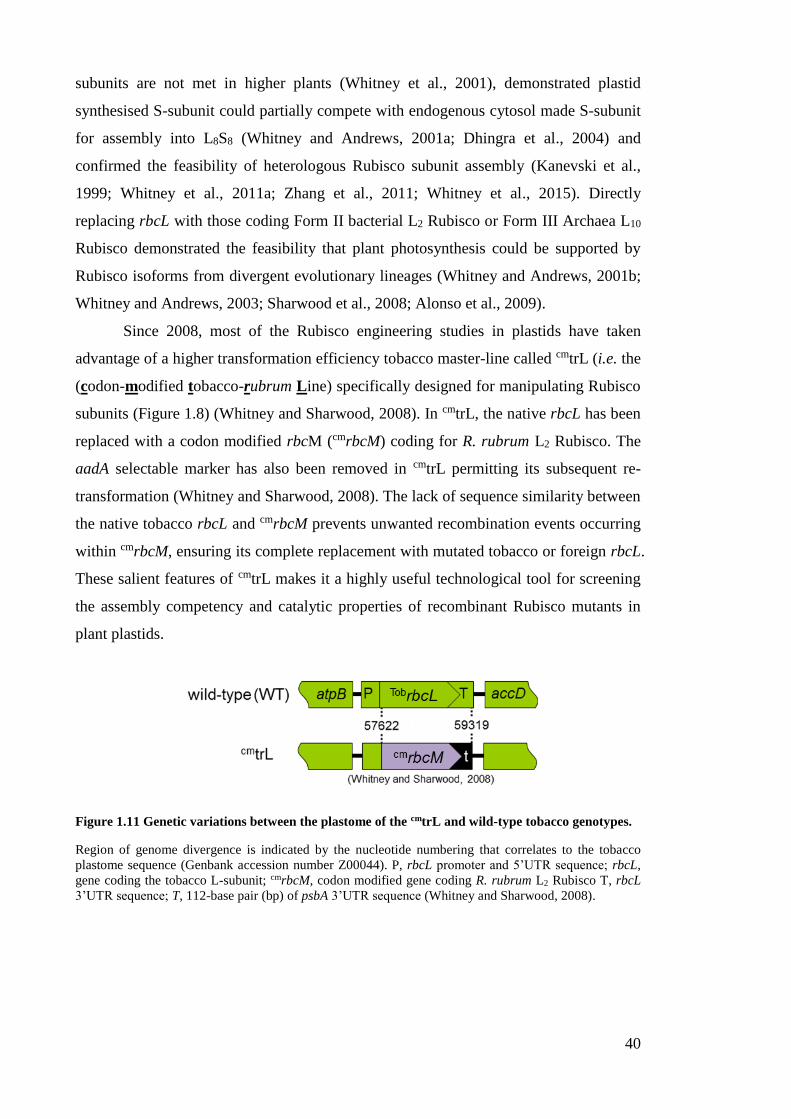
40
subunits are not met in higher plants (Whitney et al., 2001), demonstrated plastid
synthesised S-subunit could partially compete with endogenous cytosol made S-subunit
for assembly into L8S8 (Whitney and Andrews, 2001a; Dhingra et al., 2004) and
confirmed the feasibility of heterologous Rubisco subunit assembly (Kanevski et al.,
1999; Whitney et al., 2011a; Zhang et al., 2011; Whitney et al., 2015). Directly
replacing rbcL with those coding Form II bacterial L2 Rubisco or Form III Archaea L10
Rubisco demonstrated the feasibility that plant photosynthesis could be supported by
Rubisco isoforms from divergent evolutionary lineages (Whitney and Andrews, 2001b;
Whitney and Andrews, 2003; Sharwood et al., 2008; Alonso et al., 2009).
Since 2008, most of the Rubisco engineering studies in plastids have taken
advantage of a higher transformation efficiency tobacco master-line called cmtrL (i.e. the
(codon-modified tobacco-rubrum Line) specifically designed for manipulating Rubisco
subunits (Figure 1.8) (Whitney and Sharwood, 2008). In cmtrL, the native rbcL has been
replaced with a codon modified rbcM (cmrbcM) coding for R. rubrum L2 Rubisco. The
aadA selectable marker has also been removed in cmtrL permitting its subsequent re-
transformation (Whitney and Sharwood, 2008). The lack of sequence similarity between
the native tobacco rbcL and cmrbcM prevents unwanted recombination events occurring
within cmrbcM, ensuring its complete replacement with mutated tobacco or foreign rbcL.
These salient features of cmtrL makes it a highly useful technological tool for screening
the assembly competency and catalytic properties of recombinant Rubisco mutants in
plant plastids.
Figure 1.11 Genetic variations between the plastome of the cmtrL and wild-type tobacco genotypes.
Region of genome divergence is indicated by the nucleotide numbering that correlates to the tobacco
plastome sequence (Genbank accession number Z00044). P, rbcL promoter and 5’UTR sequence; rbcL,
gene coding the tobacco L-subunit; cmrbcM, codon modified gene coding R. rubrum L2 Rubisco T, rbcL
3’UTR sequence; T, 112-base pair (bp) of psbA 3’UTR sequence (Whitney and Sharwood, 2008).
41
1.5 Primary objectives of thesis
The ultimate goal of this thesis is to create novel opportunities and technologies that
expand the bioengineering potential of higher plant Rubisco. Transformation strategies
developed over the past 20 years to surpass nature’s attempt at in vivo Rubisco
evolution have instead realised our incomplete understanding of the enzymes biogenesis
requirements and difficulty in improving its catalysis. Being able to produce plant
Rubisco complexes comprising homogeneous populations of native and mutated L-
subunits and S-subunits is critical to understanding their structure-function interactions
and identifying solutions to improve catalysis. This thesis examines the feasibility of
using RNA interference (RNAi) to fully silence RbcS mRNA synthesis in the cmtrL
genotype and then test the efficiency of alternative transgenic approaches for
bioengineering recombinant Rubisco comprising chloroplast made L- and S-subunits
and their effect on L8S8 Rubisco biogenesis, catalysis and leaf photosynthesis.
To assist the reader a summary of the transplastomic tobacco genotypes studied
in this thesis and detail of their Rubisco transgenes is provided in Appendix A.
42
CHAPTER 2 – MATERIAL AND METHODS
2.1 Molecular techniques
2.1.1 E. coli growth and transformation
CaCl2 competent XL1-Blue E. coli were made for DNA transformation as described by
Sambrook et al., 1989. A 10 mL culture of XL1-Blue E. coli cells was grown for 16 h in
Luria-Bertani (LB) medium (10 g L-1 tryptone, 5 g L-1 NaCl, 5 g L-1 yeast extract, with
the pH adjusted to 7.0 with 1 M NaOH) at 37°C and used to inoculate 1 L of pre-
warmed LB medium. After incubating for 4 to 5 h at 37°C the cells reached mid-log
phase growth (OD600 of ~0.5 to 0.7) and were immediately dispensed into autoclave
sterilised GSA centrifuge tubes and placed on ice. After 10 min the tubes were placed in
a pre-chilled (4°C) GSA rotor (SORVALL®) and centrifuged 3000 g for 5 min at 4°C.
The pelleted cells were gently suspended in 10 mL of ice cold sterile 0.1 M CaCl2
containing 15% (v/v) glycerol. After 20 min on ice 0.6 mL aliquots of the cells were
dispensed into sterile 1.8 mL microfuge tubes, snap-frozen in liquid N2 and stored at -
80°C.
E. coli colonies were grown on agar solidified (1.5% [w/v]) LB medium. LB-
amp medium contained ampicillin (200 µg mL-1; made from a 1000-fold dilution of 200
mg mL-1 stock dissolved in sterile Milli-Q H2O). Transformed colonies were identified
as colonies on the LB-Amp plates grown upside-down at 37°C for 16 h. Single colonies
were picked aseptically in a laminar flow cabinet and used to inoculate 1.5 mL (for
mini-prep cultures) or 100 mL (for maxi-prep culture) of LB-amp medium and grown
for 16-20 h at 37°C in a shaking incubator (set at 180-200 opm).
2.1.2 Plasmid DNA purification
pDNA mini-preps
Plasmid DNA (pDNA) from mini-prep cultures of transformed XL1-Blue E. coli cells
was extracted and purified using the Wizard® Plus SV Minipreps DNA Purification
System Kit (Promega). All steps were carried out at room temperature (RT). Mini-prep
cultures were transferred to a sterile 1.8 mL microfuge tube and pelleted by
centrifugation (1 min, 10,000 g). The cells were suspended in 0.25 mL Cell
Resuspension Solution (50 mM Tris-HCL (pH 7.5), 10 mM EDTA, 100 µg mL-1 RNase
A) prior to adding 0.25 mL Cell Lysis Solution (0.2 M NaOH, 1% SDS) and 10 L of
alkaline protease. After 5 min at RT temperature 0.35 mL of neutralization solution
(4.09 M guanidine hydrochloride, 0.759 M potassium acetate, 2.12 M glacial acetic acid,
43
pH ~4.2) was added and the reaction mixed by repeated inversion then centrifuged (5
min, 16,000 g). The supernatant was transferred to a spin-column and the pDNA
immobilised on the membrane by centrifugation (1 min, 8,000 g) and then washed twice
with successive 0.4 mL aliquots of wash buffer (60% ethanol, 60 mM potassium acetate,
8.3 mM Tris-HCl pH 7.5, 0.04 mM EDTA pH 8.0) with intermittent centrifugation (3
min, 16,000 g). The bound pDNA was eluted from the membrane with 70 µL nuclease-
free water by centrifugation (2 min, 8 000 g).
pDNA maxi-preps
pDNA purification from 100 mL cultures of E.coli was carried out using the Plasmid
Maxi Kit (QIAGEN). Cultured cells were pelleted in 50 mL Falcon tubes by
centrifugation (4800 g, 4°C, 15 min) then suspended in 10 mL buffer (50 mM Tris-Cl,
pH 8.0, 10 mM EDTA, 100 µg mL-1 RNase A) and transferred to a SS34 centrifuge tube
before adding and equal volume of Lysis Buffer (200 mM NaOH, 1% SDS [w/v]). After
5 min at RT, 10 mL of Neutralization Buffer (3.0 M potassium acetate, pH 5.5) pre-
chilled to 4°C was added and the tube incubated on ice for 20 min to promote SDS
precipitation. The sample was centrifuged at 12,000 g, 15 min at 4°C and the
supernatant transferred to a QIAGEN-tip 500 column equilibrated with Equilibration
Buffer (750 mM NaCl, 50 mM MOPS, 15% isopropanol (v/v), 0.15% Triton® X-100
(v/v), pH 7.0). After sample application the column was rinsed with two amounts of 30
mL Wash Buffer (1.0 M NaCl, 50 mM MOPS, 15% isopropanol (v/v), pH 7.0) before
eluting the pDNA with 15 mL of Elution Buffer (1.6 M NaCl, 50 mM MOPS, 15%
isopropanol (v/v), pH 7.0) into an SS34 tube. To the pDNA sample was added 10.5 mL
isopropanol before centrifuging at 17,000 g for 30 min at 4°C. After carefully decanting
the supernatant, DNA pellet was washed with 70% ethanol (v/v) and re-centrifuged
(17,000 g, 10 min, 4°C). The purified pDNA pellet was air dried and resuspended in
500 µL TE buffer (10 mM Tris-Cl pH 8.0 and 1 mM EDTA) then stored at -20°C until
use.
2.1.3 Genomic DNA extraction
Cetyltrimethylammonium bromide (CTAB) gDNA extraction
Tobacco leaf samples (0.5 cm2) were placed in 1.8 mL microfuge tubes and
immediately snap-frozen in liquid N2 and stored at -80°C. Total genomic DNA (gDNA)
was extracted using a modified method of Saghai-Maroof et al. (1984). Leaf samples
were homogenized in 0.5 mL of 60°C heated CTAB buffer (2 % [w/v] CTAB [hexa-

44
decyl-trimethyl ammonium bromide], 20 mM Tris-Cl pH 8.0, 1.4 M NaCl, 20 mM
EDTA, 1 % [w/v] PVP-40 [polyvinylpyrrolidone] and 0.02 % [w/v] DTT) using a
stainless steel pestle attached to an electric drill (MAKITA). The lysed sample was
placed in a water bath at 60°C for 20 min before adding 0.5 mL chloroform and then
mixing by inverted shaking for 15 s. The sample was centrifuged at 16,000 g, 4°C, 15
min to allow phase separation. The upper aqueous phase (~0.45 mL) containing the
gDNA was carefully removed by pipette and added to a sterile 1.8 mL microfuge tube
containing 1 mL 100% ethanol at RT. The tube was inverted several times to mix and
then centrifuged at 16,000 g at RT for 1 min to precipitate the gDNA (and RNA). The
pelleted gDNA was washed twice with 1 mL 70% (v/v) ethanol and then the ethanol
was removed using a pipette. The pellet was left to air dry before dissolving in 0.1 mL
TE buffer. Once in solution the gDNA was stored on ice in the short term, or at -20°C
for longer storage.
QIAGEN Plant Mini gDNA extraction kit
gDNA was extracted from some leaf tissue samples by lysis with an electric drill in 0.7
mL of DNeasy® (Qiagen) Plant Mini-spin Lysis Solution, followed by further extraction
steps as specified by the manufacturer. The purified gDNA was ultimately eluted from
the spin-column membrane in 100 µL TE buffer and stored at -20°C until use.
2.1.4 Restriction enzyme digests
DNA digestion with restriction enzymes was performed as described by the various
suppliers (Promega; MBI Fermentas; and NEW ENGLAND BioLabs). A standard
digest contained 0.5 µL of restriction enzyme(s) (i.e. ~5 units) and 2 µL 10× restriction
enzyme buffer, made up to final volume of 20 µL using nuclease-free water and the
pDNA or gDNA for digest (typically 0.5 to 4µg depending on the application). Digests
were incubated at 37°C in an oven for at least 3 h. For digests of vector pDNA, 0.5 µL
of alkaline phosphatase (1 unit, Promega) was added for the final 15 min of the 37°C
incubation period to prevent self-ligation of the digested vector.
2.1.5 DNA electrophoresis
Digested DNA were analysed using 1% (w/v) agarose (Promega) gels buffered in TAE
(40 mM Tris-acetate pH 8.5, 1 mM EDTA) containing 0.01% (v/v) SYBR® Safe DNA
Gel Stain (Invitrogen). Digested DNA samples were diluted with 0.2 volume of gel-
loading buffer (GLB, 0.25% [w/v] bromophenol blue and 30% [v/v] glycerol) or the
45
Fermentas 6× loading buffer and electrophoresed along with the 1 kb GeneRuler DNA
marker (Fermentas) at 80 V for ~45 min. The separated DNA fragments were visualised
with a blue-light illuminator and their sizes estimated relative to the 1 kb ladder bands.
Where required, DNA fragments were excised from the gel using a scalpel blade, placed
in a sterile microfuge tube and the DNA isolated using the Wizard® SV Gel and PCR
Clean-Up System (Promega). This process involved adding 0.4 mL of Membrane
Binding Solution (4.5 M guanidine isothiocyanate, 0.5 M potassium acetate pH 5.0) to
the excised gel fragment then incubating at 65°C for 10 min. The solution was then
transferred to a DNA binding spin-column, centrifuged (8000 g, 1 min) and then
washed twice with Membrane Wash Solution (10 mM potassium acetate, 80% ethanol,
16.7 µM EDTA). The purified DNA was eluted from the membrane in a final volume of
30 µL sterile H2O (heated to 65°C to maximise DNA recovery).
2.1.6 DNA ligations
Ligation of gel purified restriction digested fragments of the pDNA vector and DNA
insert was achieved with T4 DNA ligase (Promega) according to manufacturer
specifications. Ligation reactions with vectors between 3.0 kb and 3.8 kb were
undertaken for 1 h at RT whereas reactions with vectors between 3.8 kb and 6.0 kb were
ligated overnight at 4°C. The ligation reactions (10 µL total volume) were transformed
into CaCl2 competent XL1-Blue cells (see Section 2.1.1)
2.1.7 Primer design and storage
Primers were typically designed to be 20 to 24 nucleotides (nt) in length, with maximal
sequence specificity, and incorporated desired restriction sites to assist in the cloning of
the amplified products. The synthesized primers (Sigma) were resuspended in nuclease-
free 0.1× TE buffer to a final concentration of 100 µM and these stocks stored at -20°C.
Working stocks of 10 µM were simultaneously made for use in PCR.
2.1.8 PCR amplification
Genes of interest were amplified using either 2×PCR Master Mix from Promega (low
fidelity) or Phusion (high fidelity). The standard thermocycling protocol was carried out
with a denaturing step at 94°C for 2 min followed by 35 cycles of three steps; 1) 94°C
for 30s (denaturing) 2) 50°C for 30s (primer annealing, with the temperature varied
depending on primer G/C content) and 3) 72°C for 1 min per kb of the target gene
(elongation). All PCRs were in volumes of 40 µL and were carried out in FTS-960
46
Thermal Sequencer (Corbett Research). PCR amplification products were separated and
visualized using agarose gel electrophoresis and the desired fragments gel purified (see
Section 2.1.5) for sequencing (see Section 2.1.9), digestion or cloning into an
appropriate vector.
2.1.9 DNA sequencing
DNA sequencing was undertaken using the BigDye® Terminator Cycle Sequencing Kit
(Life Technologies) on an ABI 3730 sequencer (Biomolecular Resource Facility (BRF),
JCSMR, ANU). A 20 µL reaction contained 1 µL of 100-200 ng of template DNA, 3.5
µL of 5× buffer (400 mM Tris-HCl, pH 9.0; 10 mM MgCl2), 1 µL of BigDye mix and
0.2 µM of a primer specific for the target gene. Sequencing reactions included a
denaturing cycle at 94°C for 5 min followed by 30 cycles of three steps; 1) 96°C for 10
s, 2) 50°C for 10 s and 3) 60°C for 4 min. Products from the sequencing reactions were
processed for analysis by adding 2 volumes of 100% ethanol and 0.1 volume of 3 M
sodium acetate (pH 5.2) to precipitate the DNA. After 15 min on ice the samples were
centrifuged (16,200 g, 5 min, 4°C) and the DNA pellet washed three times with 300 µL
of 70% (v/v) ethanol. The pelleted DNA was vacuum-dried and processed for
sequencing by the BRF (ANU).
2.1.10 [ 32P]-labelled DNA probe synthesis and hybridisation
Appropriate DNA fragments (probes) homologous to target DNA and mRNA sequences
for detection were labelled with [32P] by random incorporation of α-[32P]-dATP using
the Prime-a-Gene®Labeling System (Promega). DNA template (25 ng) diluted to 10 µL
with sterile H2O was denatured by heating to 95-100°C for 2 min in a dry block heater
(Thermoline Scientific) and then rapidly cooled on ice for 1 min before adding to a tube
containing 15 µL of reaction mix (5 µL 5 x Labelling Buffer, 2 µL dNTP mix
[containing 500 µM dCTP, dGTP and dTTP], 2 µL of 2 mg mL-1 nuclease-free BSA,
2.5-5 µL α-[32P]-dATP [50 µCi], 5 U of DNA Polymerase I Large [Klenow] fragment
and sterile H2O up to 15 µL). After incubating at RT for 30-90 min, each [32P]-labelled
DNA probe was purified using the Wizard® SV Gel and PCR Clean-Up System
(Promega) (see Section 2.1.5) and eluted in 0.1 mL sterile H2O. The probes were stored
at -20°C until use.
The DNA (see Section 2.1.3) or RNA (see Section 2.1.12) bound to nylon
membranes were incubated for 3 h at 55oC in pre-heated 5-10 mL of AlkPhos DirectTM
Hybridisation Buffer (GE Healthcare) contained within a hybridisation cylinder

47
(Hybaid). The [32P]-DNA probe was first denatured at 95oC for 2 min immediately prior
to adding ~25 μL into the hybridising membranes at 55oC. After >16 h of hybridisation,
the membranes was rinsed twice for 20 min with 200 mL of 0.5× SSC buffer (75 mM
NaCl, 7.5 mM tri-sodium citrate, pH 7.0, 0.1% [w/v] SDS) at 55 oC. The membrane was
then blot dried and wrapped in Cling wrap before exposure to a Storage Phosphor
Screen GP (Kodak). After 2-96 h the [32P]-DNA hybridisation signals were visualised
using a PharosFX Plus Molecular Imager (BIORAD) and analysed using Quantity One
software (BioRad).
2.1.11 DNA blot analyses
Extracted leaf gDNA (see Section 2.1.3) was digested at 37°C overnight with restriction
enzymes (see Section 2.1.4) and the DNA separated on 0.8% (w/v) TAE agarose gels
(see Section 2.1.5). Southern blotting was carried out as described (Sambrook et al.,
1989) prior to hybridising with a [32P]- labelled probe (see Section 2.1.10). In summary
the separated DNA was denatured by soaking the gel for 20 min in denaturing solution
(1.5 M NaCl; 0.5 M NaOH) with slight agitation before briefly rinsing twice in
ultrapure H2O. The cut gDNA was transferred to HybondTM –N+ nylon membrane (GE
Healthcare) by placing the agarose gel onto blotting paper on top of an elevated surface
(e.g. an upside-down agarose gel tray). The edges of the blotting paper overlapped into
a 0.5 L reservoir of transfer buffer (1 M ammonium acetate, 20 mM NaOH). Strips of
overhead transparency were overlayed around the edges of the gel to frame the outside
of the lanes where the DNA was loaded. Layered on top of the gel was a pre-wetted
nylon membrane and 3 layers of blot paper topped with wads of dry paper towelling.
DNA transfer from the gel onto the membrane via capillary action was allowed to occur
overnight before the membrane was removed and the DNA cross linked to the
membrane by UV irradiation using a UV X-linker (Stratagene®). The membrane was
stored in Gladwrap at 4oC until ready to probe.
2.1.12 RNA extraction
Tri-Reagent® method
Samples (0.4 cm2 leaf punches up to 0.8 mg in weight) of healthy tobacco leaves were
transferred to sterile microfuge tubes and snap-frozen in liquid N2 and stored at -80°C.
The leaf samples were homogenized into 800 µL of Tri-Reagent (Sigma) using a micro-
fuge pestle attached to an electric drill. After 10 min at RT, 160 µL of chloroform was
added to the samples and mixed by rapid shaking. RNA extraction by phase separation
48
was left to occur at RT for 5 min before centrifuging 16,400 g, 15 min at 4°C. The
aqueous upper-phase (~470 µL) was transferred to a fresh sterile microfuge tubes
containing 400 µL isopropanol. The tubes were inverted several times and left at RT for
10 min before pelleting the RNA by centrifugation (16,400 g, 10 min, 23°C). The RNA
was washed twice with 500 µL 75% (v/v) ethanol before drying for 15 min in a 50°C
oven. Dried RNA pellets were resuspended in 30 µL TE buffer and placed on ice for
immediate use or snap-frozen in liquid N2 and stored at -80°C.
QIAGEN RNA extraction kit
Leaf samples (up to 0.8 mg wet weight) were homogenized in 450 µL Buffer RLT (as
provided by kit) and vigorously mixed for 10 s by vortex. The sample was then
centrifuged (16,400 g, 10 min, 23°C) and the supernatant transferred to a QIAshredder
spin column and the remaining extraction process carried out according to the
manufacturer instructions. The purified RNA was eluted from the RNeasy spin column
in two volumes of 30 µL RNase-free water, snap-frozen in liquid N2 and then stored at -
80°C until use. RNA concentrations were quantified by absorbance with a
spectrophotometer at 260 nm with an assumed 1A260 = 40 μg mL-1 RNA
2.1.13 RNA blot analyses
RNA slot blot /RNA electrophoresis
RNA slot blot analysis was undertaken using the Bio-Dot® SF Cell (Bio-Rad). An
Immun-Blot® PVDF Membrane (BIO-RAD) was soaked in sterile water for 10 min
prior to assembly of the Bio-Dot® SF Cell apparatus. Once assembled, RNA
denaturizing solution (10 mM NaOH, 1 mM EDTA) was added to each well and
removed by a gentle vacuum applied to the apparatus. RNA samples were diluted 1:100
with sterile water and ice cold Denaturizing Solution was added to samples immediately
before use. RNA samples were added to the wells of the Bio-Dot® SF Cell and bound to
the membrane by gentle vacuum. The applied RNA was then washed with RNA
Denaturizing Solution, thoroughly dried under vacuum and then the RNA immobilised
onto the membrane by UV cross-linking using a UV Stratalinker® 1800
(STRATAGENE). The membrane was wrapped in GladWrap and stored at 4°C until
use. RNA samples for analysis by denaturing formaldehyde agarose gel electrophoresis
were prepared by adding 39.2 µL gel loading buffer (14 µL formamide, 4.6 µL
formaldehyde, 0.6 µL 0.5 M EDTA, 1.1 µL 1 M Na2HPO4, pH 6.8; 0.6 10 mg mL-1
ethidium bromide, 11.2 µL 6× DNA Loading Dye [Thermo Scientific]) to 5 µg of RNA
49
in a final volume 67.2 µL. Samples were heated at 65°C for 15 min just prior to
electrophoresis, cooled rapidly on ice for >2 min before loading on a 1.2% (w/v)
agarose solidified denaturing formaldehyde gel (autoclaved solution of 0.72 g agarose
in 55.2 mL of 21.7 mM sodium phosphate buffer (pH 6.8) to which 4.8 mL of
formaldehyde solution (37% [w/v] is added once cooled to 55°C). The gel was run at 80
V for 3 h and the gel tank buffer (5.4 mL 1 M NaHPO4, pH 6.8; 41.4 mL formaldehyde
[37%]; made up to 500 mL with sterile water) was mixed every 15 min to prevent
formation of a pH gradient.
RNA blot hybridisation and visualisation
[32P]-labelled probes were prepared and hyrbidised to the membranes as described in
Section 2.1.10. The membranes were left in cassettes for at least 48 h before [32P]
signals from labelled probes hybridized to the membrane were visualized using a
PharosFX Plus Molecular Imager and analysed using Quantity One software (BioRad).
2.2 Protein analysis techniques
2.2.1 Protein extraction and concentration assay
Leaf disc samples (0.5 cm2 in area) from either tissue cultured plantlets or soil-grown
plants were snap-frozen in liquid N2 and stored at -80°C in 1.8 mL microcentrifuge
tubes. Using 2 mL glass tissue homogenizers (Wheaton® USA) the leaf protein was
isolated in 0.5 to 1 mL ice cold extraction buffer (50 mM EPPS-NaOH pH 8.0, 1 mM
EDTA, 10 mM MgCl2, 2 mM DTT and 1% [w/v] PVPP, 0.1%
[v/v] plant protease inhibitor [Sigma]) and the soluble protein recovered by
centrifugation (16,400 g, 5 min, 4°C). Aliquots of the protein were taken for PAGE
analysis (see Sections 2.2.2 and 2.2.3), Rubisco content quantification (see Section 2.5.1)
and Rubisco catalysis measurements (see Section 2.5.2). The soluble protein content
was also determined using the Coomassie Plus Protein Assay Reagent (Pierce) against
BSA standards (0, 5 and 10 µg mL-1, Pierce) according to manufacturer instructions.
2.2.2 Non-denaturing (nd)PAGE electrophoresis
An aliquot (160 µL) of each soluble leaf protein was mixed with 40 µL non-denaturing
(nd) gel loading buffer (80% [v/v] glycerol; 0.01% [w/v] bromophenol blue) and
maintained on ice until analysed by ndPAGE. Samples and appropriate controls were
loaded onto 4-12% Tris-Glycine gels (Novex®, Life Technologies) and separated by
electrophoresis at 60 V in pre-chilled running buffer (25 mM Tris, 192 mM glycine) at
4°C for 16 h using a XCell SureLockTM Electrophoresis Cell (Novex®). The gel was
50
removed from its cassette and placed in fixing solution (50% [v/v] methanol, 10% [v/v]
glacial acetic acid in Milli-Q water) and gently shaken for 15-45 min using an orbital
shaker (BIO-LINE) before washing extensively with repeated changes of 100 mL Milli-
Q water every 15 min for 1-2 h. The separated protein bands were then visualised using
Gelcode Blue Stain Reagent (Pierce). After incubating for >1 h the excess stain was
removed by multiple washes with Milli-Q H2O. Gel images were scanned at 600 dpi
using an Epson dual illumination flatbed scanner.
2.2.3 SDS-PAGE electrophoresis and western blot analyses
SDS-PAGE electrophoresis
Samples of leaf protein (see Section 2.2.1) were also diluted 1:3 with 4× SDS-PAGE
gel loading buffer (125 mM Tris-HCl, pH 6.8; 40% [v/v] glycerol; 4% [w/v] SDS; and
75 mM 2-mercaptoethanol). Prior to electrophoresis the samples were boiled for 5 min
and then centrifuged (16,400 g, 5 min, RT). Each sample and appropriate molecular
weight marker standards were loaded onto a 4-12% Bis-Tris gel (Novex®, Life
Technologies) and separated using MES buffer (50 mM MES, 50 mM Tris, 0.1% [w/v]
SDS, 1 mM EDTA) at 200 V in an XCell SureLock Electrophoresis Cell (Novex®).
Gels intended for staining were fixed, washed and stained with Gelcode Blue Stain
Reagent as described in Section 2.2.2.
Western blot detection and visualisation
Gels intended for protein (western) blotting were placed in an XCell II Blot Module
(Novex®) adjacent to HybondTM-C Extra nitrocellulose membrane (GE Biosciences).
Assembly of the blotting “sandwich” incorporated (from the cathode (-) side of the
blotting rig) a pre-wetted sponge, a piece of blotting paper, the gel, nitrocellulose
membrane, piece of blotting paper and additional wet sponges to fill the blot module.
The “sandwich” was submerged in western transfer buffer (25 mM Bicine, 25 mM Bis-
Tris, 1 mM EDTA) and the transfer made at a constant 25 V (the current at ~150 mA
initially) for 1 h. After transfer the membrane was incubated in appropriate blocking
solution (Table 2.1) for 1 hour at room temperature (RT) or overnight at 4°C. The
membranes were then rinsed with TBS buffer (50 mM Tris-Cl, 150 mM NaCl, pH 7.6)
and appropriate dilutions of a primary (1°) antibody added. After 1 h of gentle shaking
at RT the membranes were rinsed with three 5 min washes of TBS buffer before adding
the appropriate secondary (2°) antibody (Table 2.1).
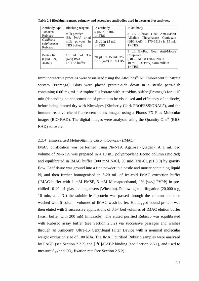
51
Table 2.1 Blocking reagent, primary and secondary antibodies used in western blot analyses.
Antibody type Blocking reagent 1° antibody 2° antibody
Tobacco
Rubisco milk powder
(5% [w/v] dried
milk powder in
TBS buffer)
5 µL in 15 mL
1× TBS 3 µL BioRad Goat Anti-Rabbit
Alkaline Phosphatase Conjugate
(BIO-RAD, # 170-6518) in 15 mL
1× TBS
Galdieria
sulphuraria
Rubisco
15 µL in 15 mL
1× TBS
Penta-His
(QIAGEN,
34460)
15 mL of 3%
(w/v) BSA
1× TBS buffer
20 µL in 15 mL 3%
BSA (w/v) in 1× TBS
3 µL BioRad Goat Anti-Mouse
Conjugate
(BIO-RAD, # 170-6520) in
10 mL 10% (w/v) skim milk in
1× TBS
Immunoreactive proteins were visualised using the AttoPhos® AP Fluorescent Substrate
System (Promega). Blots were placed protein-side down in a sterile petri-dish
containing 0.06 mg mL-1 Attophos® substrate with AttoPhos buffer (Promega) for 1-15
min (depending on concentration of protein to be visualised and efficiency of antibody)
before being blotted dry with Kimwipes (Kimberly-Clark PROFESSIONAL®), and the
immuno-reactive chemi-fluorescent bands imaged using a Pharos FX Plus Molecular
imager (BIO-RAD). The digital images were analysed using the Quantity One® (BIO-
RAD) software.
2.2.4 Immobilized Metal-Affinity Chromatography (IMAC)
IMAC purification was performed using Ni-NTA Agarose (Qiagen). A 1 mL bed
volume of Ni-NTA was prepared in a 10 mL polypropylene Econo column (BioRad)
and equilibrated in IMAC buffer (300 mM NaCl, 50 mM Tris-Cl, pH 8.0) by gravity
flow. Leaf tissue was ground into a fine powder in a pestle and mortar containing liquid
N2 and then further homogenised in 5-20 mL of ice-cold IMAC extraction buffer
(IMAC buffer with 1 mM PMSF, 5 mM Mercaptoethanol, 1% [w/v] PVPP) in pre-
chilled 10-40 mL glass homogenisers (Wheaton). Following centrifugation (20,000 x g,
10 min, at 2 oC) the soluble leaf protein was passed through the column and then
washed with 5 column volumes of IMAC wash buffer. His-tagged bound protein was
then eluted with 3 successive applications of 0.5× bed volumes of IMAC elution buffer
(wash buffer with 200 mM Imidazole). The eluted purified Rubisco was equilibrated
with Rubisco assay buffer (see Section 2.5.2) via successive passages and washes
through an Amicon® Ultra-15 Centrifugal Filter Device with a nominal molecular
weight exclusion size of 100 kDa. The IMAC purified Rubisco samples were analysed
by PAGE (see Section 2.2.2) and [14C]-CABP binding (see Section 2.5.1), and used to
measure Sc/o and CO2-fixation rate (see Section 2.5.2).
52
2.3 Plant maintenance, tissue culture techniques
2.3.1 Seed germination
Nicotiana tabacum (cv Petit Havana N,N) was the tobacco cultivar used in all
transformation experiments. When growing sterile N. tabacum plants the seed were
germinated on MS medium that contained Macro nutrients (18.5 mM KNO3, 1.5 mM
MgSO4·7H2O, 4.45 mM CaCl2·6H2O, 1.25 mM KH2PO4, and 20 mM (NH4)NO3),
Micro nutrients (0.09 mM MnSO4·2H2O, 0.1 mM H3BO4, 0.03 mM ZnSO4·7H2O,
0.005 mM KI, 0.25 µg mL-1 NaMoO4·2H2O, 0.025 µg mL-1 CuSO4·5H2O and 0.025 µg
mL-1 CoCl2·6H2O), 0.006 mM FeCl3·6H2O and 0.001 mM Na2-EDTA·2H2O. The
addition of 1 mg mL-1 thiamine, 0.1 mg mL-1 -napthaleneacetic acid [NAA, auxin], 1
mg mL-1 6-benzylaminopurine [BAP, cytokinin]) and 100 g L-1 myo-inositol to the MS
medium was used to generate Regeneration Media of Plants (RMOP) medium. The pH
of both media was adjusted to 5.7 using 1 M NaOH and 0.6% (w/v) agar added to media
prior to autoclaving. After sterilization the media was cooled to 50°C. Any required
antibiotics (sterilized through 0.2 µ filtration units) were added at this point just prior to
pouring the media. Seeds were sterilised by incubating in 1% (v/v) bleach (Na-
hypochlorite diluted in Milli-Q water) for 10 min and rinsed with repeated changes of
excess sterile water. Floating seeds, indicating a possible lack of embryo, were removed.
Once thoroughly rinsed the seeds were sown on a petri-dish containing MS medium and
grown at 25°C with 50-100 µmol photons m-2 s-1 artificial illumination on a 14:10 h L:D
cycle.
2.3.2 Maintenance of plants
Seedlings germinated for biolistic transformation were individually transferred under
aseptic conditions into sterile 750 mL tissue culture pots (JUPITER INDUSTRIES)
containing 150 mL MS medium containing 3% [w/v] sucrose (see Section 2.3.1) with
100 µg mL-1 Timentin (ticarcillin disodium and clavunalate potassium; SmithKline and
Beecham Phamaceuticals UK) added to prevent fungal contamination. The pots were
placed in a 25°C controlled environment growth cabinet with elevated CO2 conditions
(2.5% [v/v] CO2 in air) and 50-100 µmol photons m-2 s-1 artificial illumination.
Some Rubisco-deplete tobacco genotypes that were unable to be grown from
seed in soil were first germinated on MS medium and grown under elevated CO2 before
transferring to growth in sterile 750 mL tissue culture pots (see above). Once roots and
~4 to 6 true leaves were established the plants were transferred into growth in soil
(Green Wizard Premium potting mix, Debco). During transfer care was taken to gently
53
remove the plants from the tissue culture pots and the roots were carefully massaged in
water at RT to remove residual tissue culture media before they were placed in 2-5 L
pots of soil. Osmocote® (Scotts Australia) fertiliser pellets were added to the soil every
two weeks. Plants were grown in specialised Controlled Environment Rooms (CER) at
25°C, 300-450 µmol photons m-2 s-1 illumination (14:10 h L:D cycle) in air (0.04% [v/v]
CO2) or air with 1% or 2% (v/v) CO2.
2.3.3 Floral pollination processes
Care was taken to collect seed from flowers that had been artificially pollinated with
pollen from the desired tobacco genotype. Cross-pollination was undertaken by
removing the immature anthers from a newly emerging flower and pollinating the
stigma with pollen from another tobacco genotype. Pollination using pollen originating
from wild-type N. tabacum or untransformed plants was termed back-crossing. Self-
pollination was undertaken by rubbing the stigma and mature anthers of the same flower
together. Manually pollinated flowers were marked with colored strings and other
surrounding flowers excised by scalpel before the floral heads were bagged to avoid
unwanted pollination events. The resulting seed pods were harvested 4-7 weeks later
once they were suitably browned and dried. The pods were excised from the plants,
dried at 37°C for 3-5 d and then the seed collected from the pods and stored in 25 mL
glass vials at 4°C.
2.4 Plant transformation
2.4.1 Nuclear transformation using Agrobacterium tumefaciens
Growth and transformation of LBA4404 Agrobacterium cells
The psiSS transforming plasmid was a gift from Dr. Katia Wostrikoff, Cornell
University, USA following its use in the publication of Wostrikoff and Stern (2007).
The genetic details of psiSS are fully described in Chapter 3. Agrobacterium strain
LB4404 (ElectroMAXTM) (Hoekema et al., 1983) cells were grown in autoclave
sterilised YM medium (0.04% [w/v] yeast extract; 1.0% [w/v] mannitol; 1.7 mM NaCl;
0.8 mM MgSO4·H2O; 2.2 mM K2HPO4·3H2O, pH 7.0] at 28°C. Electrocompetent
LB4404 cell stocks (20 µL aliquot) in 10% (v/v) glycerol provided by Dr. Whitney were
transformed with 100 ng of psiSS in a chilled 0.1 cm electroporation cuvette using an E.
coli Pulser (BIO-RAD) at 2.5 kV, 200 Ω, 25 μF. YM medium (1 mL) was added to the
cuvette before transferring to sterile 10 mL round bottom falcon tube and incubating at
28°C for 2 h before plating on agar solidified plates of LB-Kan (30 μg mL-1 kanamycin).
54
After 2 d growth at 28°C a colony of psiSS-transformed LB4404 was used to inoculate
50 mL LB-Kan and grown at 28°C until the turbidity of culture at A600 was ~0.3 before
use for leaf transformation.
Agrotransformation and selection of putative transformants
Under aseptic conditions sterile tissue culture grown tobacco leaves were dissected into
0.5 cm × 0.5 cm pieces in a sterile petri dish. Approximately 10 mL of the psiSS-
LB4404 cells were added to the leaves for 10 min with constant, gentle agitation. Leaf
pieces were then removed, wiped on sterile filter paper and then placed abaxial-side up
on MS medium containing Macro nutrients, Micro nutrients, 3 mM FeSO4·7H2O, 10
mM Na2-EDTA·2H2O, vitamins (20 mg mL-1 inositol, 0.2 mg mL-1, nicotinic acid, 0.2
mg mL-1 pyridoxine HCI, 0.2 mg mL-1 thiamine HCl, 0.2 mg mL-1 glycine), hormones
(0.1 µg mL-1 NAA, 1 µg mL-1 BAP) and 1 mg mL-1 glufosinate (Basta) - the non-
selective herbicide used to select for tissue stably transformed with the selectable
marker bar coded in T-DNA region of psiSS. The tissue was grown at 25°C in a growth
cabinet in air with 2.5% (v/v) CO2, and 50-100 µmol photons m-2 s-1 artificial
illumination for up to 2 months. Putative transgenic callus emerged after 3-4 weeks.
Callus from different leaf sections (i.e. to ensure from independent transformation
events) were successively regenerated 5 times on MS medium containing 5 mg L-1
Basta before transferring the successfully transformed plantlets to 750 mL pots
containing MS medium and timentin (200 mg L-1). Once the plants had established
roots they were transplanted to soil and grown to fertile maturity in a controlled
environment room (CER) (see Section 2.3) and their flowers pollinated appropriately
for segregation analysis and generation of homozygous lines (detailed in Chapter 3).
2.4.2 Biolistic plastome transformation
Preparation of tungsten particles
Tungsten (50 mg, M17 BIO-RAD) was weighed into a 10 mL screw-cap tube
containing 2 mL of 100% ethanol and sterilised by heating in a water bath at 90°C for
30 min. The tube was placed on ice and the tungsten sonicated three times for 5 min
using a LABSONIC 1510 sonicator (B. BRAUN) to ensure dispersal of tungsten
aggregates. The sterilised tungsten was transferred to a sterile 1.8 mL microfuge tube
and pelleted by centrifugation at 8000 g for 15 s. The ethanol in the tube was removed
by pipette and the tungsten suspended in 1 mL of sterile water. This process was
55
repeated four times to remove all ethanol. The tungsten was finally resuspended in
sterile water at a final concentration of 50 µg mL-1 and stored on ice until use.
DNA coating of tungsten particles
Prepared tungsten (50 µL) was added to 10 µg of maxiprep-purified plasmid DNA (see
Section 2.1.2) in a sterile microfuge tube and flicked to mix. Immediately, 50 L of 2.5
M CaCl2 then 20 L of 0.1 M spermidine (Sigma-Aldrich) were sequentially added and
mixed by vortex after each addition. The mixture was then mixed at 4°C using an IKA®
M51 Minishaker (CROWN SCIENTIFIC) at 1400 opm for 30 min. The DNA coated
tungsten were pelleted by centrifugation at 2500 g for ~10 s and the supernatant was
carefully removed by pipette before rinsing with 200 L ethanol. The mixture was
resuspended by flicking. This process for washing the tungsten-DNA particles with
ethanol was repeated 6 more times to remove all spermidine and CaCl2.
Bombardment of DNA into tobacco leaf tissue
Under aseptic conditions young leaves from tissue culture grown tobacco were
dissected into ~3 × 3 cm flattened sections and placed abaxial-side up on Whatman
paper in a petri-dish of MS medium agar containing 3% (w/v) sucrose (Section 2.3.1).
Leaves were bombarded with DNA-coated tungsten using a helium (He) acceleration
gun (BioRad) using 1100 psi rupture discs. Bombarded leaves were incubated at 25°C
under elevated (2.5% [v/v]) CO2 conditions at <20 µmol photons m-2 s-1 artificial
illumination for 2 d before dissecting them into 0.5 cm × 0.5 cm pieces and gently
embedding them into RMOP-spec selective medium (RMOP containing 500 µg mL-1
spectinomycin). After 3-6 weeks the leaf sections bleached white and green
spectinomycin resistant plantlets began to emerge. These plantlets were removed and
continually passaged though further rounds of growth on RMOP-spec medium until
bona-fide transformed lines were identified and viable seed collected.
2.5 Biochemical analyses
2.5.1 [14C]-CABP determination of Rubisco content
Synthesis of RuBP and [12C]- and [14C]-CPBP
Laboratory stocks of RuBP were synthesized and purified according to Kane et al.,
(1998) by Dr. Whitney and used to synthesise carboxypentitiol-1,5-bisphosphate
(CPBP), an isomeric mixture of carboxyribitol-1,5-bisphosphate (CRBP) and the tight
binding Rubisco inhibitor carboxyarabinitol-1,5-bisphosphate (CABP). [12C]- and [14C]-
56
CPBP were made by reacting an appropriate amount of RuBP in 0.1 M Tris-acetate
buffer (pH 8.5) with a 1.1-fold molar excess amount of either K12CN or K14CN (GE
Biosciences) for 48 h in a sealed vial. The reaction was then filtered through AG50W-
X8 (H+ form, BIO-RAD) resin to remove unreacted KCN and the eluent freeze-dried.
The dried residue was dissolved in 50 mM Bicine-NaOH (pH 9.3) and stored at -20°C
(long term storage) or 4°C (for day to day use). The specific activity of the [14C]-CPBP
was 106,667 cpm nmol-1.
Rubisco content analysis (per sample)
Rubisco content was determined by chromatographic separation of Rubisco bound
[14C]-CABP from unbound [14C]-CPBP using a 12 mL bed volume of Sephadex G50
(Fine grade, GE Biosciences) gel filtration in a glass 0.7 x 30 cm Econo-column (BIO-
RAD) equilibrated in column buffer (20 mM Bicine-NaOH, pH 8.0, 75 mM NaCl). Leaf
protein samples (see Section 2.2.1, typically 100 µL) were incubated with 10 to 20 mM
NaHCO3 and 10 mM MgCl2 for 10 min at RT to “activate” each Rubisco catalytic site
(Figure 1.5) before adding 1 µL [14C]-CPBP. After a further 15-30 min at RT the entire
sampled was carefully loaded onto the column, followed by 200 µL of column buffer.
Three further aliquots of buffer (750 µL) were added to the column before any fractions
were collected. The following 7 fractions (750 µL each) were collected into separate 7
mL plastid scintillation vials (16 × 50 mm; Starstedt) and 1 mL of scintillant (Ultima-
Flo Gold, Packard) was added and mixed by vortex. Fractions 2 to 4 contained the
Rubisco-bound [14C]-CABP peak and fractions 6 and 7 the start of the unbound [14C]-
CPBP peak. The radioactivity in each sample was measured using a Tri-Carb 2800TR
Liquid Scintillation Analyzer (PerkinElmer).
Rubisco content analysis (per lane) for SDS and ndPAGE
For SDS and ndPAGE samples, Rubisco content per lane was calculated using
parameters of Rubisco content analysis per sample (Rs), volume of sample used for SDS
or ndPAGE (vs), volume of gel loading buffer (vb) and final volume of sample loaded
onto the gel (vf) using the equation
Total Rubisco content per lane, Rl = vs/(vs+vb)*Rc*v (Eq. 5)
57
2.5.2 Rubisco catalysis measurements
The Michaelis-Menten constants (Km) of Rubisco for CO2 was measured under ambient
O2 concentrations to determine a KC21%O2 value at 25ºC using RuBP-dependent 14CO2
fixation assays using rapidly extracted soluble leaf protein (see Section 2.2.1)
(Sharwood et al., 2008). To fully CO2-Mg2+ activate all the Rubisco sites in the sample
the soluble protein was diluted with an equal volume 2× Mg-14CO2 buffer (50 mM
EPPS-OH, pH 8.0, 20 mM MgCl2, 1 mM EDTA, 20 mM NaH14CO3) and incubated at
25ºC for 7-9 min. The assays were initiated by the addition of 20 μL of the activated
enzyme into septum sealed 8 mL glass scintillation vials containing 480 μl of 14CO2
fixation assay buffer (50 mM EPPS-OH pH 8.2, 1 mM EDTA, 20 mM MgCl2, 0 to ~4.1
mM NaH14CO3 [i.e. 0 to 37.4 µM 14CO2, specific activity ~1500-2000 cpm nmol-1], 10
µg mL-1 carbonic anhydrase, 0.2 mM RuBP). The Henderson-Hasslebach equation used
to calculate the CO2 (C) concentration in the assays is
where Ct is total organic carbon, the V/v is the ratio of reaction vial headspace (V) to
solution volume (v), q is the solubility of CO2 in water at 1 atm at 25°C (0.03292 Mol L-
1 atm-1), R is the universal gas constant, T is the reaction temperature (298K). pK1 and
pK2 values of 6.25 and 10.33, respectively, were used in the calculations.
RuBP-independent 14CO2 fixation control assays were run for each sample and
contained H2O in place of RuBP. After adding sample the assays were stopped after one
minute by adding 100 μl of 25 % (v/v) formic acid. The mixture was dried at 90ºC using
a heating block and the final residue dissolved in 0.5 mL water and mixed with 1 mL of
scintillant (Ultima-Gold). Acid stable radioactivity was measured in a scintillation
counter. Values for Kc21%O2 (in µM) and substrate saturated carboxylation rates(Vc max, in
units of mol CO2 fixed s-1 [vol of sample in assay]-1) were extrapolated from fitting the
data to the Michaelis-Menten equation. The carboxylation turnover rates (kCcat, in units
of s-1) was the determined by dividing Vc max by the mol Rubisco active sites in the assay
as quantified by [14C]-CABP binding (see Section 2.5.1).
2.5.3 14CO2 specific activity determination
To determine the 14CO2 specific activity of the reactions, replicate assays (with the
highest CO2 concentrations) containing known amounts of RuBP (typically only 20
nmol) were allowed to proceed for 30-90 min to ensure full RuBP fixation. The specific
[C] =
1 + + 10(pH-pK
1) + 10
(2pH‒pK1‒pK
2)
(Eq. 6)
Ct
V
vqRT
58
activity was calculated by dividing the 14C values measured by the scintillation counter
by the 20 nmol of RuBP in the assay to obtain specific activity values for the assays in
cpm (nmol CO2 fixed)-1.
2.6 Leaf gas exchange
2.6.1 Optimisation of plant stomatal conditions
Plants were transferred from their growth facility to a laboratory and left overnight in
the dark at atmospheric conditions. Gas exchange was performed the next morning on
young, fully expanded tobacco leaves (typically mid-way down the canopy of plant 30-
40 cm in height) using a Li-COR 6400-02B using a Red / Blue (10%) LED light source
and a set leaf temperature of 25°C. Illumination and CO2 concentrations (Ca) in the leaf
chamber were initially set at 110 µmol photons m-2 s-1and ~96 µbar Ca respectively to
encourage stomatal opening. The apparatus was left for 1 hour prior to measuring how
leaf CO2 assimilation rates (A) responded to changes in intercellular CO2 concentrations
(Ci) (i.e. A-Ci response curves comparable to that shown in Figure 1.09).
Once stomatal conductance (gm) was > 0.25 µmol m-2 s-1, the Ca levels in the
leaf chamber were raised to 400 ppm, illumination increased to 1200 µmol photons m-2
s-1 and the chamber air flow rate set at 500 µmol s-1. After 30 min, equilibration
measurements of A were then logged in triplicate under varying Ci levels between 50
and 1800 µbar. At the completion of the A-Ci measurements the leaf was returned to
steady state (i.e. ~400 µbar Ca) in the dark a measure of the dark respiration rate (Rd)
taken after 15 min before turning the light back on for another 15 min then rapidly
taking samples of the leaf for proteomic (see Section 2.2) and biochemical (see Section
2.5) analysis. The data were then compared with the photosynthetic rates predicted
using the models of Farquhar et al. (1980) as described in Section 1.3.2.
59
CHAPTER 3 – GENERATING A CMTRLRNAI-S
TOBACCO GENOTYPE WHERE S-SUBUNIT
SYNTHESIS IS SILENCED BY RNAI
3.1 Introduction
3.1.1 Mutagenic analysis of the RbcS multigene family in leaf chloroplasts
Initial attempts to genetically modify Rubisco in plants utilised an RbcS anti-sense (anti-
RbcS) knockdown of S-subunit synthesis in tobacco (Rodermel et al., 1988; Hudson et
al., 1992). This approach was subsequently applied to a number of other species
including Solanum lycopersicum (tomato) (Kubien and Sage, 2008), Arabidopsis
thaliana (Izumi et al., 2012), Flaveria bidentis (Furbank et al., 1996) and Oryza sativa
(rice) (Makino et al., 1997). The most potent anti-RbcS tobacco (N. tabacum) genotype
generated had Rubisco levels that were 62% lower than wild-type and therefore
struggled to grow under atmospheric CO2 (Zhang et al., 2002). By using an anti-RbcS
approach, independent transgenic genotypes have produced varied levels of L8S8
Rubisco (Rodermel et al., 1988; Hudson et al., 1992; Andrews et al., 1995).
Accordingly the growth and survival becomes increasingly compromised in the
genotypes producing lower amounts of Rubisco.
These anti-RbcS genotypes proved useful in studying the influence of Rubisco
activity on photosynthesis and growth (Stitt et al., 1991; Bernacchi et al., 2001; Kubien
et al., 2003) and testing the accuracy of the Farquhar, von Caemmerer and Berry
photosynthesis models (Farquhar et al., 1980; von Caemmerer et al., 1994; see also
Section 1.3.2). Conversely transgenic studies have also sought to examine the effect of
overproducing Rubisco by reciprocal genetic approaches to elevate RbcS mRNA levels
in leaves (i.e. a “sense-RbcS” approach). This approach in rice has produced genotypes
with up to a 20% increase in L8S8 Rubisco, which although influencing the N-
partitioning in the plant, did not improve leaf photosynthesis and plant growth (Suzuki
et al., 2007; Suzuki et al., 2009). Other analogous nucleus transformation approaches
have sought to supplement plants with heterologous RbcS to study structural aspects of
the S-subunit influence on Rubisco catalysis. For example introducing an RbcS from
Pisum sativum (pea) into Arabidopsis resulted in the synthesis of chimeric L8S8
enzymes comprising both Arabidopsis and pea S-subunits in an average 7:1 molar ratio
(Getzoff et al., 1998). Biochemical analyses indicated the Rubisco chimers had ~11%
reductions in the carboxylase activity (kCcat) and reduced catalytic site activation status
60
(carbamylation status). These findings suggested Rubisco quaternary conformation was
adversely affected by structural incompatibilities between the subunits (Getzoff et al.,
1998). In contrast a more recent effort to transplant S-subunits from C4 plant Sorghum
bicolor into rice successfully produced genotypes with chimeric rice Rubisco where 30-
80% of the total S-subunit population were from sorghum (Ishikawa et al., 2011).
Consistent with the S-subunits having an influence on catalysis (see Section 1.2.2), the
chimeric rice Rubisco isoforms showed 1.3 to 1.5-fold increases in kCcat and
corresponding reductions in CO2 affinity (i.e. an increase in the Km for CO2) –
reminiscent of the catalytic properties for Rubisco from C4 plants (see Section 1.3.1).
While an exciting step forward in Rubisco bioengineering from the perspective of the S-
subunit, these studies demonstrate a glaring limitation in the bioengineering of S-
subunits in leaf chloroplasts for structure-function analyses – the population of
recombinant L8S8 Rubiscos invariably contain varying stoichiometry of heterologous S-
subunits. As RbcS is a multigene family in plants (for example tobacco has 13 copies
(Gong et al., 2014) (see Section 3.2.1), targeted silencing of all RbcS mRNA and
replacement with a single foreign or modified version has so far been unfeasible.
3.1.2 Relocating RbcS into the tobacco chloroplast
Difficulties in targeted modification of RbcS in the nucleus of higher plants led to
efforts in engineering the gene in the chloroplast genome (plastome). Initial trials using
tobacco introduced native tobacco RbcS (coding a hepta-histidine [H7] C-terminal tag)
both with and without its chloroplast transit peptide coding sequence (Whitney and
Andrews, 2001a). Both RbcS transgenes (tpRbcSH7 and RbcSH7) were inserted into the
inverted repeat regions of the tobacco plastome to produce independently transformed
RVtpSSuH and RVSSuH tobacco genotypes (discussed in further detail in Section
4.1.4). While it was shown that plastid-synthesized S-subunits could successfully
assemble into L8S8 Rubisco, only ~1% of plastid made S-subunits assembled despite
their chloroplast RbcS mRNA levels exceeding endogenous cytosolic RbcS mRNA
levels by more than 10-fold.
Possible inefficiencies with translation of the plastid made S-subunits or/and
strong competition by the endogenous cytosol made S-subunits are possible
explanations for the paucity of plastid made S-subunits assembled (Whitney and
Andrews, 2001a; Zhang et al., 2002). Consistent with this a comparable transplastomic
approach in an anti-RbcS tobacco line found no evidence for chloroplast made S-
subunits impacting Rubisco production (Zhang et al., 2002). A subsequent attempt
61
proved more successful when transforming the same anti-RbcS tobacco genotype with
RbcS inserted within a transcriptionally active spacer region between trnI and trnA of
the plastome inverted repeat regions (Dhingra et al., 2004). The resulting transplastomic
lines showed significant improvements in Rubisco levels – almost to wild-type levels.
Whether this increase in Rubisco levels was solely attributable to plastid made S-
subunit synthesis or associated with partial silencing of anti-RbcS phenotype was not
experimentally examined. Testing for this would be feasible by confirming Rubisco
levels were still low in the progeny of wild-type plants pollinated with pollen from the
transplastomic lines.
3.1.3 cmtrL - a tobacco genotype for chloroplast-targeted Rubisco bioengineering
An approach to study the role of the S-subunit on Rubisco catalysis by coupling RNAi
silencing of RbcS mRNA production with transplastomic integration of RbcS into the
plastome has been proposed (Sharwood et al., 2008). An important consideration is that
elimination of Rubisco synthesis in plants by RNAi-RbcS would produce progeny of
poor viability and unable to grow to fertile maturity outside of tissue culture or via
grafting onto wild-type stock. Such genotypes would essentially be comparable to the
rbcL deletion plants of Kanevski and Maliga (1994). However the cmtrL tobacco
genotype (see Section 1.4.3) provides a viable alternative for eliminating tobacco S-
subunits synthesis without affecting growth phenotype. In cmtrL the tobacco Rubisco L-
subunit gene (rbcL) has been replaced with cmrbcM coding for Rhodospirillum rubrum
L2 Rubisco (Whitney and Sharwood, 2008) (Figure 3.1A). While the cmtrL line requires
growth under high-CO2 conditions to compensate for the poor catalytic properties of R.
rubrum Rubisco (see Section 1.1.5) it is readily capable of growing to maturity in soil
without detriment to phenotype in air with high-CO2 (i.e. greater than 0.5% [v/v],
Figure 3.1C). Of additional benefit is the cmtrL line poses a tailor-made genotype for
efficiently introducing rbcL coding heterologous Rubisco L-subunits from plants
(Whitney et al., 2011b) and Archaea (Alonso et al., 2009), in addition to re-inserting
mutated forms of the tobacco rbcL (Whitney and Sharwood, 2008; Whitney et al.,
2011a). The cmtrL genotype is also advantaged by the size difference between the S-
subunit lacking R. rubrum L2 Rubisco (~100 kDa) and larger L8S8 Rubisco (~520 kDa)
that make identifying L8S8 Rubisco transformed lines easily discernible by non-
denaturing PAGE (ndPAGE) (Whitney and Sharwood, 2008).
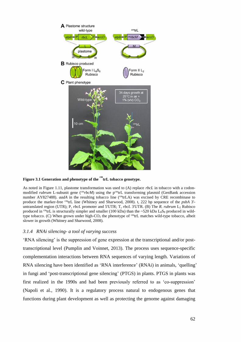
62
Figure 3.1 Generation and phenotype of the cm
trL tobacco genotype.
As noted in Figure 1.11, plastome transformation was used to (A) replace rbcL in tobacco with a codon-
modified rubrum L-subunit gene (cmrbcM) using the pcmtrL transforming plasmid (GenBank accession
number AY827488). aadA in the resulting tobacco line (cmtrLA) was excised by CRE recombinase to
produce the marker-free cmtrL line (Whitney and Sharwood, 2008). t, 222 bp sequence of the psbA 3'-
untranslated region (UTR); P, rbcL promoter and 5'UTR; T, rbcL 3'UTR. (B) The R. rubrum L2 Rubisco
produced in cmtrL is structurally simpler and smaller (100 kDa) than the ~520 kDa L8S8 produced in wild-
type tobacco. (C) When grown under high-CO2 the phenotype of cmtrL matches wild-type tobacco, albeit
slower in growth (Whitney and Sharwood, 2008).
3.1.4 RNAi silencing- a tool of varying success
‘RNA silencing’ is the suppression of gene expression at the transcriptional and/or post-
transcriptional level (Pumplin and Voinnet, 2013). The process uses sequence-specific
complementation interactions between RNA sequences of varying length. Variations of
RNA silencing have been identified as ‘RNA interference’ (RNAi) in animals, ‘quelling’
in fungi and ‘post-transcriptional gene silencing’ (PTGS) in plants. PTGS in plants was
first realized in the 1990s and had been previously referred to as ‘co-suppression’
(Napoli et al., 1990). It is a regulatory process natural to endogenous genes that
functions during plant development as well as protecting the genome against damaging

63
changes introduced by viruses and mobile DNA elements such as transposons (Voinnet,
2002; Pumplin and Voinnet, 2013).
PTGS regulates cytoplasmic RNA levels through sequence-specific degradation
involving a cascade of RNA-interacting proteins (Chen and Aravin, 2015). Degradation
is triggered by varying types of small double-stranded RNA (dsRNA) such as
microRNA (miRNA) or small-interfering RNA (siRNA). These 21-25 nucleotide RNA
molecules are made from the cleavage of dsRNA or hairpin-loop RNA structures by a
nuclease complex made of DICER-LIKE1 protein (DCL1) (Blevins et al., 2006;
Ahmadovich Bozorov et al., 2012), HYPONASTIC LEAVES1 (HYL1) (Han et al.,
2004; Kurihara et al., 2006) and SERRATE (SE) (Yang et al., 2006). As shown in
Figure 3.2, the miRNA and siRNA products are recognised by their nucleotide structure
and methylated at their 3’ ends by HUA ENHANCER1 (HEN1) (Park et al., 2002).
HEN1 is a plant miRNA methyltransferase protein that promotes binding of the small
RNAs to an RNA-induced silencing complex (RISC). The RISC complex contains an
Argonaute-1 (AGO1) protein that helps identify target mRNA for degradation or to
inhibit translation by DNA/histone modification (Sunkar and Zhu, 2007). AGO1 also
serves as an endonuclease to cleave the targeted mRNA (Martinez and Tuschl, 2004). In
addition to AGO1, RISC also has an ATP-dependent helicase-like component that
separates the double-stranded RNA at its less stable strand (usually its antisense strand),
and retains the antisense strand as a guide to target additional mRNAs for degradation
(Khvorova et al., 2003) (Figure 3.2).
3.1.5 The tobacco RbcS multigene family and its potential silencing by RNAi
The RbcS multigene family is a result of allopolypoidy and cytonuclear evolution from
the two parents (N. sylvestris♀ and N. tomentosiformis♂) of N. tabacum (see Section
1.4.1). RbcS expression appears to be light regulated by one or two repeats of a set of
light responsive elements (Jiang et al., 1994) sequences approximately 166-149 bp
upstream from the TATA box (Gupta, 2009). Differential expression of the RbcS
mRNAs may also vary due to intrinsic differences in the stability of their transcripts and
thus provide inaccurate assessment of difference in transcription rates of the varying
RbcS alleles (Izumi et al., 2012).
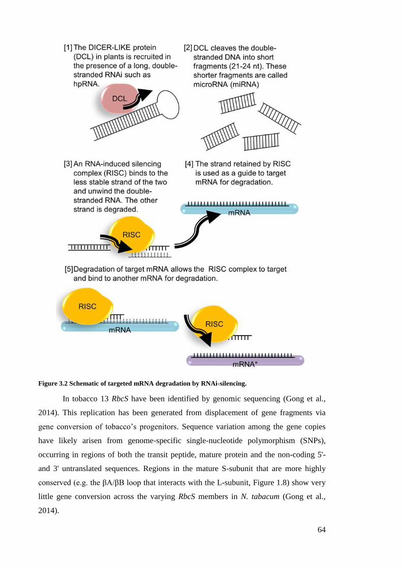
64
Figure 3.2 Schematic of targeted mRNA degradation by RNAi-silencing.
In tobacco 13 RbcS have been identified by genomic sequencing (Gong et al.,
2014). This replication has been generated from displacement of gene fragments via
gene conversion of tobacco’s progenitors. Sequence variation among the gene copies
have likely arisen from genome-specific single-nucleotide polymorphism (SNPs),
occurring in regions of both the transit peptide, mature protein and the non-coding 5'-
and 3' untranslated sequences. Regions in the mature S-subunit that are more highly
conserved (e.g. the βA/βB loop that interacts with the L-subunit, Figure 1.8) show very
little gene conversion across the varying RbcS members in N. tabacum (Gong et al.,
2014).
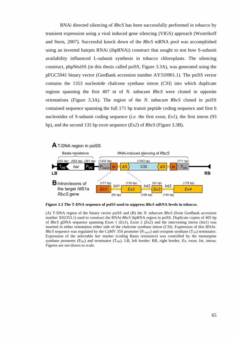
65
RNAi directed silencing of RbcS has been successfully performed in tobacco by
transient expression using a viral induced gene silencing (VIGS) approach (Wostrikoff
and Stern, 2007). Successful knock down of the RbcS mRNA pool was accomplished
using an inverted hairpin RNAi (ihpRNAi) construct that sought to test how S-subunit
availability influenced L-subunit synthesis in tobacco chloroplasts. The silencing
construct, phpNtsiSS (in this thesis called psiSS, Figure 3.3A), was generated using the
pFGC5941 binary vector (GenBank accession number AY310901.1). The psiSS vector
contains the 1353 nucleotide chalcone synthase intron (CSI) into which duplicate
regions spanning the first 407 nt of N. tabacum RbcS were cloned in opposite
orientations (Figure 3.3A). The region of the N. tabacum RbcS cloned in psiSS
contained sequence spanning the full 171 bp transit peptide coding sequence and first 6
nucleotides of S-subunit coding sequence (i.e. the first exon; Ex1), the first intron (93
bp), and the second 135 bp exon sequence (Ex2) of RbcS (Figure 3.3B).
Figure 3.3 The T-DNA sequence of psiSS used to suppress RbcS mRNA levels in tobacco.
(A) T-DNA region of the binary vector psiSS and (B) the N. tabacum RbcS (from GenBank accession
number X02353.1) used to construct the RNAi-RbcS ihpRNA region in psiSS. Duplicate copies of 405 bp
of RbcS gDNA sequence spanning Exon 1 (Ex1), Exon 2 (Ex2) and the intervening intron (Int1) was
inserted in either orientation either side of the chalcone synthase intron (CSI). Expression of this RNAi-
RbcS sequence was regulated by the CaMV 35S promoter (PCamV) and octopine synthase (TOS) terminator.
Expression of the selectable bar marker (coding Basta resistance) was controlled by the mannopine
synthase promoter (PMS) and terminator (TMS). LB, left border; RB, right border; Ex, exon; Int, intron.
Figures are not drawn to scale.

66
3.1.6 Research objective – Silencing S-subunit synthesis in cmtrL by ihpRNAi-RbcS
Being able to study specific protein-protein interactions between the S- and L-subunits
of plant Rubisco has remained an elusive goal. As indicated above, partial success has
been obtained through introduction of RbcS transgenes by nucleus and plastome
transformation approaches, although catalytic interpretation of these chimeric enzymes
is of limited use given they contain heterologous populations of S-subunits. Fully
appreciating the molecular detail of subunit interactions in higher plant Rubisco is
stymied by this heterogeneity as it precludes the generation of recombinant Rubisco
containing a homogeneous population of S-subunits. Such limitations are avoided in
Chlamydomonas due to the availability of a mutant strain where both nuclear RbcS have
been silenced by insertion mutagenesis (Khrebtukova and Spreitzer, 1996). This has
allowed for testing of specific sequence changes in both the L- and S-subunits
(Karkehabadi et al., 2005; Spreitzer et al., 2005; Genkov and Spreitzer, 2009; Genkov et
al., 2010). No comparable system currently exists in plants due to RbcS comprising a
larger multigene family. The inability to express plant L8S8 Rubisco in E. coli or other
expression host further limits structure-function studies of plant L- and S-subunits.
This chapter describes the generation of a new tobacco genotype where nuclear
S-subunit synthesis is eliminated using an RNAi-RbcS approach. As summarised in
Figure 3.4, the target tobacco genotype for transformation is the cmtrL line where the L2
R. rubrum Rubisco has no requirement for S-subunits and therefore should not be
affected by RNAi directed silencing of S-subunit synthesis.
Figure 3.4 Research objective summary – producing an RNAi-RbcS silenced tobacco line using the cmtrL genotype.
As the S-subunits have no assembly partner in cmtrL they are rapidly degraded (Whitney and Andrews
2001a). Silencing S-subunit synthesis by RNAi in this tobacco genotype is therefore not expected to
disadvantage growth.
67
3.2 Results
3.2.1 Analysis of the RbcS diversity in tobacco
Since the publication of the full genome sequence for Arabidopsis some 15 years ago
(Analysis of the genome sequence of the flowering plant Arabidopsis thaliana, 2000)
there has been an exponential increase in the number of plant genomes sequenced. It is
therefore surprising that an entire genome sequence for N. tabacum has only recently
been publically available (Gong et al., 2014) albeit not annotated. Greater progress has
been made with annotating the genome and transcriptome sequence of N. benthamiana
(http://sydney.edu.au/science/molecular_bioscience/benthamiana/), the variety mostly
used in Agro-infiltration experimental transgene applications (Goodin et al., 2008). A
bioinformatic analysis of the new N. tabacum sequence was undertaken with regard to
annotating the genetic information of its RbcS. In general, RbcS of higher plants
typically comprise two or more introns (Wolter et al., 1988). As shown in Figure 3.5,
this is also the case for the 13 RbcS in N. tabacum with 3 alleles having three introns
and the other 10 only two introns. In this analysis the genes maternally derived from the
N. sylvestris genome are represented by the prefix NtS and those paternally derived from
N. tomentosiformis are indicated by NtT.
Analysis of the 13 N. tabacum RbcS found the NtS5 (Nicotiana tabacum
Sylvestris parent RbcS 5) and NtT5 (Nicotiana tabacum Tomentosiformis parent RbcS 5)
to be paralogs as they were sufficiently divergent from the other genes by each
containing a ~3-fold larger first intron of ~500 bp (Figure 3.5) (Gong et al., 2014).
When aligned a number of the RbcS alleles were found to share 100% identity in their
mature S-subunit coding sequence (i.e. NtS1a & NtS1b; NtS3 & NtT3a & NtS4) (Figure
3.6A). In terms of nucleotide and amino acid sequence identity, all the RbcS alleles
showed greater variation in the region coding the N-terminal transit peptide pre-
sequence that spans 171-174 nucleotides (i.e. 57 or 58 amino acids) (Figure 3.6A). As
shown in Figure 3.6B, alignment of only the mature S-subunit coding sequence
identified several polymorphic residues that constitute 9 different S-subunit isoforms
(labelled Nt-SS1 to Nt-SS9) within N. tabacum. In this thesis, the S-subunit isoform
studied constitutes the Nt-SS1 isoform which is coded by the same RbcS inserted into
the nuclear transforming plasmid psiSS (Figure 3.3A) (Wostrikoff and Stern, 2007) and
transformed into the tobacco plastome to make the RVtpSSuH and RVSSuH genotypes
(Whitney and Andrews, 2001a) and described further in Chapter 4.

68
Figure 3.5 The RBCS multigene family in N. tabacum.
The nucleotide sequence of the 13 RbcS in N. tabacum vary mainly in their intron sequences. The NtS1a,
NtS1b and NtT1 alleles are the only members in the family having 3 introns, the remainder only having 2
introns. NtS1a and NtS1b (highlighted in blue) are the RbcS used in the transgenic studies described in
this thesis. NtS5 and NtT5 are paralogs that are divergent from the other family members. Genes
maternally derived from the N. sylvestris genome are represented by the prefix NtS while those paternally
derived from N. tomentosiformis are indicated by NtT. Genbank accession numbers for each member of
the RbcS multigene family can be found in Appendix B.
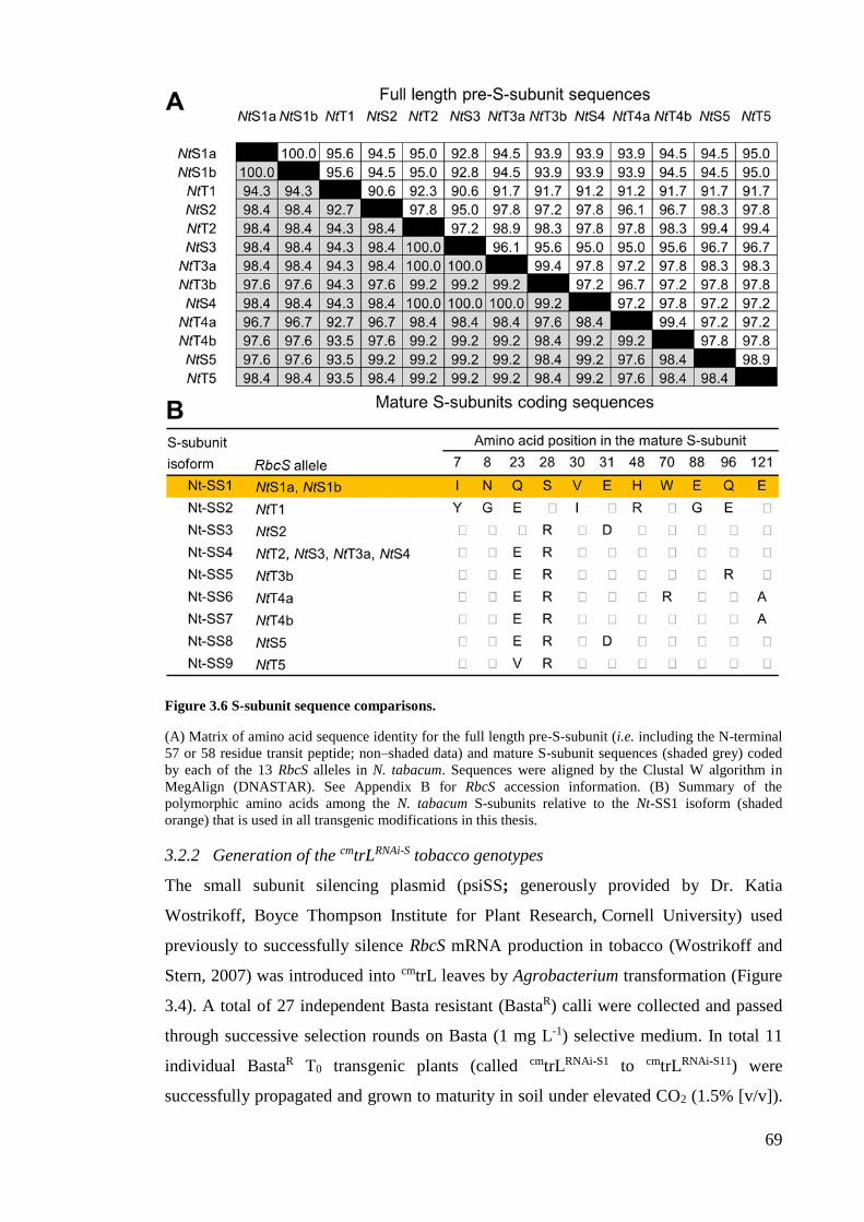
69
Figure 3.6 S-subunit sequence comparisons.
(A) Matrix of amino acid sequence identity for the full length pre-S-subunit (i.e. including the N-terminal
57 or 58 residue transit peptide; non–shaded data) and mature S-subunit sequences (shaded grey) coded
by each of the 13 RbcS alleles in N. tabacum. Sequences were aligned by the Clustal W algorithm in
MegAlign (DNASTAR). See Appendix B for RbcS accession information. (B) Summary of the
polymorphic amino acids among the N. tabacum S-subunits relative to the Nt-SS1 isoform (shaded
orange) that is used in all transgenic modifications in this thesis.
3.2.2 Generation of the cmtrLRNAi-S tobacco genotypes
The small subunit silencing plasmid (psiSS; generously provided by Dr. Katia
Wostrikoff, Boyce Thompson Institute for Plant Research, Cornell University) used
previously to successfully silence RbcS mRNA production in tobacco (Wostrikoff and
Stern, 2007) was introduced into cmtrL leaves by Agrobacterium transformation (Figure
3.4). A total of 27 independent Basta resistant (BastaR) calli were collected and passed
through successive selection rounds on Basta (1 mg L-1) selective medium. In total 11
individual BastaR T0 transgenic plants (called cmtrLRNAi-S1 to cmtrLRNAi-S11) were
successfully propagated and grown to maturity in soil under elevated CO2 (1.5% [v/v]).
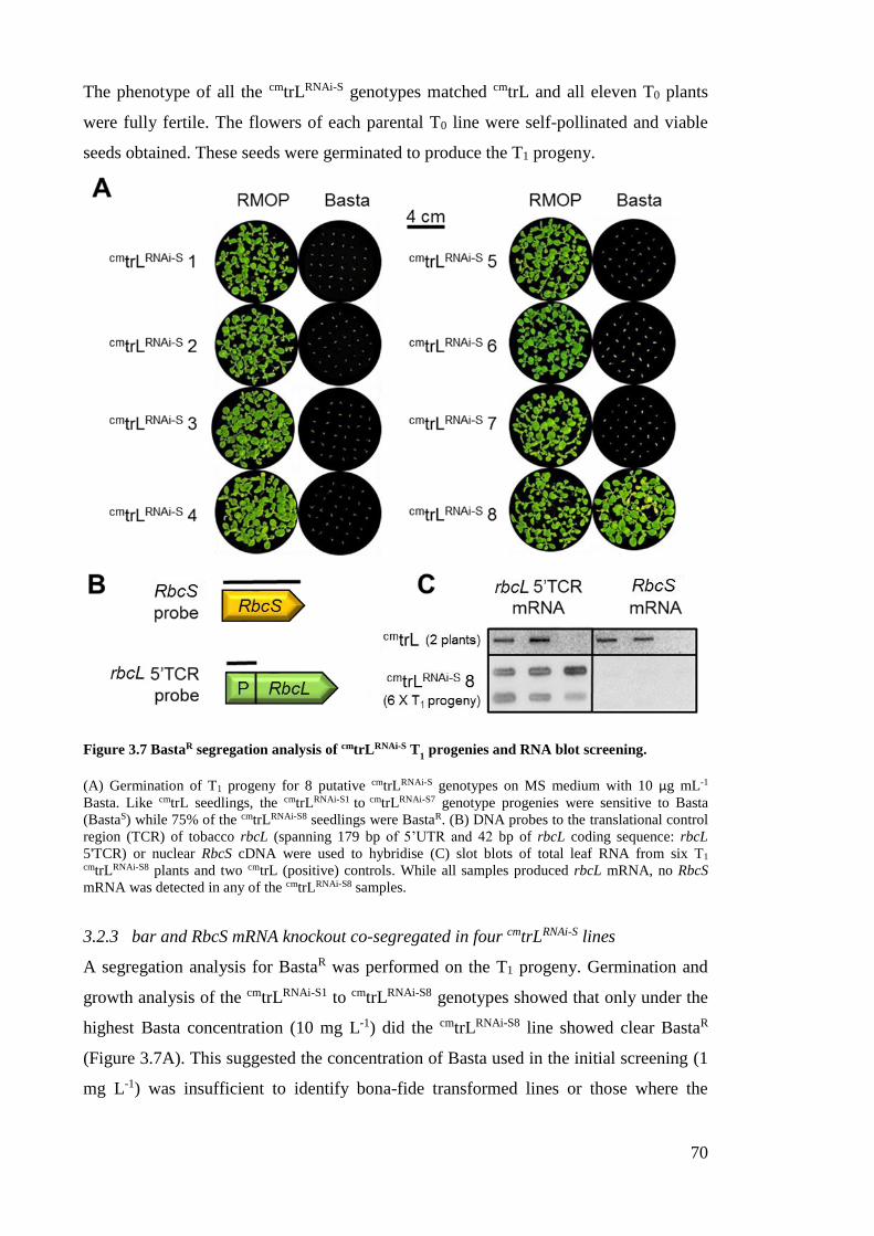
70
The phenotype of all the cmtrLRNAi-S genotypes matched cmtrL and all eleven T0 plants
were fully fertile. The flowers of each parental T0 line were self-pollinated and viable
seeds obtained. These seeds were germinated to produce the T1 progeny.
Figure 3.7 BastaR segregation analysis of cmtrLRNAi-S T1 progenies and RNA blot screening.
(A) Germination of T1 progeny for 8 putative cmtrLRNAi-S genotypes on MS medium with 10 µg mL-1
Basta. Like cmtrL seedlings, the cmtrLRNAi-S1 to cmtrLRNAi-S7 genotype progenies were sensitive to Basta
(BastaS) while 75% of the cmtrLRNAi-S8 seedlings were BastaR. (B) DNA probes to the translational control
region (TCR) of tobacco rbcL (spanning 179 bp of 5’UTR and 42 bp of rbcL coding sequence: rbcL
5'TCR) or nuclear RbcS cDNA were used to hybridise (C) slot blots of total leaf RNA from six T1 cmtrLRNAi-S8 plants and two cmtrL (positive) controls. While all samples produced rbcL mRNA, no RbcS
mRNA was detected in any of the cmtrLRNAi-S8 samples.
3.2.3 bar and RbcS mRNA knockout co-segregated in four cmtrLRNAi-S lines
A segregation analysis for BastaR was performed on the T1 progeny. Germination and
growth analysis of the cmtrLRNAi-S1 to cmtrLRNAi-S8 genotypes showed that only under the
highest Basta concentration (10 mg L-1) did the cmtrLRNAi-S8 line showed clear BastaR
(Figure 3.7A). This suggested the concentration of Basta used in the initial screening (1
mg L-1) was insufficient to identify bona-fide transformed lines or those where the
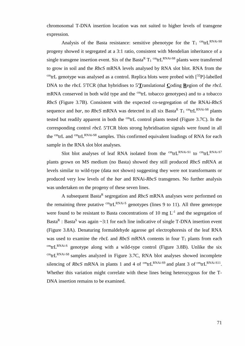
71
chromosomal T-DNA insertion location was not suited to higher levels of transgene
expression.
Analysis of the Basta resistance: sensitive phenotype for the T1 cmtrLRNAi-S8
progeny showed it segregated at a 3:1 ratio, consistent with Mendelian inheritance of a
single transgene insertion event. Six of the BastaR T1 cmtrLRNAi-S8 plants were transferred
to grow in soil and the RbcS mRNA levels analysed by RNA slot blot. RNA from the
cmtrL genotype was analysed as a control. Replica blots were probed with [32P]-labelled
DNA to the rbcL 5'TCR (that hybridises to 5'Translational Coding Region of the rbcL
mRNA conserved in both wild type and the cmtrL tobacco genotypes) and to a tobacco
RbcS (Figure 3.7B). Consistent with the expected co-segregation of the RNAi-RbcS
sequence and bar, no RbcS mRNA was detected in all six BastaR T1 cmtrLRNAi-S8 plants
tested but readily apparent in both the cmtrL control plants tested (Figure 3.7C). In the
corresponding control rbcL 5'TCR blots strong hybridisation signals were found in all
the cmtrL and cmtrLRNAi-S8 samples. This confirmed equivalent loadings of RNA for each
sample in the RNA slot blot analyses.
Slot blot analyses of leaf RNA isolated from the cmtrLRNAi-S1 to cmtrLRNAi-S7
plants grown on MS medium (no Basta) showed they still produced RbcS mRNA at
levels similar to wild-type (data not shown) suggesting they were not transformants or
produced very low levels of the bar and RNAi-RbcS transgenes. No further analysis
was undertaken on the progeny of these seven lines.
A subsequent BastaR segregation and RbcS mRNA analyses were performed on
the remaining three putative cmtrLRNAi-S genotypes (lines 9 to 11). All three genetoype
were found to be resistant to Basta concentrations of 10 mg L-1 and the segregation of
BastaR : BastaS was again ~3:1 for each line indicative of single T-DNA insertion event
(Figure 3.8A). Denaturing formaldehyde agarose gel electrophoresis of the leaf RNA
was used to examine the rbcL and RbcS mRNA contents in four T1 plants from each
cmtrLRNAi-S genotype along with a wild-type control (Figure 3.8B). Unlike the six
cmtrLRNAi-S8 samples analyzed in Figure 3.7C, RNA blot analyses showed incomplete
silencing of RbcS mRNA in plants 1 and 4 of cmtrLRNAi-S9 and plant 3 of cmtrLRNAi-S11.
Whether this variation might correlate with these lines being heterozygous for the T-
DNA insertion remains to be examined.
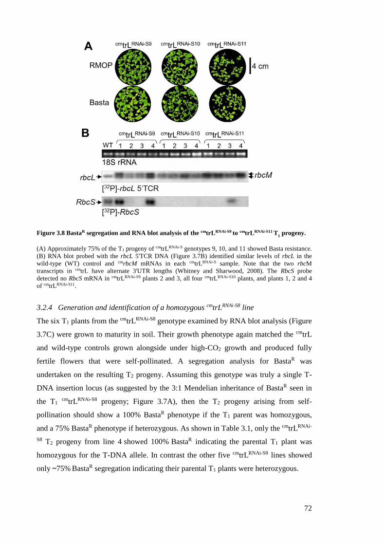
72
Figure 3.8 BastaR segregation and RNA blot analysis of the cmtrLRNAi-S9 to cmtrLRNAi-S11 T1 progeny.
(A) Approximately 75% of the T1 progeny of cmtrLRNAi-S genotypes 9, 10, and 11 showed Basta resistance.
(B) RNA blot probed with the rbcL 5'TCR DNA (Figure 3.7B) identified similar levels of rbcL in the
wild-type (WT) control and cmrbcM mRNAs in each cmtrLRNAi-S sample. Note that the two rbcM
transcripts in cmtrL have alternate 3'UTR lengths (Whitney and Sharwood, 2008). The RbcS probe
detected no RbcS mRNA in cmtrLRNAi-S9 plants 2 and 3, all four cmtrLRNAi-S10 plants, and plants 1, 2 and 4
of cmtrLRNAi-S11.
3.2.4 Generation and identification of a homozygous cmtrLRNAi-S8 line
The six T1 plants from the cmtrLRNAi-S8 genotype examined by RNA blot analysis (Figure
3.7C) were grown to maturity in soil. Their growth phenotype again matched the cmtrL
and wild-type controls grown alongside under high-CO2 growth and produced fully
fertile flowers that were self-pollinated. A segregation analysis for BastaR was
undertaken on the resulting T2 progeny. Assuming this genotype was truly a single T-
DNA insertion locus (as suggested by the 3:1 Mendelian inheritance of BastaR seen in
the T1 cmtrLRNAi-S8 progeny; Figure 3.7A), then the T2 progeny arising from self-
pollination should show a 100% BastaR phenotype if the T1 parent was homozygous,
and a 75% BastaR phenotype if heterozygous. As shown in Table 3.1, only the cmtrLRNAi-
S8 T2 progeny from line 4 showed 100% BastaR indicating the parental T1 plant was
homozygous for the T-DNA allele. In contrast the other five cmtrLRNAi-S8 lines showed
only ~75% BastaR segregation indicating their parental T1 plants were heterozygous.
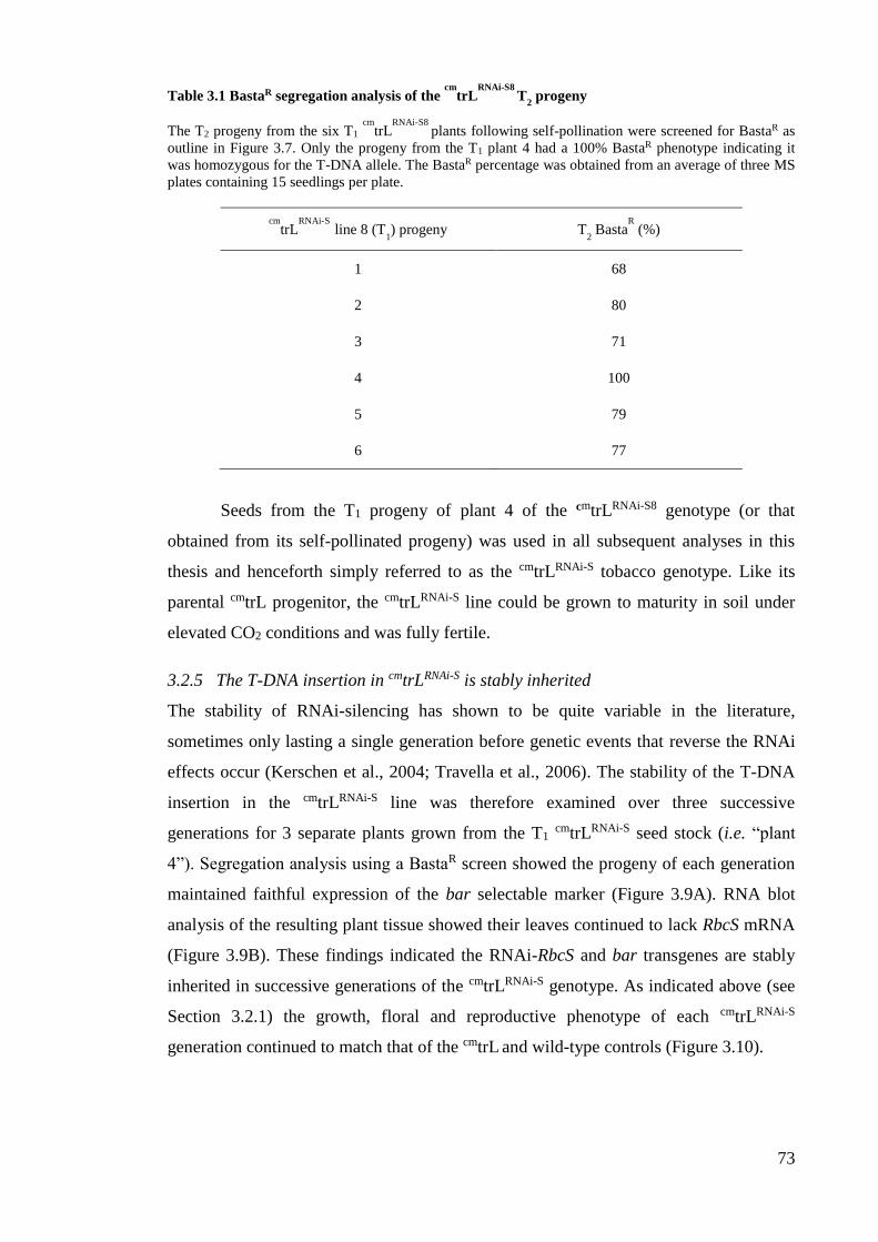
73
Table 3.1 BastaR segregation analysis of the cm
trLRNAi-S8
T2 progeny
The T2 progeny from the six T1 cm
trLRNAi-S8
plants following self-pollination were screened for BastaR as
outline in Figure 3.7. Only the progeny from the T1 plant 4 had a 100% BastaR phenotype indicating it
was homozygous for the T-DNA allele. The BastaR percentage was obtained from an average of three MS
plates containing 15 seedlings per plate.
cmtrL
RNAi-S line 8 (T
1) progeny T
2 Basta
R (%)
1 68
2 80
3 71
4 100
5 79
6 77
Seeds from the T1 progeny of plant 4 of the cmtrLRNAi-S8 genotype (or that
obtained from its self-pollinated progeny) was used in all subsequent analyses in this
thesis and henceforth simply referred to as the cmtrLRNAi-S tobacco genotype. Like its
parental cmtrL progenitor, the cmtrLRNAi-S line could be grown to maturity in soil under
elevated CO2 conditions and was fully fertile.
3.2.5 The T-DNA insertion in cmtrLRNAi-S is stably inherited
The stability of RNAi-silencing has shown to be quite variable in the literature,
sometimes only lasting a single generation before genetic events that reverse the RNAi
effects occur (Kerschen et al., 2004; Travella et al., 2006). The stability of the T-DNA
insertion in the cmtrLRNAi-S line was therefore examined over three successive
generations for 3 separate plants grown from the T1 cmtrLRNAi-S seed stock (i.e. “plant
4”). Segregation analysis using a BastaR screen showed the progeny of each generation
maintained faithful expression of the bar selectable marker (Figure 3.9A). RNA blot
analysis of the resulting plant tissue showed their leaves continued to lack RbcS mRNA
(Figure 3.9B). These findings indicated the RNAi-RbcS and bar transgenes are stably
inherited in successive generations of the cmtrLRNAi-S genotype. As indicated above (see
Section 3.2.1) the growth, floral and reproductive phenotype of each cmtrLRNAi-S
generation continued to match that of the cmtrL and wild-type controls (Figure 3.10).
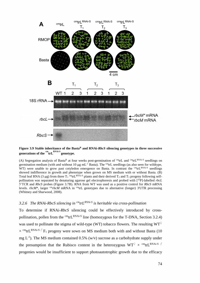
74
Figure 3.9 Stable inheritance of the BastaR and RNAi-RbcS silencing genotypes in three successive
generations of the cm
trLRNAi-S
genotype.
(A) Segregation analysis of BastaR at four weeks post-germination of cmtrL and cmtrLRNAi-S seedlings on
germination medium (with and without 10 µg mL-1 Basta). The cmtrL seedlings (as also seen for wildtype,
WT) were unable to grow past cotyledon emergence on Basta. In contrast the cmtrLRNAi-S seedlings
showed indifference in growth and phenotype when grown on MS medium with or without Basta. (B)
Total leaf RNA (3 µg) from three T1 cmtrLRNAi-S plants and their derived T2 and T3 progeny following self-
pollination was separated by denaturing agarose gel electrophoresis and probed with [32P]-labelled rbcL
5’TCR and RbcS probes (Figure 3.7B). RNA from WT was used as a positive control for RbcS mRNA
levels. rbcM*, larger cmrbcM mRNA in cmtrL genotypes due to alternative (longer) 3'UTR processing
(Whitney and Sharwood, 2008).
3.2.6 The RNAi-RbcS silencing in cmtrLRNAi-S is heritable via cross-pollination
To determine if RNAi-RbcS silencing could be effectively introduced by cross-
pollination, pollen from the cmtrLRNAi-S line (homozygous for the T-DNA, Section 3.2.4)
was used to pollinate the stigma of wild-type (WT) tobacco flowers. The resulting WT♀
× cmtrLRNAi-S ♂ F1 progeny were sown on MS medium both with and without Basta (10
mg L-1). The MS medium contained 0.5% (w/v) sucrose as a carbohydrate supply under
the presumption that the Rubisco content in the heterozygous WT♀ × cmtrLRNAi-S ♂
progenies would be insufficient to support photoautotrophic growth due to the efficacy

75
of RNAi-RbcS silencing. As expected, all the progeny were BastaR and showed a
chlorotic phenotype when compared to wild-type (Figure 3.11).
Figure 3.10 The conserved phenotype after successive generations of cm
trLRNAi-S
.
Self-pollination of T0, T1 and T2 plants of the cmtrLRNAi-S line gave progenies (T1, T2 and T3 respectively)
whose (A) vegetative growth and (B) floral phenotypes under high-CO2 conditions remained identical to
the cmtrL and wild-type tobacco genotypes.
This phenotype matched that found for tobacco genotypes making little or no Rubisco
(Whitney and Sharwood, 2008) suggesting effective silencing of RbcS mRNA and S-
subunit synthesis in the plants that correspondingly depleted L8S8 Rubisco biogenesis.
The dependency of the WT♀ × cmtrLRNAi-S♂ F1 seedlings for sucrose
supplementation for survival was tested by transplanting seedlings from the MS
medium containing Basta (Figure 3.12A) into medium either
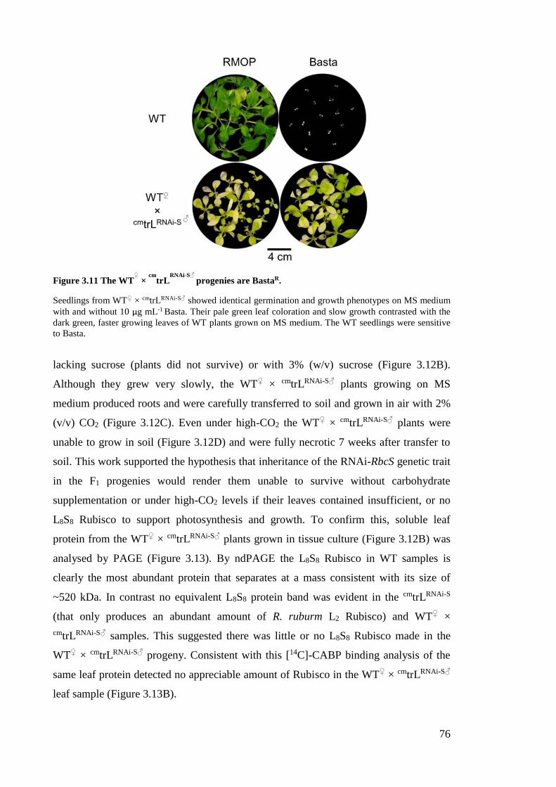
76
Figure 3.11 The WT♀ ×
cmtrL
RNAi-S♂ progenies are BastaR.
Seedlings from WT♀ × cmtrLRNAi-S♂ showed identical germination and growth phenotypes on MS medium
with and without 10 µg mL-1 Basta. Their pale green leaf coloration and slow growth contrasted with the
dark green, faster growing leaves of WT plants grown on MS medium. The WT seedlings were sensitive
to Basta.
lacking sucrose (plants did not survive) or with 3% (w/v) sucrose (Figure 3.12B).
Although they grew very slowly, the WT♀ × cmtrLRNAi-S♂ plants growing on MS
medium produced roots and were carefully transferred to soil and grown in air with 2%
(v/v) CO2 (Figure 3.12C). Even under high-CO2 the WT♀ × cmtrLRNAi-S♂ plants were
unable to grow in soil (Figure 3.12D) and were fully necrotic 7 weeks after transfer to
soil. This work supported the hypothesis that inheritance of the RNAi-RbcS genetic trait
in the F1 progenies would render them unable to survive without carbohydrate
supplementation or under high-CO2 levels if their leaves contained insufficient, or no
L8S8 Rubisco to support photosynthesis and growth. To confirm this, soluble leaf
protein from the WT♀ × cmtrLRNAi-S♂ plants grown in tissue culture (Figure 3.12B) was
analysed by PAGE (Figure 3.13). By ndPAGE the L8S8 Rubisco in WT samples is
clearly the most abundant protein that separates at a mass consistent with its size of
~520 kDa. In contrast no equivalent L8S8 protein band was evident in the cmtrLRNAi-S
(that only produces an abundant amount of R. ruburm L2 Rubisco) and WT♀ ×
cmtrLRNAi-S♂ samples. This suggested there was little or no L8S8 Rubisco made in the
WT♀ × cmtrLRNAi-S♂ progeny. Consistent with this [14C]-CABP binding analysis of the
same leaf protein detected no appreciable amount of Rubisco in the WT♀ × cmtrLRNAi-S♂
leaf sample (Figure 3.13B).

77
Figure 3.12 The WT♀ × cmtrLRNAi-S♂ progeny are unable to grow autotrophically.
(A) A four-week old WT♀ × cmtrLRNAi-S♂ seedling on MS medium-Basta (0.5% w/v sucrose) was
transferred to (B) growth on germination media containing 3% (w/v) sucrose. Although growth was very
slow, the WT♀ × cmtrLRNAi-S♂ plant produced roots and was carefully transferred to (C) growth in soil in
air with 2% (v/v) CO2 showed. (D) After four weeks no growth was evident and leaf necrosis was
becoming increasing evident.
SDS-PAGE analysis of the same leaf proteins was also undertaken (Figure
3.13C). Both L-subunits and S-subunits were clearly evident by Coomassie staining in
the WT sample. However only L-subunits were evident in cmtrLRNAi-S and no L- or S-
subunits were evident in the WT♀ × cmtrLRNAi-S♂ sample. Western blot analysis of the
protein with an antibody to tobacco Rubisco detected both subunits in the WT sample,
showed no cross-reactivity to the R. rubrum Rubisco produced in cmtrLRNAi-S and
detected only trace amounts of soluble L-subunits in the WT♀ × cmtrLRNAi-S♂ sample
(Figure 3.13C). Whether this corresponds to nascent, unassembled L-subunits or those
assembled in trace levels of L8S8 complexes was not clarified in this instance (but were
examined in subsequent analyses that are described in Chapter 4).
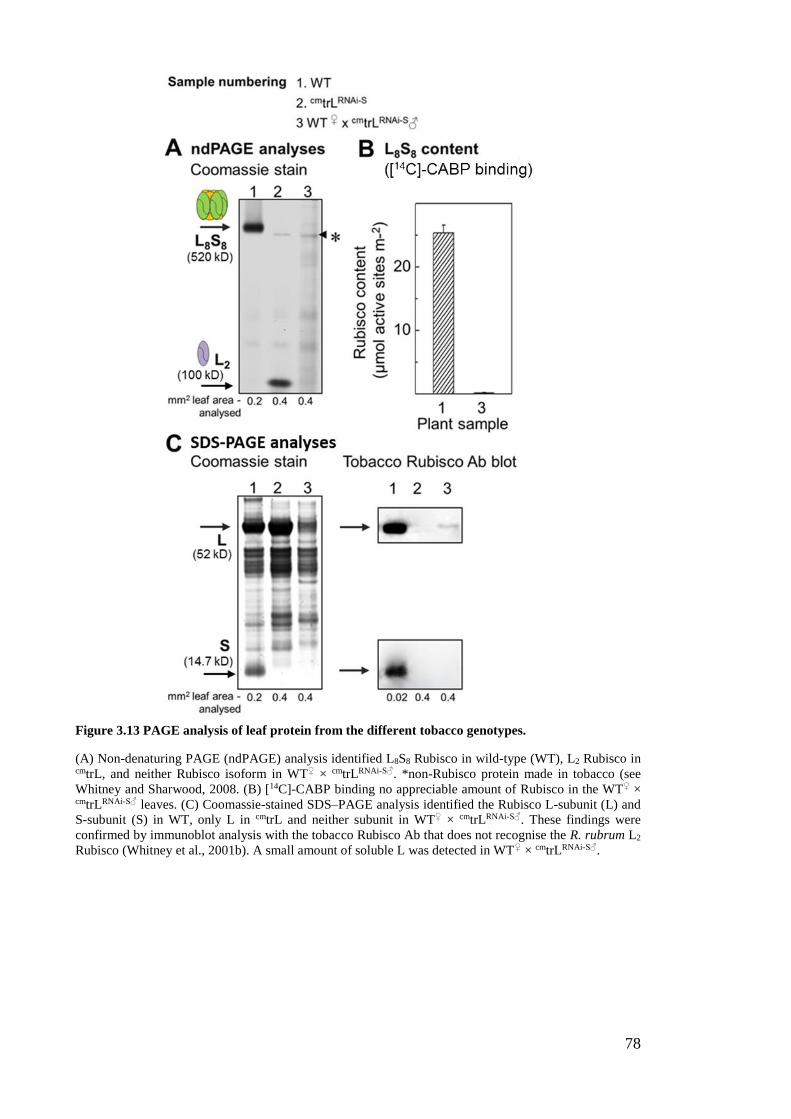
78
Figure 3.13 PAGE analysis of leaf protein from the different tobacco genotypes.
(A) Non-denaturing PAGE (ndPAGE) analysis identified L8S8 Rubisco in wild-type (WT), L2 Rubisco in cmtrL, and neither Rubisco isoform in WT♀ × cmtrLRNAi-S♂. *non-Rubisco protein made in tobacco (see
Whitney and Sharwood, 2008. (B) [14C]-CABP binding no appreciable amount of Rubisco in the WT♀ × cmtrLRNAi-S♂ leaves. (C) Coomassie-stained SDS–PAGE analysis identified the Rubisco L-subunit (L) and
S-subunit (S) in WT, only L in cmtrL and neither subunit in WT♀ × cmtrLRNAi-S♂. These findings were
confirmed by immunoblot analysis with the tobacco Rubisco Ab that does not recognise the R. rubrum L2
Rubisco (Whitney et al., 2001b). A small amount of soluble L was detected in WT♀ × cmtrLRNAi-S♂.
79
3.3 Discussion
Since the unravelling of the RNAi-gene silencing phenomenon in plants nearly twenty
years ago (Waterhouse et al., 1998) it has emerged as one of the major tools in plant
functional genomics. The application of RNAi to study gene function or use in
commercial applications to modify metabolic pathways and facilitate resistance to plant
pathogens has escalated dramatically over the last decade or so (Duan et al., 2012;
Senthil-Kumar and Mysore, 2010). This chapter describes the successful application of
RNAi in the generation of genetically stable cmtrLRNAi-S tobacco genotypes where RbcS
mRNA accumulation has been effectively suppressed to levels that preclude L8S8
Rubisco biogenesis.
3.3.1 The potency of the RNAi-RbcS transgene in all the transformed genotypes
While mechanistic detail of the T-DNA insertion process into plant genomes still
remains poorly understood, the sites of integration appear to be most frequent in
transcriptionally active regions of the genome (Filipenko et al., 2009). It is thought this
arises as these chromosome regions are more loosely packed by histones and thus are
euchromatic. In each of the transformed cmtrLRNAi-S genotypes selected in this study (i.e.
lines cmtrLRNAi-S8, cmtrLRNAi-S9,
cmtrLRNAi-S10 and cmtrLRNAi-S11) segregation analyses for
Basta resistance indicated all arose from single T-DNA insertional events (Figures 3.7
and 3.8). Ongoing backcrossing studies within the Whitney laboratory (comparable to
that shown in Figure 3.9) are uniformly showing the RNAi-RbcS genotype is stably
maintained in all the progeny in each of the cmtrLRNAi-S lines. As with the cross-
pollination study between cmtrLRNAi-S and wild-type (Figure 3.11), the same crosses with
homozygous cmtrLRNAi-S9,
cmtrLRNAi-S10 and cmtrLRNAi-S11 plants have recently been
reproduced and found to all produce progeny with little or no Rubisco (Whitney,
unpublished). These findings attest to the versatility of the RNAi-RbcS silencing
potential of plasmid psiSS, consistent with that observed by Wostrikoff and Stern (2007)
when using the same construct to silence S-subunit synthesis in wild-type tobacco.
The reason for the reproducible genetic stability and efficiency of the RNAi-
RbcS sequence is uncertain. It is possible that inclusion of the 93 bp Intron 1 sequence
in the 405 bp RbcS sequence incorporated in psiSS (Figure 3.3B) may suppress gene
silencing of the transformed T-DNA RNAi sequence. Consistent with this hypothesis,
expression of reporter proteins have shown the inclusion of intron sequences in the
transgenes reduced their targeted silencing by more than 4-fold (Christie et al., 2011).
Prior RNAi-silencing studies have surmised that for more efficient silencing RNAi
80
constructs should incorporate 300 bp or more of target sequence, with constructs
incorporating the entire target gene sequence likely to have the most efficient silencing
(Hily et al., 2007). The efficiency of RbcS mRNA silencing in the cmtrLRNAi-S genotypes
questions the accuracy of such assumptions given the RNAi-RbcS hairpin sequence in
the psiSS plasmid comprises only 57% (i.e. 312 of the 543 nucleotides) of coding
sequence of the varying RbcS transcripts found in N. tabacum (Figure 3.5).
The relinquished requirement for S-subunits in the cmtrL (Figure 3.4) may also
contribute to inherent stability of the RNAi-RbcS genotype. As indicated in Figure 3.1,
the photosynthetic CO2 assimilation dependency of cmtrL is supported by R. rubrum L2
Rubisco that does not require S-subunits (Whitney and Andrews, 2001b; Whitney and
Andrews, 2003). One method to test this hypothesis would be to transform the tobacco
rbcL back into the plastome of cmtrLRNAi-S. This would reinstate the potential for tobacco
L8S8 synthesis if silencing of the RNAi-RbcS genotype was readily reversible. A
reversion of the RNAi-RbcS silencing in this genotype however seems unlikely given
the WT♀ × cmtrLRNAi-S♂ progeny sown on MS medium (no Basta; Figure 3.11) showed
no evidence of any progeny having improved growth that would be expected if they re-
acquired the capacity for L8S8 biogenesis via destabilizing the RNAi-RbcS silencing.
3.3.2 Locating the T-DNA insertion position in cmtrLRNAi-S genomes
The recent availability of comprehensive genome sequence for N. tabacum (Gong et al.,
2014) provides impetus for accurately identifying the T-DNA insertion point within the
cmtrLRNAi-S genome. Replicating such analyses in the cmtrLRNAi-S9, cmtrLRNAi-S10 and
cmtrLRNAi-S11 is also of merit to compare their chromosomal insertion points and confirm
they all arise from independent, single insertion, transformation events (Figure 3.8).
Characterising the genome insertional point is also important for identifying potential
effects on the expression of gene(s) within inserted region as well as identifying
whether the genetic context of their insertional point might explain the potency of the
RNAi-RbcS genotype (see Section 3.3.1).
Identifying the chromosomal insertion point may prove to be somewhat of a
bioinformatic challenge given that the current publically available N. tabacum sequence
information comprises non-annotated whole genome shotgun contiguous sequences
(WGS). Nevertheless, work has now begun in the Whitney laboratory using
genome/primer walking to determine the T-DNA insertion site in each of the four
cmtrLRNAi-S genotypes produced in this thesis. As outlined in Figure 3.14, the nucleotide
template for this PCR based technique is gDNA from each genotype that has been
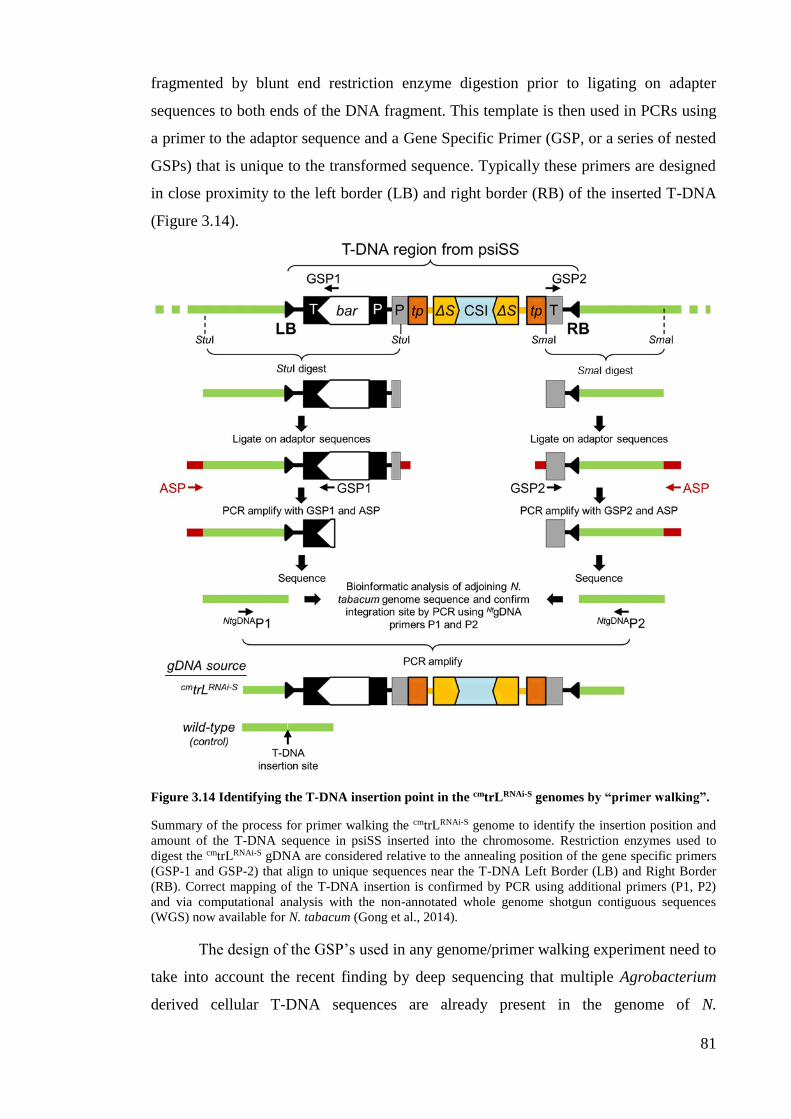
81
fragmented by blunt end restriction enzyme digestion prior to ligating on adapter
sequences to both ends of the DNA fragment. This template is then used in PCRs using
a primer to the adaptor sequence and a Gene Specific Primer (GSP, or a series of nested
GSPs) that is unique to the transformed sequence. Typically these primers are designed
in close proximity to the left border (LB) and right border (RB) of the inserted T-DNA
(Figure 3.14).
Figure 3.14 Identifying the T-DNA insertion point in the cmtrLRNAi-S genomes by “primer walking”.
Summary of the process for primer walking the cmtrLRNAi-S genome to identify the insertion position and
amount of the T-DNA sequence in psiSS inserted into the chromosome. Restriction enzymes used to
digest the cmtrLRNAi-S gDNA are considered relative to the annealing position of the gene specific primers
(GSP-1 and GSP-2) that align to unique sequences near the T-DNA Left Border (LB) and Right Border
(RB). Correct mapping of the T-DNA insertion is confirmed by PCR using additional primers (P1, P2)
and via computational analysis with the non-annotated whole genome shotgun contiguous sequences
(WGS) now available for N. tabacum (Gong et al., 2014).
The design of the GSP’s used in any genome/primer walking experiment need to
take into account the recent finding by deep sequencing that multiple Agrobacterium
derived cellular T-DNA sequences are already present in the genome of N.

82
tomentosiformis (one of the ancestral progenitors of N. tabacum) (Chen et al., 2014; see
also Section 1.4.1). This analysis validated the findings of prior genomic PCR analyses
that identified varying regions of T-DNA sequence from varying Agrobacterium
sources in many, but not all, species of Nicotiana (Intrieri and Buiatti, 2001; Mohajjel-
Shoja et al., 2011). With this in mind, the current GSP’s being utilized to analyse the
four cmtrLRNAi-S genotypes are specific to the bar selectable marker (adjacent to the LB)
and the octopine synthase terminator sequence regulating the RNAi-RbcS allele
(adjacent to the RB Figure 3.14).
3.3.3 Exploiting cmtrLRNAi-S for transplastomic study of Rubisco
Effective silencing of RbcS mRNA accumulation in the cmtrLRNAi-S line poses the first
report of successful elimination of S-subunit synthesis by RNAi in any species of higher
plant. As such the cmtrLRNAi-S lines provided a unique genotype for transgenic studies of
S-subunit biochemistry in regard to its role on L8S8 biogenesis and catalysis in tobacco
leaf chloroplasts. The following chapters describe varying transgenic tests undertaken to
test the versatility of cmtrLRNAi-S for bioengineering tobacco and heterologous S-subunits
and the development of tailored chloroplast transformation approaches for optimal
recombinant Rubisco expression in tobacco leaves.
83
CHAPTER 4 – ENABLING ASSEMBLY OF RUBISCO
COMPRISING CHLOROPLAST MADE SMALL
SUBUNITS USING THE CMTRLRNAI-S TOBACCO
GENOTYPE
4.1 Introduction
4.1.1 Rubisco assembly in higher plants
Approximately 3000 nucleus encoded proteins, including the Rubisco S-subunits, are
imported from the cytosol into leaf chloroplasts (Li and Chiu, 2010). The reason why
most, but not all, chloroplast genes have been translocated to the nucleus in higher
plants remains uncertain. Gene transfer from the chloroplast genome (plastome) to the
nucleus during evolution is thought to be facilitated by an improved allowance of gene
expansion and adaptive evolution (Coate et al., 2011). The conserved retention of some
chloroplast genes is thought to arise from the potential toxicity of their products if
produced in the cytosol or an inability to meet the levels needed for chloroplast
functioning (Timmis et al., 2004). For Rubisco, relocation of the rbcL to the nucleus
seems unfavourable with regard to being able to meet the large amounts of L-subunit
required in Rubisco biogenesis (Kanevski and Maliga, 1994). In contrast, the
evolutionary relocation and duplication of RbcS in the nucleus of plant cells has
occurred without peril (see Section 3.1.5). Accompanying this relocation has been the
incorporation of appropriate cis elements necessary for regulating RbcS expression and
a sequence for chloroplast targeting. As well, appropriate sequence changes have had to
evolve to facilitate interaction of the nascent S-subunits with the molecular partners
needed during their synthesis, folding and chaperoning to and through the chloroplast
envelope. Once in the stroma additional ancillary proteins are also required for
chaperoning S-subunit assembly with the Rubisco L-subunits into L8S8 holoenzyme
(Nishimura et al., 2008).
4.1.2 Synthesis of S-subunits in the cytosol and import into chloroplasts
While the L-subunit undergoes a variety of translational and post-translation processes
within the chloroplast stroma, RbcS in the nucleus of plant cells have evolved to code a
precursor S-subunit peptide of 20 kDa that is synthesized by cytoplasmic ribosomes
(Dobberstein et al., 1977). Once translated, the amino (N-) terminal extension (i.e. a ~6
kDa transit peptide) of the S-subunit precursor undergoes a range of post-translational
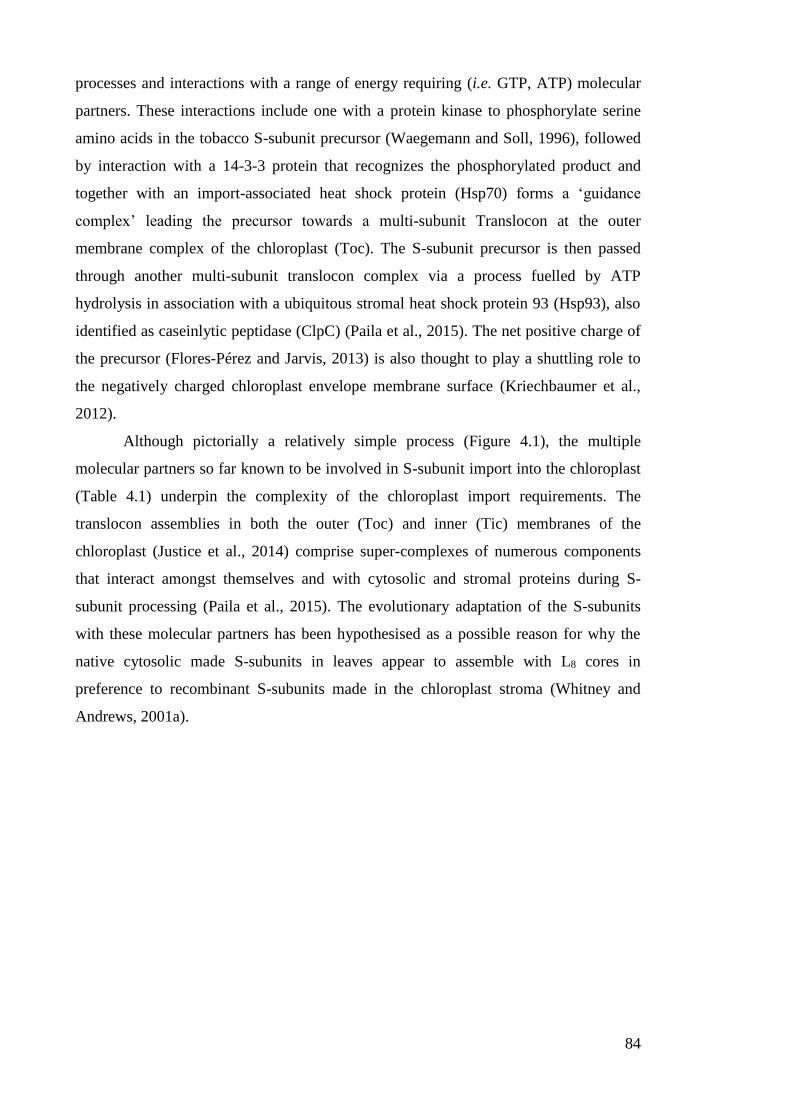
84
processes and interactions with a range of energy requiring (i.e. GTP, ATP) molecular
partners. These interactions include one with a protein kinase to phosphorylate serine
amino acids in the tobacco S-subunit precursor (Waegemann and Soll, 1996), followed
by interaction with a 14-3-3 protein that recognizes the phosphorylated product and
together with an import-associated heat shock protein (Hsp70) forms a ‘guidance
complex’ leading the precursor towards a multi-subunit Translocon at the outer
membrane complex of the chloroplast (Toc). The S-subunit precursor is then passed
through another multi-subunit translocon complex via a process fuelled by ATP
hydrolysis in association with a ubiquitous stromal heat shock protein 93 (Hsp93), also
identified as caseinlytic peptidase (ClpC) (Paila et al., 2015). The net positive charge of
the precursor (Flores-Pérez and Jarvis, 2013) is also thought to play a shuttling role to
the negatively charged chloroplast envelope membrane surface (Kriechbaumer et al.,
2012).
Although pictorially a relatively simple process (Figure 4.1), the multiple
molecular partners so far known to be involved in S-subunit import into the chloroplast
(Table 4.1) underpin the complexity of the chloroplast import requirements. The
translocon assemblies in both the outer (Toc) and inner (Tic) membranes of the
chloroplast (Justice et al., 2014) comprise super-complexes of numerous components
that interact amongst themselves and with cytosolic and stromal proteins during S-
subunit processing (Paila et al., 2015). The evolutionary adaptation of the S-subunits
with these molecular partners has been hypothesised as a possible reason for why the
native cytosolic made S-subunits in leaves appear to assemble with L8 cores in
preference to recombinant S-subunits made in the chloroplast stroma (Whitney and
Andrews, 2001a).
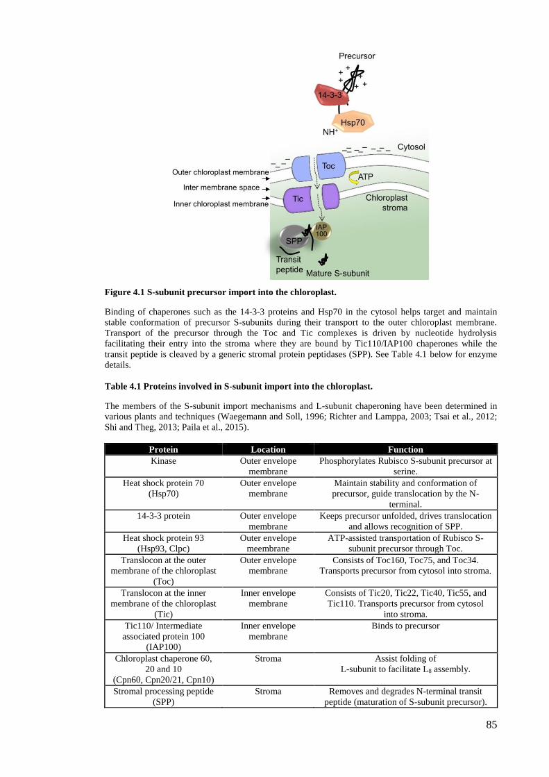
85
Figure 4.1 S-subunit precursor import into the chloroplast.
Binding of chaperones such as the 14-3-3 proteins and Hsp70 in the cytosol helps target and maintain
stable conformation of precursor S-subunits during their transport to the outer chloroplast membrane.
Transport of the precursor through the Toc and Tic complexes is driven by nucleotide hydrolysis
facilitating their entry into the stroma where they are bound by Tic110/IAP100 chaperones while the
transit peptide is cleaved by a generic stromal protein peptidases (SPP). See Table 4.1 below for enzyme
details.
Table 4.1 Proteins involved in S-subunit import into the chloroplast.
The members of the S-subunit import mechanisms and L-subunit chaperoning have been determined in
various plants and techniques (Waegemann and Soll, 1996; Richter and Lamppa, 2003; Tsai et al., 2012;
Shi and Theg, 2013; Paila et al., 2015).
Protein Location Function
Kinase Outer envelope
membrane
Phosphorylates Rubisco S-subunit precursor at
serine.
Heat shock protein 70
(Hsp70)
Outer envelope
membrane
Maintain stability and conformation of
precursor, guide translocation by the N-
terminal.
14-3-3 protein Outer envelope
membrane
Keeps precursor unfolded, drives translocation
and allows recognition of SPP.
Heat shock protein 93
(Hsp93, Clpc)
Outer envelope
meembrane
ATP-assisted transportation of Rubisco S-
subunit precursor through Toc.
Translocon at the outer
membrane of the chloroplast
(Toc)
Outer envelope
membrane
Consists of Toc160, Toc75, and Toc34.
Transports precursor from cytosol into stroma.
Translocon at the inner
membrane of the chloroplast
(Tic)
Inner envelope
membrane
Consists of Tic20, Tic22, Tic40, Tic55, and
Tic110. Transports precursor from cytosol
into stroma.
Tic110/ Intermediate
associated protein 100
(IAP100)
Inner envelope
membrane
Binds to precursor
Chloroplast chaperone 60,
20 and 10
(Cpn60, Cpn20/21, Cpn10)
Stroma Assist folding of
L-subunit to facilitate L8 assembly.
Stromal processing peptide
(SPP)
Stroma Removes and degrades N-terminal transit
peptide (maturation of S-subunit precursor).
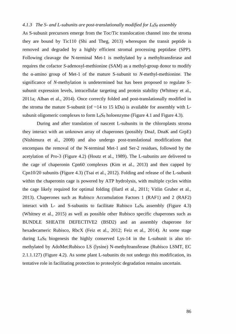
86
4.1.3 The S- and L-subunits are post-translationally modified for L8S8 assembly
As S-subunit precursors emerge from the Toc/Tic translocation channel into the stroma
they are bound by Tic110 (Shi and Theg, 2013) whereupon the transit peptide is
removed and degraded by a highly efficient stromal processing peptidase (SPP).
Following cleavage the N-terminal Met-1 is methylated by a methyltransferase and
requires the cofactor S-adenosyl-methionine (SAM) as a methyl-group donor to modify
the α-amino group of Met-1 of the mature S-subunit to N-methyl-methionine. The
significance of N-methylation is undetermined but has been proposed to regulate S-
subunit expression levels, intracellular targeting and protein stability (Whitney et al.,
2011a; Alban et al., 2014). Once correctly folded and post-translationally modified in
the stroma the mature S-subunit (of ~14 to 15 kDa) is available for assembly with L-
subunit oligomeric complexes to form L8S8 holoenzyme (Figure 4.1 and Figure 4.3).
During and after translation of nascent L-subunits in the chloroplasts stroma
they interact with an unknown array of chaperones (possibly DnaJ, DnaK and GrpE)
(Nishimura et al., 2008) and also undergo post-translational modifications that
encompass the removal of the N-terminal Met-1 and Ser-2 residues, followed by the
acetylation of Pro-3 (Figure 4.2) (Houtz et al., 1989). The L-subunits are delivered to
the cage of chaperonin Cpn60 complexes (Kim et al., 2013) and then capped by
Cpn10/20 subunits (Figure 4.3) (Tsai et al., 2012). Folding and release of the L-subunit
within the chaperonin cage is powered by ATP hydrolysis, with multiple cycles within
the cage likely required for optimal folding (Hartl et al., 2011; Vitlin Gruber et al.,
2013). Chaperones such as Rubisco Accumulation Factors 1 (RAF1) and 2 (RAF2)
interact with L- and S-subunits to facilitate Rubisco L8S8 assembly (Figure 4.3)
(Whitney et al., 2015) as well as possible other Rubisco specific chaperones such as
BUNDLE SHEATH DEFECTIVE2 (BSD2) and an assembly chaperone for
hexadecameric Rubisco, RbcX (Feiz et al., 2012; Feiz et al., 2014). At some stage
during L8S8 biogenesis the highly conserved Lys-14 in the L-subunit is also tri-
methylated by AdoMet:Rubisco LS (lysine) N-methyltransferase (Rubisco LSMT, EC
2.1.1.127) (Figure 4.2). As some plant L-subunits do not undergo this modification, its
tentative role in facilitating protection to proteolytic degradation remains uncertain.
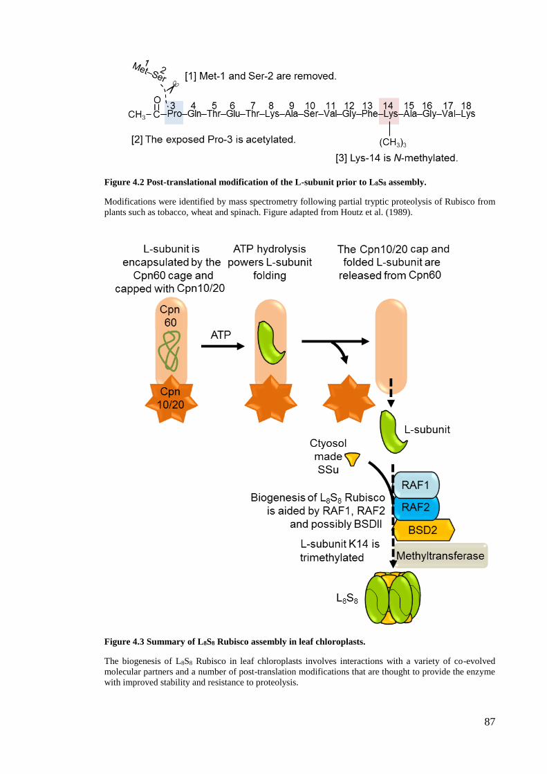
87
Figure 4.2 Post-translational modification of the L-subunit prior to L8S8 assembly.
Modifications were identified by mass spectrometry following partial tryptic proteolysis of Rubisco from
plants such as tobacco, wheat and spinach. Figure adapted from Houtz et al. (1989).
Figure 4.3 Summary of L8S8 Rubisco assembly in leaf chloroplasts.
The biogenesis of L8S8 Rubisco in leaf chloroplasts involves interactions with a variety of co-evolved
molecular partners and a number of post-translation modifications that are thought to provide the enzyme
with improved stability and resistance to proteolysis.

88
4.1.4 Expression of RbcS transformed into the tobacco plastome
Attempts to re-introduce an RbcS back into the chloroplast genome (plastome) of
tobacco showed that despite large increases in transcript abundance (Figure 4.4) very
few chloroplast made S-subunits were incorporated into L8S8 complexes (Whitney and
Andrews 2001a). The same result was obtained when the same RbcS was introduced
into anti-RbcS tobacco genotypes producing 70 to 80% less Rubisco when compared
with WT (Zhang et al., 2002). In both experiments the transplastomic tobacco
genotypes contained duplicate copies of a native N. tabacum RbcS inserted into the
inverted repeat regions of the plastome. In the Whitney and Andrews (2001a) study, the
RbcS encoded a transit peptide (tp) (RVtpSSuH) or no tp (RVSSuH) (Figure 4.7).
Expression of the transgenes was regulated by identical psbA promoter and terminator
regulatory sequences that had successfully facilitated high levels of recombinant protein
expression (Staub and Maliga, 1994; Whitney and Andrews, 2001a). Each RbcS
transgene included a C-terminal hepta-histidine-encoding sequence (SH7) to allow the
identification and purification by nickel-nitrilotriacetic acid (Ni-NTA) of protein
complexes comprising chloroplast made SH7-subunits. Analyses revealed the
RVtpSSuH genotype produced higher amounts of SH7 than the RVSSuH plants
(consistent with the higher levels of tpRbcS mRNA in RVtpSSuH leaves, Figure 4.4).
Despite the >10-fold higher levels of chloroplast RbcS mRNAs made relative to the
cytosol RbcS mRNA levels, the chloroplast made SH7-subunits only accounted for ~1.2%
of the total S-subunit pool assembled into Rubisco (Staub and Maliga, 1994; Whitney
and Andrews, 2001a).
Figure 4.4 RNA blot analyses showing the high abundance of plastome made RbcS mRNA in the
transplastomic RVtpSSuH and RVSSuH tobacco genotypes relative to wild-type (WT)
RNA hybridized with a [32P]-labelled RbcS DNA probe showing the chloroplast made RbcS mRNA in the
RVtpSSuH (pttpRbcS) and RVSSuH (cpRbcS) tobacco lines were more than five-fold higher than the
cytosolic RbcS mRNA levels (nucRbcS). Figure modified from Whitney and Andrews (2001).
Increasing the incorporation of plastid made S-subunits into L8S8 complexes was
obtained by directing RbcS transgene insertion into a transcriptional hot spot in the
plastome inverted repeat region of an anti-RbcS tobacco genotype (Dhingra et al., 2004;
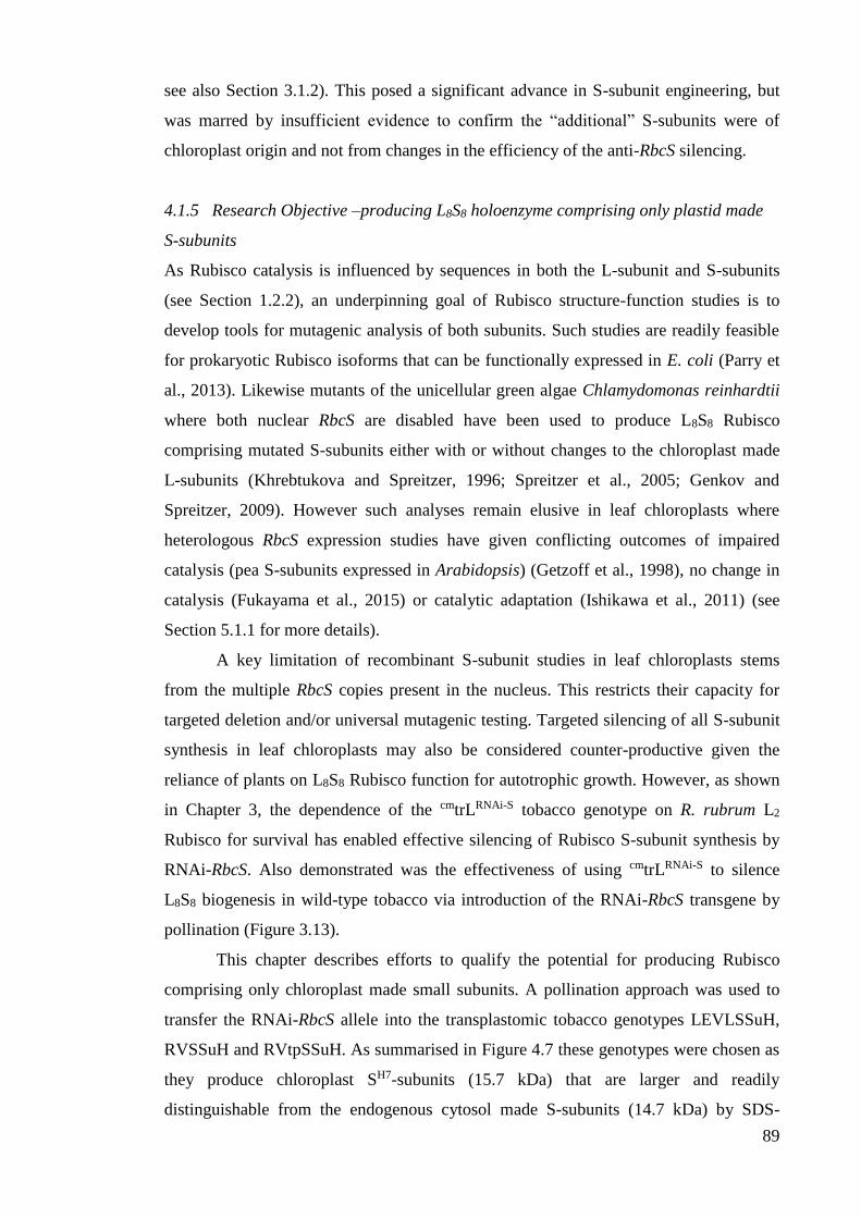
89
see also Section 3.1.2). This posed a significant advance in S-subunit engineering, but
was marred by insufficient evidence to confirm the “additional” S-subunits were of
chloroplast origin and not from changes in the efficiency of the anti-RbcS silencing.
4.1.5 Research Objective –producing L8S8 holoenzyme comprising only plastid made
S-subunits
As Rubisco catalysis is influenced by sequences in both the L-subunit and S-subunits
(see Section 1.2.2), an underpinning goal of Rubisco structure-function studies is to
develop tools for mutagenic analysis of both subunits. Such studies are readily feasible
for prokaryotic Rubisco isoforms that can be functionally expressed in E. coli (Parry et
al., 2013). Likewise mutants of the unicellular green algae Chlamydomonas reinhardtii
where both nuclear RbcS are disabled have been used to produce L8S8 Rubisco
comprising mutated S-subunits either with or without changes to the chloroplast made
L-subunits (Khrebtukova and Spreitzer, 1996; Spreitzer et al., 2005; Genkov and
Spreitzer, 2009). However such analyses remain elusive in leaf chloroplasts where
heterologous RbcS expression studies have given conflicting outcomes of impaired
catalysis (pea S-subunits expressed in Arabidopsis) (Getzoff et al., 1998), no change in
catalysis (Fukayama et al., 2015) or catalytic adaptation (Ishikawa et al., 2011) (see
Section 5.1.1 for more details).
A key limitation of recombinant S-subunit studies in leaf chloroplasts stems
from the multiple RbcS copies present in the nucleus. This restricts their capacity for
targeted deletion and/or universal mutagenic testing. Targeted silencing of all S-subunit
synthesis in leaf chloroplasts may also be considered counter-productive given the
reliance of plants on L8S8 Rubisco function for autotrophic growth. However, as shown
in Chapter 3, the dependence of the cmtrLRNAi-S tobacco genotype on R. rubrum L2
Rubisco for survival has enabled effective silencing of Rubisco S-subunit synthesis by
RNAi-RbcS. Also demonstrated was the effectiveness of using cmtrLRNAi-S to silence
L8S8 biogenesis in wild-type tobacco via introduction of the RNAi-RbcS transgene by
pollination (Figure 3.13).
This chapter describes efforts to qualify the potential for producing Rubisco
comprising only chloroplast made small subunits. A pollination approach was used to
transfer the RNAi-RbcS allele into the transplastomic tobacco genotypes LEVLSSuH,
RVSSuH and RVtpSSuH. As summarised in Figure 4.7 these genotypes were chosen as
they produce chloroplast SH7-subunits (15.7 kDa) that are larger and readily
distinguishable from the endogenous cytosol made S-subunits (14.7 kDa) by SDS-

90
PAGE (Whitney and Andrews, 2001a). The content and subunit composition of Rubisco
in the resulting progeny was analysed to compare the effectiveness of chloroplast made
SH7-subunit assembly into L8S8 holoenzyme. Such knowledge is pivotal towards
developing an optimal transgenic approach for mutagenic study of Rubisco structure
and function in leaf chloroplasts.
Figure 4.5 Schematic demonstration of the transgenic crosses tested in this study.
Schematic showing the aim of crossed-pollinating cmtrLRNAi-S with varying transplastomic lines producing
chloroplast made S-subunits to generate RNAi-RbcS progeny where only the chloroplast made S-subunits
are available for L8S8 biogenesis.
4.2 Results
4.2.1 Generating the LEVLSSuH transplastomic line
The RVSSuH and RVtpSSuH tobacco genotypes used in this study were those
previously generated (Whitney and Andrews, 2001a). In contrast the LEVLSSuH
tobacco lines are uncharacterised transplastomic genotypes generated by Dr. Whitney
where the N. tabacum RbcS transgene is inserted downstream of rbcL (Figure 4.6).
Three homoplasmic LEVLSSuH lines were generated by transforming the cmtrL
genotype (Figure 1.11) with plasmid pLevLSSuH, a derivative of the pLEV1
transforming plasmid (Whitney et al., 1999). As indicated in Figure 4.6, the transformed
LEVLSSuH tobacco genotypes contained the same tobacco RbcS transformed into
RVtpSSuH (i.e. inclusive of its N-terminal transit peptide coding sequence) that was
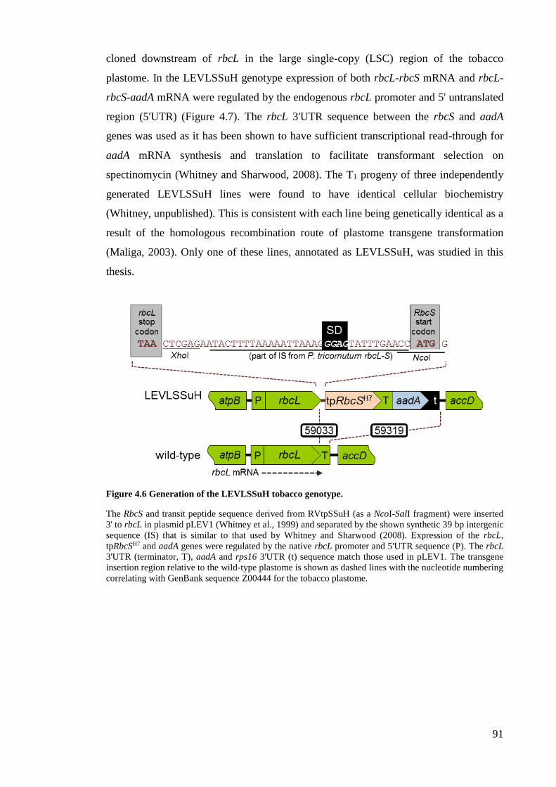
91
cloned downstream of rbcL in the large single-copy (LSC) region of the tobacco
plastome. In the LEVLSSuH genotype expression of both rbcL-rbcS mRNA and rbcL-
rbcS-aadA mRNA were regulated by the endogenous rbcL promoter and 5' untranslated
region (5'UTR) (Figure 4.7). The rbcL 3'UTR sequence between the rbcS and aadA
genes was used as it has been shown to have sufficient transcriptional read-through for
aadA mRNA synthesis and translation to facilitate transformant selection on
spectinomycin (Whitney and Sharwood, 2008). The T1 progeny of three independently
generated LEVLSSuH lines were found to have identical cellular biochemistry
(Whitney, unpublished). This is consistent with each line being genetically identical as a
result of the homologous recombination route of plastome transgene transformation
(Maliga, 2003). Only one of these lines, annotated as LEVLSSuH, was studied in this
thesis.
Figure 4.6 Generation of the LEVLSSuH tobacco genotype.
The RbcS and transit peptide sequence derived from RVtpSSuH (as a NcoI-SalI fragment) were inserted
3' to rbcL in plasmid pLEV1 (Whitney et al., 1999) and separated by the shown synthetic 39 bp intergenic
sequence (IS) that is similar to that used by Whitney and Sharwood (2008). Expression of the rbcL,
tpRbcSH7 and aadA genes were regulated by the native rbcL promoter and 5'UTR sequence (P). The rbcL
3'UTR (terminator, T), aadA and rps16 3'UTR (t) sequence match those used in pLEV1. The transgene
insertion region relative to the wild-type plastome is shown as dashed lines with the nucleotide numbering
correlating with GenBank sequence Z00444 for the tobacco plastome.
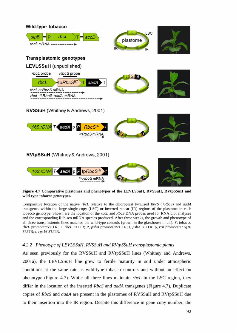
92
Figure 4.7 Comparative plastomes and phenotypes of the LEVLSSuH, RVSSuH, RVtpSSuH and
wild-type tobacco genotypes.
Comparitive location of the native rbcL relative to the chloroplast localised RbcS (cpRbcS) and aadA
transgenes within the large single copy (LSC) or inverted repeat (IR) regions of the plastome in each
tobacco genotype. Shown are the location of the rbcL and RbcS DNA probes used for RNA blot analyses
and the corresponding Rubisco mRNA species produced. After three weeks, the growth and phenotype of
all three transplastomic lines matched the wild-type controls (grown in the glasshouse in air). P, tobacco
rbcL promoter/5'UTR; T, rbcL 3'UTR; P, psbA promoter/5'UTR; t, psbA 3'UTR; p, rrn promoter/T7g10
5'UTR; t, rps16 3'UTR.
4.2.2 Phenotype of LEVLSSuH, RVSSuH and RVtpSSuH transplastomic plants
As seen previously for the RVSSuH and RVtpSSuH lines (Whitney and Andrews,
2001a), the LEVLSSuH line grew to fertile maturity in soil under atmospheric
conditions at the same rate as wild-type tobacco controls and without an effect on
phenotype (Figure 4.7). While all three lines maintain rbcL in the LSC region, they
differ in the location of the inserted RbcS and aadA transgenes (Figure 4.7). Duplicate
copies of RbcS and aadA are present in the plastomes of RVSSuH and RVtpSSuH due
to their insertion into the IR region. Despite this difference in gene copy number, the
93
amount of plastid made S-subunits assembled into Rubisco in LEVLSSuH closely
matched that of RVtpSSuH (Whitney unpublished). That is, plastid made S-subunits
only accounted for ~1% of the total S-subunit pool assembled into L8S8 Rubisco in
LEVLSSuH, which is equivalent to the ~1.2% measured in RVtpSSuH (Whitney and
Andrews, 2001a).
4.2.3 Inheritance of the RNAi-RbcS T-DNA via pollination with cmtrLRNAi-S pollen
Flowers from RVSSuH, RVtpSSuH and LEVLSSuH T2 plants were pollinated with
pollen from the cmtrLRNAi-S line. The inheritance frequency (segregation analysis) of the
bar (nuclear transgene) and aadA (plastome transgene) selectable markers in the
resulting transplastome♀ × cmtrLRNAi-S♂ F1 progenies were examined (Figure 4.8A).
Seeds sown on MS medium (i.e. with no selection antibiotic or herbicide) showed 100%
viability. Compared to the wild-type controls, all the transgenic progeny showed
impaired growth on MS medium. In particular the growth of the wild-type (WT)♀ ×
cmtrLRNAi-S♂ progeny was greatly impeded with their survival dependant on the inclusion
of sucrose in the MS medium (see also Figure 3.12). All the progeny from the cmtrLRNAi-
S crosses survived growth on media containing Basta (10 mg L-1) consistent with
Mendelian inheritance of the RNAi-RbcS T-DNA (that includes bar) from the
homozygous cmtrLRNAi-S line. On media containing spectinomycin (500 mg L-1), only
progeny of the plastome-transformed lines showed resistance with their growth rate and
phenotype mirroring those of the progeny grown on media containing Basta (Figure
4.8A). These findings agree with the nuclear location of bar in chromosomes inherited
from cmtrLRNAi-S♂ and the maternal inheritance of aadA in the plastome of the
LEVLSSuH, RVSSuH and RVtpSSuH transplastomic genotypes.
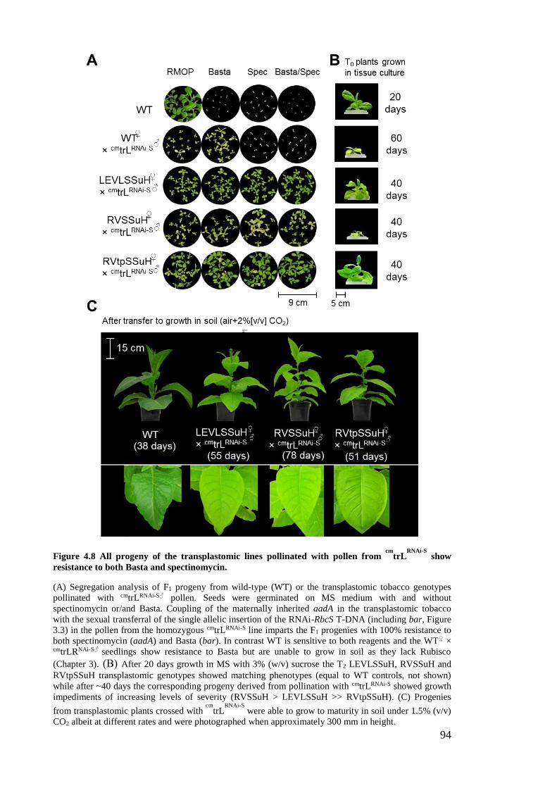
94
Figure 4.8 All progeny of the transplastomic lines pollinated with pollen from cm
trLRNAi-S
show
resistance to both Basta and spectinomycin.
(A) Segregation analysis of F1 progeny from wild-type (WT) or the transplastomic tobacco genotypes
pollinated with cmtrLRNAi-S♂ pollen. Seeds were germinated on MS medium with and without
spectinomycin or/and Basta. Coupling of the maternally inherited aadA in the transplastomic tobacco
with the sexual transferral of the single allelic insertion of the RNAi-RbcS T-DNA (including bar, Figure
3.3) in the pollen from the homozygous cmtrLRNAi-S line imparts the F1 progenies with 100% resistance to
both spectinomycin (aadA) and Basta (bar). In contrast WT is sensitive to both reagents and the WT♀ × cmtrLRNAi-S♂
seedlings show resistance to Basta but are unable to grow in soil as they lack Rubisco
(Chapter 3). (B) After 20 days growth in MS with 3% (w/v) sucrose the T2 LEVLSSuH, RVSSuH and
RVtpSSuH transplastomic genotypes showed matching phenotypes (equal to WT controls, not shown)
while after ~40 days the corresponding progeny derived from pollination with cmtrLRNAi-S showed growth
impediments of increasing levels of severity (RVSSuH > LEVLSSuH >> RVtpSSuH). (C) Progenies
from transplastomic plants crossed with cm
trLRNAi-S
were able to grow to maturity in soil under 1.5% (v/v)
CO2 albeit at different rates and were photographed when approximately 300 mm in height.
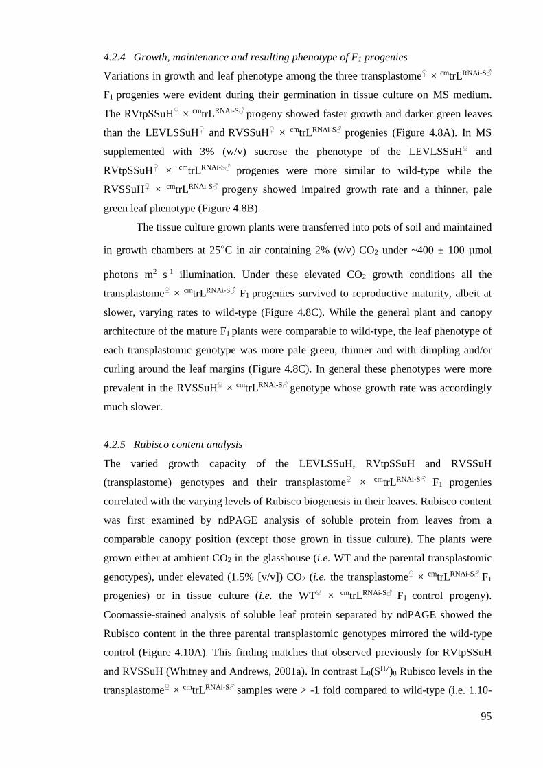
95
4.2.4 Growth, maintenance and resulting phenotype of F1 progenies
Variations in growth and leaf phenotype among the three transplastome♀ × cmtrLRNAi-S♂
F1 progenies were evident during their germination in tissue culture on MS medium.
The RVtpSSuH♀ × cmtrLRNAi-S♂ progeny showed faster growth and darker green leaves
than the LEVLSSuH♀ and RVSSuH♀ × cmtrLRNAi-S♂ progenies (Figure 4.8A). In MS
supplemented with 3% (w/v) sucrose the phenotype of the LEVLSSuH♀ and
RVtpSSuH♀ × cmtrLRNAi-S♂ progenies were more similar to wild-type while the
RVSSuH♀ × cmtrLRNAi-S♂ progeny showed impaired growth rate and a thinner, pale
green leaf phenotype (Figure 4.8B).
The tissue culture grown plants were transferred into pots of soil and maintained
in growth chambers at 25°C in air containing 2% (v/v) CO2 under ~400 ± 100 µmol
photons m2 s-1 illumination. Under these elevated CO2 growth conditions all the
transplastome♀ × cmtrLRNAi-S♂ F1 progenies survived to reproductive maturity, albeit at
slower, varying rates to wild-type (Figure 4.8C). While the general plant and canopy
architecture of the mature F1 plants were comparable to wild-type, the leaf phenotype of
each transplastomic genotype was more pale green, thinner and with dimpling and/or
curling around the leaf margins (Figure 4.8C). In general these phenotypes were more
prevalent in the RVSSuH♀ × cmtrLRNAi-S♂ genotype whose growth rate was accordingly
much slower.
4.2.5 Rubisco content analysis
The varied growth capacity of the LEVLSSuH, RVtpSSuH and RVSSuH
(transplastome) genotypes and their transplastome♀ × cmtrLRNAi-S♂ F1 progenies
correlated with the varying levels of Rubisco biogenesis in their leaves. Rubisco content
was first examined by ndPAGE analysis of soluble protein from leaves from a
comparable canopy position (except those grown in tissue culture). The plants were
grown either at ambient CO2 in the glasshouse (i.e. WT and the parental transplastomic
genotypes), under elevated (1.5% [v/v]) CO2 (i.e. the transplastome♀ × cmtrLRNAi-S♂ F1
progenies) or in tissue culture (i.e. the WT♀ × cmtrLRNAi-S♂ F1 control progeny).
Coomassie-stained analysis of soluble leaf protein separated by ndPAGE showed the
Rubisco content in the three parental transplastomic genotypes mirrored the wild-type
control (Figure 4.10A). This finding matches that observed previously for RVtpSSuH
and RVSSuH (Whitney and Andrews, 2001a). In contrast L8(SH7)8 Rubisco levels in the
transplastome♀ × cmtrLRNAi-S♂ samples were > -1 fold compared to wild-type (i.e. 1.10-
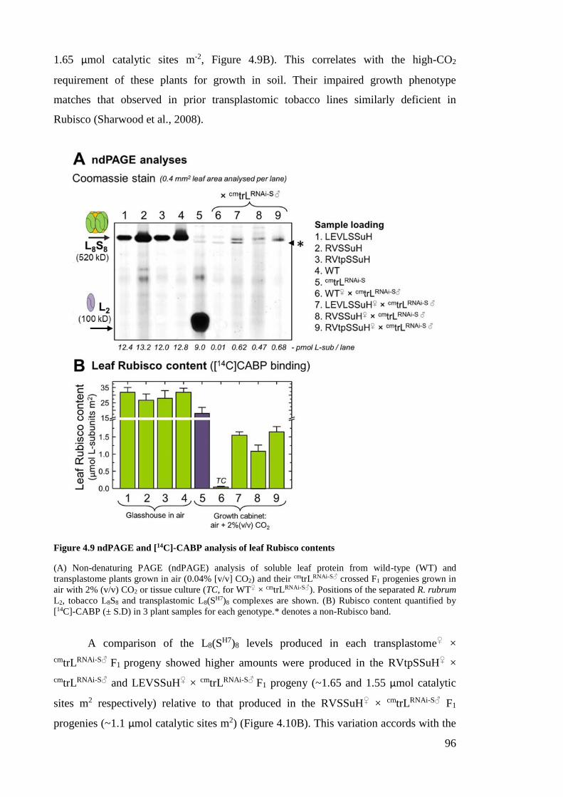
96
1.65 µmol catalytic sites m-2, Figure 4.9B). This correlates with the high-CO2
requirement of these plants for growth in soil. Their impaired growth phenotype
matches that observed in prior transplastomic tobacco lines similarly deficient in
Rubisco (Sharwood et al., 2008).
Figure 4.9 ndPAGE and [14C]-CABP analysis of leaf Rubisco contents
(A) Non-denaturing PAGE (ndPAGE) analysis of soluble leaf protein from wild-type (WT) and
transplastome plants grown in air (0.04% [v/v] CO2) and their cmtrLRNAi-S♂ crossed F1 progenies grown in
air with 2% (v/v) CO2 or tissue culture (TC, for WT♀ × cmtrLRNAi-S♂). Positions of the separated R. rubrum
L2, tobacco L8S8 and transplastomic L8(SH7)8 complexes are shown. (B) Rubisco content quantified by
[14C]-CABP (± S.D) in 3 plant samples for each genotype.* denotes a non-Rubisco band.
A comparison of the L8(SH7)8 levels produced in each transplastome♀ ×
cmtrLRNAi-S♂ F1 progeny showed higher amounts were produced in the RVtpSSuH♀ ×
cmtrLRNAi-S♂ and LEVSSuH♀ × cmtrLRNAi-S♂ F1 progeny (~1.65 and 1.55 µmol catalytic
sites m2 respectively) relative to that produced in the RVSSuH♀ × cmtrLRNAi-S♂ F1
progenies (~1.1 µmol catalytic sites m2) (Figure 4.10B). This variation accords with the
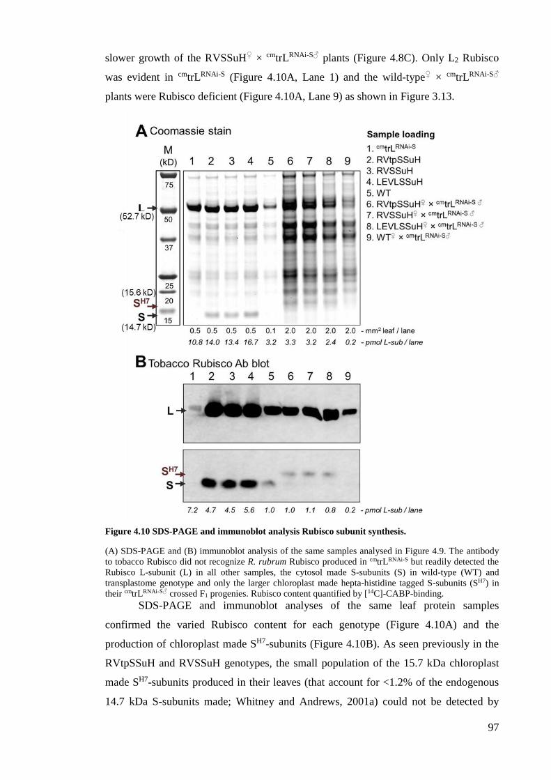
97
slower growth of the RVSSuH♀ × cmtrLRNAi-S♂ plants (Figure 4.8C). Only L2 Rubisco
was evident in cmtrLRNAi-S (Figure 4.10A, Lane 1) and the wild-type♀ × cmtrLRNAi-S♂
plants were Rubisco deficient (Figure 4.10A, Lane 9) as shown in Figure 3.13.
Figure 4.10 SDS-PAGE and immunoblot analysis Rubisco subunit synthesis.
(A) SDS-PAGE and (B) immunoblot analysis of the same samples analysed in Figure 4.9. The antibody
to tobacco Rubisco did not recognize R. rubrum Rubisco produced in cmtrLRNAi-S but readily detected the
Rubisco L-subunit (L) in all other samples, the cytosol made S-subunits (S) in wild-type (WT) and
transplastome genotype and only the larger chloroplast made hepta-histidine tagged S-subunits (SH7) in
their cmtrLRNAi-S♂ crossed F1 progenies. Rubisco content quantified by [14C]-CABP-binding.
SDS-PAGE and immunoblot analyses of the same leaf protein samples
confirmed the varied Rubisco content for each genotype (Figure 4.10A) and the
production of chloroplast made SH7-subunits (Figure 4.10B). As seen previously in the
RVtpSSuH and RVSSuH genotypes, the small population of the 15.7 kDa chloroplast
made SH7-subunits produced in their leaves (that account for <1.2% of the endogenous
14.7 kDa S-subunits made; Whitney and Andrews, 2001a) could not be detected by
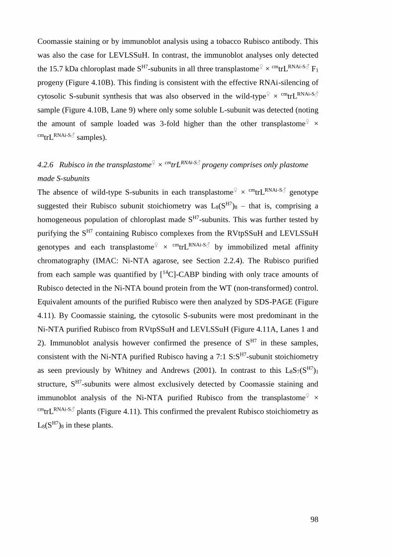
98
Coomassie staining or by immunoblot analysis using a tobacco Rubisco antibody. This
was also the case for LEVLSSuH. In contrast, the immunoblot analyses only detected
the 15.7 kDa chloroplast made SH7-subunits in all three transplastome♀ × cmtrLRNAi-S♂ F1
progeny (Figure 4.10B). This finding is consistent with the effective RNAi-silencing of
cytosolic S-subunit synthesis that was also observed in the wild-type♀ × cmtrLRNAi-S♂
sample (Figure 4.10B, Lane 9) where only some soluble L-subunit was detected (noting
the amount of sample loaded was 3-fold higher than the other transplastome♀ ×
cmtrLRNAi-S♂ samples).
4.2.6 Rubisco in the transplastome♀ × cmtrLRNAi-S♂ progeny comprises only plastome
made S-subunits
The absence of wild-type S-subunits in each transplastome♀ × cmtrLRNAi-S♂ genotype
suggested their Rubisco subunit stoichiometry was L8(SH7)8 – that is, comprising a
homogeneous population of chloroplast made SH7-subunits. This was further tested by
purifying the SH7 containing Rubisco complexes from the RVtpSSuH and LEVLSSuH
genotypes and each transplastome♀ × cmtrLRNAi-S♂ by immobilized metal affinity
chromatography (IMAC: Ni-NTA agarose, see Section 2.2.4). The Rubisco purified
from each sample was quantified by [14C]-CABP binding with only trace amounts of
Rubisco detected in the Ni-NTA bound protein from the WT (non-transformed) control.
Equivalent amounts of the purified Rubisco were then analyzed by SDS-PAGE (Figure
4.11). By Coomassie staining, the cytosolic S-subunits were most predominant in the
Ni-NTA purified Rubisco from RVtpSSuH and LEVLSSuH (Figure 4.11A, Lanes 1 and
2). Immunoblot analysis however confirmed the presence of SH7 in these samples,
consistent with the Ni-NTA purified Rubisco having a 7:1 S:SH7-subunit stoichiometry
as seen previously by Whitney and Andrews (2001). In contrast to this L8S7(SH7)1
structure, SH7-subunits were almost exclusively detected by Coomassie staining and
immunoblot analysis of the Ni-NTA purified Rubisco from the transplastome♀ ×
cmtrLRNAi-S♂ plants (Figure 4.11). This confirmed the prevalent Rubisco stoichiometry as
L8(SH7)8 in these plants.
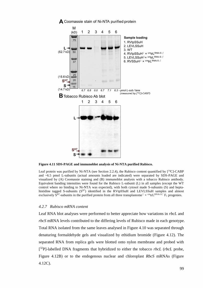
99
Figure 4.11 SDS-PAGE and immunoblot analysis of Ni-NTA purified Rubisco.
Leaf protein was purified by Ni-NTA (see Section 2.2.4), the Rubisco content quantified by [14C]-CABP
and ~6.5 pmol L-subunits (actual amounts loaded are indicated) were separated by SDS-PAGE and
visualized by (A) Coomassie staining and (B) immunoblot analysis with a tobacco Rubisco antibody.
Equivalent banding intensities were found for the Rubisco L-subunit (L) in all samples (except the WT
control where no binding to Ni-NTA was expected), with both cytosol made S-subunits (S) and hepta-
histidine tagged S-subunits (SH7) identified in the RVtpSSuH and LEVLSSuH samples and almost
exclusively SH7-subunits in the purified protein from all three transplastome♀ × cmtrLRNAi-S♂ F1 progenies.
4.2.7 Rubisco mRNA content
Leaf RNA blot analyses were performed to better appreciate how variations in rbcL and
rbcS mRNA levels contributed to the differing levels of Rubisco made in each genotype.
Total RNA isolated from the same leaves analysed in Figure 4.10 was separated through
denaturing formaldehyde gels and visualized by ethidium bromide (Figure 4.12). The
separated RNA from replica gels were blotted onto nylon membrane and probed with
[32P]-labelled DNA fragments that hybridized to either the tobacco rbcL (rbcL probe,
Figure 4.12B) or to the endogenous nuclear and chloroplast RbcS mRNAs (Figure
4.12C).
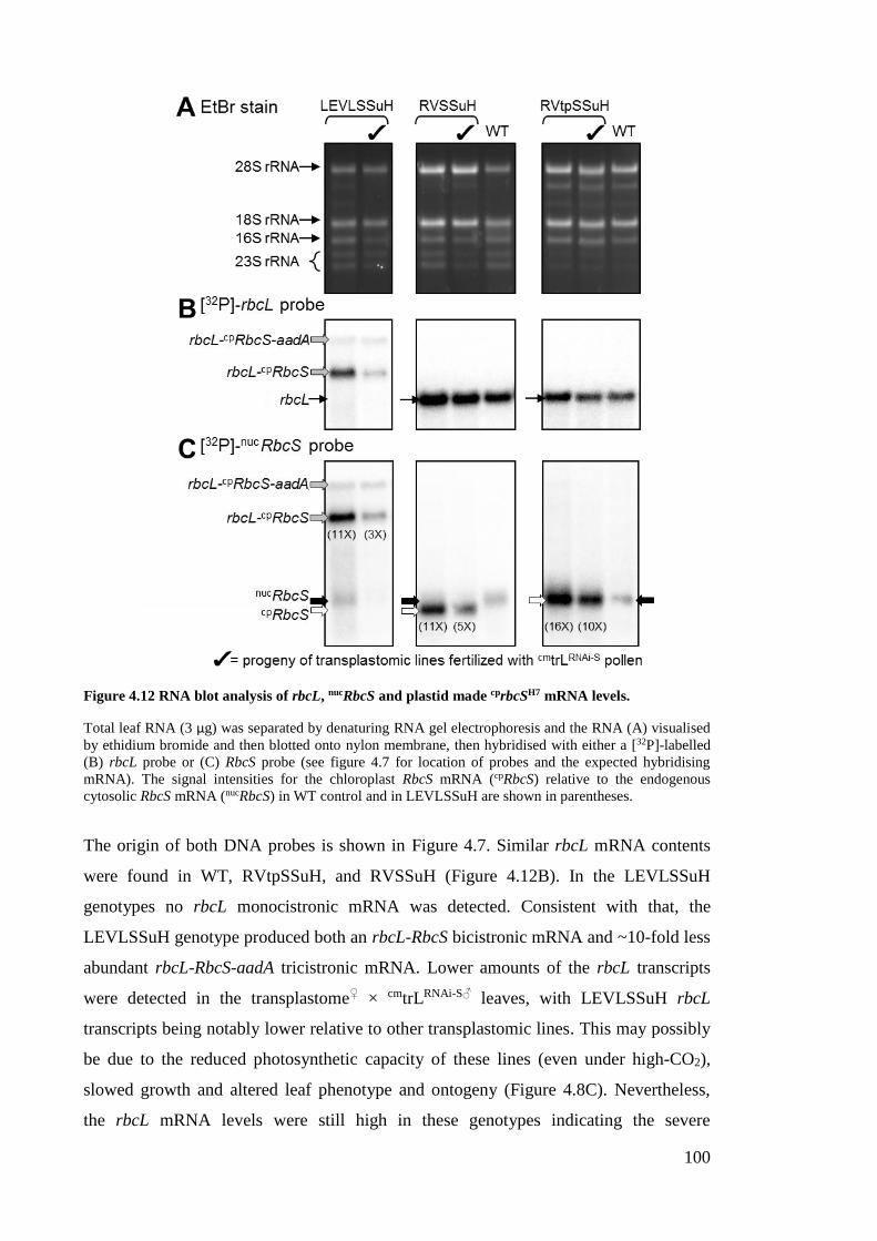
100
Figure 4.12 RNA blot analysis of rbcL, nucRbcS and plastid made cprbcSH7 mRNA levels.
Total leaf RNA (3 µg) was separated by denaturing RNA gel electrophoresis and the RNA (A) visualised
by ethidium bromide and then blotted onto nylon membrane, then hybridised with either a [32P]-labelled
(B) rbcL probe or (C) RbcS probe (see figure 4.7 for location of probes and the expected hybridising
mRNA). The signal intensities for the chloroplast RbcS mRNA (cpRbcS) relative to the endogenous
cytosolic RbcS mRNA (nucRbcS) in WT control and in LEVLSSuH are shown in parentheses.
The origin of both DNA probes is shown in Figure 4.7. Similar rbcL mRNA contents
were found in WT, RVtpSSuH, and RVSSuH (Figure 4.12B). In the LEVLSSuH
genotypes no rbcL monocistronic mRNA was detected. Consistent with that, the
LEVLSSuH genotype produced both an rbcL-RbcS bicistronic mRNA and ~10-fold less
abundant rbcL-RbcS-aadA tricistronic mRNA. Lower amounts of the rbcL transcripts
were detected in the transplastome♀ × cmtrLRNAi-S♂ leaves, with LEVLSSuH rbcL
transcripts being notably lower relative to other transplastomic lines. This may possibly
be due to the reduced photosynthetic capacity of these lines (even under high-CO2),
slowed growth and altered leaf phenotype and ontogeny (Figure 4.8C). Nevertheless,
the rbcL mRNA levels were still high in these genotypes indicating the severe
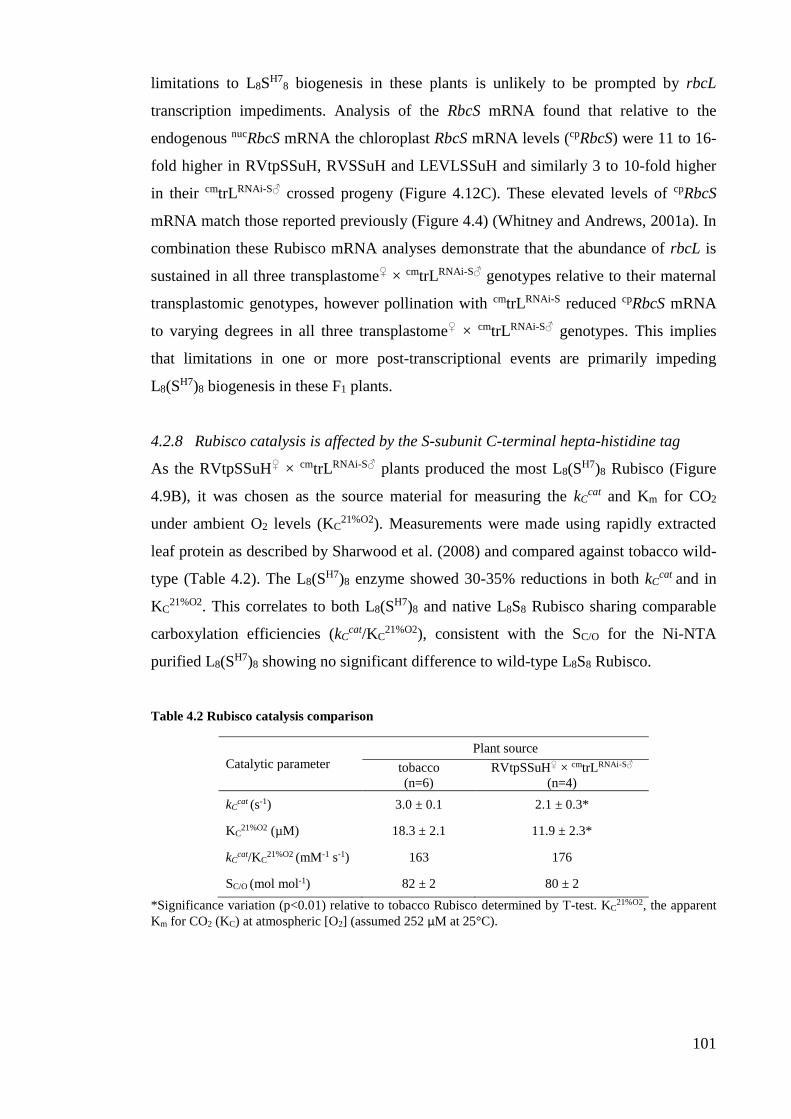
101
limitations to L8SH7
8 biogenesis in these plants is unlikely to be prompted by rbcL
transcription impediments. Analysis of the RbcS mRNA found that relative to the
endogenous nucRbcS mRNA the chloroplast RbcS mRNA levels (cpRbcS) were 11 to 16-
fold higher in RVtpSSuH, RVSSuH and LEVLSSuH and similarly 3 to 10-fold higher
in their cmtrLRNAi-S♂ crossed progeny (Figure 4.12C). These elevated levels of cpRbcS
mRNA match those reported previously (Figure 4.4) (Whitney and Andrews, 2001a). In
combination these Rubisco mRNA analyses demonstrate that the abundance of rbcL is
sustained in all three transplastome♀ × cmtrLRNAi-S♂ genotypes relative to their maternal
transplastomic genotypes, however pollination with cmtrLRNAi-S reduced cpRbcS mRNA
to varying degrees in all three transplastome♀ × cmtrLRNAi-S♂ genotypes. This implies
that limitations in one or more post-transcriptional events are primarily impeding
L8(SH7)8 biogenesis in these F1 plants.
4.2.8 Rubisco catalysis is affected by the S-subunit C-terminal hepta-histidine tag
As the RVtpSSuH♀ × cmtrLRNAi-S♂ plants produced the most L8(SH7)8 Rubisco (Figure
4.9B), it was chosen as the source material for measuring the kCcat and Km for CO2
under ambient O2 levels (KC21%O2). Measurements were made using rapidly extracted
leaf protein as described by Sharwood et al. (2008) and compared against tobacco wild-
type (Table 4.2). The L8(SH7)8 enzyme showed 30-35% reductions in both kC
cat and in
KC21%O2. This correlates to both L8(S
H7)8 and native L8S8 Rubisco sharing comparable
carboxylation efficiencies (kCcat/KC
21%O2), consistent with the SC/O for the Ni-NTA
purified L8(SH7)8 showing no significant difference to wild-type L8S8 Rubisco.
Table 4.2 Rubisco catalysis comparison
Catalytic parameter
Plant source
tobacco
(n=6)
RVtpSSuH♀ × cmtrLRNAi-S♂
(n=4)
kCcat (s-1) 3.0 ± 0.1 2.1 ± 0.3*
KC21%O2 (µM) 18.3 ± 2.1 11.9 ± 2.3*
kCcat/KC
21%O2 (mM-1 s-1) 163 176
SC/O (mol mol-1) 82 ± 2 80 ± 2
*Significance variation (p<0.01) relative to tobacco Rubisco determined by T-test. KC21%O2, the apparent
Km for CO2 (KC) at atmospheric [O2] (assumed 252 µM at 25°C).
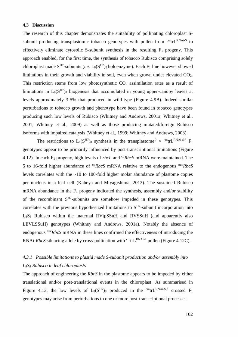
102
4.3 Discussion
The research of this chapter demonstrates the suitability of pollinating chloroplast S-
subunit producing transplastomic tobacco genotypes with pollen from cmtrLRNAi-S to
effectively eliminate cytosolic S-subunit synthesis in the resulting F1 progeny. This
approach enabled, for the first time, the synthesis of tobacco Rubisco comprising solely
chloroplast made SH7-subunits (i.e. L8(SH7)8 holoenzyme). Each F1 line however showed
limitations in their growth and viability in soil, even when grown under elevated CO2.
This restriction stems from low photosynthetic CO2 assimilation rates as a result of
limitations in L8(SH7)8 biogenesis that accumulated in young upper-canopy leaves at
levels approximately 3-5% that produced in wild-type (Figure 4.9B). Indeed similar
perturbations to tobacco growth and phenotype have been found in tobacco genotypes
producing such low levels of Rubisco (Whitney and Andrews, 2001a; Whitney et al.,
2001; Whitney et al., 2009) as well as those producing mutated/foreign Rubisco
isoforms with impaired catalysis (Whitney et al., 1999; Whitney and Andrews, 2003).
The restrictions to L8(SH7)8 synthesis in the transplastome♀ × cmtrLRNAi-S♂ F1
genotypes appear to be primarily influenced by post-transcriptional limitations (Figure
4.12). In each F1 progeny, high levels of rbcL and cpRbcS mRNA were maintained. The
5 to 16-fold higher abundance of cpRbcS mRNA relative to the endogenous nucRbcS
levels correlates with the ~10 to 100-fold higher molar abundance of plastome copies
per nucleus in a leaf cell (Kabeya and Miyagishima, 2013). The sustained Rubisco
mRNA abundance in the F1 progeny indicated the synthesis, assembly and/or stability
of the recombinant SH7-subunits are somehow impeded in these genotypes. This
correlates with the previous hypothesized limitations to SH7-subunit incorporation into
L8S8 Rubisco within the maternal RVtpSSuH and RVSSuH (and apparently also
LEVLSSuH) genotypes (Whitney and Andrews, 2001a). Notably the absence of
endogenous nucRbcS mRNA in these lines confirmed the effectiveness of introducing the
RNAi-RbcS silencing allele by cross-pollination with cmtrLRNAi-S pollen (Figure 4.12C).
4.3.1 Possible limitations to plastid made S-subunit production and/or assembly into
L8S8 Rubisco in leaf chloroplasts
The approach of engineering the RbcS in the plastome appears to be impeded by either
translational and/or post-translational events in the chloroplast. As summarised in
Figure 4.13, the low levels of L8(SH7)8 produced in the cmtrLRNAi-S♂ crossed F1
genotypes may arise from perturbations to one or more post-transcriptional processes.
103
Figure 4.13 Summary of Rubisco subunit expression in cmtrLRNAi-S♂ crossed F1 genotypes and
unknown limitations in subunit processing in the chloroplast.
Possible limitations imposed by the additional C-terminal hepta-histidine (H7) tag on the capacity of SH7-
subunits to assemble into L8S8 holoenzyme seem unlikely. A prior comparison of tranplastomic tobacco
producing plastid made S-subunits without H7 tags showed they shared the same assembly limitations as
the SH7-subunits (Whitney and Andrews, 2001a). This would suggest limitations in translational
processing of chloroplast made S-subunits (see Section 4.3.1.1) or an increased propensity for their
proteolytic degradation (see Section 4.3.1.2) in explaining their limited capacity for incorporation during
L8S8 biogenesis.
4.3.1.1 Limitations to translational processing of the RbcSH7 mRNA
Translational processing of chloroplast mRNA’s is highly influenced by the structure
and stability of the mRNA, its capacity to engage a wide range of regulatory mRNA
elements and a host of requisite protein translation factors coded by genes in both the
chloroplast and nucleus (Yamaguchi et al., 2000; Zerges, 2000). RNA elements
identified as regulatory components in the translation of chloroplast messages are
primarily located in the 5′UTR. These elements include Shine-Dalgarno (SD) sequences,
stem-loop structures, and A/U rich elements. While nearly all bacterial mRNAs use
base pairing between a SD sequence and a complementary sequence located near the 3′
end of the 16S rRNA, the positioning of the SD sequence in chloroplast mRNA 5’UTR
is highly variable. For example the SD in the tobacco psbA mRNA is 33-36 nt upstream
of the D1 protein initiator AUG codon (Bonham-Smith and Bourque, 1989). A common
impediment to reliably expressing recombinant proteins in leaf chloroplasts is the
pervasive influence the introduced gene coding sequence has on mRNA structure and its
translational processing (Maliga, 2004). Significant variations in recombinant protein
104
expression have been observed for different genes using the same chloroplast gene
regulatory sequences (i.e. promoter, 5'- and 3'UTR) (Maliga, 2003). For example, the
same psbA gene elements used in this thesis to regulate RbcSH7 translation in the
RVSSuH and RVtpSSuH genotypes (Figure 4.7) have been used to drive the production
of the cyanobacterial BicA membrane protein (Pengelly et al., 2014), GUS (Staub and
Maliga, 1993) and rbcL-S operons of Galdieria sulphuraria (red algae) and
Phaeodactylum tricornutum (diatom) Rubisco (Whitney et al., 2001) in tobacco
chloroplasts. Each protein was produced in differing amounts ranging from 0.1%, 2%
and 35% of the leaf soluble cellular protein respectively. Similarly the use of the same
rrn promoter, T7g10 5’UTR and rps16 3’UTR sequences have produced up to 30-fold
differences in the levels of recombinant protein expression (Maliga, 2003). These
examples highlight the necessity to better understand promoter and terminator element
function and their translation machineries in greater molecular detail. Such information
is critical if we are to more reliably modulate recombinant protein translation initiation
and elongation rates in the chloroplast (Mayfield et al., 1995; Somanchi and Mayfield,
1999; Maliga and Bock, 2011).
The structural similarity between the translation machinery of chloroplasts and
prokaryotes has led to the belief that the patterns of codon usage in chloroplasts might
show evidence of bias that can be linked to levels of protein expression (Sugiura, 1992).
Understanding the biased usage of synonymous codons has been of particular benefit
for optimising recombinant protein expression in E. coli (Angov, 2011). For E. coli, the
codon usage efficiency closely reflects the differential composition of the genomic
tRNA pool. In chloroplasts, only around 30 tRNA species are coded by the plastome.
Highly translated proteins in the chloroplasts therefore tend to show strong codon use
preference for these tRNA (Alkatib et al., 2012). Increasing evidence from plastome
transformation studies seems to increasingly support this assertion. By incorporating the
biased usage of particular synonymous codons comparable to those used by highly
expressed chloroplast genes the production of recombinant proteins such as R. rubrum
Rubisco (Whitney and Sharwood, 2008), S. verticillus Ble (Heitzer et al., 2007), B.
thuringiensis cryIA(b) (Perlak et al., 1991) and C. tetani TetC (Tregoning et al., 2003)
have been enhanced by 3 to 10-fold. This enhancement is presumed to occur as a
consequence of faster rates of translation elongation. With regard to this current study,
the higher GC content of nuclear tobacco RbcS incorporated into each transplastomic
line produces a codon use that varies considerably from the AT rich tobacco rbcL
(Table 4.3). Examination of the differential codon use however sees the CUC (coding
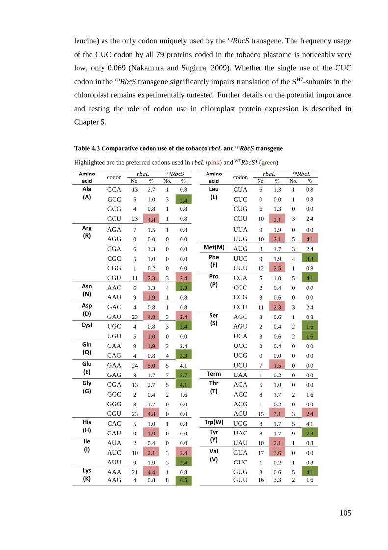
105
leucine) as the only codon uniquely used by the cpRbcS transgene. The frequency usage
of the CUC codon by all 79 proteins coded in the tobacco plastome is noticeably very
low, only 0.069 (Nakamura and Sugiura, 2009). Whether the single use of the CUC
codon in the cpRbcS transgene significantly impairs translation of the SH7-subunits in the
chloroplast remains experimentally untested. Further details on the potential importance
and testing the role of codon use in chloroplast protein expression is described in
Chapter 5.
Table 4.3 Comparative codon use of the tobacco rbcL and cpRbcS transgene
Highlighted are the preferred codons used in rbcL (pink) and WTRbcS* (green)
Amino acid
codon rbcL cpRbcS
Amino acid
codon rbcL cpRbcS
No. % No. %
No. % No. %
Ala (A)
GCA 13 2.7 1 0.8
Leu (L)
CUA 6 1.3 1 0.8
GCC 5 1.0 3 2.4 CUC 0 0.0 1 0.8
GCG 4 0.8 1 0.8
CUG 6 1.3 0 0.0
GCU 23 4.8 1 0.8
CUU 10 2.1 3 2.4
Arg (R)
AGA 7 1.5 1 0.8
UUA 9 1.9 0 0.0
AGG 0 0.0 0 0.0
UUG 10 2.1 5 4.1
CGA 6 1.3 0 0.0
Met(M) AUG 8 1.7 3 2.4
CGC 5 1.0 0 0.0
Phe (F)
UUC 9 1.9 4 3.3
CGG 1 0.2 0 0.0
UUU 12 2.5 1 0.8
CGU 11 2.3 3 2.4
Pro (P)
CCA 5 1.0 5 4.1
Asn (N)
AAC 6 1.3 4 3.3
CCC 2 0.4 0 0.0
AAU 9 1.9 1 0.8
CCG 3 0.6 0 0.0
Asp (D)
GAC 4 0.8 1 0.8
CCU 11 2.3 3 2.4
GAU 23 4.8 3 2.4
Ser (S)
AGC 3 0.6 1 0.8
CysI UGC 4 0.8 3 2.4
AGU 2 0.4 2 1.6
UGU 5 1.0 0 0.0
UCA 3 0.6 2 1.6
Gln (Q)
CAA 9 1.9 3 2.4
UCC 2 0.4 0 0.0
CAG 4 0.8 4 3.3
UCG 0 0.0 0 0.0
Glu (E)
GAA 24 5.0 5 4.1
UCU 7 1.5 0 0.0
GAG 8 1.7 7 5.7
Term UAA 1 0.2 0 0.0
Gly (G)
GGA 13 2.7 5 4.1
Thr (T)
ACA 5 1.0 0 0.0
GGC 2 0.4 2 1.6
ACC 8 1.7 2 1.6
GGG 8 1.7 0 0.0
ACG 1 0.2 0 0.0
GGU 23 4.8 0 0.0
ACU 15 3.1 3 2.4
His (H)
CAC 5 1.0 1 0.8
Trp(W) UGG 8 1.7 5 4.1
CAU 9 1.9 0 0.0
Tyr (Y)
UAC 8 1.7 9 7.3
Ile (I)
AUA 2 0.4 0 0.0
UAU 10 2.1 1 0.8
AUC 10 2.1 3 2.4
Val (V)
GUA 17 3.6 0 0.0
AUU 9 1.9 3 2.4
GUC 1 0.2 1 0.8
Lys (K)
AAA 21 4.4 1 0.8
GUG 3 0.6 5 4.1
AAG 4 0.8 8 6.5
GUU 16 3.3 2 1.6
106
4.3.1.2 Are the translated SH7-subunits prone to proteolysis?
As the R. rubrum L2 Rubisco in the cmtrL tobacco genotype has no requirements for S-
subunits, it is assumed the cytosolic made S-subunits are still made but are quickly
degraded in the chloroplast stroma as no evidence for their accumulation has been found
(Whitney and Andrews, 2001a; Whitney and Andrews, 2003; Whitney and Sharwood,
2008). The same is also assumed for L-subunits in anti-RbcS genotypes where the
detection of unassembled L-subunits or oligomeric assemblies of L-subunits have not
been observed (Hudson et al., 1992; Andrews et al., 1995b; Wostrikoff and Stern, 2007;
Kubien and Sage, 2008). This apparent rapid degradation of “unused” proteins in the
stroma likely stems from the highly efficient degradation pathways that exist in
chloroplasts (Xie et al., 2015). The most notable proteases are DegP (a serine protease)
and the multi-subunit ClpP and FtsH homologs. These proteases function via ATPase
hydrolysis with varying levels of substrate recognition specificity (Kuroda and Maliga,
2003; Adam et al., 2011). Overall chloroplast proteases play critical roles in chloroplast
biogenesis and maintaining homeostasis and function. Their roles in maintaining
homeostasis are particularly important in response to cellular stresses (e.g. heat, high
light) where the requirement for degradation and recycling of damaged proteins, such as
the D1 protein of photosystem II, is elevated (Haußühl et al., 2001).
In the chloroplast, is the assumed co-translational formylation of the nascent
peptide Met-1. Peptide deformylase subsequently removes this modification to facilitate
direct post-translational modification of Met-1 by N-acetyltransferase (NAT) activity,
N-methylation or its removal by methionine amino peptidase (MAP) before
modification of the new N-terminal amino acid (i.e. amino acid at position 2). The
capacity of MAP to undertake this process is thought to be dependent on the size and
charge of the amino acid at position 2. For cytosol made proteins targeted to the stroma,
processing of the transit peptide leader sequence (see Section 4.1.2) is facilitated by a
~140 kDa stromal processing peptidase (SPP; a metalloprotease) (Richter et al., 2005).
Consistent with that seen previously by Whitney and Andrews (2001) none of the
chloroplast made SH7-subunits in RVtpSSuH, LEVLSSuH or their cmtrLRNAi-S♂ crossed
F1 progeny contained the 57 amino acid leader sequence (Figure 4.10) reaffirming SPP
processing of transit peptides occurs within the chloroplast stroma.
An underpinning question is whether the limited capacity to incorporate stroma
synthesized SH7-subunit into L8S8 complexes stems from their spatial segregation away
from the co-evolved chaperone-facilitated S-subunit assembly pathway? As indicated in
Figure 4.1, for cytosol made S-subunits this pathway begins near the Tic entry point of
107
the chloroplast envelope where a range of evolved molecular partners are located. In
contrast the SH7-subunits are likely synthesized by thylakoid-bound or stroma-located
chloroplast polysomes. This places the SH7-subunits spatially remote from the envelope-
associated molecular partners, likely exposing the SH7-subunits to heightened rates of
proteolysis. Prior [35S]-Met pulse-chase analyses however showed no significant
difference in the synthesis or degradation of the SH7-subunits in either RVtpSSuH or
LEVLSSuH (Whitney and Andrews, 2001a). Admittedly the nature of these analyses
only detected the SH7-subunits that successfully assembled into L8S7(SH7)1 complexes as
the low levels of SH7-subunits measurable necessitated selective enrichment by IMAC
(using Ni-NTA). Repeating this [35S]-Met pulse-chase analysis in the transplastome♀ ×
cmtrLRNAi-S♂ F1 progeny would unlikely require IMAC enrichment given the higher (and
homogeneous) population of SH7-subunits incorporate the L8(SH7)8 Rubisco made. This
would improve the accuracy and sensitivity of measuring whether the SH7-subunits
show differences in their synthesis, assembly or stability relative to the cytosol made S-
subunits in wild-type controls. Such [35S]-Met pulse-chase analyses remain to be
undertaken.
A key difference between the cytosol made S-subunits and stroma made SH7-
subunits are the post-translational modification of their mature Met-1 residue. As
indicated in Section 4.1.3, following import of cytosol made precursor S-subunits into
the stroma, their tp sequence is removed and the exposed Met-1 is N-methylated. The
function of this modification remains unknown (Houtz et al., 2008). In contrast, the
Met-1 of the chloroplast made SH7-subunits are acetylated (Whitney, unpublished)
presumably via NAT activity for which there are multiple isoforms in chloroplasts. N-
terminal acetylation is considered a modification that provides chloroplast proteins
(such as the L-subunit) protection from proteolysis (Houtz et al., 2008) (Figure 4.2).
With this in mind one might speculate there may be limitations to the post-translational
acetylation of stromal made SH7-subunits by NAT – with only a limited population of
SH7-subunits correctly modified and able to resist proteolysis and assemble into L8S8
complexes. The future [35S]-Met pulse-chase analyses proposed above for the
transplastome♀ × cmtrLRNAi-S♂ F1 progeny will enable experimental testing of this
hypothesis.
108
4.3.2 The RNAi-S genotype is stably inherited
A key deficiency of many RNAi studies has been the instability of T-DNA integration.
As shown in Chapter 3, the heritable bar conferring Basta resistance in the cmtrLRNAi-S
line has proven an ideal marker gene for nuclear transformation and the identification in
transplastome♀ × cmtrLRNAi-S♂ F1 progeny inheriting the paternal chromosome
containing bar (Figure 4.8A) and RNAi-RbcS silencing T-DNA (Figure 4.12C).
Subsequent selfing of the F1 genotypes studied in this chapter have shown that the
RNAi-RbcS silencing phenotype is stably inherited in F3 progeny (data not shown).
4.3.3 The RNAi-processing machinery is not present in chloroplasts
A key finding of this chapter is the RVtpSSuH, RVSSuH and LEVLSSuH (where it is
primarily an rbcL-cpRbcS transcript) and their corresponding cmtrLRNAi-S crossed F1
progeny produced comparable levels of cpRbcS mRNA (Figure 4.12). Notably the codon
use of the plastome cpRbcS transgene matches the region of nucRbcS sequence used in the
RNAi-RbcS T-DNA (Figure 3.5). This provides strong evidence that the efficient
RNAi-processing machinery present in the cytosol (Figure 3.2) does not function in the
chloroplast stroma. This supports the findings of Zhang et al. (2015) who found no
evidence of RNAi silencing of recombinant double-stranded RNAs (dsRNAs) in potato
chloroplasts. This is somewhat not unexpected as the genes coding the dicer enzymes
or/and other protein components of the RNA-induced silencing complexes do not code
for appropriate N-terminal chloroplast targeting sequences. The discovery that
chloroplast gene expression is immune to RNAi-silencing may pose an advantage in
chloroplast bioengineering applications where silencing of cytosolic enzymes/pathways
may be beneficial. For example, many subunits of the protein complexes involved in
photosynthetic electron transport are coded in the nucleus (Leister, 2012). Future
structure-function studies of these protein components via a plastome transformation
approach would necessitate silencing of the nucleus genes by RNAi – a goal that is now
clearly achievable.
4.3.4 Future directions
The data presented in this chapter represent experiments undertaken to qualify the
potential for producing Rubisco complexes fully comprising plastome made S-subunits.
This poses a significant step forward with regard to being able to modify Rubisco
catalysis through co-ordinated changes to both the L- and S-subunits. Limitations in the
capacity to produce and assemble chloroplast made S-subunits continue to hinder

109
progress toward such goals. Identifying the causes and potential solutions to these
challenges are addressed in the following two chapters.
110
CHAPTER 5 – TRANSPLASTOMIC PRODUCTION OF
HYBRID RUBISCO COMPRISING TOBACCO L-
SUBUNITS AND ALTERNATIVE S-SUBUNITS
5.1 Introduction
During the evolution of plants and green algae multiple RbcS copies in the nucleus have
been acquired as a consequence of gene transfer and duplication from the rbcL-rbcS
operon within the genome of the cyanobacterial progenitor of plastids (Martin and
Kowallik, 1999; Bogorad, 2012). Within the nucleus, the numbers of RbcS copies have
multiplied and with ensuing mutagenic events, led to plants and algae producing
varying numbers of RbcS copies that typically code different S-subunit sequences.
Differential expression of the varying S-subunit isoforms in response to environmental
cues are hypothesised to potentially modulate Rubisco catalysis in some plant species
(Morita et al., 2014). In support of this are the rising numbers of studies on Rubisco
L8S8 chimeras comprising alternative mixtures of heterologous S-subunits that lead to
varying effects on catalysis (Table 5.1). Clearly the goals of being able to modulate
Rubisco catalysis through directed amino acid changes necessitates a better
understanding of interactions between the L- and S-subunit. This goal, hampered by the
nuclear location and multiple RbcS copies in plants have prevented their directed
deletion and mutagenesis (Whitney et al., 2011a; Parry et al., 2013). As evident from
Chapter 3 and Chapter 4, the cmtrLRNAi-S genotype has the potential to circumvent these
limitations.
5.1.1 Expression of rbcL-rbcS operons in higher plants chloroplasts
The Rubisco genes in proteobacteria, non-green algae and cyanobacteria remain
encoded as a single, co-transcribed dicistronic rbcL-rbcS (rbcL-S) mRNA (Assali et al.,
1991; Badger and Bek, 2008). In the non-green algae the Rubisco operon still resides in
the plastome with the genes separated by an intergenic region (IR) of varying lengths
(typically between 30-60 nucleotides in length). The IR typically contains a Shine-
Dalgarno (SD) sequence 7-10 nucleotides upstream of the rbcS initiator codon to
facilitate ribosome engagement for S-subunit translation (Whitney and Andrews,
2001b). These genetic elements in the IR are readily recognised by the translational
machinery within E .coli. For example the native rbcL-S sequences from proteobacteria
(e.g. Rhodobacter sphaeroides, Alcaligenes eutrophus), cyanobacteria (e.g.
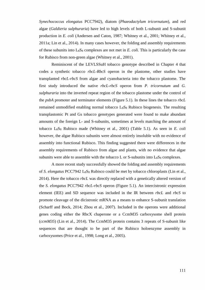
111
Synechococcus elongatus PCC7942), diatom (Phaeodactylum tricornutum), and red
algae (Galdieria sulphuraria) have led to high levels of both L-subunit and S-subunit
production in E. coli (Andersen and Caton, 1987; Whitney et al., 2001; Whitney et al.,
2011a; Lin et al., 2014). In many cases however, the folding and assembly requirements
of these subunits into L8S8 complexes are not met in E. coli. This is particularly the case
for Rubisco from non-green algae (Whitney et al., 2001).
Reminiscent of the LEVLSSuH tobacco genotype described in Chapter 4 that
codes a synthetic tobacco rbcL-RbcS operon in the plastome, other studies have
transplanted rbcL-rbcS from algae and cyanobacteria into the tobacco plastome. The
first study introduced the native rbcL-rbcS operon from P. tricornutum and G.
sulphuraria into the inverted repeat region of the tobacco plastome under the control of
the psbA promoter and terminator elements (Figure 5.1). In these lines the tobacco rbcL
remained unmodified enabling normal tobacco L8S8 Rubisco biogenesis. The resulting
transplastomic Pt and Gs tobacco genotypes generated were found to make abundant
amounts of the foreign L- and S-subunits, sometimes at levels matching the amount of
tobacco L8S8 Rubisco made (Whitney et al., 2001) (Table 5.1). As seen in E. coli
however, the algae Rubisco subunits were almost entirely insoluble with no evidence of
assembly into functional Rubisco. This finding suggested there were differences in the
assembly requirements of Rubisco from algae and plants, with no evidence that algae
subunits were able to assemble with the tobacco L or S-subunits into L8S8 complexes.
A more recent study successfully showed the folding and assembly requirements
of S. elongatus PCC7942 L8S8 Rubisco could be met by tobacco chloroplasts (Lin et al.,
2014). Here the tobacco rbcL was directly replaced with a genetically altered version of
the S. elongatus PCC7942 rbcL-rbcS operon (Figure 5.1). An intercistronic expression
element (IEE) and SD sequence was included in the IR between rbcL and rbcS to
promote cleavage of the dicistronic mRNA as a means to enhance S-subunit translation
(Scharff and Bock, 2014; Zhou et al., 2007). Included in the operons were additional
genes coding either the RbcX chaperone or a CcmM35 carboxysome shell protein
(ccmM35) (Lin et al., 2014). The CcmM35 protein contains 3 repeats of S-subunit like
sequences that are thought to be part of the Rubisco holoenzyme assembly in
carboxysomes (Price et al., 1998; Long et al., 2005).
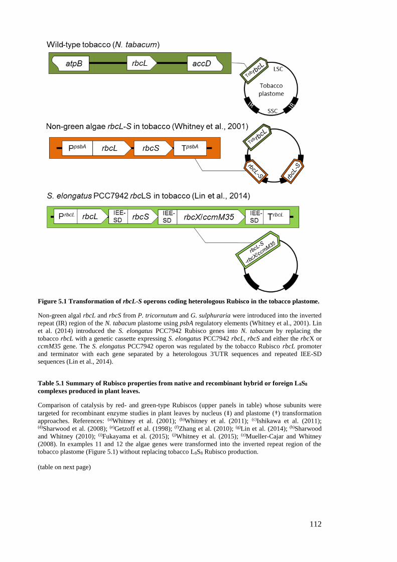
112
Figure 5.1 Transformation of rbcL-S operons coding heterologous Rubisco in the tobacco plastome.
Non-green algal rbcL and rbcS from P. tricornutum and G. sulphuraria were introduced into the inverted
repeat (IR) region of the N. tabacum plastome using psbA regulatory elements (Whitney et al., 2001). Lin
et al. (2014) introduced the S. elongatus PCC7942 Rubisco genes into N. tabacum by replacing the
tobacco rbcL with a genetic cassette expressing S. elongatus PCC7942 rbcL, rbcS and either the rbcX or
ccmM35 gene. The S. elongatus PCC7942 operon was regulated by the tobacco Rubisco rbcL promoter
and terminator with each gene separated by a heterologous 3'UTR sequences and repeated IEE-SD
sequences (Lin et al., 2014).
Table 5.1 Summary of Rubisco properties from native and recombinant hybrid or foreign L8S8
complexes produced in plant leaves.
Comparison of catalysis by red- and green-type Rubiscos (upper panels in table) whose subunits were
targeted for recombinant enzyme studies in plant leaves by nucleus (‡) and plastome (†) transformation
approaches. References: (a)Whitney et al. (2001); (b)Whitney et al. (2011); (c)Ishikawa et al. (2011); (d)Sharwood et al. (2008); (e)Getzoff et al. (1998); (f)Zhang et al. (2010); (g)Lin et al. (2014); (h)Sharwood
and Whitney (2010); (i)Fukayama et al. (2015); (j)Whitney et al. (2015); (z)Mueller-Cajar and Whitney
(2008). In examples 11 and 12 the algae genes were transformed into the inverted repeat region of the
tobacco plastome (Figure 5.1) without replacing tobacco L8S8 Rubisco production.
(table on next page)
113
Native L8S8 Rubisco
Rubisco
type
Organism
type Plant/algae species(reference)
Catalysis
kCcat Kc Kc
21%O2 Sc/o
(s-1) (µM) (µM) (mol mol-1)
Red Red algae
Griffithsia monilis (a) 2.6 9.3 12.6 167
Galdieria sulphuraria (a) 1.2 3.3 5.5 166
Diatom Phaeodactylum tricornutum (a) 3.4 27.9 42.9 133
Green
cyanobacteria Synechococcus elongatus PCC 7942 (z) 11.8 200.0 250.8 42
C3 Nicotiana tabacum (tobacco) (b) 3.0 12.6 18.3 82
C3 Oryza sativa (rice) (c) 1.7 17.3 N/A 117
C3 Helianthus annuus (sunflower) (d) 2.9 8.2 23.9 84
C4 Flaveria bidentis (b) 4.8 20.4 33 81
C4 Sorghum bicolor (sorghum) (c) 4.1 41.5 N/A N/A
C4 Flaveria bidentis (b) 4.8 20.4 32.6 81 Hybrid or foreign L8S8 Rubisco
Ex
am
ple
Name of plant
genotype(reference)
Rubisco subunit source recombinant mRNA
content*
Rubisco
content
Catalysis
kCcat Kc Kc
21%O2 Sc/o
L-subunit S-subunit rbcL RbcS (% of WT) (s-1) (µM) (µM) (mol mol-1)
1‡ T7.3 (e) A. thaliana P. sativum native 18% 89% ~85% WT n.m n.m n.m
2† LLS4 (f) S. lycopersicum N. tabacum n.m native <20% 2.8 n.m n.m n.m
3† Se7942 (g) Synechococcus
PCC7942 N. tabacum n.m. (rbcL-S mRNA) <15% >8.0 n.m n.m n.m
4† tobpring (b) F. pringlei N. tabacum 80-100% native 20% 3.5 13.0 23.3 80
5† tobflo (b) F. floridana N. tabacum 80-100% native 50% 3.7 14.5 24.7 81
6† tobbid (b) F. bidentis N. tabacum 80-100% native 35% 4.7 19.9 32.2 79
7† tobRst (d) H. annuus N. tabacum 15% native 11% 3.3 N/A 20.8 84
8† tRstL7 (h) H. annuus N. tabacum 60% native 17%
9† tobAtL (j) A. thaliana N. tabacum 70-75% native 25% 2.3 8.6 18.4 80
10‡ SS5 (c) O. sativa S. bicolor native 79% 124% 2.5 24.0 N/A 103
11‡ PS23 (i) O. sativa P. pratense native 33% ~107% 1.8 22.4 N/A 126
12† Pt2 (a) P. tricornutum P. tricornutum 38% (rbcL-S mRNA) 8-30% The algae Rubisco subunits produced were insoluble
13† Gs (a) G. sulphuraria G. sulphuraria 3% (rbcL-S mRNA) 5% The algae Rubisco subunits produced were insoluble
114
The resulting SeLSX and SeLSM35 tobacco genotypes produced by Lin et al.,
(2014) accumulated low and varying amounts of S. elongatus PCC7942 Rubisco. At
most the cyanobacteria Rubisco accumulated to only ~15% that of the wild-type
tobacco controls (Table 5.1). Due to these low levels and S. elongatus PCC7942
Rubisco’s poor affinity and specificity for CO2, the SeLSX and SeLSM35 genotypes
required high-CO2 supplementation to support growth in soil. Apparent structural
incompatibilities between the S. elongatus PCC7942 S-subunits and the endogenous
tobacco S-subunits precluded assembly of the latter in the Rubisco complexes made in
SeLSX and SeLSM35. This incompatibility may explain the observation of a prior
transplastomic study where replacement of the tobacco rbcL with only the S. elongatus
PCC7942 rbcL resulted in no Rubisco production (Kanevski et al., 1999).
5.1.2 Catalytic variability of higher plant hybrid Rubisco
While generally it is evident the S-subunit can influence catalysis, the greater sequence
diversity between S-subunits (relative to L-subunits) appears to produce unpredictable
patterns as to the potential for S-subunits to assemble with heterologous L8 cores and
their influence on holoenzyme catalysis. For example, different algae S-subunits were
able to assemble with Rhodobacter sphaeroides L-subunits into “red”-type L8S8
complexes, however the carboxylation rate (kCcat) of each hybrid enzyme was slowed
>100-fold (Joshi et al., 2015). In contrast, assembly of tobacco S-subunits with L-
subunits from sunflower, tomato and various Flaveria species has no significant
influence on catalysis (Sharwood et al., 2008; Sharwood and Whitney, 2010), but slow
down the carboxylation rate of Arabidopsis Rubisco (Whitney et al., 2011b; Zhang et al.,
2011; Whitney et al., 2015) (see Section 5.1.2). Likewise, the assembly of Sorghum S-
subunits into rice Rubisco produced L8S8 chimers with increased kCcat and Km for CO2
(KC) (Ishikawa et al., 2011), while assembly of pea S-subunits into Arabidopsis Rubisco
decreased kCcat (Getzoff et al., 1998).
The inability to assemble vascular plant Rubisco in E. coli has also led to
numerous structure-function studies by bioengineering hybrid higher plant Rubisco (i.e.
complexes comprising heterologous subunit compositions) via plastome and nuclear
transformation approaches (Table 5.1). While the catalytic properties of some hybrid
enzymes suggest some S-subunits have little or no influence on catalysis, others can
evince a dramatic change. For example, assembly of pea Rubisco S-subunits into A.
thaliana Rubisco generated a hybrid enzyme where kCcat was reduced by ~12% (Getzoff
et al., 1998) while a S-subunit from Phleum pratense had no effect on rice Rubisco
115
catalysis (Fukayama et al., 2015). In contrast, introducing a S-subunit from the C4 plant
sorghum into rice had a significant effect on Rubisco catalysis – raising kCcat and KC by
47% and 39% respectively (Ishikawa et al., 2011).
Targeted changes to the L-subunit source in tobacco via chloroplast
transformation has also led to varying effects on catalysis. Replacement of the tobacco
rbcL with the gene from sunflower (Sharwood et al., 2008), tomato (Zhang et al., 2011)
and different Flaveria species (Whitney et al., 2011b) had little effect on the catalytic
properties of the hybrid enzymes that closely matched that of the Rubisco from which
the L-subunit originated. This suggests the tobacco S-subunits although having little
influence on hybrid enzyme catalysis, are required to maintain functional Rubisco
catalysis. However, recent work by Whitney et al. (2015) show that the kCcat of hybrid
Rubisco comprising Arabidopsis L-subunits and tobacco S-subunits is impaired by
~25%. Identifying how sequences with the Rubisco L- and S-subunits from different
plant sources interact to affect catalysis poses a critical avenue for further study if we
are to identify ways in which to reliably bioengineer improved forms of Rubisco (Parry
et al., 2013).
5.1.3 Differential assembly of hybrid Rubisco in tobacco chloroplasts
As highlighted in Table 5.1, the biogenesis of hybrid Rubisco comprising tobacco S-
subunits and different plant L-subunits varies considerably. In genotypes producing
sunflower L-subunits the hybrid enzyme accumulated in tissue culture grown plants to
levels of ~30% that of wild-type (Kanevski et al., 1999). In soil, this genotype required
elevated CO2 to survive where hybrid Rubisco levels were only ~10% that of wild-type
(Sharwood et al., 2008). The reduced levels of hybrid Rubisco resulted in unique leaf
phenotypes in the juvenile plants. Even in genotypes producing 4-fold higher levels of
sunflower rbcL, the level of hybrid Rubisco produced only increased by ~70%
(Sharwood and Whitney, 2010) indicating biogenesis of the hybrid L8S8 complex was
primarily impeded by limitations in the folding and/or assembly of the sunflower L-
subunits with the tobacco S-subunits. A parallel study of Whitney et al. (2011) using L-
subunits from 3 different Flaveria species also showed 20-50% limitations in hybrid
Rubisco production compared to wild-type (Table 5.1). Although the transplanted L-
subunits only varied in 3 amino acid residues, the level of hybrid Rubisco produced
varied more than 2-fold – attesting to how single amino acid changes in the L-subunit
can strongly influence holoenzyme biogenesis.
116
The limited capacity of tobacco genotypes producing tomato L-subunit to
assemble hybrid Rubisco poses a conundrum (Zhang et al., 2011) as both tobacco and
tomato are Solanaceae species with L-subunits that vary only by 4 amino acid residues
(Genbank accession numbers ABC56308.1 and CAA77361.1). The use of the rrn
promoter by Zhang et al. (2011) in place of the native rbcL promoter/5’UTR sequence
was found to perturb tobacco Rubisco biogenesis dramatically as shown by a control
genotype where the native tobacco rbcL is controlled by the rrn genetic elements
(Whitney, unpublished). The cause of this unexpected result has yet to be determined.
As indicated in Table 5.1, common to all current heterologous rbcL replacement
studies has been impediments to the biogenesis of the hybrid or foreign Rubisco
complexes. In general this deficiency appears attributable to incompatibilities in the
chloroplast such as inefficiencies in mRNA translation, subunit misfolding and/or
incompatible assembly requirements. As a consequence no foreign or hybrid L8S8
Rubisco has been produced in amounts nearing wild-type levels. This has typically
necessitated additional CO2 to support autotrophic growth of these transplastomic lines.
5.1.4 Research Objective – Producing hybrid Rubisco comprising tobacco L-subunits
and heterologous S-subunits
As indicated above, technological limitations in the capacity to effectively engineer the
Rubisco S-subunits in planta and the puzzling variability in the catalytic properties of
the recombinant hybrid enzymes produced has so far frustrated Rubisco S-subunit
mutagenic studies. The surgical precision of plastome transformation provides a
tantalizing opportunity to use the cmtrLRNAi-S genotype as a vector to produce Rubisco
complexes by transplanting in an rbcL-rbcS operon to replace rbcM. The strategy is
summarised in Figure 5.2.
By this approach the assembled Rubisco would comprise a homogenous
population of L- and S-subunits and avoid the need for propagating and testing multiple
transgenic lines currently undertaken following nucleus transformation approaches
(Suzuki et al., 2007; Suzuki et al., 2009; Suzuki and Makino, 2012). Research described
in this chapter was aimed at determining the capacity of phylogenetically different S-
subunit isoforms to assemble with tobacco L8 cores and their consequence on catalysis.
Using chloroplast transformation in cmtrLRNAi-S, rbcM was replaced with synthetic rbcL-
rbcS operons comprising tobacco rbcL and rbcS coding Rubisco S-subunits from
tobacco (control), Sorghum bicolor, Flaveria bidentis (both C4 plants) and the red algae
Griffithsia monilis (Figure 5.2). Transforming plasmid design, the transforming process

117
and identification of transplastomic genotypes followed by an analysis of the Rubisco
biochemistry in each genotype is examined to explain their growth phenotype.
Figure 5.2 Strategy for introducing rbcL-rbcS operons into cmtrLRNAi-S via plastome transformation.
5.2 Results
5.2.1 Choice of S-subunits for transformation
S-subunits of varying structural compatibility to tobacco Rubisco were used in this
study to test how they influenced catalysis if capable of assembly with tobacco L8 cores.
The S-subunit from Griffithsia monilis was chosen as the Rubisco from this filamentous
red algae possesses the most efficient L8S8 enzyme characterised so far (Table 5.1). G.
monilis Rubisco has a 2-fold higher CO2/O2 specificity (Sc/o) and 26% higher
carboxylation efficiency (kCcat/KC
21%O2; 206 mM-1 s-1) relative to tobacco Rubisco (176
mM-1 s-1) (Whitney et al., 2001). Typical of S-subunits from non-green algae, the βA/βB
loop of G. monilis S-subunits (10 amino acids) is shorter than higher plant S-subunits
(22 amino acids) and has an extended C-terminus that includes a βE-βF loop that is
unique to “red”-type Rubisco (Spreitzer, 2003) (Figure 5.3A). The S-subunits from the
C4 species Flaveria bidentis is studied as its Rubisco shows differential increases of 57%
and 76% in kCcat and KC
21%O2 respectively, relative to tobacco Rubisco (Table 5.1). Of
particular interest was whether the increases in KC observed for rice Rubisco
incorporating sorghum S-subunits (Ishikawa et al., 2011) (Table 5.1) were emulated
when assembled with tobacco L-subunits. A comparison of the vascular plant S-subunit
identities showed all three isoforms shared ~71-78% identity while only having 36-39%
identity to the G. monilis S-subunit (Figure 5.3B).
118
5.2.2 Modifying the codon use of the transplanted RbcS
The objectives of this work were run in parallel with efforts to transform the nucleus of
cmtrLRNAi-S with a synthetic tobacco RbcS (TobRbcS) (Birch and Whitney, unpublished).
To ensure TobRbcS was not also a substrate for the introduced RNAi-RbcS silencing in
cmtrLRNAi-S its codon use was modified to diverge from the native RbcS mRNA
transcripts. As shown in Table 5.2, the codon use of TobRbcS varied significantly from
the native nuclear RbcS (WTRbcS) previously transformed into tobacco (Whitney and
Andrews, 2001a) (Figure 4.7). As a result of the altered codon use the TobRbcS and
WTRbcS shared only 71% identity (Figure 5.4B) compared to the >94% homology
shared by the varied RbcS mRNAs in tobacco (Figure 3.6A). While analyses of the
cmtrLRNAi-S cross-pollinated with RVSSuH, RVtpSSuH and LEVLSSuH genotypes
showed the chloroplasts are immune to cytosol directed RNAi-RbcS silencing (see
Section 4.3.3), this was not known prior to initiation of this project.
The synthetic RbcS for the F. bidentis, S. bicolor and G. monilis S-subunit were
generated using the TobRbcS as a template (Table 5.2). Where possible, identical
nucleotide sequences were incorporated. The goal of maintaining maximum sequence
identity was to minimise potential variations in the stability of each mRNA and their
translational potential. As shown in Figure 5.4B, resulting synthetic SorgRbcS and
BidRbcS showed high levels of identity with TobRbcS (82-89%), consistent with their
high level of amino acid homology (72-78%) (Figure 5.3B). In contrast the high amino
acid divergence between the G. monilis S-subunit and the plant S-subunits (only 36-38%
identity) (Figure 5.3B) led to low identities (only 36-40%) between GmRbcS and
synthetic plant RbcS (Figure 5.4B, noting that due to the extensive structural and
sequence divergence of the GmS-subunit in Figure 5.3, the alignment of GmRbcS is not
provided in Figure 5.4A).
5.2.3 Transforming RbcS into the plastome as an rbcL-rbcS operon
Introducing the TobRbcS into the transforming plasmid pLEV4 took advantage of a
unique NdeI cloning site in tobacco rbcL (Figure 5.5A). A GenScript synthetic gene
cloned into pUC57 was made that coded the 3'end of tobacco rbcL (starting at the NdeI
site) and an adjoining 52 bp intergenic sequence (IS). This sequence is 19 bp longer
than that used in LEVLSSuH (Whitney and Sharwood, 2008) and incorporates less of
the native P. tricornutum rbcL-S intergenic sequence (Figure 5.5B). The IS sequence
also varied with the SD sequence positioned at nucleotides -10 to -7 upstream of the
TobRbcS AUG initiator codon compared with its position at -15 to -11 in LEVLSSuH.
119
This variation was introduced to facilitate the incorporation of a unique SphI cloning
site found naturally in the tobacco RbcS at the transit peptide scission site. A SalI site
was introduced adjacent to the TAA stop codon in synthetic TobRbcS to enable cloning
of the 720 bp ΔtobL-IS- TobRbcS sequence directly into the NdeI-SalI sites of pLEV4 to
make the transforming plasmids pLEVL-TobSH6 (Figure 5.5). The synthesized SorgRbcS,
BidRbcS and GmRbcS (Figure 5.5A) that coded a S-subunit for Rubisco from S. bicolor, F.
bidentis, and G. monilis (Figure 5.4A) incorporated complementary SphI and SalI sites
to facilitate their cloning into pLEVL-TobSH6 to generate the transforming plasmids
pLEVL-SorgSH6, pLEVL-BidSH6 and pLEVL-GmSH6, respectively. Each RbcS coded a
C-terminal 6×His-tag so they were readily distinguishable from endogenous tobacco S-
subunits.
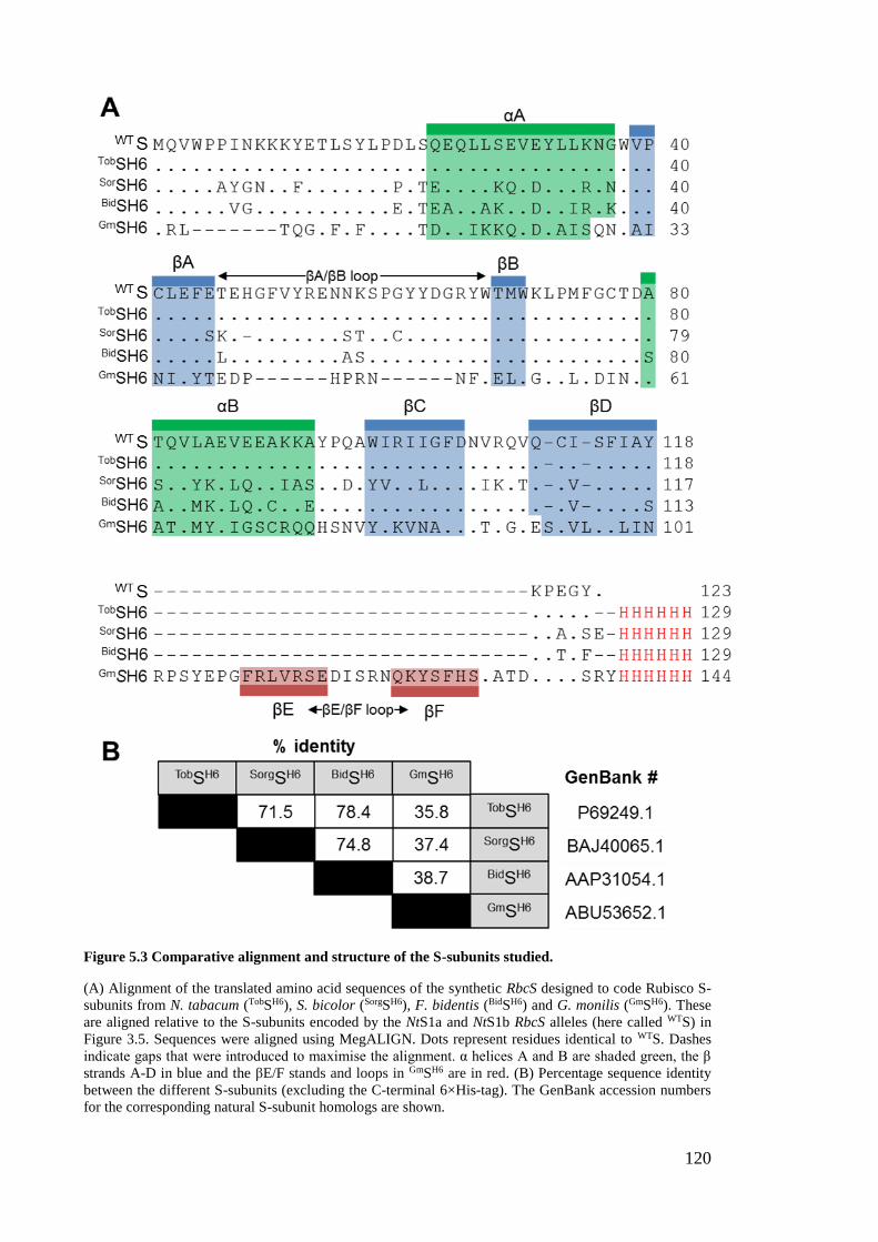
120
Figure 5.3 Comparative alignment and structure of the S-subunits studied.
(A) Alignment of the translated amino acid sequences of the synthetic RbcS designed to code Rubisco S-
subunits from N. tabacum (TobSH6), S. bicolor (SorgSH6), F. bidentis (BidSH6) and G. monilis (GmSH6). These
are aligned relative to the S-subunits encoded by the NtS1a and NtS1b RbcS alleles (here called WTS) in
Figure 3.5. Sequences were aligned using MegALIGN. Dots represent residues identical to WTS. Dashes
indicate gaps that were introduced to maximise the alignment. α helices A and B are shaded green, the β
strands A-D in blue and the βE/F stands and loops in GmSH6 are in red. (B) Percentage sequence identity
between the different S-subunits (excluding the C-terminal 6×His-tag). The GenBank accession numbers
for the corresponding natural S-subunit homologs are shown.
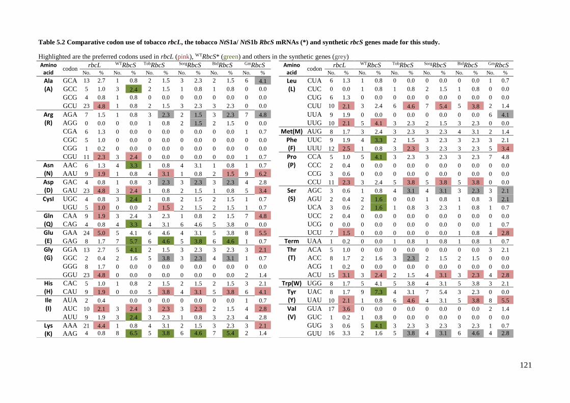
121
Table 5.2 Comparative codon use of tobacco rbcL, the tobacco NtS1a/ NtS1b RbcS mRNAs (*) and synthetic rbcS genes made for this study.
Highlighted are the preferred codons used in rbcL (pink), WTRbcS* (green) and others in the synthetic genes (grey)
Amino
acid codon
rbcL WTRbcS TobRbcS SorgRbcS BidRbcS GmRbcS
Amino acid
codon rbcL WTRbcS TobRbcS SorgRbcS BidRbcS GmRbcS
No. % No. % No. % No. % No. % No. %
No. % No. % No. % No. % No. % No. %
Ala (A)
GCA 13 2.7 1 0.8 2 1.5 3 2.3 2 1.5 6 4.1
Leu (L)
CUA 6 1.3 1 0.8 0 0.0 0 0.0 0 0.0 1 0.7
GCC 5 1.0 3 2.4 2 1.5 1 0.8 1 0.8 0 0.0
CUC 0 0.0 1 0.8 1 0.8 2 1.5 1 0.8 0 0.0
GCG 4 0.8 1 0.8 0 0.0 0 0.0 0 0.0 0 0.0
CUG 6 1.3 0 0.0 0 0.0 0 0.0 0 0.0 0 0.0
GCU 23 4.8 1 0.8 2 1.5 3 2.3 3 2.3 0 0.0
CUU 10 2.1 3 2.4 6 4.6 7 5.4 5 3.8 2 1.4
Arg (R)
AGA 7 1.5 1 0.8 3 2.3 2 1.5 3 2.3 7 4.8
UUA 9 1.9 0 0.0 0 0.0 0 0.0 0 0.0 6 4.1
AGG 0 0.0 0 0.0 1 0.8 2 1.5 2 1.5 0 0.0
UUG 10 2.1 5 4.1 3 2.3 2 1.5 3 2.3 0 0.0
CGA 6 1.3 0 0.0 0 0.0 0 0.0 0 0.0 1 0.7
Met(M) AUG 8 1.7 3 2.4 3 2.3 3 2.3 4 3.1 2 1.4
CGC 5 1.0 0 0.0 0 0.0 0 0.0 0 0.0 0 0.0
Phe (F)
UUC 9 1.9 4 3.3 2 1.5 3 2.3 3 2.3 3 2.1
CGG 1 0.2 0 0.0 0 0.0 0 0.0 0 0.0 0 0.0
UUU 12 2.5 1 0.8 3 2.3 3 2.3 3 2.3 5 3.4
CGU 11 2.3 3 2.4 0 0.0 0 0.0 0 0.0 1 0.7
Pro (P)
CCA 5 1.0 5 4.1 3 2.3 3 2.3 3 2.3 7 4.8
Asn (N)
AAC 6 1.3 4 3.3 1 0.8 4 3.1 1 0.8 1 0.7
CCC 2 0.4 0 0.0 0 0.0 0 0.0 0 0.0 0 0.0
AAU 9 1.9 1 0.8 4 3.1 1 0.8 2 1.5 9 6.2
CCG 3 0.6 0 0.0 0 0.0 0 0.0 0 0.0 0 0.0
Asp (D)
GAC 4 0.8 1 0.8 3 2.3 3 2.3 3 2.3 4 2.8
CCU 11 2.3 3 2.4 5 3.8 5 3.8 5 3.8 0 0.0
GAU 23 4.8 3 2.4 1 0.8 2 1.5 1 0.8 5 3.4
Ser (S)
AGC 3 0.6 1 0.8 4 3.1 4 3.1 3 2.3 3 2.1
CysI UGC 4 0.8 3 2.4 1 0.8 2 1.5 2 1.5 1 0.7
AGU 2 0.4 2 1.6 0 0.0 1 0.8 1 0.8 3 2.1
UGU 5 1.0 0 0.0 2 1.5 2 1.5 2 1.5 1 0.7
UCA 3 0.6 2 1.6 1 0.8 3 2.3 1 0.8 1 0.7
Gln (Q)
CAA 9 1.9 3 2.4 3 2.3 1 0.8 2 1.5 7 4.8
UCC 2 0.4 0 0.0 0 0.0 0 0.0 0 0.0 0 0.0
CAG 4 0.8 4 3.3 4 3.1 6 4.6 5 3.8 0 0.0
UCG 0 0.0 0 0.0 0 0.0 0 0.0 0 0.0 1 0.7
Glu (E)
GAA 24 5.0 5 4.1 6 4.6 4 3.1 5 3.8 8 5.5
UCU 7 1.5 0 0.0 0 0.0 0 0.0 1 0.8 4 2.8
GAG 8 1.7 7 5.7 6 4.6 5 3.8 6 4.6 1 0.7
Term UAA 1 0.2 0 0.0 1 0.8 1 0.8 1 0.8 1 0.7
Gly (G)
GGA 13 2.7 5 4.1 2 1.5 3 2.3 3 2.3 3 2.1
Thr (T)
ACA 5 1.0 0 0.0 0 0.0 0 0.0 0 0.0 3 2.1
GGC 2 0.4 2 1.6 5 3.8 3 2.3 4 3.1 1 0.7
ACC 8 1.7 2 1.6 3 2.3 2 1.5 2 1.5 0 0.0
GGG 8 1.7 0 0.0 0 0.0 0 0.0 0 0.0 0 0.0
ACG 1 0.2 0 0.0 0 0.0 0 0.0 0 0.0 0 0.0
GGU 23 4.8 0 0.0 0 0.0 0 0.0 0 0.0 2 1.4
ACU 15 3.1 3 2.4 2 1.5 4 3.1 3 2.3 4 2.8
His (H)
CAC 5 1.0 1 0.8 2 1.5 2 1.5 2 1.5 3 2.1
Trp(W) UGG 8 1.7 5 4.1 5 3.8 4 3.1 5 3.8 3 2.1
CAU 9 1.9 0 0.0 5 3.8 4 3.1 5 3.8 6 4.1
Tyr (Y)
UAC 8 1.7 9 7.3 4 3.1 7 5.4 3 2.3 0 0.0
Ile (I)
AUA 2 0.4
0.0 0 0.0 0 0.0 0 0.0 1 0.7
UAU 10 2.1 1 0.8 6 4.6 4 3.1 5 3.8 8 5.5
AUC 10 2.1 3 2.4 3 2.3 3 2.3 2 1.5 4 2.8
Val (V)
GUA 17 3.6 0 0.0 0 0.0 0 0.0 0 0.0 2 1.4
AUU 9 1.9 3 2.4 3 2.3 1 0.8 3 2.3 4 2.8
GUC 1 0.2 1 0.8 0 0.0 0 0.0 0 0.0 0 0.0
Lys (K)
AAA 21 4.4 1 0.8 4 3.1 2 1.5 3 2.3 3 2.1
GUG 3 0.6 5 4.1 3 2.3 3 2.3 3 2.3 1 0.7
AAG 4 0.8 8 6.5 5 3.8 6 4.6 7 5.4 2 1.4
GUU 16 3.3 2 1.6 5 3.8 4 3.1 6 4.6 4 2.8
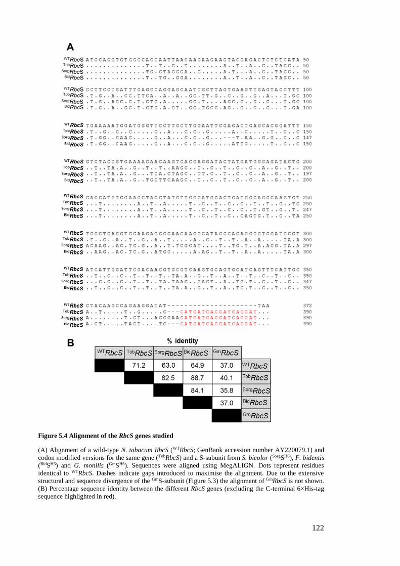
122
Figure 5.4 Alignment of the RbcS genes studied
(A) Alignment of a wild-type N. tabacum RbcS (WTRbcS; GenBank accession number AY220079.1) and codon modified versions for the same gene (TobRbcS) and a S-subunit from S. bicolor (SorgSH6), F. bidentis
(BidSH6) and G. monilis (GmSH6). Sequences were aligned using MegALIGN. Dots represent residues
identical to WTRbcS. Dashes indicate gaps introduced to maximise the alignment. Due to the extensive
structural and sequence divergence of the GmS-subunit (Figure 5.3) the alignment of GmRbcS is not shown.
(B) Percentage sequence identity between the different RbcS genes (excluding the C-terminal 6×His-tag
sequence highlighted in red).
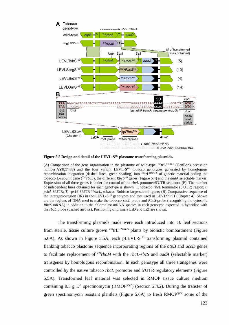
123
Figure 5.5 Design and detail of the LEVL-SH6 plastome transforming plasmids.
(A) Comparison of the gene organisation in the plastome of wild-type, cmtrLRNAi-S (GenBank accession
number AY827488) and the four variant LEVL-SH6 tobacco genotypes generated by homologous
recombination integration (dashed lines, green shading) into cmtrLRNAi-S of genetic material coding the
tobacco L-subunit gene (TobrbcL), the different RbcSH6 genes (Figure 5.4) and the aadA selectable marker.
Expression of all three genes is under the control of the rbcL promoter/5'UTR sequence (P). The number
of independent lines obtained for each genotype is shown. T, tobacco rbcL terminator (3'UTR) region; t,
psbA 3'UTR; T, rps16 3'UTR tobrbcL, tobacco Rubisco large subunit gene; (B) Comparative sequence of
the intergenic-region (IR) in the LEVL-SH6 genotypes and that used in LEVLSSuH (Chapter 4). Shown
are the regions of DNA used to make the tobacco rbcL probe and RbcS probe (recognising the cytosolic
RbcS mRNA) in addition to the chloroplast mRNA species in each genotype expected to hybridise with
the rbcL probe (dashed arrows). Positioning of primers LsD and LsZ are shown.
The transforming plasmids made were each introduced into 10 leaf sections
from sterile, tissue culture grown cmtrLRNAi-S plants by biolistic bombardment (Figure
5.6A). As shown in Figure 5.5A, each pLEVL-SH6 transforming plasmid contained
flanking tobacco plastome sequence incorporating regions of the atpB and accD genes
to facilitate replacement of cmrbcM with the rbcL-rbcS and aadA (selectable marker)
transgenes by homologous recombination. In each genotype all three transgenes were
controlled by the native tobacco rbcL promoter and 5'UTR regulatory elements (Figure
5.5A). Transformed leaf material was selected in RMOP tissue culture medium
containing 0.5 g L-1 spectinomycin (RMOPspec) (Section 2.4.2). During the transfer of
green spectinomycin resistant plantlets (Figure 5.6A) to fresh RMOPspec some of the

124
soluble leaf protein was analysed by ndPAGE (Figure 5.6B). Correctly transformed
lines were identified as those making L8S8 Rubisco and/or those no longer making the R.
rubrum L2 Rubisco encoded by the displaced cmrbcM in cmtrLRNAi-S (Figure 5.5A). Four
to 14 independently transformed lines were obtained for each genotype. After two to
four rounds of further selection on RMOPspec the transformed region of the plastome
was PCR amplified (using primers LsD and LsZ, Figure 5.5B) from three lines of each
genotype and fully sequenced (results obtained not shown). As expected from the
precision of homologous recombination no unwanted nucleotide differences were
identified in any of the genotypes. The three lines for each genotype were therefore
genetically identical and accordingly showed matching growth phenotypes and cellular
biochemistry. The following data depicts that measured for a representative T0 line of
each genotype.
Figure 5.6 Transforming cmtrLRNAi-S and selecting for the transplastomic genotypes.
(A) The variant pLEVL-SH6 transforming plasmids were coated onto tungsten and introduced into cmtrLRNAi-S chloroplasts by biolistic transformation. The bombarded leaves were embedded into selective
RMOPspec medium and after ~4-7 weeks at 25 ̊C under ~20-60 µmol photons m2 s-1 illumination in air
containing 1.5% (v/v) CO2, green plantlet tissues emerged from the bleached white leaf sections. (B)
Example of ndPAGE screening of soluble leaf protein from the spectinomycin resistant plantlets.
Correctly transformed lines (green ticks) were identified as those making (small amounts of) recombinant
L8S8 Rubisco (red asterix) or less/no R. rubrum L2 Rubisco.
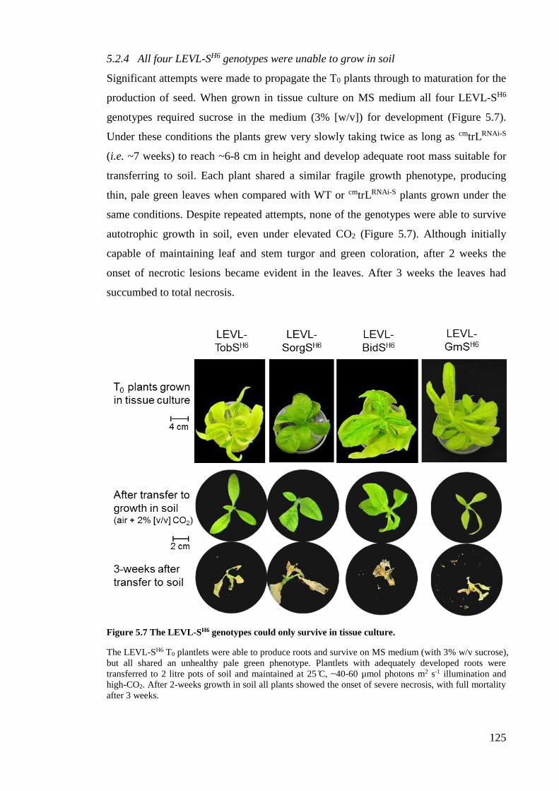
125
5.2.4 All four LEVL-SH6 genotypes were unable to grow in soil
Significant attempts were made to propagate the T0 plants through to maturation for the
production of seed. When grown in tissue culture on MS medium all four LEVL-SH6
genotypes required sucrose in the medium (3% [w/v]) for development (Figure 5.7).
Under these conditions the plants grew very slowly taking twice as long as cmtrLRNAi-S
(i.e. ~7 weeks) to reach ~6-8 cm in height and develop adequate root mass suitable for
transferring to soil. Each plant shared a similar fragile growth phenotype, producing
thin, pale green leaves when compared with WT or cmtrLRNAi-S plants grown under the
same conditions. Despite repeated attempts, none of the genotypes were able to survive
autotrophic growth in soil, even under elevated CO2 (Figure 5.7). Although initially
capable of maintaining leaf and stem turgor and green coloration, after 2 weeks the
onset of necrotic lesions became evident in the leaves. After 3 weeks the leaves had
succumbed to total necrosis.
Figure 5.7 The LEVL-SH6 genotypes could only survive in tissue culture.
The LEVL-SH6 T0 plantlets were able to produce roots and survive on MS medium (with 3% w/v sucrose),
but all shared an unhealthy pale green phenotype. Plantlets with adequately developed roots were
transferred to 2 litre pots of soil and maintained at 25 ̊C, ~40-60 µmol photons m2 s-1 illumination and
high-CO2. After 2-weeks growth in soil all plants showed the onset of severe necrosis, with full mortality
after 3 weeks.
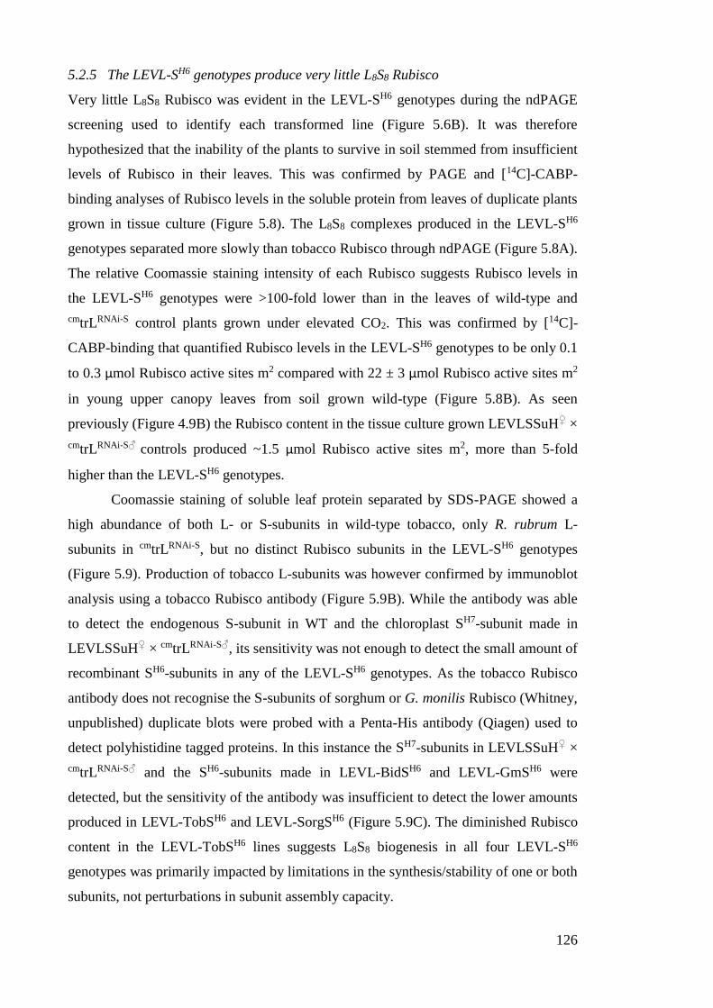
126
5.2.5 The LEVL-SH6 genotypes produce very little L8S8 Rubisco
Very little L8S8 Rubisco was evident in the LEVL-SH6 genotypes during the ndPAGE
screening used to identify each transformed line (Figure 5.6B). It was therefore
hypothesized that the inability of the plants to survive in soil stemmed from insufficient
levels of Rubisco in their leaves. This was confirmed by PAGE and [14C]-CABP-
binding analyses of Rubisco levels in the soluble protein from leaves of duplicate plants
grown in tissue culture (Figure 5.8). The L8S8 complexes produced in the LEVL-SH6
genotypes separated more slowly than tobacco Rubisco through ndPAGE (Figure 5.8A).
The relative Coomassie staining intensity of each Rubisco suggests Rubisco levels in
the LEVL-SH6 genotypes were >100-fold lower than in the leaves of wild-type and
cmtrLRNAi-S control plants grown under elevated CO2. This was confirmed by [14C]-
CABP-binding that quantified Rubisco levels in the LEVL-SH6 genotypes to be only 0.1
to 0.3 µmol Rubisco active sites m2 compared with 22 ± 3 µmol Rubisco active sites m2
in young upper canopy leaves from soil grown wild-type (Figure 5.8B). As seen
previously (Figure 4.9B) the Rubisco content in the tissue culture grown LEVLSSuH♀ ×
cmtrLRNAi-S♂ controls produced ~1.5 µmol Rubisco active sites m2, more than 5-fold
higher than the LEVL-SH6 genotypes.
Coomassie staining of soluble leaf protein separated by SDS-PAGE showed a
high abundance of both L- or S-subunits in wild-type tobacco, only R. rubrum L-
subunits in cmtrLRNAi-S, but no distinct Rubisco subunits in the LEVL-SH6 genotypes
(Figure 5.9). Production of tobacco L-subunits was however confirmed by immunoblot
analysis using a tobacco Rubisco antibody (Figure 5.9B). While the antibody was able
to detect the endogenous S-subunit in WT and the chloroplast SH7-subunit made in
LEVLSSuH♀ × cmtrLRNAi-S♂, its sensitivity was not enough to detect the small amount of
recombinant SH6-subunits in any of the LEVL-SH6 genotypes. As the tobacco Rubisco
antibody does not recognise the S-subunits of sorghum or G. monilis Rubisco (Whitney,
unpublished) duplicate blots were probed with a Penta-His antibody (Qiagen) used to
detect polyhistidine tagged proteins. In this instance the SH7-subunits in LEVLSSuH♀ ×
cmtrLRNAi-S♂ and the SH6-subunits made in LEVL-BidSH6 and LEVL-GmSH6 were
detected, but the sensitivity of the antibody was insufficient to detect the lower amounts
produced in LEVL-TobSH6 and LEVL-SorgSH6 (Figure 5.9C). The diminished Rubisco
content in the LEVL-TobSH6 lines suggests L8S8 biogenesis in all four LEVL-SH6
genotypes was primarily impacted by limitations in the synthesis/stability of one or both
subunits, not perturbations in subunit assembly capacity.
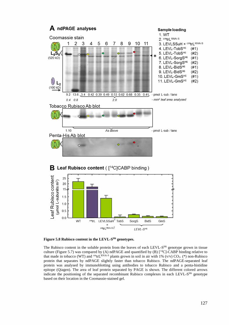
127
Figure 5.8 Rubisco content in the LEVL-SH6 genotypes.
The Rubisco content in the soluble protein from the leaves of each LEVL-SH6 genotype grown in tissue
culture (Figure 5.7) was compared by (A) ndPAGE and quantified by (B) [14C]-CABP binding relative to
that made in tobacco (WT) and cmtrLRNAi-S plants grown in soil in air with 1% (v/v) CO2. (*) non-Rubisco
protein that separates by ndPAGE slightly faster than tobacco Rubisco. The ndPAGE-separated leaf
protein was analysed by immunoblotting using antibodies to tobacco Rubisco and a penta-histidine
epitope (Qiagen). The area of leaf protein separated by PAGE is shown. The different colored arrows
indicate the positioning of the separated recombinant Rubisco complexes in each LEVL-SH6 genotype
based on their location in the Coomassie-stained gel.
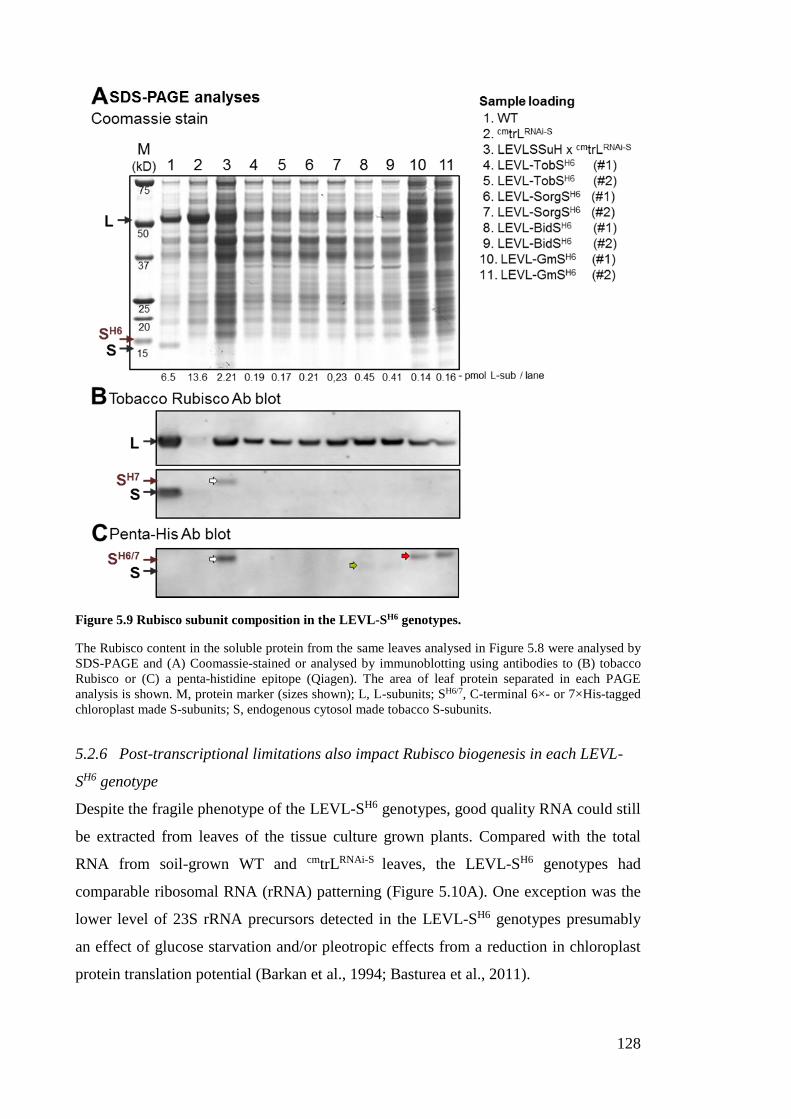
128
Figure 5.9 Rubisco subunit composition in the LEVL-SH6 genotypes.
The Rubisco content in the soluble protein from the same leaves analysed in Figure 5.8 were analysed by
SDS-PAGE and (A) Coomassie-stained or analysed by immunoblotting using antibodies to (B) tobacco
Rubisco or (C) a penta-histidine epitope (Qiagen). The area of leaf protein separated in each PAGE
analysis is shown. M, protein marker (sizes shown); L, L-subunits; SH6/7, C-terminal 6×- or 7×His-tagged
chloroplast made S-subunits; S, endogenous cytosol made tobacco S-subunits.
5.2.6 Post-transcriptional limitations also impact Rubisco biogenesis in each LEVL-
SH6 genotype
Despite the fragile phenotype of the LEVL-SH6 genotypes, good quality RNA could still
be extracted from leaves of the tissue culture grown plants. Compared with the total
RNA from soil-grown WT and cmtrLRNAi-S leaves, the LEVL-SH6 genotypes had
comparable ribosomal RNA (rRNA) patterning (Figure 5.10A). One exception was the
lower level of 23S rRNA precursors detected in the LEVL-SH6 genotypes presumably
an effect of glucose starvation and/or pleotropic effects from a reduction in chloroplast
protein translation potential (Barkan et al., 1994; Basturea et al., 2011).
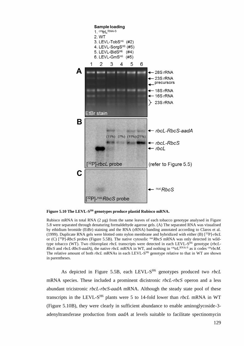
129
Figure 5.10 The LEVL-SH6 genotypes produce plastid Rubisco mRNA.
Rubisco mRNA in total RNA (2 µg) from the same leaves of each tobacco genotype analysed in Figure
5.8 were separated through denaturing formaldehyde agarose gels. (A) The separated RNA was visualised
by ethidium bromide (EtBr) staining and the RNA (rRNA) banding annotated according to Claros et al.
(1999). Duplicate RNA gels were blotted onto nylon membrane and hybridized with either (B) [32P]-rbcL
or (C) [32P]-RbcS probes (Figure 5.5B). The native cytosolic nucRbcS mRNA was only detected in wild-
type tobacco (WT). Two chloroplast rbcL transcripts were detected in each LEVL-SH6 genotype (rbcL-
RbcS and rbcL-RbcS-aadA), the native rbcL mRNA in WT, and nothing in cmtrLRNAi-S as it codes cmrbcM.
The relative amount of both rbcL mRNAs in each LEVL-SH6 genotype relative to that in WT are shown
in parentheses.
As depicted in Figure 5.5B, each LEVL-SH6 genotypes produced two rbcL
mRNA species. These included a prominent dicistronic rbcL-rbcS operon and a less
abundant tricistronic rbcL-rbcS-aadA mRNA. Although the steady state pool of these
transcripts in the LEVL-SH6 plants were 5 to 14-fold lower than rbcL mRNA in WT
(Figure 5.10B), they were clearly in sufficient abundance to enable aminoglycoside-3-
adenyltransferase production from aadA at levels suitable to facilitate spectinomycin
130
resistance during selection. Importantly the reduced abundance of these mRNA levels
are less severe than the 100-fold lower levels of Rubisco produced in the LEVL-SH6
genotypes (Figure 5.8B). This implies post-transcriptional limitations are likely the key
factor impeding L8S8 biogenesis in the LEVL-SH6 genotypes. Consistent with the
efficient RNAi-RbcS targeted silencing in the paternal cmtrLRNAi-S genotype (Chapter 3)
no cytosolic RbcS mRNA was detected in the LEVL-SH6 genotypes (Figure 5.10C).
5.3 Discussion
Comparable to the high plastome transformation efficiency of the “tobacco master line”
cmtrL (Whitney and Sharwood, 2008), the cmtrLRNAi-S line was found to be similarly
efficient for deriving independently transformed lines (Figure 5.5A). This efficiency
appears independent of photosynthetic viability as all LEVL-SH6 genotypes produced
very little hybrid L8S8 enzyme (Figure 5.8) and was incapable of supporting autotrophic
plant growth (Figure 5.7). This finding matches that observed by Sharwood and
Whitney (2008) who readily produced tobacco genotypes that made no Rubisco or non-
catalytic tobacco Rubisco via plastome transformation of cmtrL. Despite only producing
low levels of Rubisco, the LEVL-SH6 genotypes produced provide the first examples of
transgenic plant producing functional hybrid Rubisco comprising tobacco L-subunits
assembled with a homogenous population of S-subunits from either S. bicolor, F.
bidentis or the red algae G. monilis. Rubisco production via this rbcL-rbcS
transplantation approach however appears predominantly impeded by one or more post-
transcriptional events. As discussed below, the confronting challenge is to identify the
post-transcriptional process(es) impeding hybrid Rubisco biogenesis and design
solutions to circumvent them.
5.3.1 Lower Rubisco mRNA levels in LEVL-SH6 – cause or effect of low hybrid enzyme
biogenesis?
The limitation to hybrid Rubisco levels in the LEVL-SH6 genotypes accorded with their
inability to survive without carbohydrate supplementation (e.g. sucrose) in tissue culture.
All the LEVL-SH6 progeny produced low levels of Rubisco mRNA. These steady state
mRNA pools comprised a rbcL-rbcS-aadA transcript and a >2-fold more abundant
shorter rbcL-rbcS transcript. In total these bi- and tricistronic mRNAs were more than
5-fold lower in relative abundance than the rbcL mRNA content in wild-type (Figure
5.10). As shown previously in transplastomic tobacco genotypes where Rubisco content
is adversely limited, the steady state levels of rbcL mRNA can be reduced (e.g. 2 to 5-
131
fold in the tobRSt and tRstL7 genotypes) (Sharwood et al., 2008; Sharwood and Whitney,
2010) (Table 5.1). Plausible causes of this reduction are variations in mRNA synthesis
and/or stability – both of which are likely sensitive to cellular homeostasis which
appears highly compromised in Rubisco deficient transgenic plants, even when grown
in tissue culture media supplemented with sucrose (Figure 5.7).
The mRNA’s sequence strongly influences its quaternary folding and capacity to
interact with molecular factors that are essential to its correct, stabilising maturation and
translation. One example of Rubisco mRNA maturation includes the pentatricopeptide
repeat protein (PPR) MRL1 that processes the rbcL 5’UTR sequence to produce a
shortened transcript with higher stability to 5'-3’ degradation (Johnson et al., 2010).
Likewise, equipping rbcL with its native rbcL 3'UTR appears important for maintaining
wild-type rbcL mRNA abundance (Whitney and Andrews, 2003). The rbcL 3'UTR is
maintained in the LEVL-SH6 genotypes and is located downstream of RbcS. Use of this
gene organisation seems practical as it had little impact on the rbcL-cpRbcS mRNA
levels produced in the LEVLSSuH genotype (Figure 4.10). Only in the LEVLSSuH♀ ×
cmtrLRNAi-S♂ F1 progeny where photosynthesis and growth were perturbed was a 4-fold
reduction in Rubisco mRNA observed. Recent work has also found 5-fold reductions in
the levels of Rubisco chloroplast mRNA in tobAtL-R1 genotypes (Whitney et al., 2015).
In this genotype, a gene coding RAF1 from Arabidopsis is situated in a comparable
place to RbcS in the LEVL-SH6 genotypes. The growth of the tobAtL-R1 plants was also
slowed relative to wild-type due to reductions in Rubisco content.
The variation in Rubisco transcript levels in the LEVL-SH6 genotypes and prior
transplastomic lines highlights two issues in the context of the ongoing challenges in
plastome transformation studies. Firstly, it emphasises the unpredictable influence of
changes to the 5’UTR, coding and 3’UTR sequences on mRNA synthesis and stability.
This uncertainty often necessitates optimisation via trialling different sequence
combinations (Maliga, 2004). Secondly, it questions the extent to which changes in the
cell of transplastomic genotypes influence mRNA transcription and stability. This
appears particularly pertinent to genotypes where photosynthesis (and thus growth)
have been perturbed. For example, the diminished cellular viability of the LEVL-SH6
genotypes likely contributes strongly to their low Rubisco mRNA levels. This contrasts
with other genotypes with Rubisco activity limitations where the rbcL mRNA levels can
be increased by enhancing cellular viability by growing the plants under elevated CO2
or in tissue culture. As seen in Table 5.1, such growth conditions enabled near wild-type
rbcL mRNA levels to be produced in the tobpring, tobflo and tobbid genotypes grown
132
under high-CO2 (Whitney et al., 2011b). Improving the growth potential of the LEVL-
SH6 genotypes by such approaches proved untenable as the level of hybrid Rubisco
biogenesis was too low to attain suitable levels of autotrophic growth by high-CO2 or
tissue culture (Figure 5.7).
The application of exogenous hormones such as cytokinin, auxin or gibberellin
(GA3) have been used to rescue perturbed growth development and fertility in tobacco
transgenes (Abbasourr et al., 2012; Huang et al., 2003; Reutter et al., 1998) however the
relationship between exogenous and plant endogenous hormones is complex (Black et
al., 1994). To what extent such hormone treatments can rescue, if at all, the
carbohydrate limitated growth of plants lacking Rubisco activity remains untested.
Certainly the slow growth of Rubisco deficient plants can be partially mitigated when
grown in medium containing 0.5% sucrose (Figure 5.7), but to what extent their growth
can be improved by growing in vitro in media with higher carbohydrate (e.g 2% glucose
(Chen et al., 2016)) has yet to be examined.
Pale green leaves are commonly attributed to a reduced leaf chlorophyll content
that may or may not be related to photosynthethic efficiency (Havaux & Tardy, 1999).
Chloroplast genes such as EGY1 that control’s leaf senescence (Chen et al., 2016) as
well as genes like RBD1 (Wang et al., 2016), S6K1 (Sun et al., 2016) and RPS5 (Zhang
et al., 2016) that regulate protein translation when manipulated have produced plants
showcasing low chlorophyll content with pale green phenotype. The quest to pinpoint
specific genes affected by engineering the LEVL-SH6 genotypes and their relative levels
in an induced chlorophyll-limited environment remains to be embarked upon.
5.3.2 Post-transcriptional limitations to hybrid Rubisco biogenesis in LEVL-SH6
While the lower rbcL mRNA levels in the LEVL-SH6 genotypes likely contribute to the
scarcity of hybrid Rubisco produced, it appears perturbations to one or more post-
transcriptional processes further limit the enzymes’ production. As evident from the
growing plastome transformation literature, driving expression of different transgenes
with a common promoter/5’UTR regulatory does not guarantee reproducible levels of
recombinant protein production (Maliga, 2003; Maliga, 2004; Scharff and Koop, 2007).
For example, experiments using the same rRNA operon promoter (Prrn) have led to
more than a 10,000-fold variation in the levels of recombinant protein produced due to
varying post-transcriptional issues (Maliga, 2003). In the case of the LEVL-SH6
genotypes it seems unlikely the impediment to Rubisco biogenesis arises from an
133
obstruction to tobacco L-subunit synthesis, in particular since L8S8 Rubisco biogenesis
in the genetically similar LEVLSSuH genotype matches wild-type (Figure 4.9B).
The versatility of the pLEV vectors in successfully bioengineering Rubisco is
well documented (Whitney et al., 2011a) (Table 5.1). Using pLEV the expression of
rbcL transgenes (and promoter-less aadA) is regulated by the native tobacco rbcL
promoter and 5’UTR sequences (Figure 5.5). The resulting genotypes have all
succeeded in producing L-subunits for L8S8 biogenesis, but with varying levels of
success (Table 5.1). For example, tobacco lines transformed with pLEV4 (that
generated the genotype tobLEV4) produced wild-type levels of Rubisco and rbcL mRNA
(Whitney et al., 2011b). In contrast, introducing sunflower rbcL (rbcLsun) produced the
tobRst genotype where the total rbcLsun mRNA pool and hybrid (sunL)8(tobS)8 Rubisco
levels were both reduced ~9-fold (Sharwood et al., 2008). In a subsequent tRstL7
genotype the rbcLsun mRNA pool was increased 4-fold, but the level of (sunL)8(tobS)8
Rubisco made only increased 50% (Sharwood and Whitney, 2010) (Table 5.1). Clearly
post-transcriptional issues can influence heterologous L-subunit synthesis in tobacco
chloroplasts. However these issues are likely dampened in each LEVL-SH6 genotype as
they share the native tobacco rbcL coding and promoter/5’UTR regulatory sequences. It
therefore seems unlikely that problems with translation of the tobacco L-subunit are
causing the >100-fold lower levels of L8S8 production in the LEVL-SH6 genotypes.
The spectinomycin resistant phenotype of the LEVL-SH6 lines implies sufficient
levels of aminoglycoside-3-adenyltransferase (aadA) are produced. That is, translation
processing of the aadA component of the larger, less abundant rbcL-rbcS-aadA mRNA
is feasible. Questions arise however as to how much aadA protein was made in each of
the LEVL-SH6 genotypes and what amount is needed to confer a spectinomycin resistant
phenotype? These issues remain to be quantified in all pLEV-derived genotypes, and in
all tobacco plastome transformation events in general. One approach would be to
quantify and compare aadA levels in varying transplastomic lines using a new antibody
from Agrisera. This is an objective for future consideration.
Taken together, these justifications suggest a significant impediment to hybrid
Rubisco production in the LEVL-SH6 lines arising from a heightened hindrance in the
synthesis of the heterologous Rubisco S-subunits or/and their capacity to assemble with
the tobacco L-subunits. The efficient functioning of proteases within chloroplasts may
also contribute to the paucity of hybrid Rubisco biogenesis, in particular if problems
with Rubisco subunit production, folding or assembly into stable L8S8 complexes
increase their propensity for proteolysis.
134
5.3.2.1 Problems with initiating chloroplast S-subunit synthesis in the LEVL-SH6 lines
As in bacterial systems, protein translation in chloroplasts requires the successful
assembly of a ribosome on an mRNA (initiation) before synthesis of a polypeptide can
proceed (elongation) and reach its full length and trigger protein release (termination).
Impeding any of these three phases slows the translational processing of a protein
(Woodson and Chory, 2008). In bacteria the rate-limiting step often occurs at the point
of initiation with the strength of ribosome binding strongly correlating with the amount
of protein synthesis (O’Connor et al., 2013). In particular, changes to the Shine-
Dalgarno (SD) sequence (i.e. the ribosome binding site) have a dramatic influence on
the strength (i.e. effectiveness) of ribosome engagement with the mRNA. Using a gfp
marker the mechanistic differences in the SD recognition machinery between E. coli
and tobacco chloroplasts has been tested by modifying the position and number of SD
sequences placed upstream of the AUG initiator codon (Drechsel and Bock, 2011). The
results suggested that closely located SD sequences upstream of an AUG initiation
codon in a plastome impede translation initiation by competitively impeding 30S
ribosomal subunit engagement. A key finding was that the 30S ribosome preferentially
bound to the most 5' SD sequence, suggesting that the chloroplast translational
machinery may be inefficient at recognising SD sequences within polycistronic
transcripts – in direct contrast to E. coli (Drechsel and Bock, 2011). This finding
provided reasoning for the hypothesis that many chloroplast polycistronic mRNA’s
undergo inter-cistronic cleavage into monocistronic mRNAs to increase their translation
efficiency. This requirement for this cleavage however does not appear to be absolute as
some chloroplast oligocistronic mRNAs do not undergo scission (e.g. the psaA-psaB
mRNA) and neither do some transgenes (e.g. rbcL-rbcS operon from algae) which can
still produce very high levels of L- and S-subunit synthesis in tobacco chloroplasts
(Whitney and Andrews, 2001a) (example 12 and 13, Table 5.1).
Similarly, other elements such as mRNA secondary structure, small RNA
(sRNA) binding sites and start codon positioning all strongly influence translation
initiation (Storz et al., 2011). Compared with bacterial systems however, chloroplast
mRNA’s show significant variation in the positioning of their SD sequence within the
5’UTR relative to the AUG initiator codon (typically 12-7 nucleotides upstream of
AUG [Maliga, 2003]). At the extreme is the psbA mRNA where the SD sequence
(GGAG) is located 33 nt upstream of the AUG start codon (Kim and Mullet, 1994). In
this instance the chloroplast S1 ribosomal protein binds to the psbA 5'UTR to fold the
mRNA and bring the SD sequence to the consensus position for ribosome engagement
135
and initiation. In most instances the plastid mRNAs share SD sequences that are
complementary to the 3' end of the 16S rRNA.
Table 5.3 Variation in the intergenic sequences used in rbcL-rbcS transgene studies in the tobacco
plastome.
Details of Rubisco mRNA and L8S8 contents are summarised in Table 5.1. The Shine-Dalgarno (SD)
GGAG and ATG/GTG initiator codons for the rbcS transgenes are shown in bold.{1} Whitney and
Sharwood, 2008; {2} Whitney and Andrews, 2001; {3} Lin et al., 2014. Part of the 5’UTR sequence and
SD for rbcL is provided as a comparison.
An examination of prior rbcL-rbcS operons transformed into the tobacco
plastome shows variation in the positioning of the SD sequence relative to the AUG
codon of the rbcS transgene (Table 5.3). A distinct difference in the LEVL-SH6
genotypes is the closer location of the SD sequence to the AUG start codon of the
synthetic cpRbcS transgenes. However the position of the SD -11 to -7 nucleotides in the
LEVL-SH6 lines matches that used by the tobacco rbcL (Figure 5.10). As noted by
Kuroda and Maliga (2001), the translational control region (TCR) of the rbcL mRNA is
dependent on sequence within the 5'UTR and adjoining coding sequence. Work within
the Whitney lab has found synonymous single nucleotide changes within the first three
codons of the coding sequence (i.e. nucleotide changes that maintain amino acid
sequence) that were predicted to alter mRNA folding (“secondary structure”) leading to
significant perturbations in L-subunit synthesis (Orr, 2013). Work in this thesis
136
demonstrated that maintaining mRNA folding, and not amino acid sequence, was
important for maintaining L-subunit synthesis.
This finding has little relevance on most of the rbcL transgene substitution
studies so far undertaken as they have generally maintained the wild-type tobacco rbcL
sequence for up to 42 nucleotides downstream of the AUG codon. As indicated in
Whitney et al. (2011), the first 50 nucleotides of 5' coding sequence of plant rbcL are
highly conserved. This suggests the differences observed in the capacity for foreign
Rubisco biogenesis in tobacco chloroplasts (Table 5.1) is independent of recombinant
L-subunit translation initiation.
Problems with the mRNA quaternary structure of the cpRbcS transgenes in the
LEVL-SH6 therefore seem a likely contributor to the low amount of hybrid Rubisco
produced if S-subunit translation initiation and/or elongation is impeded. Potential
problems with initiation could be tested by altering the SD location and intervening
sequence to the AUG to match that used naturally by other chloroplast transcripts. As
with tobacco rbcL, this may need to include a short region of the native sequence
downstream of the AUG codon that naturally facilitates translation (a possibility
experimentally tested in Chapter 6). As discussed in Section 5.3.2.2, problems with
translation elongation might be examined by altering the cp RbcS sequence to match the
codon use of other chloroplast genes (also tested in Chapter 6).
5.3.2.2 Synthesis of the S-subunit is slowed by poor codon use
Slowing the elongation process of translation would expectantly also influence the
levels of protein synthesis in chloroplasts. Extensive analyses in bacterial systems have
highlighted how the use, clustering, and repetition of rare codons in an mRNA can slow
the elongation rate (Gustafsson et al., 2004; Proshkin et al., 2010). This slowing
typically correlates with impediments in the cellular availability of cognate transfer
RNA (tRNA) that trigger pausing of ribosomes on the mRNA (Buchan and Stansfield,
2007). Sucrose gradient centrifugation analysis of mRNA-polysome associations (i.e.
mRNA’s with multiple ribosomes bound) is often used to gauge how well an mRNA is
being translated. Efficiently translated mRNAs typically have higher numbers of
ribosomes attached and therefore sediment further down the gradient (Mašek et al.,
2011). A comparison of the polysome sedimentation profile for the rbcL-RbcS and
rbcL-RbcS-aadA mRNA’s in the LEVLSSuH♀ × cmtrLRNAi-S♂ and LEVL-TobSH6
genotypes may therefore prove useful in identifying differences in S-subunit translation.
For example, if translational processing of the mRNA’s was more impeded in LEVL-
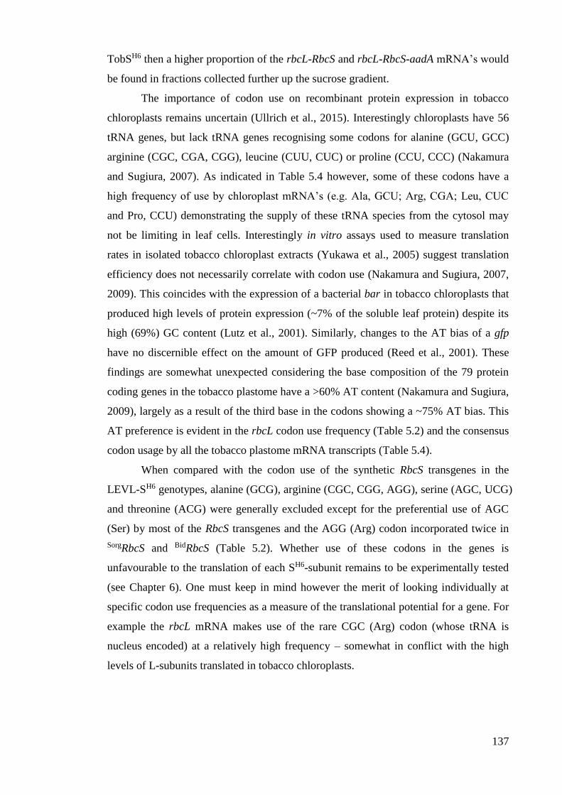
137
TobSH6 then a higher proportion of the rbcL-RbcS and rbcL-RbcS-aadA mRNA’s would
be found in fractions collected further up the sucrose gradient.
The importance of codon use on recombinant protein expression in tobacco
chloroplasts remains uncertain (Ullrich et al., 2015). Interestingly chloroplasts have 56
tRNA genes, but lack tRNA genes recognising some codons for alanine (GCU, GCC)
arginine (CGC, CGA, CGG), leucine (CUU, CUC) or proline (CCU, CCC) (Nakamura
and Sugiura, 2007). As indicated in Table 5.4 however, some of these codons have a
high frequency of use by chloroplast mRNA’s (e.g. Ala, GCU; Arg, CGA; Leu, CUC
and Pro, CCU) demonstrating the supply of these tRNA species from the cytosol may
not be limiting in leaf cells. Interestingly in vitro assays used to measure translation
rates in isolated tobacco chloroplast extracts (Yukawa et al., 2005) suggest translation
efficiency does not necessarily correlate with codon use (Nakamura and Sugiura, 2007,
2009). This coincides with the expression of a bacterial bar in tobacco chloroplasts that
produced high levels of protein expression (~7% of the soluble leaf protein) despite its
high (69%) GC content (Lutz et al., 2001). Similarly, changes to the AT bias of a gfp
have no discernible effect on the amount of GFP produced (Reed et al., 2001). These
findings are somewhat unexpected considering the base composition of the 79 protein
coding genes in the tobacco plastome have a >60% AT content (Nakamura and Sugiura,
2009), largely as a result of the third base in the codons showing a ~75% AT bias. This
AT preference is evident in the rbcL codon use frequency (Table 5.2) and the consensus
codon usage by all the tobacco plastome mRNA transcripts (Table 5.4).
When compared with the codon use of the synthetic RbcS transgenes in the
LEVL-SH6 genotypes, alanine (GCG), arginine (CGC, CGG, AGG), serine (AGC, UCG)
and threonine (ACG) were generally excluded except for the preferential use of AGC
(Ser) by most of the RbcS transgenes and the AGG (Arg) codon incorporated twice in
SorgRbcS and BidRbcS (Table 5.2). Whether use of these codons in the genes is
unfavourable to the translation of each SH6-subunit remains to be experimentally tested
(see Chapter 6). One must keep in mind however the merit of looking individually at
specific codon use frequencies as a measure of the translational potential for a gene. For
example the rbcL mRNA makes use of the rare CGC (Arg) codon (whose tRNA is
nucleus encoded) at a relatively high frequency – somewhat in conflict with the high
levels of L-subunits translated in tobacco chloroplasts.
138
Table 5.4 Codon use by the 79 mRNAs produced in tobacco chloroplasts (Nakamura and Sugiura,
2009)
An examination of the codon preferences for the tobacco chloroplast genes showed the lowest frequencies
of use are for alanine (GCG), arginine (CGC, CGG, AGG), serine (AGC, UCG) and threonine (ACG).
5.3.2.3 An increased propensity for recombinant Rubisco subunit proteolysis?
Proteases are active in all regions of the chloroplast to assist in protein maturation,
damaged protein recycling and general maintenance of protein quality control (van Wijk,
2015). While a dozen or so types of protease are known to operate in different
compartments of chloroplasts, their mechanisms for substrate selection remain poorly
understood, despite many showing evolutionary linkages with well-studied bacterial
proteases (Olinares et al., 2011). The absence of detectable S-subunits in cmtrLRNAi-S
(Figure 5.9B) attests to efficiency of stromal proteases for degrading unassembled
proteins. As discussed above, the use of atypical codons in the RbcS transgenes tested in
this study (see Section 5.3.2.2) and possible problems arising from unfavourable mRNA
folding (see Section 5.3.2.1) likely impeded translational processing of the recombinant
S-subunits in the LEVL-TobSH6 genotypes. A consequence of this may be a slowing of
S-subunit translational processing that, hypothetically, may increase exposure of the
emerging nascent peptides as substrates for protease attack. A technique often employed
to measure protein translation, maturation, modification and stability is pulse-chase with
[35S]-Met/unlabelled-Met (Pohl and Hasilik, 2015; Whitney et al., 2015). This has
previously been used to measure the stability of plastid made SH7-subunits incorporated
into L8S8 complexes in the RVSSuH and RVtpSSuH genotypes (Whitney and Andrews,
2001a) (Figure 4.7). The method was however not sensitive enough to measure the
synthesis and degradation of the nascent chloroplast made SH7-peptides, especially
considering the efficiency of proteolysis in chloroplasts. This approach would therefore

139
be even more ineffective in the LEVL-SH6 genotypes given they produce even lower
levels of plastid made S-subunits.
Prior attempts to express non-green algae Rubisco in tobacco chloroplasts found
no evidence for “red”-L8S8 assembly, however the heterologous L- and S-subunits were
found to accumulate as insoluble protein in the Pt2 and Gs tobacco genotypes (Whitney
et al., 2001) (Table 5.1). Notably this finding prompted comparative SDS-PAGE and
immunoblot analysis of the soluble and the total cellular protein (i.e. both soluble and
insoluble protein) in the LEVL-SH6 leaves to confirm tobacco L-subunits and varied S-
subunits were not accumulating as insoluble protein (data not shown). In another PhD
project in the Whitney lab, the flowers of both Pt2 and Gs tobacco genotypes were
pollinated with pollen from cmtrLRNAi-S to knock out tobacco L8S8 production (analogous
to the process used in Chapter 4). As expected the resulting Pt2♀ × cmtrLRNAi-S♂ and Gs♀
× cmtrLRNAi-S♂ progeny produced no tobacco Rubisco resulting in plants that could only
grow slowly in tissue culture (i.e. comparable to the LEVL-SH6 genotypes) (Figure 5.7).
Curiously, despite the high levels of insoluble algae L- and S-subunit units made in the
parental Pt2 and Gs genotypes, none were detected in the cmtrLRNAi-S crossed progeny
except for trace levels of soluble algal S-subunits (Gunn, 2014). This finding
demonstrates the efficiency of protease activity in tobacco chloroplasts, especially in
plants where growth and resource availability are greatly impeded. It also demonstrates
that algae S-subunits seem less prone to chloroplast protease degradation, as also
reported by Whitney et al. (2001). This improved stability might explain why the
GmSH6-subunit levels detected in the soluble protein of LEVL-GmSH6 leaves appears to
accumulate at higher than the S-subunits in the other LEVL-SH6 genotypes (Figure
5.9C), that did not correlate with the amount of hybrid (TobL)8(GmS)8 Rubisco made in
the same protein sample (Figure 5.8).
The role of proteolysis in the effect of low Rubisco contents in the LEVL-SH6
genotypes remains unclear. As shown in Chapter 4, the LEVLSSuH♀ × cmtrLRNAi-S♂
plants make ~1.5 µmol L-subunits m2 of L8(SH7)8 Rubisco (Figures 4.9 and Figure 5.8)
which can support autotrophic growth to full maturity in soil under high-CO2 (Figure
4.8C). In contrast the LEVL-TobSH6 genotype only managed to produce <0.4 µmol L-
subunits m2 which relegated their survival dependency to growth in tissue culture
(Figure 5.7). When considering the identical subunit amino acid sequence analogies
between the LEVL-TobSH6 and LEVLSSuH♀ × cmtrLRNAi-S♂ genotypes it seems hard to
envisage that variations in proteolytic activity or reduced S-subunit chaperoning can
entirely account for their >4-fold differences in Rubisco content (Figure 5.8).

140
5.3.3 Varying structural incompatibilities between heterologous L- and S-subunit
effect on holoenzyme assembly and catalysis
There is an extensive number of amino acid interactions between the S- and L-subunit
in an L8S8 complex (Figure 1.8). As demonstrated in the example alignment in Figure
5.3, there is significant divergence among S-subunit sequences, especially between the
“green” and “red” Rubisco lineages (Andersson, 2008). An important structural
differentiation in the S-subunits between these lineages is the extended C-terminal β-
hairpin extension (i.e. the βE/βF loop) that structurally occupies the ends of the central
solvent channel that traverses the centre of L8S8 complexes (Spreitzer, 2003).
Expression studies in E. coli demonstrated the red-type Rubisco S-subunits from a
diatom could assemble with L8 cores of cyanobacteria Rubisco (Joshi et al., 2015). The
level of the hybrid L8S8 enzyme made was however extremely low. This might be
explained by the recent demonstration by Joshi et al. (2015) as to the role of the C-
terminal β-hairpin extension in “red” S-subunits in forming critical interactions in the
holoenzyme central channel that are necessary for stable L8S8 assembly and activity.
Despite these apparent subunit compatibility issues, the LEVL-GmSH6 progeny
successfully demonstrated for the first time the biogenesis of L8S8 complexes
comprising tobacco L-subunits and the G. monilis S-subunits (Figure 5.8). Notably, the
catalytic viability of this hybrid enzyme is totally compromised – further attesting to the
influence of the S-subunit on Rubisco catalysis. This finding is in agreement with the
results of Joshi et al. (2015) that showed hybrid L8S8 comprising the L-subunits of the
“red”-type Rubisco from Rhodobacter sphaeroides and the S-subunits from red algae
Rubisco were also catalytically inactive.
Incompatibilities in amino acid sequences between L- and S-subunits from
heterologous higher plant sources seem less impactful on hybrid L8S8 assembly. As
shown in Table 5.1, hybrid Rubisco holoenzyme comprising cytosol made heterologous
plant S-subunits (e.g. from Arabidopsis, S. bicolor or P. pratense Rubisco) or tobacco
S-subunits and alternative plant L-subunits (e.g. sunflower, tomato, Flaveria and
Arabidopsis) can be readily produced in leaf chloroplasts, although with varying effects
on catalysis. Prior analysis of rice Rubisco containing varying stoichiometries of rice
and sorghum S-subunits showed, respectively 50% and 40% increases in kCcat and KC
relative to wild-type rice Rubisco (Ishikawa et al., 2011) (see also Table 5.1).
Unfortunately the low amounts of Rubisco produced in the LEVL-SH6 genotypes in this
thesis prevented rapid extraction methods typically undertaken to accurately measure
kCcat and KC (Sharwood et al., 2008). Consistent with this need for rapid analysis of
141
freshly extracted Rubisco, the measured kCcat rates for the Ni-NTA purified enzymes
from the LEVL-SH6 genotypes are low (results not shown) and likely are compromised
by the inclusion of a polyhistidine tag on the S-subunits that appears to result in
ambiguous changes to the catalytic properties of Rubisco (Table 4.2). Nevertheless the
activity measurements serve to highlight that the Rubisco L8S8 hybrid enzymes
comprising S. bicolor or F. bidentis S-subunits retained catalytic activity while those
comprising G. monilis S-subunits did not.
The synthesis of cytosolic made S-subunits appears to be stably and efficiently
silenced in the cmtrLRNAi-S line and its plastome-transformed progeny. This brings into
question the necessity for including distinguishable peptide tags on the re-introduced S-
subunit transgenes. Exclusion of tags would help to better understand the influence of
S-subunits on the catalytic properties – a consideration for future S-subunit engineering
in cmtrLRNAi-S (discussed in Chapter 7).
5.3.4 Future goals – increasing incorporation of plastid made S-subunits into L8S8
Rubisco
As hypothesized by Whitney and Andrews (2001) the relocation of RbcS to the nucleus
appears to have led to adaptive changes in S-subunit evolution that influence the
biogenesis pathway of Rubisco that appear to limit the incorporation of chloroplast
made S-subunits. The validity of this hypothesis requires further testing using plastome
transformation to identify solutions for increasing S-subunit synthesis in the plastid. As
indicated in Section 5.3.2 above, one potential solution may be to increase translational
processing of the plastid S-subunits through modification of its codon use to match that
of other chloroplast made proteins – an objective tested in Chapter 6. An overall critical
component of engineering the S-subunit is better understanding the natural import and
chaperoning processes of cytosol made S-subunits. This understanding may provide
clues as to why they may be preferentially incorporated during L8S8 biogenesis in leaf
chloroplasts. Such considerations are important if transformations of heterologous S-
subunits into other plant species are to be effectively undertaken as a means to
bioengineer changes to Rubisco catalysis. Future possibilities for utilising the cmtrLRNAi-
S genotype to study components of the cytosolic S-subunit maturation process are
discussed in Chapter 7.
142
CHAPTER 6 – ENHANCING HYBRID RUBISCO
PRODUCTION IN TOBACCO CHLOROPLASTS
6.1 Introduction
The prior chapters of this thesis have highlighted the potential utility of the cmtrLRNAi-S
master line for engineering both the Rubisco L- and S-subunits simultaneously in higher
plants chloroplasts. Pollinating transplastomic lines producing chloroplast made S-
subunit with pollen from the cmtrLRNAi-S genotype was successful in producing Rubisco
isoforms comprising solely chloroplast made L- and S-subunits (Chapter 4). An initial
attempt to directly transform the cmtrLRNAi-S plastome with synthetic Rubisco operons
comprising tobacco rbcL and heterologous rbcS genes proved less effective at
producing hybrid, recombinant Rubisco (Chapter 5). Such anomalies in the differential
expression potential of transgenes is common to chloroplast transformation applications
as the technology remains encumbered by uncertainties on how to reliably predict
recombinant protein production (Ullrich et al., 2015). As indicated in Section 4.3.1.1
and Section 5.3.2, expression of different proteins driven only recorded by identical
regulatory elements (e.g. the rrn promoter and T7g10 5’UTR) can differ by as much as
10,000-fold in their levels of expression (Maliga, 2003). To counter these uncertainties
it is common practice for transplastomic application to pursue a try-it-and-see approach
by testing the effect of different regulatory sequences or modifications in codon use on
transgene expression (see Section 5.3.2.2).
6.1.1 Chaperone incompatibilities and proteolysis influence Rubisco biogenesis in E.
coli and in chloroplasts
In any recombinant protein expression system both the intrinsic folding potential of the
target protein and its requirements for compatible molecular partners (e.g. chaperones,
chaperonin, accessory proteins and subunits) for folding and assembly strongly
influence the level of functional protein expression (i.e. its biogenesis). As a result of
folding and assembly incompatibilities the expression of the Rubisco subunits from any
plant or algae in E. coli results in the accumulation of mis-folded, insoluble L- and S-
subunits that aggregate as insoluble inclusion bodies (Whitney et al., 2011a; Parry et al.,
2013). With regard to recombinant protein expression in leaf chloroplasts however, the
detection of insoluble red algae Rubisco L- and S-subunits in the tobacco Pt and
tobacco Gs genotypes (Table 5.1) poses the only recorded instance identifying insoluble
photosynthetic recombinant protein accumulation in chloroplasts (Whitney et al., 2001).
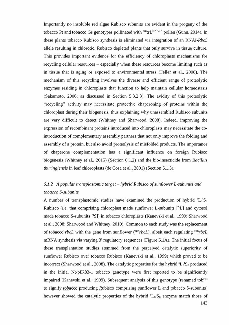
143
Importantly no insoluble red algae Rubisco subunits are evident in the progeny of the
tobacco Pt and tobacco Gs genotypes pollinated with cmtrLRNAi-S pollen (Gunn, 2014). In
these plants tobacco Rubisco synthesis is eliminated via integration of an RNAi-RbcS
allele resulting in chlorotic, Rubisco depleted plants that only survive in tissue culture.
This provides important evidence for the efficiency of chloroplasts mechanisms for
recycling cellular resources – especially when these resources become limiting such as
in tissue that is aging or exposed to environmental stress (Feller et al., 2008). The
mechanism of this recycling involves the diverse and efficient range of proteolytic
enzymes residing in chloroplasts that function to help maintain cellular homeostasis
(Sakamoto, 2006; as discussed in Section 5.3.2.3). The avidity of this proteolytic
“recycling” activity may necessitate protective chaperoning of proteins within the
chloroplast during their biogenesis, thus explaining why unassembled Rubisco subunits
are very difficult to detect (Whitney and Sharwood, 2008). Indeed, improving the
expression of recombinant proteins introduced into chloroplasts may necessitate the co-
introduction of complementary assembly partners that not only improve the folding and
assembly of a protein, but also avoid proteolysis of misfolded products. The importance
of chaperone complementation has a significant influence on foreign Rubisco
biogenesis (Whitney et al., 2015) (Section 6.1.2) and the bio-insecticide from Bacillus
thuringiensis in leaf chloroplasts (de Cosa et al., 2001) (Section 6.1.3).
6.1.2 A popular transplastomic target – hybrid Rubisco of sunflower L-subunits and
tobacco S-subunits
A number of transplastomic studies have examined the production of hybrid sL8tS8
Rubisco (i.e. that comprising chloroplast made sunflower L-subunits [SL] and cytosol
made tobacco S-subunits [tS]) in tobacco chloroplasts (Kanevski et al., 1999; Sharwood
et al., 2008; Sharwood and Whitney, 2010). Common to each study was the replacement
of tobacco rbcL with the gene from sunflower (sunrbcL), albeit each regulating sunrbcL
mRNA synthesis via varying 3' regulatory sequences (Figure 6.1A). The initial focus of
these transplantation studies stemmed from the perceived catalytic superiority of
sunflower Rubisco over tobacco Rubisco (Kanevski et al., 1999) which proved to be
incorrect (Sharwood et al., 2008). The catalytic properties for the hybrid sL8tS8 produced
in the initial Nt-pIK83-1 tobacco genotype were first reported to be significantly
impaired (Kanevski et al., 1999). Subsequent analysis of this genotype (renamed tobRst
to signify tobacco producing Rubisco comprising sunflower L and tobacco S-subunits)
however showed the catalytic properties of the hybrid sL8tS8 enzyme match those of

144
sunflower Rubisco (Table 5.1) in accordance with the measured leaf gas exchange
properties of the plant (Sharwood et al., 2008).
Figure 6.1 Key features of tobacco genotypes producing sunflower L-subunits.
(A) Different transplastomic replacement strategies have replaced the tobacco rbcL with the sunflower
rbcL to produce the genotypes Nt-pIK83-1 (aKanevski et al, 1999 – renamed tobRst by bSharwood et al.,
2008), tRstLA7 and tRstL (cSharwood and Whitney 2010). P, rbcL promoter/5'UTR sequence; T, tobacco
rbcL 3'UTR; T, psbA 3'UTR; t, rps16 3'UTR; p, rrn promoter/T7g10 5'UTR; white triangles, 34 nt loxP
sequences. (B) Only low levels of hybrid SL8tS8 Rubisco (comprising sunflower L-subunits and tobacco
S-subunits) were made in each genotype, even in the tRstL lines where sunrbcL mRNA levels were
increased 4-fold. (C) Each genotype could be grown to maturity in air with 0.5% (v/v) CO2 producing
slower growing plants with pale green leaves with (D) abnormal curling around the edges that was more
prominent in lines producing less SL8tS8 Rubisco. Figure adapted from Sharwood et al. (2008, 2010).
145
All the transplastomic tobacco genotypes producing hybrid sL8tS8 enzyme
showed significant limitations on the enzyme’s biogenesis in tobacco chloroplasts
(Figure 6.1B). At best the amount of sL8tS8 produced was only ~17% the Rubisco levels
made in tobacco (wild-type) controls. Even increasing the sunrbcL mRNA content 4-fold
only led to a modest 40-50% increase in sL8tS8 production (when comparing the tRstLA7
and tRstL7 genotypes in Figure 6.1B) suggesting hybrid sL8tS8 synthesis was primarily
impeded during one or more post-transcriptional events. As a result of the reduced
Rubisco levels, the growth of each sL8tS8 producing genotype was slower than wild-type,
necessitating high-CO2 for growth in soil. Even under high-CO2 all the genotypes
shared a pale green leaf phenotype and their juvenile progeny produced abnormal, thin,
strap-like shaped leaves (Figure 6.1C). The leaves of more mature plants were of similar
shape to the wild-type controls, but were thinner with pronounced dimpling around the
leaf margins (Figure 6.1D). The severity of this dimpling phenotype was less evident in
the tRstL7 genotype that produced the most sL8tS8 enzyme and thus had improved leaf
CO2 assimilation rates and grew more quickly than the tRst and tRstLA7 genotypes
(Sharwood et al., 2008; Sharwood and Whitney, 2010).
The reason(s) for the low levels of sL8tS8 biogenesis remain speculative. The
finding that sL8tS8 levels were not greatly enhanced by increasing sunrbcL mRNA levels
strongly favours limitations to one or more events limiting synthesis of the hybrid
enzyme. Analyses of [35S]-Methionine labelled sL8tS8 complexes showed their turnover
rates matched that of wild-type tobacco Rubisco (Sharwood et al., 2008). This indicated
the hybrid sL8tS8 enzyme was not susceptible to a higher rate of proteolysis. As both the
tobrbcL and sunrbcL share >92% sequence identity (with their 5' sequences being
identical for the first 56 bp) it was difficult to advocate that incompatibility in
translation initiation or elongation significantly impeded SL-subunit synthesis. It was
proposed that structural incompatibilities in the quaternary conformations of the
assembled SL-subunit octameric cores (i.e. SL8) may hinder tS-subunit assembly into
L8S8 complexes (Sharwood and Whitney, 2010). It was also hypothesized this
impediment may also increase the susceptibility of the subunits to proteolysis. Overall it
was postulated the compounding limitation impeding hybrid sL8tS8 biogenesis in
tobacco chloroplasts resulted from incompatibilities in the chaperoning requirements of
the SL-subunit (Sharwood et al., 2008; Sharwood and Whitney, 2010). This conclusion
is now supported by recent work demonstrating the importance of chaperone
compatibility on hybrid Rubisco biogenesis in leaf chloroplasts (Whitney et al., 2015)
(see also Section 6.1.3)
146
6.1.3 Considerations for increasing recombinant protein production in chloroplasts
As emulated in hybrid sL8tS8 studies and other recombinant protein expression work
using chloroplast transformation, it is not simply a case that the steady state pool of a
transgene mRNA levels reflects the amount of recombinant protein produced (Maliga,
2003). A number of early plastome transformation studies in particular paid close
attention to the high levels of transgene mRNA production with little consideration to
the level of recombinant protein made (Ohtani et al., 1991; Richter et al., 2000;
Outchkourov et al., 2003). Deciphering the underpinning reason(s) for this mismatch in
the abundance of transgene transcript and protein is rarely scrutinized experimentally
due to the costs (in both time and money) of chloroplast transformation. For example, as
discussed in Chapter 5, a comprehensive study on the relative importance of codon use
on transgene expression in chloroplasts remains incomplete (see Section 5.3.2.2). The
potential influence of the highly efficient proteolytic mechanisms in chloroplasts to
recombinant protein synthesis in leaf chloroplasts is also poorly understood (see Section
5.3.2.3 and paragraph above). Although some appreciation is required for the necessity
for genetic complementarity of an mRNA and the translational machinery for efficient
initiation and elongation processes of translation (see Section 5.3.2.1), these rules do not
appear absolute as there is significant natural variation found among the endogenous
chloroplast mRNA species (Choquet and Wollman, 2002; Peeters and Hanson, 2002;
Juszczak et al., 2012).
As first hypothesised by Whitney and Andrews (2001), problems with the
production of recombinant Rubisco in leaf chloroplasts likely stem from
incompatibilities with their folding and assembly requirements. This is now supported
by recent tests on comparable tobacco genotypes transformed with the Arabidopsis rbcL
with and without Arabidopsis raf1. In the transformed lines making the Arabidopsis
RAF1 Rubisco chaperone, the levels of hybrid ALtS enzyme (comprising Arabidopsis L-
subunits and tS) were increased more than 7-fold. The RAF1 chaperone forms a homo-
dimer complex and functions to assemble the post-chaperonin folded L-subunits into
Rubisco complexes (Feiz et al., 2014). The benefits of chaperone co-expression were
also seen in tobacco genotypes expressing recombinant Bt-protein where inclusion of
the putative chaperone, ORF 2, in the transformed cry2Aa2 operon helped attain Bt
expression levels of ~40% (w/w) of the leaf protein (de Cosa et al., 2001).
147
6.1.4 New insights into polycistronic mRNA design
There is increasing interest to exploit the inherent capacity of chloroplasts to produce
polycistronic mRNA’s in recombinant protein studies. A favoured application of this
approach has been to try and introduce novel metabolic pathways to enable the
synthesis of a particular product of metabolic or commercial interest. For example to
synthesize the biodegradable polyester polyhydroxybutyrate (PHB) in tobacco
chloroplasts, a polycistron encoding the enzymes phbC, phbA and phbB was
successfully introduced into the plastome (Nakashita et al., 2001). Similarly, expression
of the insecticide Bacillus thuringiensis (Bt) in Btcry2Aa2 as a three-gene operon in
tobacco resulted in the production of high levels of Bt toxin and accompanying
chaperones (de Cosa et al., 2001). With regard to recombinant Rubisco engineering, the
use of the IEE element was used in the synthetic polycistronic mRNA driving
expression of Synechococcus PCC7942 L-subunit, S-subunit and CcmM35/RbcX
synthesis in tobacco leaves (Lin et al., 2014; see also Figure 5.1). Unfortunately,
quantitative analyses of the mRNA levels of the cyanobacteria transgenes in the Se7942
tobacco genotypes was not examined (Table 5.1) making it difficult to meaningfully
gauge the effectiveness of incorporating the IEE sequences.
6.1.5 Research Objective – testing alternative transgene structures to modulate hybrid
Rubisco synthesis in cmtrLRNAi-S
Difficulties in engineering Rubisco S-subunits in planta have thus limited the feasibility
of studying S-subunit interactions with chaperones and elements upon entry into the
chloroplast stroma, and as to the dependency of these interactions for assembly with L-
subunits into L8S8 holoenzyme. Furthermore, the inability to produce plant or algae
L8S8 Rubisco in E. coli (Nishimura et al., 2008; Parry et al., 2013) or efficiently
manipulate the S-subunit in leaf chloroplasts (see Section 4.1.4) has impaired
meaningful study on the structural complementarity between plant Rubisco L- and S-
subunits and its consequences to Rubisco catalysis. In a bid to begin addressing the
former issue, this chapter describes utilisation of a unified chloroplast transforming
plasmid design to introduce into the plastome of cmtrLRNAi-S with varying combinations
of tobacco and sunflower L- and S-subunits. Rubisco production and catalysis in the
resulting four transplastomic genotypes is examined and the corresponding effects on
leaf photosynthesis, plant growth and phenotype tested. The outcomes are examined in
the context of better understanding the constraints to Rubisco biogenesis when using
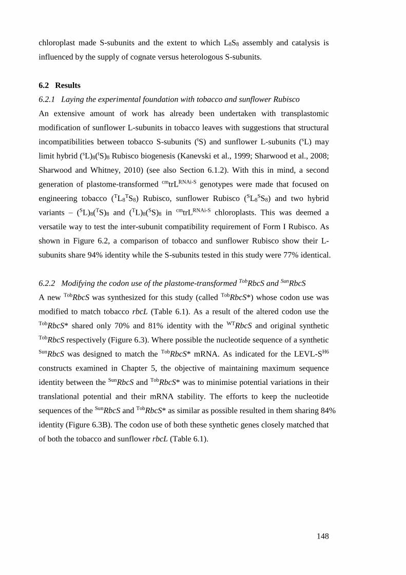
148
chloroplast made S-subunits and the extent to which L8S8 assembly and catalysis is
influenced by the supply of cognate versus heterologous S-subunits.
6.2 Results
6.2.1 Laying the experimental foundation with tobacco and sunflower Rubisco
An extensive amount of work has already been undertaken with transplastomic
modification of sunflower L-subunits in tobacco leaves with suggestions that structural
incompatibilities between tobacco S-subunits (tS) and sunflower L-subunits (sL) may
limit hybrid (sL)8(tS)8 Rubisco biogenesis (Kanevski et al., 1999; Sharwood et al., 2008;
Sharwood and Whitney, 2010) (see also Section 6.1.2). With this in mind, a second
generation of plastome-transformed cmtrLRNAi-S genotypes were made that focused on
engineering tobacco (TL8TS8) Rubisco, sunflower Rubisco (SL8
SS8) and two hybrid
variants – (SL)8(TS)8 and (TL)8(
SS)8 in cmtrLRNAi-S chloroplasts. This was deemed a
versatile way to test the inter-subunit compatibility requirement of Form I Rubisco. As
shown in Figure 6.2, a comparison of tobacco and sunflower Rubisco show their L-
subunits share 94% identity while the S-subunits tested in this study were 77% identical.
6.2.2 Modifying the codon use of the plastome-transformed TobRbcS and SunRbcS
A new TobRbcS was synthesized for this study (called TobRbcS*) whose codon use was
modified to match tobacco rbcL (Table 6.1). As a result of the altered codon use the
TobRbcS* shared only 70% and 81% identity with the WTRbcS and original synthetic
TobRbcS respectively (Figure 6.3). Where possible the nucleotide sequence of a synthetic
SunRbcS was designed to match the TobRbcS* mRNA. As indicated for the LEVL-SH6
constructs examined in Chapter 5, the objective of maintaining maximum sequence
identity between the SunRbcS and TobRbcS* was to minimise potential variations in their
translational potential and their mRNA stability. The efforts to keep the nucleotide
sequences of the SunRbcS and TobRbcS* as similar as possible resulted in them sharing 84%
identity (Figure 6.3B). The codon use of both these synthetic genes closely matched that
of both the tobacco and sunflower rbcL (Table 6.1).
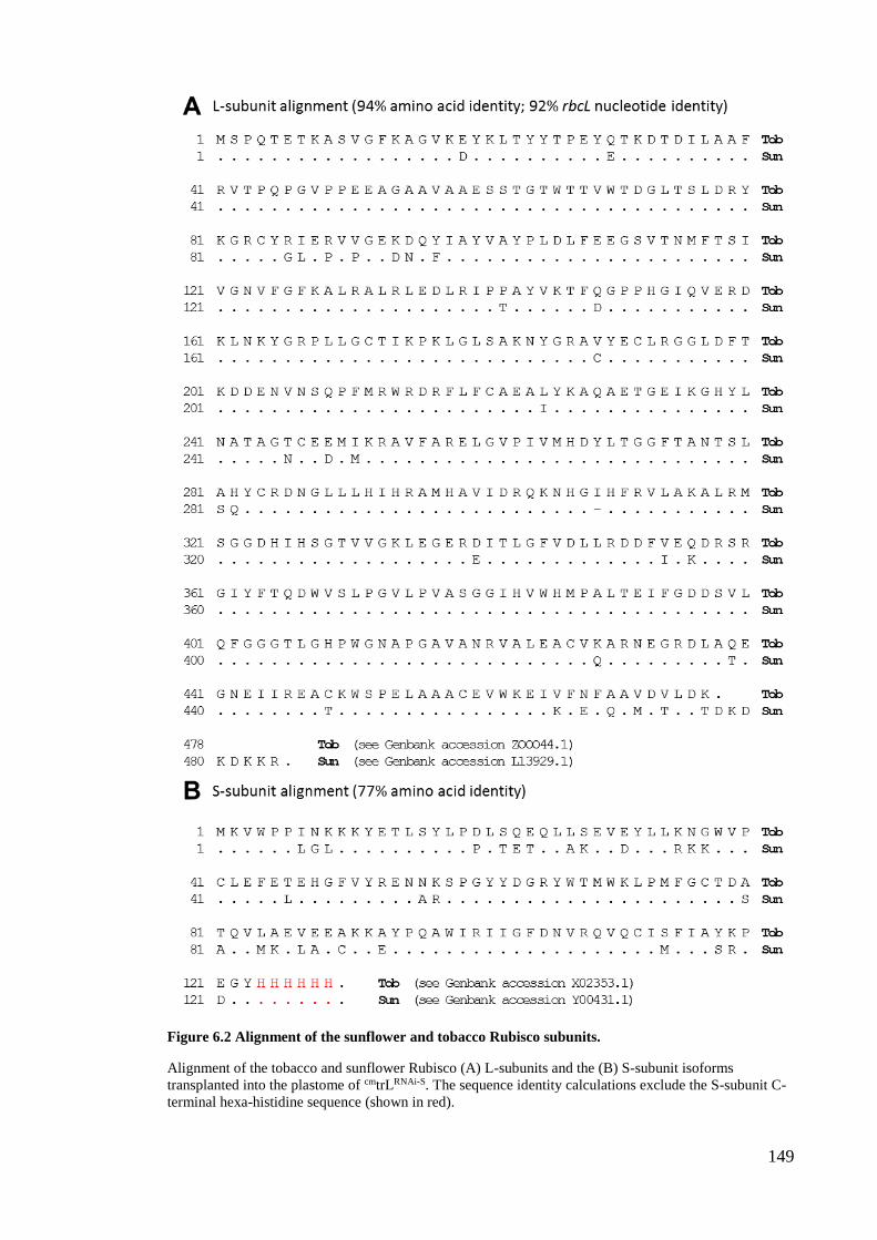
149
Figure 6.2 Alignment of the sunflower and tobacco Rubisco subunits.
Alignment of the tobacco and sunflower Rubisco (A) L-subunits and the (B) S-subunit isoforms
transplanted into the plastome of cmtrLRNAi-S. The sequence identity calculations exclude the S-subunit C-
terminal hexa-histidine sequence (shown in red).
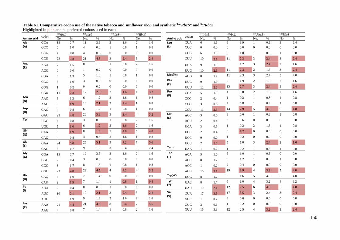
150
Table 6.1 Comparative codon use of the native tobacco and sunflower rbcL and synthetic TobRbcS* and SunRbcS.
Highlighted in pink are the preferred codons used in each.
Amino acid codon
TobrbcL SunrbcL TobRbcS* SunRbcS
Amino acid codon
TobrbcL SunrbcL TobRbcS* SunRbcS
No. % No. % No. % No. %
No. % No. % No. % No. %
Ala (A)
GCA 13 2.7 11 2.3 2 1.6 2 1.6
Leu (L)
CUA 6 1.3 9 1.9 1 0.8 3 2.4
GCC 5 1.0 4 0.8 1 0.8 1 0.8
CUC 0 0.0 0 0.0 0 0.0 0 0.0
GCG 4 0.8 4 0.8 0 0.0 0 0.0
CUG 6 1.3 5 1.0 1 0.8 1 0.8
GCU 23 4.8 21 4.3 3 2.4 3 2.4
CUU 10 2.1 11 2.3 3 2.4 3 2.4
Arg (R)
AGA 7 1.5 8 1.6 1 0.8 2 1.6
UUA 9 1.9 6 1.2 3 2.4 2 1.6
AGG 0 0.0 1 0.2 0 0.0 0 0.0
UUG 10 2.1 11 2.3 2 1.6 3 2.4
CGA 6 1.3 5 1.0 1 0.8 1 0.8
Met(M) AUG 8 1.7 11 2.3 3 2.4 5 4.0
CGC 5 1.0 3 0.6 0 0.0 0 0.0
Phe (F)
UUC 9 1.9 9 1.9 2 1.6 2 1.6
CGG 1 0.2 0 0.0 0 0.0 0 0.0
UUU 12 2.5 13 2.7 3 2.4 3 2.4
CGU 11 2.3 12 2.5 2 1.6 4 3.2
Pro (P)
CCA 5 1.0 4 0.8 2 1.6 2 1.6
Asn (N)
AAC 6 1.3 6 1.2 2 1.6 1 0.8
CCC 2 0.4 1 0.2 1 0.8 1 0.8
AAU 9 1.9 10 2.1 3 2.4 1 0.8
CCG 3 0.6 4 0.8 1 0.8 1 0.8
Asp (D)
GAC 4 0.8 6 1.2 1 0.8 1 0.8
CCU 11 2.3 14 2.9 5 4.0 6 4.8
GAU 23 4.8 26 5.3 3 2.4 4 3.2
Ser (S)
AGC 3 0.6 3 0.6 1 0.8 1 0.8
CysI UGC 4 0.8 3 0.6 1 0.8 2 1.6
AGU 2 0.4 3 0.6 0 0.0 0 0.0
UGU 5 1.0 6 1.2 2 1.6 2 1.6
UCA 3 0.6 1 0.2 2 1.6 1 0.8
Gln (Q)
CAA 9 1.9 8 1.6 5 4.0 5 4.0
UCC 2 0.4 6 1.2 0 0.0 0 0.0
CAG 4 0.8 4 0.8 2 1.6 1 0.8
UCG 0 0.0 1 0.2 0 0.0 0 0.0
Glu (E)
GAA 24 5.0 25 5.1 9 7.2 7 5.6
UCU 7 1.5 5 1.0 3 2.4 2 1.6
GAG 8 1.7 9 1.9 3 2.4 3 2.4
Term UAA 1 0.2 1 0.2 1 0.8 1 0.8
Gly (G)
GGA 13 2.7 12 2.5 2 1.6 2 1.6
Thr (T)
ACA 5 1.0 5 1.0 1 0.8 0 0.0
GGC 2 0.4 3 0.6 0 0.0 0 0.0
ACC 8 1.7 6 1.2 1 0.8 1 0.8
GGG 8 1.7 8 1.6 1 0.8 1 0.8
ACG 1 0.2 2 0.4 0 0.0 0 0.0
GGU 23 4.8 22 4.5 4 3.2 4 3.2
ACU 15 3.1 19 3.9 4 3.2 5 4.0
His (H)
CAC 5 1.0 7 1.4 0 0.0 0 0.0
Trp(W) UGG 8 1.7 8 1.6 5 4.0 5 4.0
CAU 9 1.9 7 1.4 1 0.8 1 0.8
Tyr (Y)
UAC 8 1.7 5 1.0 4 3.2 4 3.2
Ile (I)
AUA 2 0.4 0 0.0 1 0.8 0 0.0
UAU 10 2.1 12 2.5 6 4.8 5 4.0
AUC 10 2.1 10 2.1 3 2.4 3 2.4
Val (V)
GUA 17 3.6 17 3.5 3 2.4 3 2.4
AUU 9 1.9 9 1.9 2 1.6 2 1.6
GUC 1 0.2 3 0.6 0 0.0 0 0.0
Lys (K)
AAA 21 4.4 21 4.3 8 6.4 7 5.6
GUG 3 0.6 1 0.2 0 0.0 0 0.0
AAG 4 0.8 7 1.4 1 0.8 2 1.6
GUU 16 3.3 12 2.5 4 3.2 3 2.4
151
Figure 6.3 Sequence comparison of the RbcS transgenes transformed into the tobacco plastome.
(see next page for figure legend)
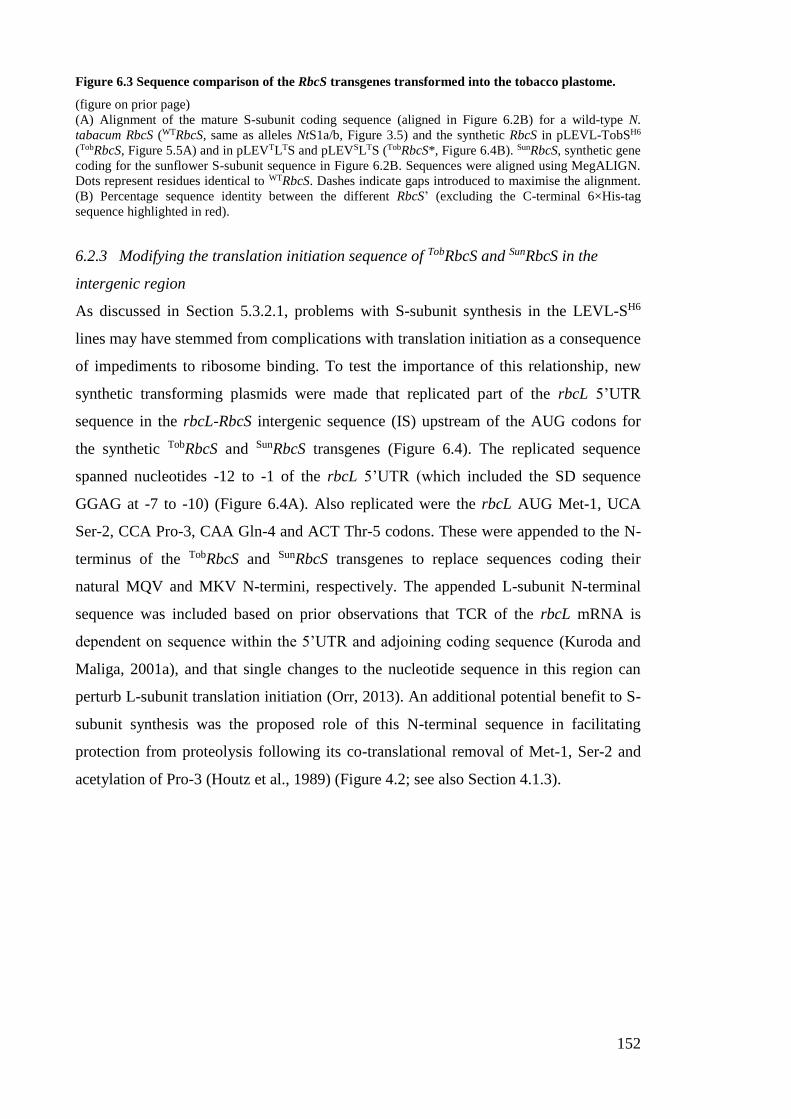
152
Figure 6.3 Sequence comparison of the RbcS transgenes transformed into the tobacco plastome.
(figure on prior page)
(A) Alignment of the mature S-subunit coding sequence (aligned in Figure 6.2B) for a wild-type N.
tabacum RbcS (WTRbcS, same as alleles NtS1a/b, Figure 3.5) and the synthetic RbcS in pLEVL-TobSH6
(TobRbcS, Figure 5.5A) and in pLEVTLTS and pLEVSLTS (TobRbcS*, Figure 6.4B). SunRbcS, synthetic gene
coding for the sunflower S-subunit sequence in Figure 6.2B. Sequences were aligned using MegALIGN.
Dots represent residues identical to WTRbcS. Dashes indicate gaps introduced to maximise the alignment.
(B) Percentage sequence identity between the different RbcS’ (excluding the C-terminal 6×His-tag
sequence highlighted in red).
6.2.3 Modifying the translation initiation sequence of TobRbcS and SunRbcS in the
intergenic region
As discussed in Section 5.3.2.1, problems with S-subunit synthesis in the LEVL-SH6
lines may have stemmed from complications with translation initiation as a consequence
of impediments to ribosome binding. To test the importance of this relationship, new
synthetic transforming plasmids were made that replicated part of the rbcL 5’UTR
sequence in the rbcL-RbcS intergenic sequence (IS) upstream of the AUG codons for
the synthetic TobRbcS and SunRbcS transgenes (Figure 6.4). The replicated sequence
spanned nucleotides -12 to -1 of the rbcL 5’UTR (which included the SD sequence
GGAG at -7 to -10) (Figure 6.4A). Also replicated were the rbcL AUG Met-1, UCA
Ser-2, CCA Pro-3, CAA Gln-4 and ACT Thr-5 codons. These were appended to the N-
terminus of the TobRbcS and SunRbcS transgenes to replace sequences coding their
natural MQV and MKV N-termini, respectively. The appended L-subunit N-terminal
sequence was included based on prior observations that TCR of the rbcL mRNA is
dependent on sequence within the 5’UTR and adjoining coding sequence (Kuroda and
Maliga, 2001a), and that single changes to the nucleotide sequence in this region can
perturb L-subunit translation initiation (Orr, 2013). An additional potential benefit to S-
subunit synthesis was the proposed role of this N-terminal sequence in facilitating
protection from proteolysis following its co-translational removal of Met-1, Ser-2 and
acetylation of Pro-3 (Houtz et al., 1989) (Figure 4.2; see also Section 4.1.3).
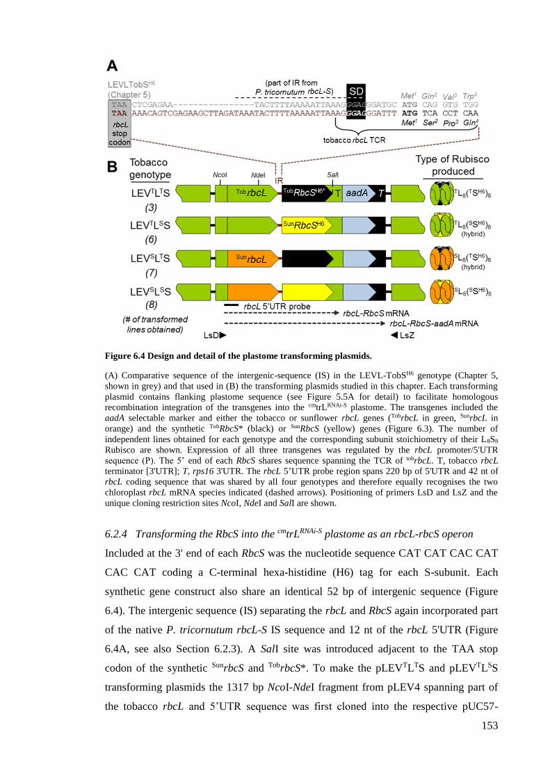
153
Figure 6.4 Design and detail of the plastome transforming plasmids.
(A) Comparative sequence of the intergenic-sequence (IS) in the LEVL-TobSH6 genotype (Chapter 5,
shown in grey) and that used in (B) the transforming plasmids studied in this chapter. Each transforming
plasmid contains flanking plastome sequence (see Figure 5.5A for detail) to facilitate homologous
recombination integration of the transgenes into the cmtrLRNAi-S plastome. The transgenes included the
aadA selectable marker and either the tobacco or sunflower rbcL genes (TobrbcL in green, SunrbcL in
orange) and the synthetic TobRbcS* (black) or SunRbcS (yellow) genes (Figure 6.3). The number of
independent lines obtained for each genotype and the corresponding subunit stoichiometry of their L8S8
Rubisco are shown. Expression of all three transgenes was regulated by the rbcL promoter/5'UTR
sequence (P). The 5’ end of each RbcS shares sequence spanning the TCR of tobrbcL. T, tobacco rbcL
terminator [3'UTR]; T, rps16 3'UTR. The rbcL 5’UTR probe region spans 220 bp of 5'UTR and 42 nt of
rbcL coding sequence that was shared by all four genotypes and therefore equally recognises the two
chloroplast rbcL mRNA species indicated (dashed arrows). Positioning of primers LsD and LsZ and the
unique cloning restriction sites NcoI, NdeI and SalI are shown.
6.2.4 Transforming the RbcS into the cmtrLRNAi-S plastome as an rbcL-rbcS operon
Included at the 3' end of each RbcS was the nucleotide sequence CAT CAT CAC CAT
CAC CAT coding a C-terminal hexa-histidine (H6) tag for each S-subunit. Each
synthetic gene construct also share an identical 52 bp of intergenic sequence (Figure
6.4). The intergenic sequence (IS) separating the rbcL and RbcS again incorporated part
of the native P. tricornutum rbcL-S IS sequence and 12 nt of the rbcL 5'UTR (Figure
6.4A, see also Section 6.2.3). A SalI site was introduced adjacent to the TAA stop
codon of the synthetic SunrbcS and TobrbcS*. To make the pLEVTLTS and pLEVTLSS
transforming plasmids the 1317 bp NcoI-NdeI fragment from pLEV4 spanning part of
the tobacco rbcL and 5’UTR sequence was first cloned into the respective pUC57-
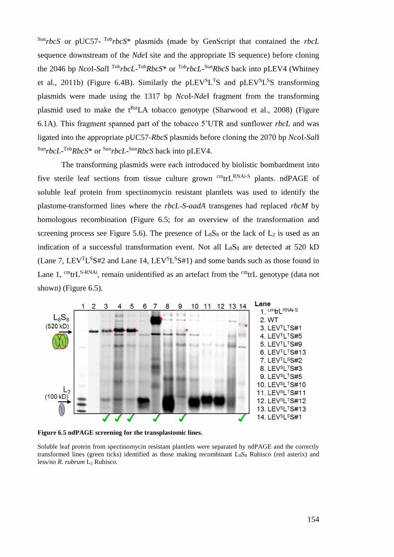
154
SunrbcS or pUC57- TobrbcS* plasmids (made by GenScript that contained the rbcL
sequence downstream of the NdeI site and the appropriate IS sequence) before cloning
the 2046 bp NcoI-SalI TobrbcL-TobRbcS* or TobrbcL-SunRbcS back into pLEV4 (Whitney
et al., 2011b) (Figure 6.4B). Similarly the pLEVSLTS and pLEVSLSS transforming
plasmids were made using the 1317 bp NcoI-NdeI fragment from the transforming
plasmid used to make the tRstLA tobacco genotype (Sharwood et al., 2008) (Figure
6.1A). This fragment spanned part of the tobacco 5’UTR and sunflower rbcL and was
ligated into the appropriate pUC57-RbcS plasmids before cloning the 2070 bp NcoI-SalI
SunrbcL-TobRbcS* or SunrbcL-SunRbcS back into pLEV4.
The transforming plasmids were each introduced by biolistic bombardment into
five sterile leaf sections from tissue culture grown cmtrLRNAi-S plants. ndPAGE of
soluble leaf protein from spectinomycin resistant plantlets was used to identify the
plastome-transformed lines where the rbcL-S-aadA transgenes had replaced rbcM by
homologous recombination (Figure 6.5; for an overview of the transformation and
screening process see Figure 5.6). The presence of L8S8 or the lack of L2 is used as an
indication of a successful transformation event. Not all L8S8 are detected at 520 kD
(Lane 7, LEVTLSS#2 and Lane 14, LEVSLSS#1) and some bands such as those found in
Lane 1, cmtrLS-RNAi, remain unidentified as an artefact from the cmtrL genotype (data not
shown) (Figure 6.5).
Figure 6.5 ndPAGE screening for the transplastomic lines.
Soluble leaf protein from spectinomycin resistant plantlets were separated by ndPAGE and the correctly
transformed lines (green ticks) identified as those making recombinant L8S8 Rubisco (red asterix) and
less/no R. rubrum L2 Rubisco.
155
Similar to the LEVL-SH6 genotypes made in Chapter 5, the three transgenes
(rbcL, RbcS and aadA) that were transformed into the resulting LEVTLTS, LEVTLSS,
LEVSLTS and LEVSLSS genotypes were all controlled by the native tobacco rbcL
promoter, 5'UTR and 3'UTR regulatory elements (Figure 5.5A). Three to eight
independently transformed lines were obtained for each genotype (Figure 6.4B).
6.2.5 Growth, maintenance and resulting phenotype the different tobacco genotypes
After 2-4 rounds of further selection on RMOPspec the transformed region of the
plastome was PCR amplified (using primers LsD and LsZ, Figure 6.4) from three lines
of each genotype and fully sequenced to confirm each independently transformed line
was genetically identical (data not shown). At least three T0 lines for each genotype
were transferred from RMOPspec regeneration medium into tissue culture pots
containing MS medium (with 3% [w/v] sucrose and 500 mg L-1 spectinomycin) to
stimulate normal root and shoot development. After five weeks growing at 25°C under
2% (v/v) CO2 and ~40 µmol photons m2 s-1 illumination, the phenotype of the LEVTLTS
and LEVTLSS genotypes resembled wild-type. In contrast, the sunflower L-subunit
producing LEVSLTS and LEVSLSS lines struggled to grow despite sucrose supplement
in MS medium (Figure 6.6). These lines shared a pale green phenotype, had fragile, thin
leaves and grew very slowly – even more impaired than that observed previously for the
tobRst, tRstLA and tRstL7 genotypes grown in tissue culture (Whitney and Sharwood,
2008; Sharwood and Whitney, 2010).
After developing suitable root mass in tissue culture, the plants were transferred
into soil to grow to maturity in air containing 2% (v/v) CO2. In soil, the LEVTLTS and
LEVTLSS lines maintained their wild-type like phenotype and were able to produce
normal flowers that were pollinated with wild-type pollen. The resulting seeds were
collected for further study of the T1 progeny. Despite repeated attempts the LEVSLTS
and LEVSLSS lines were however unable to grow in soil under elevated CO2. This
restricted subsequent studies of their leaf biochemistry to material from T0 plants grown
after several rounds of selection and regeneration in RMOP-spec medium.
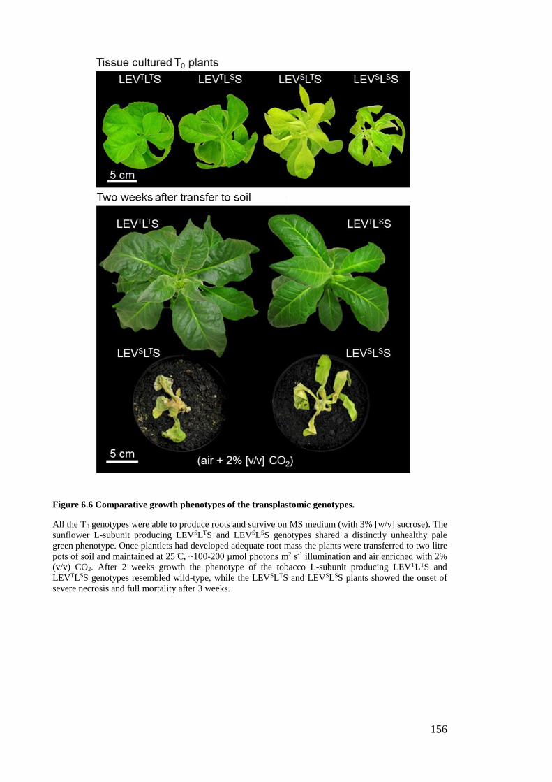
156
Figure 6.6 Comparative growth phenotypes of the transplastomic genotypes.
All the T0 genotypes were able to produce roots and survive on MS medium (with 3% [w/v] sucrose). The
sunflower L-subunit producing LEVSLTS and LEVSLSS genotypes shared a distinctly unhealthy pale
green phenotype. Once plantlets had developed adequate root mass the plants were transferred to two litre
pots of soil and maintained at 25 C̊, ~100-200 µmol photons m2 s-1 illumination and air enriched with 2%
(v/v) CO2. After 2 weeks growth the phenotype of the tobacco L-subunit producing LEVTLTS and
LEVTLSS genotypes resembled wild-type, while the LEVSLTS and LEVSLSS plants showed the onset of
severe necrosis and full mortality after 3 weeks.

157
6.2.6 Recombinant L8S8 Rubisco production was dependent on the L-subunit source
Analyses of Rubisco content in three independently obtained lines for each genotype
were undertaken. The T1 LEVTLTS and LEVTLSS progeny were grown in a growth
cabinet (25°C under ambient CO2 and 400 ± 100 μmol photons m2 s-1 illumination,
14:10h L:D cycle) and at 35 cm in height leaf #5 from the apical meristem (i.e. the
youngest near fully expanded leaf) was sampled for protein analyses. Leaf samples from
the LEVSLTS and LEVSLSS lines were obtained from T0 tissue culture plants growing in
MS containing 3% (w/v) sucrose. Analysis of the soluble leaf protein by ndPAGE
showed abundant levels of Rubisco were produced in the LEVTLTS and LEVTLSS
genotypes and very little in the sunflower L-subunit (SL) producing LEVSLTS and
LEVSLSS lines (Figure 6.7A). The identities of the Rubisco complexes were confirmed
by immunoblot analysis using antibodies to tobacco Rubisco and a penta-histidine
antigen. Only the Rubisco in the transplastomic lines reacted with the latter antibody,
confirming they incorporated the chloroplast made recombinant sunflower (SSH6) or
tobacco (TSH6) S-subunits.
A comparison of the separation rates through ndPAGE for the different Rubisco
isoforms showed those incorporating the sunflower SSH6-subunits migrated substantially
slower. In contrast, the TL8(TSH6)8 and SL8(
TSH6)8 complexes produced in the LEVTLTS
and LEVSLTS genotypes separated at the same rate as wild-type tobacco Rubisco. This
suggests the sunflower SSH6-subunits impart differences in the quaternary packing when
assembled with either the tobacco L-subunits (TL) or sunflower L-subunits (SL). In all
the lines analysed, no R. rubrum L2 Rubisco was detectable by ndPAGE confirming
their homoplasmicity (Figure 6.7A).
The Rubisco levels in each genotype were quantified by [14C]-CABP binding
analyses (Figure 6.7B). Relative to the Rubisco content in control wild-type (WT)
tobacco leaves, the Rubisco levels in LEVTLTS and LEVTLSS was reduced
approximately 40% and 70% respectively. In contrast the Rubisco content in LEVSLTS
and LEVSLSS leaves were reduced by more than 96% resembling those produced in the
LEVL-SH6 progeny (i.e. <1 μmol active sites m2, Figure 5.8B).
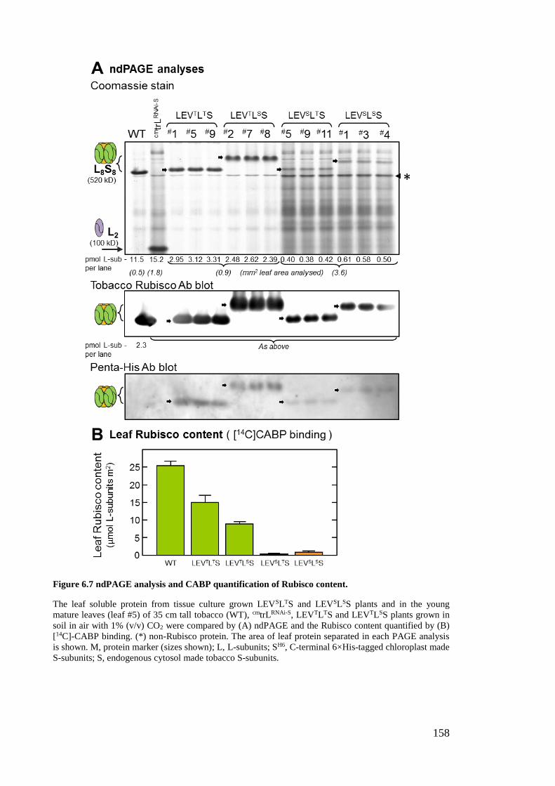
158
Figure 6.7 ndPAGE analysis and CABP quantification of Rubisco content.
The leaf soluble protein from tissue culture grown LEVSLTS and LEVSLSS plants and in the young
mature leaves (leaf #5) of 35 cm tall tobacco (WT), cmtrLRNAi-S, LEVTLTS and LEVTLSS plants grown in
soil in air with 1% (v/v) CO2 were compared by (A) ndPAGE and the Rubisco content quantified by (B)
[14C]-CABP binding. (*) non-Rubisco protein. The area of leaf protein separated in each PAGE analysis
is shown. M, protein marker (sizes shown); L, L-subunits; SH6, C-terminal 6×His-tagged chloroplast made
S-subunits; S, endogenous cytosol made tobacco S-subunits.
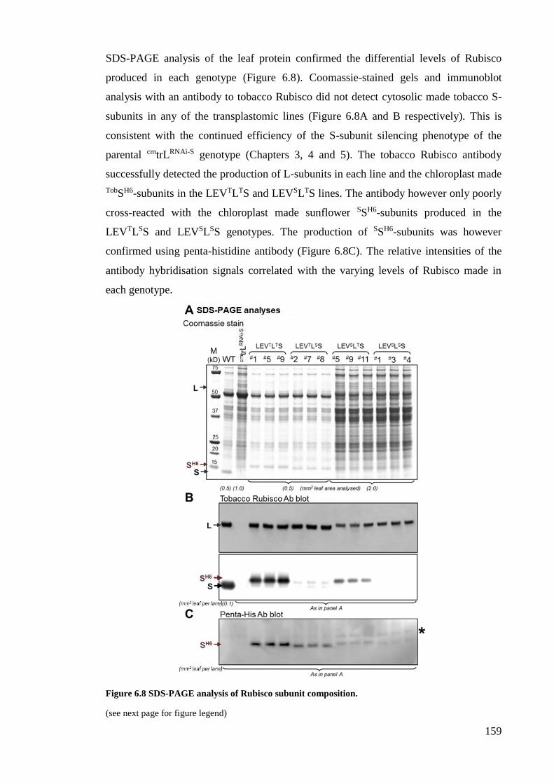
159
SDS-PAGE analysis of the leaf protein confirmed the differential levels of Rubisco
produced in each genotype (Figure 6.8). Coomassie-stained gels and immunoblot
analysis with an antibody to tobacco Rubisco did not detect cytosolic made tobacco S-
subunits in any of the transplastomic lines (Figure 6.8A and B respectively). This is
consistent with the continued efficiency of the S-subunit silencing phenotype of the
parental cmtrLRNAi-S genotype (Chapters 3, 4 and 5). The tobacco Rubisco antibody
successfully detected the production of L-subunits in each line and the chloroplast made
TobSH6-subunits in the LEVTLTS and LEVSLTS lines. The antibody however only poorly
cross-reacted with the chloroplast made sunflower SSH6-subunits produced in the
LEVTLSS and LEVSLSS genotypes. The production of SSH6-subunits was however
confirmed using penta-histidine antibody (Figure 6.8C). The relative intensities of the
antibody hybridisation signals correlated with the varying levels of Rubisco made in
each genotype.
Figure 6.8 SDS-PAGE analysis of Rubisco subunit composition.
(see next page for figure legend)
160
Figure 6.8 SDS-PAGE analysis of Rubisco subunit composition.
(figure on prior page)
The leaf soluble protein from Figure 6.7 was analysed by SDS-PAGE and (A) Coomassie-stained or
immunoblotted against antibodies to (B) tobacco Rubisco or (C) a penta-histidine epitope (Qiagen). The
area of leaf protein separated in each PAGE analysis is shown. M, protein marker (sizes shown); L, L-
subunits; SH6, C-terminal 6×His-tagged chloroplast made tobacco or sunflower S-subunits; S, endogenous
cytosol made tobacco S-subunits. Asterix (*) indicates a protein in tobacco leaves at ~16.5 kDa that
cross-reacts with the penta-histidine antibody.
6.2.7 Post-transcriptional limitations to Rubisco biogenesis in LEVSLTS and LEVSLSS
After transferral to nylon membrane the RNA was probed with the [32P]-labelled rbcL
5’UTR probe that hybridises to the 5' sequence conserved in both the TobrbcL and
SunrbcL mRNA (Figure 6.4B). Each transplastomic genotype produced two rbcL mRNA
species; a prominent rbcL-rbcS (dicistronic) operon and a ~50% less abundant rbcL-
rbcS-aadA (tricistronic) mRNA. The latter transcript is produced as a consequence of
inefficient termination by the rbcL 3'UTR located between RbcS and aadA (Whitney
and Sharwood, 2008).
Analysis of levels of both endogenous chloroplast rbcL mRNA (Figure 6.9B)
and nuclear RbcS mRNAs (Figure 6.9C) in the tissue culture grown wild-type showed
reduction by ~10% relative to that in the high-CO2 sample. In contrast no RbcS mRNAs
were detected in any of the transplastomic lines, consistent with the efficiency of the
RNAi-targeted silencing of these transcripts in the maternal cmtrLRNAi-S genotype
(Chapter 3). The same probe did not hybridise with the abundant chloroplast made
rbcL-RbcS transcripts comprising either the tobRbcS* or sunRbcS due to their <70%
sequence identity with WTRbcS (Figure 6.3).
The total pool of rbcL-RbcS transcripts in the LEVTLTS and LEVTLSS lines
were both reduced by around one-third relative to the rbcL mRNA levels in WT. This
implies that limitations in transcript abundance likely contribute to the ~40% and ~70%
lower levels of Rubisco made, respectively, in the LEVTLTS and LEVTLSS genotypes
(Figure 6.7B). In contrast the SunrbcL-RbcS mRNA levels in the LEVSLTS and LEVSLSS
genotypes were, respectively, ~80% and ~90% lower than the rbcL transcript content in
WT (Figure 6.9B). While these mRNA levels are low, they do not match the more than
25-fold lower levels of Rubisco produced in these genotypes (Figure 6.7B). This
implies that further limitations during the post-transcriptional processing of the SunrbcL
mRNA are impeding L8S8 biogenesis in the LEVSLTS and LEVSLSS genotypes.
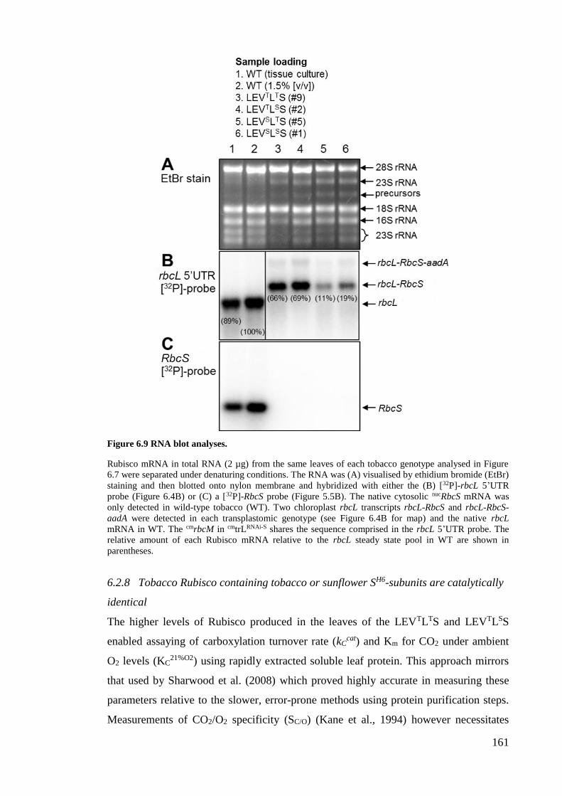
161
Figure 6.9 RNA blot analyses.
Rubisco mRNA in total RNA (2 µg) from the same leaves of each tobacco genotype analysed in Figure
6.7 were separated under denaturing conditions. The RNA was (A) visualised by ethidium bromide (EtBr)
staining and then blotted onto nylon membrane and hybridized with either the (B) [32P]-rbcL 5’UTR
probe (Figure 6.4B) or (C) a [32P]-RbcS probe (Figure 5.5B). The native cytosolic nucRbcS mRNA was
only detected in wild-type tobacco (WT). Two chloroplast rbcL transcripts rbcL-RbcS and rbcL-RbcS-
aadA were detected in each transplastomic genotype (see Figure 6.4B for map) and the native rbcL
mRNA in WT. The cmrbcM in cmtrLRNAi-S shares the sequence comprised in the rbcL 5’UTR probe. The
relative amount of each Rubisco mRNA relative to the rbcL steady state pool in WT are shown in
parentheses.
6.2.8 Tobacco Rubisco containing tobacco or sunflower SH6-subunits are catalytically
identical
The higher levels of Rubisco produced in the leaves of the LEVTLTS and LEVTLSS
enabled assaying of carboxylation turnover rate (kCcat) and Km for CO2 under ambient
O2 levels (KC21%O2) using rapidly extracted soluble leaf protein. This approach mirrors
that used by Sharwood et al. (2008) which proved highly accurate in measuring these
parameters relative to the slower, error-prone methods using protein purification steps.
Measurements of CO2/O2 specificity (SC/O) (Kane et al., 1994) however necessitates
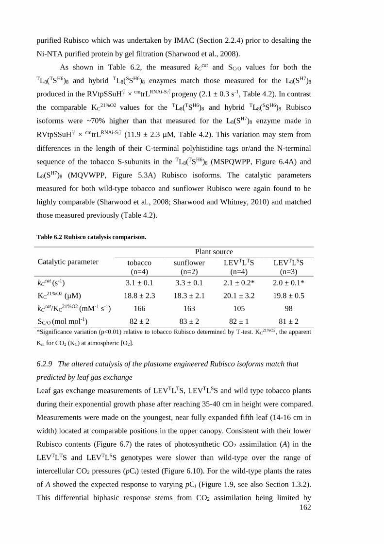
162
purified Rubisco which was undertaken by IMAC (Section 2.2.4) prior to desalting the
Ni-NTA purified protein by gel filtration (Sharwood et al., 2008).
As shown in Table 6.2, the measured kCcat and SC/O values for both the
TL8(TSH6)8 and hybrid TL8(
SSH6)8 enzymes match those measured for the L8(SH7)8
produced in the RVtpSSuH♀ × cmtrLRNAi-S♂ progeny (2.1 ± 0.3 s-1, Table 4.2). In contrast
the comparable KC21%O2 values for the TL8(
TSH6)8 and hybrid TL8(SSH6)8 Rubisco
isoforms were ~70% higher than that measured for the L8(SH7)8 enzyme made in
RVtpSSuH♀ × cmtrLRNAi-S♂ (11.9 ± 2.3 µM, Table 4.2). This variation may stem from
differences in the length of their C-terminal polyhistidine tags or/and the N-terminal
sequence of the tobacco S-subunits in the TL8(TSH6)8 (MSPQWPP, Figure 6.4A) and
L8(SH7)8 (MQVWPP, Figure 5.3A) Rubisco isoforms. The catalytic parameters
measured for both wild-type tobacco and sunflower Rubisco were again found to be
highly comparable (Sharwood et al., 2008; Sharwood and Whitney, 2010) and matched
those measured previously (Table 4.2).
Table 6.2 Rubisco catalysis comparison.
Catalytic parameter
Plant source
tobacco
(n=4)
sunflower
(n=2)
LEVTLTS
(n=4)
LEVTLSS
(n=3)
kCcat (s-1) 3.1 ± 0.1 3.3 ± 0.1 2.1 ± 0.2* 2.0 ± 0.1*
KC21%O2 (µM) 18.8 ± 2.3 18.3 ± 2.1 20.1 ± 3.2 19.8 ± 0.5
kCcat/KC
21%O2 (mM-1 s-1) 166 163 105 98
SC/O (mol mol-1) 82 ± 2 83 ± 2 82 ± 1 81 ± 2
*Significance variation (p<0.01) relative to tobacco Rubisco determined by T-test. KC21%O2, the apparent
Km for CO2 (KC) at atmospheric [O2].
6.2.9 The altered catalysis of the plastome engineered Rubisco isoforms match that
predicted by leaf gas exchange
Leaf gas exchange measurements of LEVTLTS, LEVTLSS and wild type tobacco plants
during their exponential growth phase after reaching 35-40 cm in height were compared.
Measurements were made on the youngest, near fully expanded fifth leaf (14-16 cm in
width) located at comparable positions in the upper canopy. Consistent with their lower
Rubisco contents (Figure 6.7) the rates of photosynthetic CO2 assimilation (A) in the
LEVTLTS and LEVTLSS genotypes were slower than wild-type over the range of
intercellular CO2 pressures (pCi) tested (Figure 6.10). For the wild-type plants the rates
of A showed the expected response to varying pCi (Figure 1.9, see also Section 1.3.2).
This differential biphasic response stems from CO2 assimilation being limited by
163
Rubisco activity under lower pCi (e.g. below ~400 µbar for wild-type) and then
increasingly restricted by the rate of electron transport through the light reactions of
photosynthesis as the pCi exceeds ~500 µbar (Farquhar et al., 1980) as shown in Figure
6.10. In the LEVTLTS and LEVTLSS leaves however the CO2 assimilation rates
remained limited by Rubisco activity over the pCi range tested (Figure 6.10). This
occurs due to the lower Rubisco content in their leaves (Figure 6.7) and slower Rubisco
carboxylation rates (Table 6.2).
The A-pCi data for each genotype was compared with those predicted using the
Michaelis-Menten rate equation from Farquhar et al. (1980) fitted with the catalytic
parameters measured for each Rubisco isoform (Table 6.2) and the measured Rubisco
content (quantified by [14C]-CABP binding). As shown in Figure 6.10, for the LEVTLTS
and LEVTLSS genotypes the modelled CO2 assimilation rates closely match those
measured, confirming the accuracy of the slower carboxylation properties (i.e. kCcat and
kCcat/KC
21%O2 ) measured for both the TL8(TSH6)8 and hybrid TL8(
SSH6)8 Rubisco isoforms.
For the wild-type samples the measured carboxylase limiting CO2 assimilation
rates (i.e. under low pCi) were lower than those modelled (Figure 6.10). This
overestimation likely stems from the rates of the CO2 assimilation model not taking into
account variations in the activation status of Rubisco in wild-type plants grown under
elevated CO2 – which can be reduced by up to 40% (Whitney et al., 1999; Whitney and
Andrews, 2001b; Sharwood et al., 2008), as well as light limitations that impaired CO2
assimilation beyond 400 pCi of CO2. Unfortunately the leaf discs taken for analysis
following leaf gas exchange in this study were not analysed for Rubisco activation
status – a critical oversight considering how Rubisco catalytic content has a pervasive
influence on modelling predictions using the equations of Farquhar et al. (1980) as
depicted by the B term in the equation in the legend to Figure 6.10.
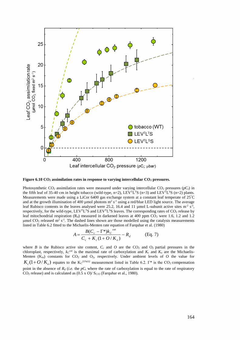
164
Figure 6.10 CO2 assimilation rates in response to varying intercellular CO2 pressures.
Photosynthetic CO2 assimilation rates were measured under varying intercellular CO2 pressures (pCi) in
the fifth leaf of 35-40 cm in height tobacco (wild-type, n=2), LEVTLTS (n=3) and LEVTLSS (n=2) plants.
Measurements were made using a LiCor 6400 gas exchange system at a constant leaf temperate of 25°C
and at the growth illumination of 400 µmol photons m2 s-1 using a red/blue LED light source. The average
leaf Rubisco contents in the leaves analysed were 25.2, 16.4 and 11 µmol L-subunit active sites m-2 s-1,
respectively, for the wild-type, LEVTLTS and LEVTLSS leaves. The corresponding rates of CO2 release by
leaf mitochondrial respiration (Rd) measured in darkened leaves at 400 ppm CO2 were 1.6, 1.2 and 1.2
μmol CO2 released m2 s-1. The dashed lines shown are those modelled using the catalysis measurements
listed in Table 6.2 fitted to the Michaelis-Menten rate equation of Farquhar et al. (1980)
d
occ
cat
Cc RKOKC
kCBA
)/1(
*)( (Eq. 7)
where B is the Rubisco active site content, Cc and O are the CO2 and O2 partial pressures in the
chloroplast, respectively, kCcat is the maximal rate of carboxylation and Kc and Ko are the Michaelis-
Menten (Km) constants for CO2 and O2, respectively. Under ambient levels of O the value for
)/1( oc KOK equates to the KC21%O2 measurement listed in Table 6.2. Γ* is the CO2 compensation
point in the absence of Rd (i.e. the pCi where the rate of carboxylation is equal to the rate of respiratory
CO2 release) and is calculated as (0.5 x O)/ SC/O (Farquhar et al., 1980).
165
6.3 Discussion
The results of this chapter affirm the versatility of the cmtrLRNAi-S line as a novel tobacco
genotype for bioengineering Rubisco in leaf chloroplasts. Shown is the successful
implementation of a dual L- and S-subunit transplastomic approach for producing
tobacco genotypes capable of making adequate levels of Rubisco to support plant
growth in air. The genetic precision and simultaneous modification of this rbcL-rbcS
co-engineering approach provided a novel method for bioengineering Rubisco isoforms
that comprised a homogenous population of L- and S-subunits – the key objective of
this thesis (see Section 1.5). The novel method worked best for the Rubisco isoforms
comprising tobacco L-subunits, suggesting the need to adhere to the specialised
assembly requirements of this subunit. Limitations to S-subunit synthesis and possible
structural complementarity issues with heterologous L-subunits also appear to impact
the production of recombinant Rubisco in leaf chloroplasts. Overcoming these
experimental hurdles pose significant challenges in fine tuning recombinant Rubisco
bioengineering efforts.
6.3.1 The varying limitations to L- and S-subunit synthesis in chloroplasts
6.3.1.1 Meeting the chaperone requirements of the L –subunit is a core requirement
As summarised in Section 1.4.3, transplastomic studies on bioengineering Rubisco in
tobacco chloroplasts have paid particular attention to replacing/modifying the plastome
rbcL (Parry et al., 2013). Transformed genotypes producing wild-type or mutated
tobacco rbcL that slow catalysis show little or no change in rbcL mRNA and L8S8
contents under conditions where autotrophic growth can be supported (e.g. high-CO2)
(Whitney et al., 1999; Whitney and Sharwood, 2008; Whitney et al., 2011b). In contrast,
in genotypes where the tobacco rbcL is replaced with an alternative plant rbcL, highly
varied amounts of hybrid Rubisco are produced (see Table 5.1 and Figure 6.1). In this
thesis, the production of sunflower SL8(SSH6)8 and hybrid SL8(
tSH6)8 Rubisco in the
LEVSLTS and LEVSLSS genotypes (Figure 6.9) proved to be more impeded than prior
sunrbcL mRNA transplastomic studies (Figure 6.1). The recurring impediment to
sunflower L-subunit synthesis in tobacco chloroplasts is not likely to stem from
problems with SunrbcL mRNA translation initiation as its 5’UTR sequence and first 54
nt of coding sequence are identical to the TobrbcL mRNA. As indicated in Sharwood et
al. (2008), this level of homology also ensures the sunflower L-subunits undergo the
same N-terminal post translational modifications (PTMs, i.e. Met-1, Ser-2 cleavage-
166
Pro-3 acetylation, Lys-14 trimethylation) that are thought to protect against proteolysis
(Houtz et al., 1989; Nishimura et al., 2008; also see Section 4.3.1.2).
The highly comparable codon use of TobrbcL and SunrbcL provide little evidence
for differences in translation elongation of both transcripts. Indeed a comparison of their
5'UTR sequences (Genbank accession number DQ383815.1 for H. annuus SunrbcL) also
shows high sequence identity (91%). In general, the multidimensional effects from
synonymous site modification of genes for optimised chloroplast expression remain
unclear (Ullrich et al., 2015). It is also important to remember that although mRNA acts
as a direct source for protein translation, its levels are not always indicative of protein
content (Bitton et al., 2008; Nagaraj et al., 2011). For example, the expression of protein
antibiotics in tobacco plastids produced incoherent mRNA and protein expression levels
that signified limitations in mRNA translational capacity (Oey et al., 2009). Testing for
possible perturbations in translation initiation or elongation of SunrbcL mRNA by
sucrose gradient polysome analyses is yet to be examined in any sunflower L-subunit
producing tobacco genotype and is clearly an important objective for future studies.
Extending such analyses to compare the polysome association of the synthetic tobacco
rbcL-rbcS mRNAs in the LEVLSSuH♀ × cmtrLRNAi-S♂ and LEVL-TobSH6 genotypes
(Chapter 5) relative to the LEVTLTS and LEVTLSS lines is also important as this would
help decipher to what extent the “chloroplast optimised” codon use in the synthetic
SunRbcS and TobRbcS* (Table 6.1) improved chloroplast S-subunit synthesis.
Recent breakthroughs in deciphering the Rubisco biogenesis pathway in leaf
chloroplasts indicate correct folding and assembly of the L-subunit is dependent on
structural complementation with folding and assembly chaperones. Evolutionary
divergence in the sequence of the sunflower and tobacco L-subunits appear to have
perturbed this chaperone-complimentarity requirement to restrict sunflower L-subunit
assembly in tobacco. One chaperone of particular impotance is the Rubisco specific
chaperone RAF1. Characterisation of RAF1 form and function began through its
discovery in a maize photosynthetic mutant library (PML) where it was postulated to
facilitate assembly of the post-chaperonin folded L-subunits into L8 core complexes for
S-subunit assembly (Feiz et al., 2012). More recent work on another maize PML mutant
suggests RAF1 may function with the aid of other Rubisco specialised chaperones
called RAF2 and BSD2 (Brutnell et al., 1999; Feiz et al., 2014). To better understand
the role of the RAF1 homo-dimer complex in leaf chloroplasts, recent work using a
plastome transformation approach confirmed the dependency of complementarity
between L-subunits and RAF1 for optimal L8S8 biogenesis (Whitney et al., 2015). That
167
is, in tobacco leaves producing Arabidopsis L-subunits the amount of hybrid AL8tS8
Rubisco (comprising Arabidopsis L- and tobacco S-subunits) was increased ~2-fold
from ~3.5 µmol catalytic sites m2 to ~7.5 µmol catalytic sites m2 by co-expression of an
Arabidopsis RAF1 isoform. Co-insertion of raf1 in an operon with rbcL unfortunately
compromised the rbcL-raf1 mRNA levels by ~3-fold. When normalised relative to
mRNA contents it was found RAF1 had increased AL8tS8 Rubisco biogenesis by more
than 7-fold (Whitney et al., 2015).
The potential for improving the biogenesis of hybrid Rubisco comprising
sunflower SL-subunits via co-expressing RAF1 from sunflower serves as a primary
target for future testing. Current efforts have been stymied so far as sequences for raf1
from sunflower remain to be determined. Facilitating further improvement to
heterologous plant Rubisco production may extend further than RAF1 to include other
L-subunit and S-subunit assembly chaperones (for potential examples see Table 4.1 and
Figure 4.3). For example, recent success in producing cyanobacteria Rubisco in tobacco
chloroplasts (i.e. the Se7942 tobacco genotypes, Table 5.1) found Rubisco assembly
was augmented in those lines co-producing the complimentary CcmM35 protein (Lin et
al., 2014). In cyanobacteria the CcmM forms the key shell protein of carboxysomes and
has a C-terminus that shares sequence homology with the S-subunit which functions to
co-ordinate Rubisco assembly inside the carboxysome (Long et al., 2007; Rae et al.,
2012; Price and Howitt, 2014). Further discussion on potential Rubisco co-chaperone
expression testing in the future is included in Section 7.6.
6.3.1.2 The contrasting restraints on L8S8 biogenesis in each tobacco genotype
The significant amount of recombinant Rubisco produced in LEVTLTS and LEVSLTS
implies the alternative rbcL-rbcS operon structure utilised in this study (Figure 6.4A)
was suitable for facilitating comparatively high levels of chloroplast S-subunit synthesis.
This would suggest that severely limited levels of Rubisco produced in LEVSLTS and
LEVSLSS lines was not limited by chloroplast S-subunit availability. This is consistent
with that described in the section above where the primary restriction to Rubisco
biogenesis is the capacity of sunflower SL-subunit to assemble into octameric cores
(Sharwood and Whitney, 2010; see also Section 5.1.3). Impeded production of
sunflower-L8 cores in the LEVSLTS and LEVSLSS genotypes may therefore render the
chloroplast made TS- and SS-subunits more prone to proteolysis. Prior Rubisco
bioengineering work has shown the efficiency of protein proteolysis in the stroma. In
cmtrL that only makes L2-Rubisco, rapid degradation of unassembled tobacco S-subunits

168
by stromal proteases prevents their detection (Whitney and Andrews, 2001b; Whitney
and Andrews, 2003; Whitney and Sharwood, 2008).
As described in Section 6.2.3, an MSPQT substitution was made to the N-
terminus of the TS- and SS-subunits in a bid to safeguard them against proteolysis
(Houtz et al., 1989). Future experiments aim to investigate the level of protection
afforded by these amino acid changes by firstly determining the S-subunit N-terminal
sequence in the LEVTLTS, LEVTLSS, LEVSLTS and LEVSLSS genotypes using
established mass spectrometry methods (Whitney et al., 2009). Secondly, rates of
Rubisco translation and stability in the leaf chloroplasts of LEVTLTS and LEVTLSS (that
make sufficient levels of Rubisco) will be undertaken via pulse-chase with [35S]-
Met/unlabelled-Met (Whitney et al., 2015; also outlined in Section 5.3.2.3). These
experiments will determine if there are potential correlations between PTM-induced
improvements in Rubisco subunit synthesis and stability. Such measurements may help
decipher whether the acetyl-Pro modification of plant L-subunits might only provide
proteolysis protection to subunits assembled within L8S8 complexes. This would support
the hypothesis that the association of nascent and unfolded L- and S-subunit with one of
their many complementary pre-assembly chaperones (Figures 4.1 and 4.3; Table 4.1) is
required for their protection from proteolysis (Nakamura et al., 2001).
6.3.1.3 Rubisco mRNA limitations confine L8S8 biogenesis in LEVSLTS and LEVSLSS.
The amount of Rubisco made in the LEVSLTS and LEVSLSS genotypes (~0.1 to 0.4
μmol active sites m2, Figure 6.7B) is ~10-fold lower relative to other tobacco genotypes
producing hybrid Rubisco comprising sunflower SL-subunits and cytosol made tobacco
S-subunits (Figure 6.1B). This suggests that additional factors other than SL-subunit
chaperone complementarity limitations were affecting Rubisco biogenesis in these lines.
They also shared the same reduced cellular viability and tissue culture dependency as
the LEVL-SH6 genotypes. As discussed in Section 5.3.1, the reduced fitness of these
plants likely contributed to the reduced mRNA levels. At the other end of the spectrum,
the wild-type like growth phenotype of the LEVTLTS and LEVTLSS genotypes could be
supported in soil under elevated CO2 (Figure 6.6). Accordingly their pools of rbcL-rbcS
mRNAs more closely matched the rbcL mRNA level produced in WT (Figure 6.9B).

169
6.3.2 L8S8 biogenesis is influenced by structural complementarity between the L-
subunit and S-subunits
As mentioned above (see Section 6.3.1.2) the new rbcL-S transgene structure tested in
this chapter appears capable of producing relatively high levels of Rubisco comprising a
homogeneous population of chloroplast made Rubisco L- and S-subunits. While folding
and assembly of heterologous L-subunits appears to interfere with recombinant Rubisco
biogenesis in leaf chloroplasts (see Section 6.3.1), structural complementarity
requirements between the subunits also appear important determinants of assembly
capacity. As recently shown by Joshi et al. (2015) the assembly of R. sphaeroides “red”
Rubisco L-subunits with S-subunits from cyanobacteria or algae was dependent on the
inclusion of an extended C-terminal βE/βF loop (Figure 5.3A). Interestingly the
chimeric L8S8 Rubiscos showed very little or no catalytic activity. The pioneering
mutagenic studies on Rubisco performed in E. coli in the 80’s and 90’s showed similar
impediments in the catalytic potential and holoenzyme assembly of cyanobacteria
Rubisco L-subunits (that can fold and assemble as L8 cores in E. coli) with S-subunits
sources from either plants (e.g. tobacco, rice, wheat and spinach) (Andrews and Lorimer,
1985; Wang et al., 2001), algae (e.g. Prochloron, P. tricornutum and Cylindrotheca)
(Andrews and Ballment, 1984; Read and Tabita, 1992) or proteobacteria (Alcaligenes
euthrophus). According to the Rubisco contents shown in Figure 6.7B, these subunit
complementation requirements appear to also extend to the tobacco lines described in
this chapter. That is, the hybrid Rubisco levels made in LEVTLSS and LEVSLTS are,
respectively, reduced 2 to 3-fold relative to the LEVTLTS and LEVSLSS genotypes
where the S-subunits are cognate to the L-subunit.
An explanation for the reduced assembly capacity of the heterologous tobacco
and sunflower L- and S- subunits may stem from disturbances in the L- and S-subunit
ionic interactions that are important in correct quaternary structure assembly (van Lun et
al., 2011). For example at least 10 of the 29 differences in residues between the tobacco
and sunflower L-subunit (Figure 6.2A) are located in the L/S-subunit interface region.
Potential limitations in the interactivity of tobacco chaperones and the sunflower S-
subunit in the stroma may also limit the duration required for proper folding/assembly.
For example the differences at L-subunit residues 25 (Glu to Gln) and 28 (Ser to Lys)
are at positions shown to interact with residues in the S-subunit (Kanevski et al., 1999).
An obvious next step is therefore to test the extent to which this compatibility
requirement between subunits influences the biogenesis of other hybrid plant Rubisco in
tobacco chloroplasts by applying the same rbcL-rbcS co-engineering strategy shown in
170
Figure 6.4. More rigorous testing in the longer term might target specific structural
regions within the S-subunit to more clearly define those that impart assembly
specificity with the L8 core.
6.3.3 Modification to the S-subunit C-terminus can slow catalysis.
Prior mutagenic studies of plant S-subunits have produced contrasting results with
regard to their influence on catalysis (summarised in Section 1.4.2 and Section 3.1.1).
As most mutagenic studies on Rubisco have focused on the L-subunit and the S-subunit
βA/βA and βE/βF loops (Kellogg and Juliano, 1997; Spreitzer, 2003; Joshi et al., 2015)
the influence of modifications to the S-subunit C-terminus on Rubisco catalysis has not
been investigated. An attempt by Whitney and Andrews (2001) to produce tobacco
Rubisco with SH7-subunits containing a hepta-histidine (H7) C-terminal extension was
unable to measure any effect on catalysis as the Rubisco produced primarily comprised
the endogenous cytosolic made cytS-subunits instead of chloroplast made SH7-subunits
(detailed in Section 4.1.4). As shown in Chapter 4, pollination of these transplastomic
genotypes (e.g. RVtpSSuH) with cmtrLRNAi-S pollen successfully silenced cytS-subunit
synthesis in their progeny to produce L8(SH7)8. The additional H7 sequence on the S-
subunit slowed down the enzyme without effect on SC/O (Table 4.2) as seen here for the
TL8(TSH6)8 and hybrid TL8(
SSH6)8 enzymes produced in LEVTLTS and LEVTLSS
respectively (Table 6.2). Measures of the Km for CO2 under ambient O2 levels (i.e.
KC21%O2) of the three differing Rubisco forms did vary. For the TL8(
TSH6)8 and hybrid
TL8(SSH6)8 enzymes the modified S-subunits had no significant influence of Rubisco
CO2-affinity as their KC21%O2 values matched those measured for wild-type tobacco and
sunflower Rubisco (Table 6.2). The accuracy of these values are consistent with the
rates of photosynthesis measured in the LEVTLTS and LEVTLSS leaves (Figure 6.10).
The lower KC21%O2 values measured for the L8(S
H7)8 enzyme are therefore likely a
consequence of experimental error associated with the 14CO2-fixation assays used (see
Section 2.5.3). This primary source of error appears associated with the low amounts of
enzyme produced in the RVtpSSuH♀ × cmtrLRNAi-S♂ genotype. This resulted in low
measurable carboxylase activities, particularly in the assays measured under lower, sub-
saturating CO2 levels. The low carboxylase rates measured compromised accurate
extrapolation of KC21%O2 using the Michaelis-Menten equation. This finding highlights
the importance of optimising the enzyme levels in these CO2-fixing assays to ensure
suitable rates of activity can be measured.
171
From the perspective of the Rubisco quaternary structure it is difficult to
rationalise how the appended C-terminal polyhistidine sequences slowed catalysis.
Nevertheless the location of the S-subunit C-terminus is distant from the catalytic sites
that are found at the interface of adjoined L-subunits and located nearer to the central
equatorial circumferences of the enzyme (Andersson and Backlund, 2008; van Lun et al.,
2011). Interestingly comparable 6×His modifications to cyanobacteria and Rhodobacter
sphaeroides S-subunits also lead to reductions in kCcat (Whitney et al., unpublished).
This common compromise to Rubisco activity cautions against incorporating affinity
tags on the S-subunit C-terminus in future structure-function experiments on Rubisco.
In the context of bioengineering plant Rubisco in tobacco chloroplasts, the continued
stability and efficiency of S-subunit silencing in the cmtrLRNAi-S genotype suggests that
future use of such tags is unwarranted.
6.3.4 A new pathway for studying Rubisco structure-function.
The plastome transformation approach in cmtrLRNAi-S used to generate LEVTLTS and
LEVTLSS genotypes affirms the potential for producing Rubisco complexes in tobacco
chloroplasts comprising homogeneous populations of L- and S-subunits made
exclusively in the chloroplast. As described above (and explored further in Chapter 7)
there is room for improvement. These include adapting the advances made in recent
years on polycistronic operon design to incorporate co-expression with other molecular
partners involved in Rubisco biogenesis (e.g. RAF1, RAF2, BSD2, RbcX, Cpn60; see
Figure 4.3), elements that optimise mRNA stability and translation (e.g. inclusion of
IEE elements) (Zhou et al., 2007; Lin et al., 2014) as well as strategies to facilitate
recycling of the aadA marker to allow for subsequent plastome transformation
opportunities (Day and Goldschmidt-Clermont, 2011). Tailoring such improvements
into the plastome transformation undertakings in cmtrLRNAi-S are key to future mutagenic
studies of tobacco and foreign Rubisco L- and S-subunits to identify structural features
in each subunit that define their assembly complementarity (see Section 6.3.2) and
potential to impact on the enzymes catalytic properties. As detailed in Section 6.2.9, this
transplastomic approach is benefited by being able to detect changes in Rubisco content
and catalysis by both biochemical methods (using isolated enzyme) and in a
translational context in how leaf photosynthesis and plant growth is influenced.
172
CHAPTER 7 – GENERAL DISCUSSION
The versatility of bioengineering Rubisco in leaf chloroplasts has advanced significantly
over the last 15 years. These advances have been somewhat restricted to manipulation
of the larger, catalytic L-subunit through targeted changes to rbcL in the plastome
(Parry et al., 2013). Extending these recombinant enzyme applications to encompass the
S-subunit has been limited by the alternative location of RbcS in the nucleus (Whitney
et al., 2011; see also Section 1.4.2). The apparent preferential assembly of the
endogenous cytosol made S-subunits over recombinant alternatives made in the
chloroplast have also constrained their bioengineering in leaves. Eliminating this
constraint by silencing RbcS mRNA synthesis using an RNAi targeted approach has
obvious consequences to plant viability since Rubisco catalysis forms the cornerstone of
photosynthetic CO2 assimilation and thus is crucial for growth.
As described in Chapter 3 the research of this thesis took an alternative approach
that proved highly successful. This approach targeted RNAi silencing of the multiple
RbcS transcripts made in the cmtrL tobacco genotype (Figure 3.6). As this genotype
produces the bacterial R. rurbum L2 Rubisco it does not require S-subunits (Figure 7.1).
The resulting generation and analysis of the homozygous cmtrLRNAi-S genotype showed it
efficiently, and stably, silenced accumulation of RbcS mRNA without effect on plant
phenotype (Figure 3.10). The stability of the RNAi-RbcS genotype through successive
cmtrLRNAi-S generations (Figure 3.9) was exploited using two complementation strategies
that successfully produced varying genotypes making recombinant Rubisco isoforms
that comprised homogenous populations of chloroplast made L- and S-subunits. As
summarized in Figure 7.1 this included a cross-pollination approach to transfer the
RNAi-RbcS allele into transplastomic lines already expressing recombinant chloroplast
made S-subunits (Chapter 4). The alternative transgenic approach tested directly
replaced rbcM in the plastome of cmtrLRNAi-S (that codes R. rubrum L2 Rubisco) with
synthetic rbcL-rbcS operons coding alternative plant and algae L- and S-subunits
(Chapters 5 and 6).
While successful in meeting the objective of developing an efficient
bioengineering platform for targeted manipulation of tobacco and foreign L- and S-
subunits in leaf chloroplasts, the findings highlighted a number of future challenges. As
summarized in Figure 7.2 these challenges include understanding the combinatorial
effects of transcript abundance/stability, subunit synthesis, folding, and compatibility
requirements for heterologous subunit assembly. These factors were found to
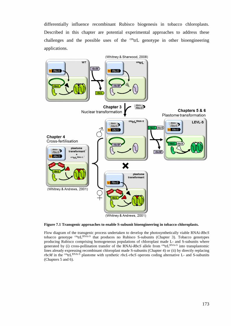
173
differentially influence recombinant Rubisco biogenesis in tobacco chloroplasts.
Described in this chapter are potential experimental approaches to address these
challenges and the possible uses of the cmtrL genotype in other bioengineering
applications.
Figure 7.1 Transgenic approaches to enable S-subunit bioengineering in tobacco chloroplasts.
Flow diagram of the transgenic process undertaken to develop the photosynthetically viable RNAi-RbcS
tobacco genotype cmtrLRNAi-S that produces no Rubisco S-subunits (Chapter 3). Tobacco genotypes
producing Rubisco comprising homogeneous populations of chloroplast made L- and S-subunits where
generated by (i) cross-pollination transfer of the RNAi-RbcS allele from cmtrLRNAi-S into transplastomic
lines already expressing recombinant chloroplast made S-subunits (Chapter 4) or (ii) by directly replacing
rbcM in the cmtrLRNAi-S plastome with synthetic rbcL-rbcS operons coding alternative L- and S-subunits
(Chapters 5 and 6).
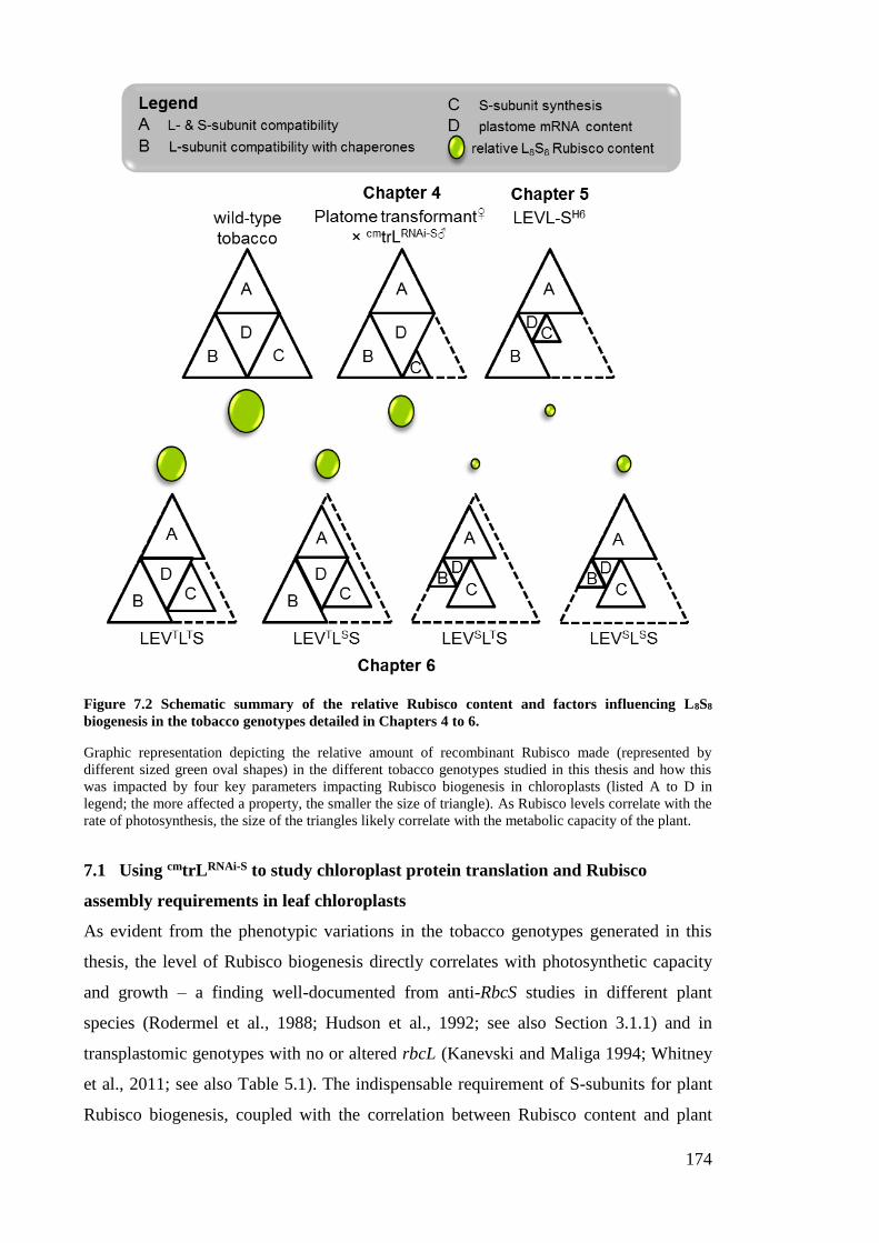
174
Figure 7.2 Schematic summary of the relative Rubisco content and factors influencing L8S8
biogenesis in the tobacco genotypes detailed in Chapters 4 to 6.
Graphic representation depicting the relative amount of recombinant Rubisco made (represented by
different sized green oval shapes) in the different tobacco genotypes studied in this thesis and how this
was impacted by four key parameters impacting Rubisco biogenesis in chloroplasts (listed A to D in
legend; the more affected a property, the smaller the size of triangle). As Rubisco levels correlate with the
rate of photosynthesis, the size of the triangles likely correlate with the metabolic capacity of the plant.
7.1 Using cmtrLRNAi-S to study chloroplast protein translation and Rubisco
assembly requirements in leaf chloroplasts
As evident from the phenotypic variations in the tobacco genotypes generated in this
thesis, the level of Rubisco biogenesis directly correlates with photosynthetic capacity
and growth – a finding well-documented from anti-RbcS studies in different plant
species (Rodermel et al., 1988; Hudson et al., 1992; see also Section 3.1.1) and in
transplastomic genotypes with no or altered rbcL (Kanevski and Maliga 1994; Whitney
et al., 2011; see also Table 5.1). The indispensable requirement of S-subunits for plant
Rubisco biogenesis, coupled with the correlation between Rubisco content and plant
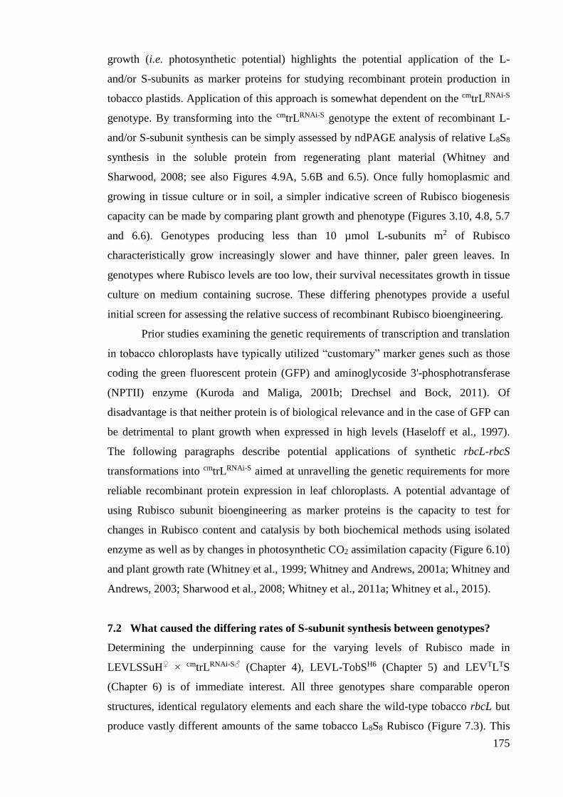
175
growth (i.e. photosynthetic potential) highlights the potential application of the L-
and/or S-subunits as marker proteins for studying recombinant protein production in
tobacco plastids. Application of this approach is somewhat dependent on the cmtrLRNAi-S
genotype. By transforming into the cmtrLRNAi-S genotype the extent of recombinant L-
and/or S-subunit synthesis can be simply assessed by ndPAGE analysis of relative L8S8
synthesis in the soluble protein from regenerating plant material (Whitney and
Sharwood, 2008; see also Figures 4.9A, 5.6B and 6.5). Once fully homoplasmic and
growing in tissue culture or in soil, a simpler indicative screen of Rubisco biogenesis
capacity can be made by comparing plant growth and phenotype (Figures 3.10, 4.8, 5.7
and 6.6). Genotypes producing less than 10 µmol L-subunits m2 of Rubisco
characteristically grow increasingly slower and have thinner, paler green leaves. In
genotypes where Rubisco levels are too low, their survival necessitates growth in tissue
culture on medium containing sucrose. These differing phenotypes provide a useful
initial screen for assessing the relative success of recombinant Rubisco bioengineering.
Prior studies examining the genetic requirements of transcription and translation
in tobacco chloroplasts have typically utilized “customary” marker genes such as those
coding the green fluorescent protein (GFP) and aminoglycoside 3'-phosphotransferase
(NPTII) enzyme (Kuroda and Maliga, 2001b; Drechsel and Bock, 2011). Of
disadvantage is that neither protein is of biological relevance and in the case of GFP can
be detrimental to plant growth when expressed in high levels (Haseloff et al., 1997).
The following paragraphs describe potential applications of synthetic rbcL-rbcS
transformations into cmtrLRNAi-S aimed at unravelling the genetic requirements for more
reliable recombinant protein expression in leaf chloroplasts. A potential advantage of
using Rubisco subunit bioengineering as marker proteins is the capacity to test for
changes in Rubisco content and catalysis by both biochemical methods using isolated
enzyme as well as by changes in photosynthetic CO2 assimilation capacity (Figure 6.10)
and plant growth rate (Whitney et al., 1999; Whitney and Andrews, 2001a; Whitney and
Andrews, 2003; Sharwood et al., 2008; Whitney et al., 2011a; Whitney et al., 2015).
7.2 What caused the differing rates of S-subunit synthesis between genotypes?
Determining the underpinning cause for the varying levels of Rubisco made in
LEVLSSuH♀ × cmtrLRNAi-S♂ (Chapter 4), LEVL-TobSH6 (Chapter 5) and LEVTLTS
(Chapter 6) is of immediate interest. All three genotypes share comparable operon
structures, identical regulatory elements and each share the wild-type tobacco rbcL but
produce vastly different amounts of the same tobacco L8S8 Rubisco (Figure 7.3). This
176
difference does not arise from intercistronic processing of their rbcL-RbcS and rbcL-
RbcS-aadA polycistronic mRNA’s. The difference in S-subunit synthesis must therefore
arise from the differences in their RbcS sequences (i.e. codon use) or/and the 18
nucleotide differences in the intergenic sequence downstream of the SD of RbcS. In the
LEVTLTS genotype this region replicated the rbcL TCR that extended into the first four
codons of S-subunit coding sequence (Figure 6.4). Potentially these sequence
differences may influence the stability of the rbcL-RbcS and rbcL-RbcS-aadA mRNAs.
Indeed the abundance of these transcripts in the genotypes producing less Rubisco were
reduced 3 to 10-fold. However, as these polycistronic mRNA’s did not undergo
intercistronic processing (i.e. RNA cutting) then they would possess identical 5′- and
3′UTR sequences. These are the regions most associated with forming stabilising
secondary structures and interacting with stabilising accessory binding proteins to
prevent exonuclease activity (Barkan et al., 1994). As discussed in Section 5.3.1, it is
more likely the reduced mRNA levels may simply arise as a consequence of the
impaired cellular viability in the genotypes producing little or no Rubisco.
Figure 7.3 Identifying the elements that increase chloroplast S-subunit synthesis.
Summary of putative follow-up plastome transformation experiments to test how differences in the RbcS
codon use and/or inclusion of the rbcL translational coding region (TCR: signified by an asterix, *) in the
intergenic sequence (IS) led to the large difference in S-subunit synthesis (and hence L8S8 Rubisco
content) in the LEVTLTS genotypes compared to the LEVLSSuH♀ × cmtrLRNAi-S♂ and LEVL-TobSH6 lines.
See text for more details.
Differences in the translational potential of the varying RbcS mRNA sequences
in the different tobacco genotypes shown in Figure 7.3 therefore seem more likely to be
responsible for the difference in S-subunit synthesis (parameter C in Figure 7.2). The
relatively high levels of S-subunits (and hence Rubisco) made in LEVTLTS indicates
that (i) RbcS in this line was efficiently translated without need of intercistronic
cleavage and (ii) its SD was efficiently recognized by the chloroplast translational
177
components. Potential starting points for testing the impact of the rbcL TCR and
differing codon use on S-subunit synthesis could commence with the generation of two
additional transplastomic lines (Figure 7.3). The importance of codon use would be
supported if Rubisco levels could be enhanced in LEVL-TobSH6 by altering the codon
use of its RbcS to match that used in LEVTLTS (option A in Figure 7.3). A reciprocal
test would involve swapping the TCR domain in LEVTLTS with the corresponding
sequence from LEVL-TobSH6 (option B in Figure 7.3). Increases in Rubisco production
in this genotype would signify the importance of translation initiation in plastid S-
subunit synthesis and that of other recombinant protein synthesis in general. The
findings of these preliminary tests would set the scene for subsequent mutagenic testing.
For example if inclusion of the TCR sequence was a key driver for increased S-subunit
synthesis, then directed mutagenesis of nucleotides in this region might help pinpoint
what nucleotide sequence(s) are essential for optimizing translation initiation.
Examining computational predictions of RNA secondary structure to ascertain
mutations that favour ribosome engagement have yet to be tested. Such tests could build
on the SD mutagenic studies of Drechsel and Bock (2011) who identified SD expression
elements useful for high-level recombinant expression.
7.3 Can chloroplast S-subunit synthesis be enhanced further?
7.3.1 Via a cmtrLRNAi-S cross pollination approach
Prior success in increasing the level of chloroplast made S-subunit synthesis was
achieved by inserting tobacco RbcS into the trnI-trnA intergenic region within the 25 kb
inverted repeat region of the plastome (Dhingra, 2004). The trnI-trnA is located between
the small (rrn16) and large (rrn23) rRNA subunit genes that are co-transcribed by
promoters upstream of rrn16 as a polycistronic rrn operon (Daniell, 2005; Lutz, 2007).
As the rrn operon mRNA is highly abundant and efficiently processed, transgenic
mRNA inserted between trnI and trnA are produced in high abundance, especially when
an additional promoter is included to drive transgene expression. Consequently,
insertion of the RbcS transgene into this region produced more RbcS mRNA than in
genotypes where RbcS was inserted into the trnV-3′rps12 region of the plastid genome
(Whitney and Andrews, 2001) using earlier generation pPRV transforming vectors
(Zoubenko, 1994). This would suggest that pollinating the two trnI-RbcS-trnA tobacco
genotypes produced by Dhingra et al. (2004) (called Nt-pLDADpsbARbcS and Nt-
pLDADg10RbcS) with pollen from cmtrLRNAi-S would produce progeny making more
tobacco Rubisco (comprising chloroplast made S- and L-subunits) relative to that made
178
in the RVtpSSuH♀ × cmtrLRNAi-S♂ plants described in Chapter 4. This experiment
remains to be undertaken.
A potential drawback of bioengineering into the trnI-trnA region is the desire
and the likely need to develop a plastome transformation process for co-engineering
changes to both rbcL and rbcS. While experimentally feasible for duplicating the copy
number of a gene of interest (GOI), the process of generating an RNAi-RbcS genotype
comparable to cmtrLRNAi-S would require a number of transgenic steps. A first step would
be to introduce an R. rubrum rbcM into the trnI-trnA region of the tobacco genome,
preferentially a genotype where rbcL has already been excised; for example the ΔrbcL
tobacco genotypes of Kode et al. (2006) or Kanevski and Maliga (1994). Recycling of
aadA would likely need to be considered for which there are now a range of strategies
available for marker gene recycling (Day and Goldschmidt-Clermont, 2011). Like the
cmtrLRNAi-S genotype made in this thesis (Chapter 3) the resulting L2-Rubisco expressing
genotype would be amenable to RNAi directed silencing of RbcS without detriment to
its phenotype. The final product would be an RNAi-RbcS genotype amenable to
plastome transformation directed replacement of the rbcM transgene in the trnI-trnA
region with rbcL-rbcS transgenes. Whether such a protracted engineering process is of
benefit will depend on whether future approaches can enhance tobacco and foreign
Rubisco production in cmtrLRNAi-S to levels comparable to wild-type. The approaches
include increasing RbcS mRNA levels, improving S-subunit translation (see Section
7.3.2) and/or enhancing foreign L-subunit assembly (see Section 7.4).
7.3.2 Stimulating rbcL-rbcS mRNA cleavage to increase RbcS mRNA production and
translation.
As discussed in see Section 5.3.2.1, possible limitations in the capacity of the
chloroplast translational machinery to recognise SD sequences within polycistronic
transcripts may explain why chloroplast polycistronic mRNA’s often undergo
intercistronic cleavage to produce monocistronic mRNAs that are more amenable to
translation (Drechsel and Bock, 2011). In support of this hypothesis, a few
transplastomic studies have found merit in using the small 50 nt RNA element coined
the intercistronic expression element (IEE, Zhou et al., 2007; see also Figure 7.4A) to
stably and correctly process di- and polycistronic transgene mRNAs (Figure 7.4B).
Transplastomic uses of the IEE include the introduction of genes into the tocopherol
pathway of tobacco and tomato chloroplasts (Lu et al., 2013) as well as helping to
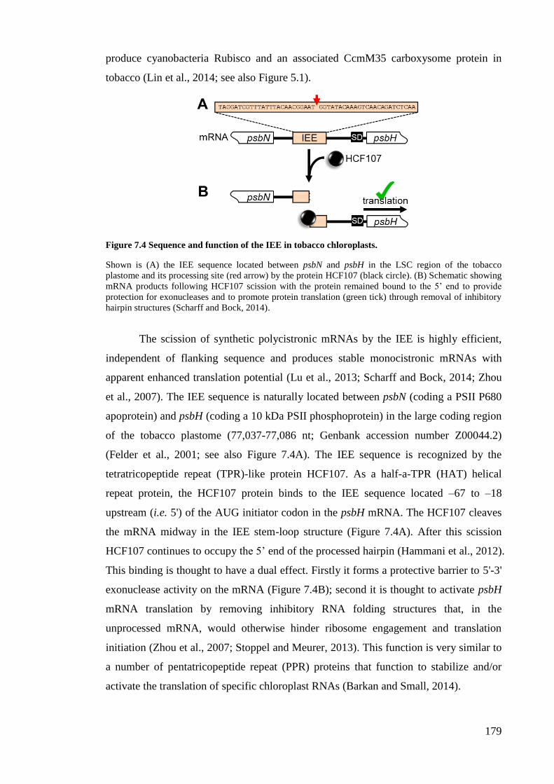
179
produce cyanobacteria Rubisco and an associated CcmM35 carboxysome protein in
tobacco (Lin et al., 2014; see also Figure 5.1).
Figure 7.4 Sequence and function of the IEE in tobacco chloroplasts.
Shown is (A) the IEE sequence located between psbN and psbH in the LSC region of the tobacco
plastome and its processing site (red arrow) by the protein HCF107 (black circle). (B) Schematic showing
mRNA products following HCF107 scission with the protein remained bound to the 5’ end to provide
protection for exonucleases and to promote protein translation (green tick) through removal of inhibitory
hairpin structures (Scharff and Bock, 2014).
The scission of synthetic polycistronic mRNAs by the IEE is highly efficient,
independent of flanking sequence and produces stable monocistronic mRNAs with
apparent enhanced translation potential (Lu et al., 2013; Scharff and Bock, 2014; Zhou
et al., 2007). The IEE sequence is naturally located between psbN (coding a PSII P680
apoprotein) and psbH (coding a 10 kDa PSII phosphoprotein) in the large coding region
of the tobacco plastome (77,037-77,086 nt; Genbank accession number Z00044.2)
(Felder et al., 2001; see also Figure 7.4A). The IEE sequence is recognized by the
tetratricopeptide repeat (TPR)-like protein HCF107. As a half-a-TPR (HAT) helical
repeat protein, the HCF107 protein binds to the IEE sequence located –67 to –18
upstream (i.e. 5') of the AUG initiator codon in the psbH mRNA. The HCF107 cleaves
the mRNA midway in the IEE stem-loop structure (Figure 7.4A). After this scission
HCF107 continues to occupy the 5’ end of the processed hairpin (Hammani et al., 2012).
This binding is thought to have a dual effect. Firstly it forms a protective barrier to 5'-3'
exonuclease activity on the mRNA (Figure 7.4B); second it is thought to activate psbH
mRNA translation by removing inhibitory RNA folding structures that, in the
unprocessed mRNA, would otherwise hinder ribosome engagement and translation
initiation (Zhou et al., 2007; Stoppel and Meurer, 2013). This function is very similar to
a number of pentatricopeptide repeat (PPR) proteins that function to stabilize and/or
activate the translation of specific chloroplast RNAs (Barkan and Small, 2014).

180
As outlined in Figure 7.5A, exploiting the potential benefits of using the IEE
sequence to enhance chloroplast S-subunit synthesis needs to be tested in cmtrLRNAi-S.
Initial testing would focus on generating a transplastomic line where the intergenic
sequence used in LEVTLTS is replaced with a terminator sequence and IEE sequence
(Figure 7.5A). Tested example terminator sequences that could be introduced
downstream of tobacco rbcL include the rbcL 3’UTR sequence from Chlamydomonas
reinhardtii (Lu et al., 2013), the 3’UTR sequence from Arabidopsis rps16 (Kuroda and
Sugiura, 2014) or psbA (Coloapa-Soto et al., 2012). A comparison of the RbcS and rbcL
mRNA levels and L8S8 Rubisco contents relative to those made in the LEVTLTS
genotype would be used to gauge the relative merit of using the IEE transgenic
approach to enhancing chloroplast S-subunit synthesis in cmtrLRNAi-S.
Figure 7.5 Increasing recombinant Rubisco biogenesis in cmtrLRNAi-S.
Possible future strategies to test how to improve L- and S-subunit synthesis in chloroplasts include (A)
increasing S-subunit production by replacing the synthetic intergenic sequence (IS*) between rbcL and
synthetic RbcS in LEVTLTS with a ‘weak’ terminator (T, light green) and IEE sequence (see text in
Section 7.3.2 for more detail). (B) Strategies for increasing foreign L-subunit assembly include co-
transforming in genes coding a cognate S-subunit (FRbcS) and one or more complimentary assembly
chaperones (e.g. RAF1, RAF2, BSD2, RbcX,) or chaperonin subunit (Cpn60α) (see Figure 4.3 and
Sections 6.3.1 and 7.4 for more details). Testing the efficiency of transgene expression and Rubisco
production might compare transplastomic lines producing a polycistronic FrbcL-FRbcS-Fraf1 mRNA
(example [i]), separate and inverted genes coding FrbcL-FRbcS and Fraf1-aadA mRNAs (example [ii]) or
monocistronic FrbcL, FRbcS and Fraf1 mRNA’s by incorporating differing “weak” terminator (shaded
light green and blue) and IEE sequences between the transgenes (examples [iii] and [iv]) to increase
mRNA stability and enhance translation.
181
7.4 Enhancing L-subunit synthesis to increase recombinant L8S8 assembly
A key impediment to foreign L-subunit synthesis in tobacco chloroplasts appears
associated with differences in their folding and assembly requirements. As indicated in
Section 5.1.1, the folding and assembly requirements of Rubisco subunits from algae
cannot be met in leaf chloroplasts (Whitney and Andrews, 2001b). Even L-subunits
derived from plant species show differing levels of compatibility with assembling in
tobacco chloroplasts (Parry et al., 2013) – in some cases due to single amino acid
differences (Whitney et al., 2011b). One reason for these incompatibilities stem from
differences in the chaperoning requirements between species. Bioinformatics sequence
comparisons in a recent study showed strong complementarity in the evolution
pathways of the L-subunit and its assembly chaperone RAF1 (Whitney et al., 2015).
This was experimentally tested using a transplastomic approach to produce genotypes
expressing Arabidopsis Rubisco L-subunits alone or in combination with a
complementary RAF1. As detailed in Section 6.1.3, Rubisco biogenesis was enhanced
in the genotypes co-producing the cognate Arabidopsis RAF1 despite the very low
amounts of polycistronic rbcL-raf1-aadA mRNA produced (Whitney et al., 2015). This
demonstrated the L-subunit assembly role of RAF1 and how foreign Rubisco biogenesis
in tobacco leaves can be enhanced by its co-expression.
Increasing (or enabling) foreign Rubisco biogenesis in tobacco chloroplasts
clearly needs to address the requirements for co-expression of complementary folding
and assembly chaperone(s)/chaperonin proteins. In addition to RAF1, these might
include other Rubisco specialized chaperones such as RAF2, BSD2, RbcX (Feiz et al.,
2012; Parry et al., 2013; Feiz et al., 2014; see also Figure 4.3 and Section 6.3.1) and
possibly the chloroplast targeted chaperonin 60 alpha subunit (Cpn60α) (Barkan, 1993;
Kim et al., 2013; see also Figure 7.5B). Four possible plastome transformation
approaches for co-introducing genes of foreign Rubisco subunits and their homologous
chaperones into the cmtrLRNAi-S plastome are shown in Figure 7.5B. These include
replacing rbcM in cmtrLRNAi-S with a genetic structure that produces either a single
FrbcL-FRbcS-Fchaperone1 polycistronic mRNA and independent aadA mRNA (option
[i]) or separate FrbcL-FRbcS and Fchaperone1-aadA mRNAs (option [ii]). Alternative
options might be to integrate “weak” terminator (shaded light green and blue) and IEE
sequences (options [iii] and [iv]) to produce monocistronic FrbcL, FRbcS and
Fchaperone mRNA’s. A comparison of mRNA levels and Rubisco content in all four
genotypes would determine to what extent (1) incorporating the T-IEE sequences
enhanced transgene mRNA content and their translation and (2) which chaperones can
182
augment foreign Rubisco biogenesis in tobacco leaf chloroplasts. This approach would
identify the ancillary proteins considered vital for Rubisco biogenesis in plastids and
provide potential genetic approaches for transforming the genes for one or more of these
proteins with the rbcL and RbcS of their cognate Rubisco.
7.5 The effect of the S-subunit on catalysis
As discussed in Section 6.3.2, sequence differences between heterologous L- and S-
subunits can impede L8S8 biogenesis, but not necessarily catalysis. As shown in Table
6.2, L8S8 Rubisco comprising tobacco L-subunits and either 6×His-tagged tobacco or
sunflower S-subunits share the same catalytic properties. As summarized in Table 5.1,
this finding is akin to finding that the assembly of tobacco S-subunits with L-subunits
from sunflower, Flaveria and tomato Rubisco have little or no influence on catalysis
(Kanevski et al., 1999; Whitney et al., 2001b; Zhang et al., 2011). More recently
however the assembly of tobacco S-subunits with Arabidopsis L-subunits was found to
slow catalysis by ~25% (Whitney et al., 2015). These contrasting influences of
heterologous S-subunits on hybrid Rubisco catalysis highlight the need to better
understand the structure-function interactions between L- and S-subunits.
Analyses of crystal structure information for varying L8S8 isoforms provide
valuable insights into amino acid interactions at the L/L-, L/S- and S/S-subunit
interfaces (van Lun et al., 2011). A deficiency of such structural analyses is being able
to ascertain the functional significance of these interactions and any subunit sequence
differences. This requires experimental testing. As presented in Section 1.2.2, the
majority of mutagenic studies examining S- and L-subunit interactions have been
undertaken using Chlamydomonas reinhardtii (Spreitzer, 2003; Karkehabadi et al.,
2005; Genkov and Spreitzer, 2009) or forms that can be functionally expressed in E.
coli (e.g. Rubisco from cyanobacteria and photosynthetic proteobacteria) (Read and
Tabita, 1994; Kellogg and Juliano, 1997; Joshi et al., 2015). The cmtrLRNAi-S genotype
developed in thesis however now provides a viable opportunity to start expanding these
structure-function studies to the L- and S-subunits of higher plant Rubisco (although
likely with some optimization as described above in Sections 7.3 and 7.4). The proven
efficiency and stability of the RNAi-RbcS silencing of S-subunit synthesis through
multiple generations of the cmtrLRNAi-S genotype (Figure 3.9) is of particular benefit as it
negates the need to include an affinity tag on the recombinant S-subunit, which was
shown to slow Rubisco catalysis by ~one-third (see Section 6.3.3).
183
While the cmtrLRNAi-S genotype poses new opportunities to surgically unravel L-
and S-subunit structural interactions via targeted mutagenesis using chloroplast
transformation, the challenge is to identify the starting point(s) for this mutagenic
testing. The work of Spreitzer et al. (2005) highlights the potential benefit of altering
Chlamydomonas Rubisco catalysis by directed changes to the S-subunit βA/βB loop and
the adjoining L-subunit amino acids. Identifying changes to make in the comparable
regions of higher plant Rubisco L- and S-subunits poses a potentially good starting
point – if viable alternatives were apparent. Identifying beneficial amino acid
modifications may however require a more strategic approach. For example a more
detailed study of the natural variation in Rubisco catalysis and their L- and S-subunit
sequences may provide better guidance on amino acid mutations to test (Parry et al.,
2013). A more focused approach may simply concentrate on the significance of the
natural S-subunit diversity within a species. For example, the four S-subunit isoforms
produced in Arabidopsis (that are each 123 amino acids in length) vary by up to 8
amino acids (The Arabidopsis Initiative, 2000). Whether these S-subunit differences
alter Rubisco catalysis remains unknown. A preliminary analysis of the Phytozome
plant genome database (http://www.phytozome.net) for RbcS sequences in eucalyptus
(Eucalyptus grandis), potato (Solanum tuberosum) and cassava (Manihot esculenta)
(representative species whose L-subunits can fold and assemble in tobacco chloroplasts)
(Kapralov and Whitney, unpublished) identified varying degrees of sequence diversity
in their mature S-subunits (Figure 7.6). The number of RbcS alleles were identified: five
in eucalyptus, four in potato and three in cassava. A comparison of the translated amino
acid sequence for the mature coding region of each allele found they each coded
different S-subunit sequences of varying diversity. The S-subunits in cassava show
between 54 and 87% identity, in potato between 59 and 96% identity and in eucalyptus
between 59 and 89% identity (Figure 7.6). This large sequence variation in S-subunit
sequence within these three species contrasts with the modest amino acid diversity (i.e.
only 7.3%) found among the 9 different S-subunit isoforms produced in tobacco (N.
tabacum) (Figure 3.6B).
Compelling questions that remain untested include how the S-subunit sequence
diversity in eucalyptus, potato and cassava (and to a lesser extent Arabidopsis and
tobacco) influence the catalytic properties of plant Rubisco and can this help identify
catalytic switches in S-subunits and/or L-subunits? Testing these questions is now
experimentally feasible using the cmtrLRNAi-S genotype given its capacity to produce
L8S8 Rubisco comprising a homogeneous population of L- and S-subunits – both made
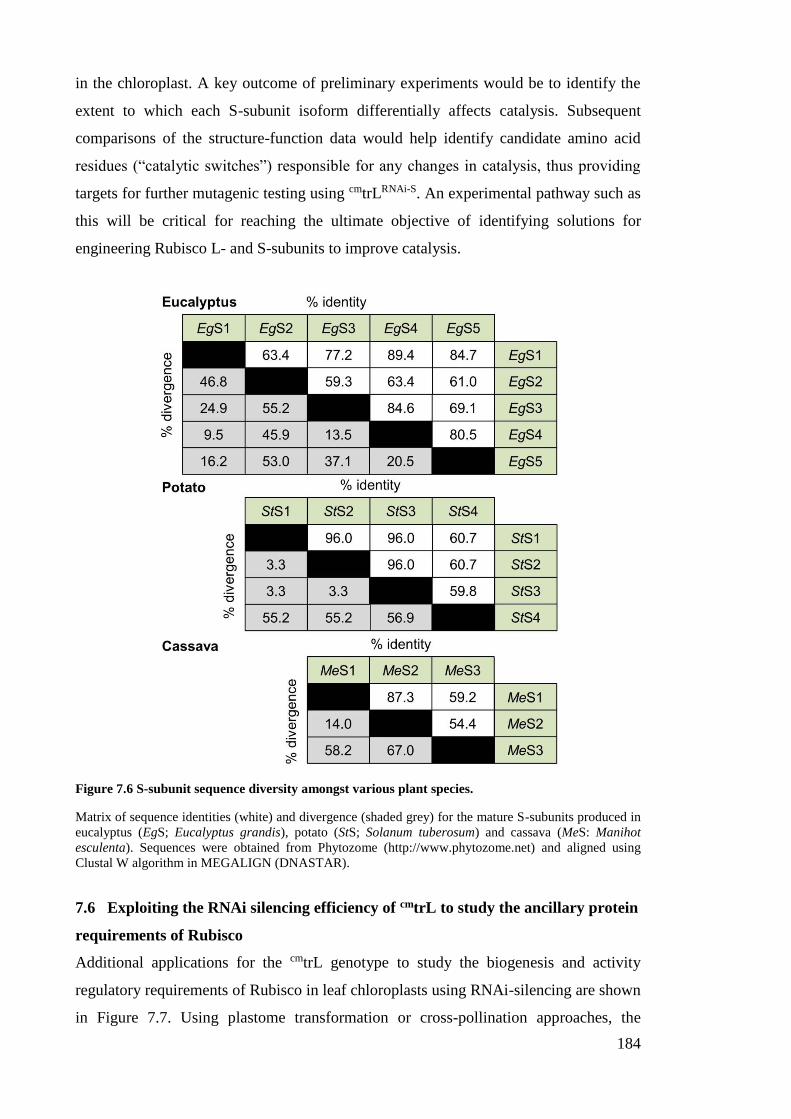
184
in the chloroplast. A key outcome of preliminary experiments would be to identify the
extent to which each S-subunit isoform differentially affects catalysis. Subsequent
comparisons of the structure-function data would help identify candidate amino acid
residues (“catalytic switches”) responsible for any changes in catalysis, thus providing
targets for further mutagenic testing using cmtrLRNAi-S. An experimental pathway such as
this will be critical for reaching the ultimate objective of identifying solutions for
engineering Rubisco L- and S-subunits to improve catalysis.
Figure 7.6 S-subunit sequence diversity amongst various plant species.
Matrix of sequence identities (white) and divergence (shaded grey) for the mature S-subunits produced in
eucalyptus (EgS; Eucalyptus grandis), potato (StS; Solanum tuberosum) and cassava (MeS: Manihot
esculenta). Sequences were obtained from Phytozome (http://www.phytozome.net) and aligned using
Clustal W algorithm in MEGALIGN (DNASTAR).
7.6 Exploiting the RNAi silencing efficiency of cmtrL to study the ancillary protein
requirements of Rubisco
Additional applications for the cmtrL genotype to study the biogenesis and activity
regulatory requirements of Rubisco in leaf chloroplasts using RNAi-silencing are shown
in Figure 7.7. Using plastome transformation or cross-pollination approaches, the
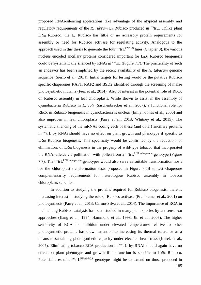
185
proposed RNAi-silencing applications take advantage of the atypical assembly and
regulatory requirements of the R. rubrum L2 Rubisco produced in cmtrL. Unlike plant
L8S8 Rubisco, the L2 Rubisco has little or no accessory protein requirements for
assembly or need for Rubisco activase for regulating activity. Analogous to the
approach used in this thesis to generate the four cmtrLRNAi-S lines (Chapter 3), the various
nucleus encoded ancillary proteins considered important for L8S8 Rubisco biogenesis
could be systematically silenced by RNAi in cmtrL (Figure 7.7). The practicality of such
an endeavor has been simplified by the recent availability of the N. tabacum genome
sequence (Sierro et al., 2014). Initial targets for testing would be the putative Rubisco
specific chaperones RAF1, RAF2 and BSD2 identified through the screening of maize
photosynthetic mutants (Feiz et al., 2014). Also of interest is the potential role of RbcX
on Rubisco assembly in leaf chloroplasts. While shown to assist in the assembly of
cyanobacteria Rubisco in E. coli (Saschenbrecker et al., 2007), a functional role for
RbcX in Rubisco biogenesis in cyanobacteria is unclear (Emlyn-Jones et al., 2006) and
also unproven in leaf chloroplasts (Parry et al., 2013; Whitney et al., 2015). The
systematic silencing of the mRNAs coding each of these (and other) ancillary proteins
in cmtrL by RNAi should have no effect on plant growth and phenotype if specific to
L8S8 Rubisco biogenesis. This specificity would be confirmed by the reduction, or
elimination, of L8S8 biogenesis in the progeny of wild-type tobacco that incorporated
the RNAi-alleles via pollination with pollen from a cmtrLRNAi-chaperone genotype (Figure
7.7). The cmtrLRNAi-chaperone genotypes would also serve as suitable transformation hosts
for the chloroplast transformation tests proposed in Figure 7.5B to test chaperone
complementarity requirements for heterologous Rubisco assembly in tobacco
chloroplasts subunits.
In addition to studying the proteins required for Rubisco biogenesis, there is
increasing interest in studying the role of Rubisco activase (Premkumar et al., 2001) on
photosynthesis (Parry et al., 2013; Carmo-Silva et al., 2014). The importance of RCA in
maintaining Rubisco catalysis has been studied in many plant species by antisense-rca
approaches (Jiang et al., 1994; Hammond et al., 1998; Jin et al., 2006). The higher
sensitivity of RCA to inhibition under elevated temperatures relative to other
photosynthetic proteins has drawn attention to increasing its thermal tolerance as a
means to sustaining photosynthetic capacity under elevated heat stress (Kurek et al.,
2007). Eliminating tobacco RCA production in cmtrL by RNAi should again have no
effect on plant phenotype and growth if its function is specific to L8S8 Rubisco.
Potential uses of a cmtrLRNAi-RCA genotype might be to extend on those proposed in
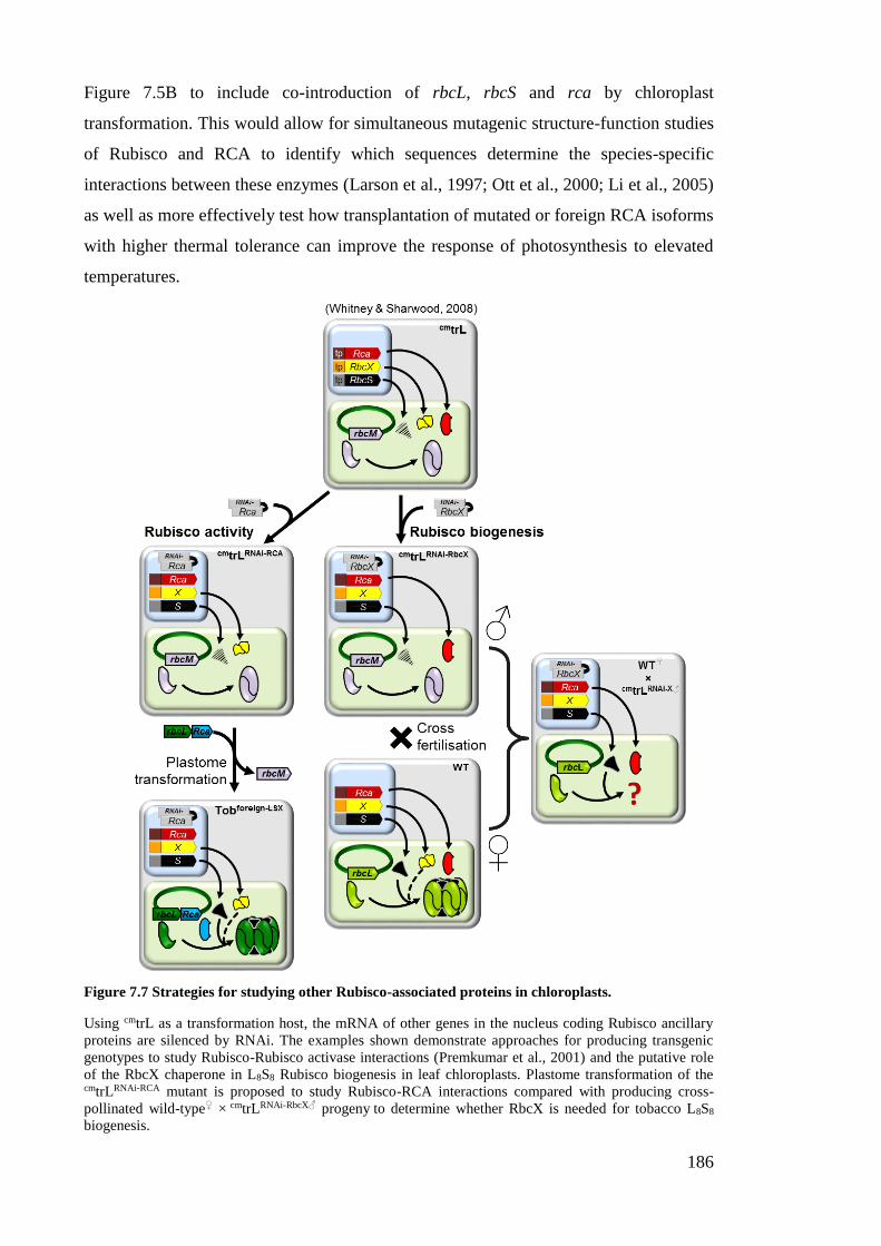
186
Figure 7.5B to include co-introduction of rbcL, rbcS and rca by chloroplast
transformation. This would allow for simultaneous mutagenic structure-function studies
of Rubisco and RCA to identify which sequences determine the species-specific
interactions between these enzymes (Larson et al., 1997; Ott et al., 2000; Li et al., 2005)
as well as more effectively test how transplantation of mutated or foreign RCA isoforms
with higher thermal tolerance can improve the response of photosynthesis to elevated
temperatures.
Figure 7.7 Strategies for studying other Rubisco-associated proteins in chloroplasts.
Using cmtrL as a transformation host, the mRNA of other genes in the nucleus coding Rubisco ancillary
proteins are silenced by RNAi. The examples shown demonstrate approaches for producing transgenic
genotypes to study Rubisco-Rubisco activase interactions (Premkumar et al., 2001) and the putative role
of the RbcX chaperone in L8S8 Rubisco biogenesis in leaf chloroplasts. Plastome transformation of the cmtrLRNAi-RCA mutant is proposed to study Rubisco-RCA interactions compared with producing cross-
pollinated wild-type♀ × cmtrLRNAi-RbcX♂ progeny to determine whether RbcX is needed for tobacco L8S8
biogenesis.
187
7.7 What is the minimal Rubisco amount needed to grow tobacco in soil?
A question among Rubisco bioengineers often raised is the lower limit of Rubisco
needed for plants to be grown in soil under elevated CO2. Obviously this will vary
depending on the catalytic properties of the Rubisco in question (i.e. such that the
slower the enzyme, the more that will be needed). Indeed one objective of putting a
faster, more efficient Rubisco into plants is so less of the enzyme would be required to
maintain current photosynthetic rates. This is seen as a strategy for potentially
improving the nitrogen use of the plant (see Section 1.3.1) as seen in C4 plants
(Ghannoum et al., 2005). In the context of this thesis’s findings and those of other
transplastomic studies (Table 5.1 and Figure 6.1) the minimal amount of tobacco
Rubisco (or catalytically comparable isoform) required for growth in soil at high-CO2 is
~1 µmol Rubisco catalytic sites m2. This value is derived from the observed success in
being able to grow the RVSSuH♀ × cmtrLRNAi-S♂ genotype in soil under high-CO2
(Figure 4.8C, ~1.2 µmol Rubisco catalytic sites m2; see also Figure 4.9B) but not the
LEVL-TobSH6 lines (Figure 5.7, <0.3 µmol Rubisco catalytic sites m2; see also Figure
5.8B). Notably the growth rate of tobacco producing 1 µmol Rubisco catalytic sites m2
would be very slow, even in air with 2% (v/v) CO2 (Figure 4.8C).
7.8 Concluding remarks
Efforts to increase photosynthesis by modifying Rubisco catalysis have gained great
momentum over recent years with the development of novel plastome transformation
techniques and expansion of the technology to a range of crop plants (Whitney et al.,
2011a; Parry et al., 2013). This thesis describes the development of new, innovative
plastome transformation capabilities that will greatly aid these goals. The new cmtrLRNAi-
S genotype (or “2nd generation master-line”) (Whitney and Sharwood, 2008) opens up
new avenues for targeted studies of both the L- and S-subunits of plant Rubisco. With
further experimental refinement of the transformation process (see Section 7.3), and
better understanding of Rubisco biogenesis (see Sections 7.4 and 7.6), the cmtrLRNAi-S
genotype promises new frontiers in the bioengineering of Rubisco in higher plant
chloroplasts.
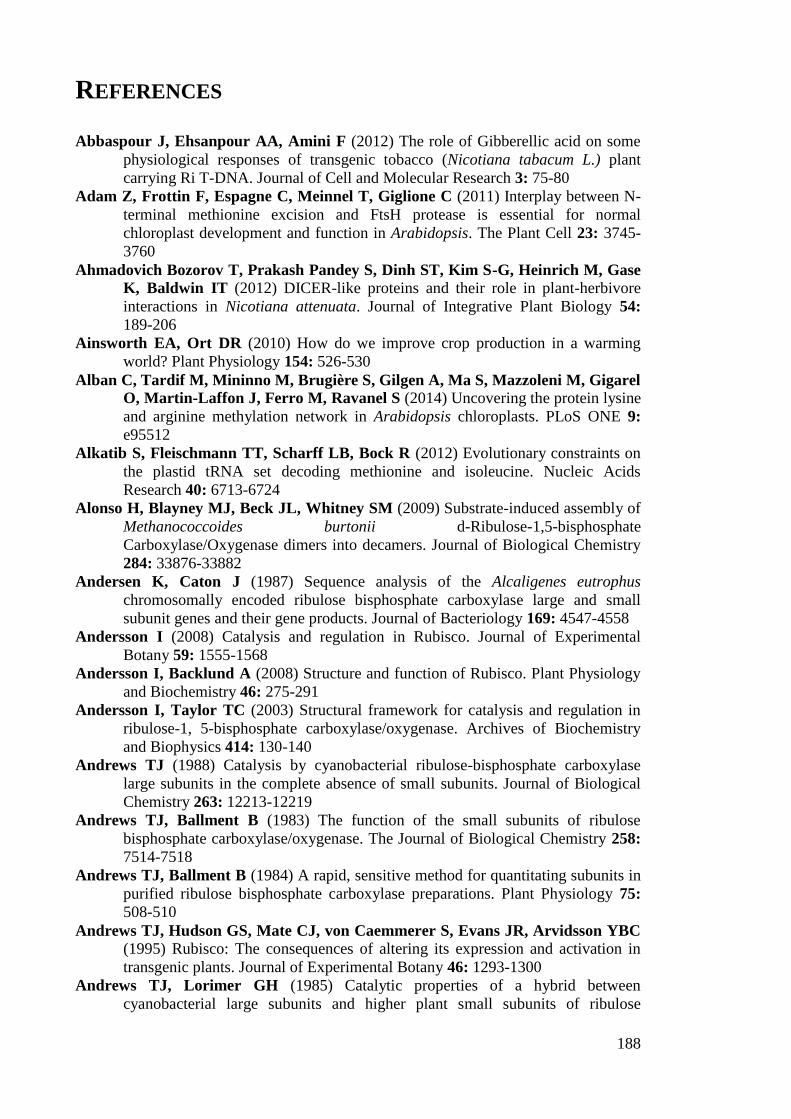
188
REFERENCES
Abbaspour J, Ehsanpour AA, Amini F (2012) The role of Gibberellic acid on some
physiological responses of transgenic tobacco (Nicotiana tabacum L.) plant
carrying Ri T-DNA. Journal of Cell and Molecular Research 3: 75-80
Adam Z, Frottin F, Espagne C, Meinnel T, Giglione C (2011) Interplay between N-
terminal methionine excision and FtsH protease is essential for normal
chloroplast development and function in Arabidopsis. The Plant Cell 23: 3745-
3760
Ahmadovich Bozorov T, Prakash Pandey S, Dinh ST, Kim S-G, Heinrich M, Gase
K, Baldwin IT (2012) DICER-like proteins and their role in plant-herbivore
interactions in Nicotiana attenuata. Journal of Integrative Plant Biology 54:
189-206
Ainsworth EA, Ort DR (2010) How do we improve crop production in a warming
world? Plant Physiology 154: 526-530
Alban C, Tardif M, Mininno M, Brugière S, Gilgen A, Ma S, Mazzoleni M, Gigarel
O, Martin-Laffon J, Ferro M, Ravanel S (2014) Uncovering the protein lysine
and arginine methylation network in Arabidopsis chloroplasts. PLoS ONE 9:
e95512
Alkatib S, Fleischmann TT, Scharff LB, Bock R (2012) Evolutionary constraints on
the plastid tRNA set decoding methionine and isoleucine. Nucleic Acids
Research 40: 6713-6724
Alonso H, Blayney MJ, Beck JL, Whitney SM (2009) Substrate-induced assembly of
Methanococcoides burtonii d-Ribulose-1,5-bisphosphate
Carboxylase/Oxygenase dimers into decamers. Journal of Biological Chemistry
284: 33876-33882
Andersen K, Caton J (1987) Sequence analysis of the Alcaligenes eutrophus
chromosomally encoded ribulose bisphosphate carboxylase large and small
subunit genes and their gene products. Journal of Bacteriology 169: 4547-4558
Andersson I (2008) Catalysis and regulation in Rubisco. Journal of Experimental
Botany 59: 1555-1568
Andersson I, Backlund A (2008) Structure and function of Rubisco. Plant Physiology
and Biochemistry 46: 275-291
Andersson I, Taylor TC (2003) Structural framework for catalysis and regulation in
ribulose-1, 5-bisphosphate carboxylase/oxygenase. Archives of Biochemistry
and Biophysics 414: 130-140
Andrews TJ (1988) Catalysis by cyanobacterial ribulose-bisphosphate carboxylase
large subunits in the complete absence of small subunits. Journal of Biological
Chemistry 263: 12213-12219
Andrews TJ, Ballment B (1983) The function of the small subunits of ribulose
bisphosphate carboxylase/oxygenase. The Journal of Biological Chemistry 258:
7514-7518
Andrews TJ, Ballment B (1984) A rapid, sensitive method for quantitating subunits in
purified ribulose bisphosphate carboxylase preparations. Plant Physiology 75:
508-510
Andrews TJ, Hudson GS, Mate CJ, von Caemmerer S, Evans JR, Arvidsson YBC (1995) Rubisco: The consequences of altering its expression and activation in
transgenic plants. Journal of Experimental Botany 46: 1293-1300
Andrews TJ, Lorimer GH (1985) Catalytic properties of a hybrid between
cyanobacterial large subunits and higher plant small subunits of ribulose
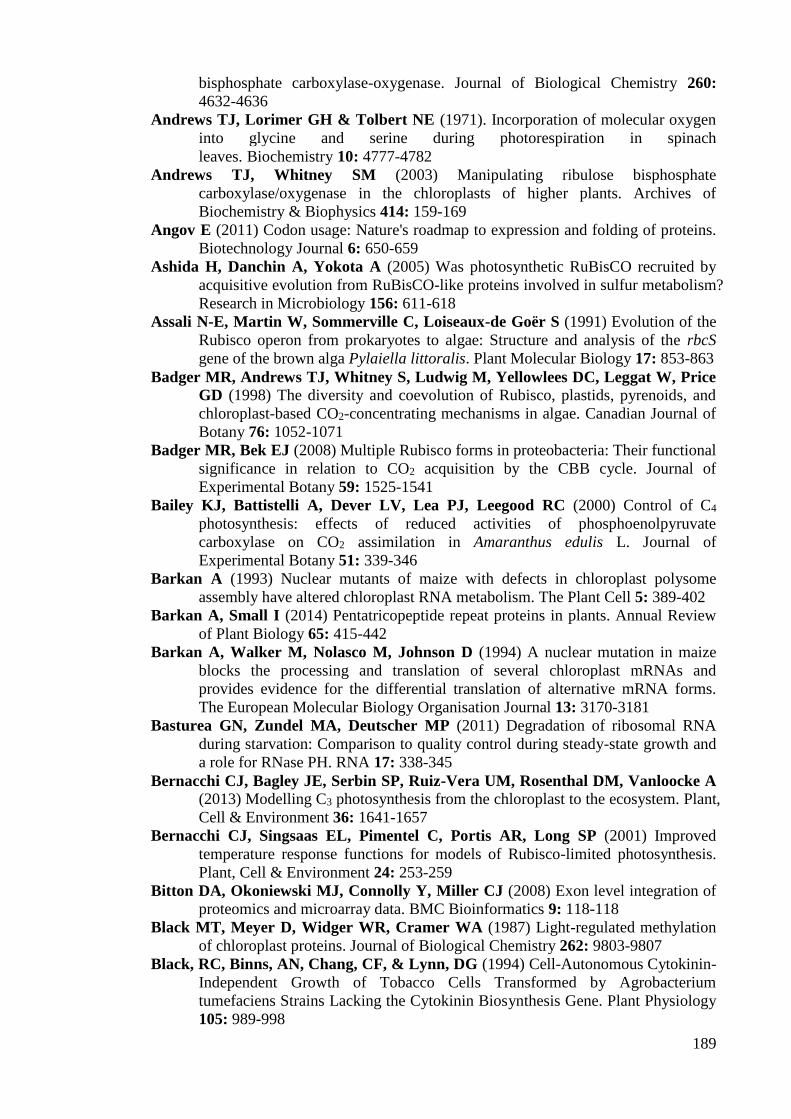
189
bisphosphate carboxylase-oxygenase. Journal of Biological Chemistry 260:
4632-4636
Andrews TJ, Lorimer GH & Tolbert NE (1971). Incorporation of molecular oxygen
into glycine and serine during photorespiration in spinach
leaves. Biochemistry 10: 4777-4782
Andrews TJ, Whitney SM (2003) Manipulating ribulose bisphosphate
carboxylase/oxygenase in the chloroplasts of higher plants. Archives of
Biochemistry & Biophysics 414: 159-169
Angov E (2011) Codon usage: Nature's roadmap to expression and folding of proteins.
Biotechnology Journal 6: 650-659
Ashida H, Danchin A, Yokota A (2005) Was photosynthetic RuBisCO recruited by
acquisitive evolution from RuBisCO-like proteins involved in sulfur metabolism?
Research in Microbiology 156: 611-618
Assali N-E, Martin W, Sommerville C, Loiseaux-de Goër S (1991) Evolution of the
Rubisco operon from prokaryotes to algae: Structure and analysis of the rbcS
gene of the brown alga Pylaiella littoralis. Plant Molecular Biology 17: 853-863
Badger MR, Andrews TJ, Whitney S, Ludwig M, Yellowlees DC, Leggat W, Price
GD (1998) The diversity and coevolution of Rubisco, plastids, pyrenoids, and
chloroplast-based CO2-concentrating mechanisms in algae. Canadian Journal of
Botany 76: 1052-1071
Badger MR, Bek EJ (2008) Multiple Rubisco forms in proteobacteria: Their functional
significance in relation to CO2 acquisition by the CBB cycle. Journal of
Experimental Botany 59: 1525-1541
Bailey KJ, Battistelli A, Dever LV, Lea PJ, Leegood RC (2000) Control of C4
photosynthesis: effects of reduced activities of phosphoenolpyruvate
carboxylase on CO2 assimilation in Amaranthus edulis L. Journal of
Experimental Botany 51: 339-346
Barkan A (1993) Nuclear mutants of maize with defects in chloroplast polysome
assembly have altered chloroplast RNA metabolism. The Plant Cell 5: 389-402
Barkan A, Small I (2014) Pentatricopeptide repeat proteins in plants. Annual Review
of Plant Biology 65: 415-442
Barkan A, Walker M, Nolasco M, Johnson D (1994) A nuclear mutation in maize
blocks the processing and translation of several chloroplast mRNAs and
provides evidence for the differential translation of alternative mRNA forms.
The European Molecular Biology Organisation Journal 13: 3170-3181
Basturea GN, Zundel MA, Deutscher MP (2011) Degradation of ribosomal RNA
during starvation: Comparison to quality control during steady-state growth and
a role for RNase PH. RNA 17: 338-345
Bernacchi CJ, Bagley JE, Serbin SP, Ruiz-Vera UM, Rosenthal DM, Vanloocke A (2013) Modelling C3 photosynthesis from the chloroplast to the ecosystem. Plant,
Cell & Environment 36: 1641-1657
Bernacchi CJ, Singsaas EL, Pimentel C, Portis AR, Long SP (2001) Improved
temperature response functions for models of Rubisco-limited photosynthesis.
Plant, Cell & Environment 24: 253-259
Bitton DA, Okoniewski MJ, Connolly Y, Miller CJ (2008) Exon level integration of
proteomics and microarray data. BMC Bioinformatics 9: 118-118
Black MT, Meyer D, Widger WR, Cramer WA (1987) Light-regulated methylation
of chloroplast proteins. Journal of Biological Chemistry 262: 9803-9807
Black, RC, Binns, AN, Chang, CF, & Lynn, DG (1994) Cell-Autonomous Cytokinin-
Independent Growth of Tobacco Cells Transformed by Agrobacterium
tumefaciens Strains Lacking the Cytokinin Biosynthesis Gene. Plant Physiology
105: 989-998

190
Blankenship RE (2002) Molecular mechanisms of photosynthesis. Blackwell Science,
Cornwall, Great Britain.
Blayney MJ, Whitney SM, Beck JL (2011) NanoESI mass spectrometry of Rubisco
and Rubisco activase structures and their interactions with nucleotides and sugar
phosphates. Journal of the American Society for Mass Spectrometry 22: 1588-
1601
Blevins T, Rajeswaran R, Shivaprasad PV, Beknazariants D, Si-Ammour A, Park
H-S, Vazquez F, Robertson D, Frederick Meins J, Hohn T, Pooggin MM (2006) Four plant Dicers mediate viral small RNA biogenesis and DNA virus
induced silencing. Nucleic Acids Research 34: 6233–6246
Block MD, Botterman J, Vandewiele M, Dockx J, Thoen C, Gosselé V, Movva NR,
Thompson C, Montagu MV, Leemans J (1987) Engineering herbicide
resistance in plants by expression of a detoxifying enzyme. The European
Molecular Biology Organisation Journal 6: 2513-2518
Bogorad L, Vasil IK (1991) The photosynthetic apparatus: Molecular biology and
operation, Vool 7B. Elsevier Science, San Diego, California.
Bonham-Smith PC, Bourque DP (1989) Translation of chloroplast-encoded mRNA:
Potential initiation and termination signals. Nucleic Acids Research 17: 2057-
2080
Bowes G, Ogren WL, Hageman RH (1971) Phosphoglycolate production catalyzed by
ribulose diphosphate carboxylase. Biochemical and Biophysical Research
Communications 45: 716-722
Boynton JE, Gillham NW, Harris EH, Hosler JP, Johnson AM, Jones AR,
Randolph. BL, Robertso. D, Klein TM, Shark KB (1988) Chloroplast
transformation in Chlamydomonas with high-velocity microprojectiles. Science
240: 1534-1538
Bracher A, Sharma A, Starling-Windhof A, Hartl FU, Hayer-Hartl M (2015)
Degradation of potent Rubisco inhibitor by selective sugar phosphatase. Nature
Plants 1
Bracher A, Starling-Windhof A, Hartl FU, Hayer-Hartl M (2011) Crystal structure
of a chaperone-bound assembly intermediate of form I Rubisco. Nature
Structural & Molecular Biology 18: 875-880
Bräsen C, Esser D, Rauch B, Siebers B (2014) Carbohydrate metabolism in archaea:
Current insights into unusual enzymes and pathways and their regulation.
Microbiology and Molecular Biology Reviews 78: 89-175
Brutnell TP, Sawers RJH, Mant A, Langdale JA (1999) BUNDLE SHEATH
DEFECTIVE2, a novel protein required for post-translational regulation of the
rbcL gene of maize. Plant Cell 11: 849-864
Buchan JR, Stansfield I (2007) Halting a cellular production line: Responses to
ribosomal pausing during translation. Biology of the Cell 99: 475-487
Carmo-Silva E, Scales JC, Madgwick PJ, Parry MAJ (2014) Optimizing Rubisco
and its regulation for greater resource use efficiency. Plant, Cell & Environment
Chen C, Wang J, Zhao X (2016). Leaf senescence induced by EGY1 defection was
partially restored by glucose in Arabidopsis thaliana. Botanical Studies, 57: 1-9.
Chen K, Dorlhac de Borne F, Szegedi E, Otten L (2014) Deep sequencing of the
ancestral tobacco species Nicotiana tomentosiformis reveals multiple T‐DNA
inserts and a complex evolutionary history of natural transformation in the genus
Nicotiana. The Plant Journal 80: 669-682
Chen Y-C, Aravin A (2015) Non-coding RNAs in transcriptional regulation. Current
Molecular Biology Reports: 1-9
191
Choquet Y, Wollman F-A (2002) Translational regulations as specific traits of
chloroplast gene expression. Federation of European Biochemical Societies
Letters 529: 39-42
Christie M, Croft LJ, Carroll BJ (2011) Intron splicing suppresses RNA silencing in
Arabidopsis. The Plant Journal 68: 159-167
Coate JE, Schlueter JA, Whaley AM, Doyle JJ (2011) Comparative evolution of
photosynthetic genes in response to polyploid and nonpolyploid duplication.
Plant Physiology 155: 2081-2095
Coloapa-Soto D, Vargas-Suárez M, Loza-Tavera H (2012) Purification of an
Arabidopsis chloroplast extract with in vitro RNA processing activity on psbA
and petD 3′-untranslated regions. Journal of Plant Physiology 169: 429-433
Daniell H, Kumar S, Dufourmantel N (2005) Breakthrough in chloroplast genetic
engineering of agronomically important crops. Trends in Biotechnology 23:
238-245
Day A, Goldschmidt-Clermont M (2011) The chloroplast transformation toolbox:
Selectable markers and marker removal. Plant Biotechnology Journal 9: 540-
553
de Cosa B, Moar W, Lee S-B, Miller M, Daniell H (2001) Overexpression of the Bt
cry2Aa2 operon in chloroplasts leads to formation of insecticidal crystals.
Nature Biotechnology 19: 71-74
Delwiche CF (1999) Tracing the thread of plastid diversity through the tapestry of life.
The American Naturalist 154: S164-S177
Dhingra A, Portis AR, Daniell H (2004) Enhanced translation of a chloroplast-
expressed RbcS gene restores small subunit levels and photosynthesis in nuclear
RbcS antisense plants. Proceedings of the National Academy of Sciences of the
United States of America 101: 6315-6320
Dobberstein B, Blobel G, Chua NH (1977) In vitro synthesis and processing of a
putative precursor for the small subunit of ribulose-1,5-bisphosphate
carboxylase of Chlamydomonas reinhardtii. Proceedings of the National
Academy of Sciences of the United States of America 74: 1082-1085
Drechsel O, Bock R (2011) Selection of Shine-Dalgarno sequences in plastids. Nucleic
Acids Research 39: 1427-1438
Duan C-G, Wang C-H, Guo H-S (2012) Application of RNA silencing to plant disease
resistance. Silence 3: 5
Durão P, Aigner H, Nagy P, Mueller-Cajar O, Hartl FU, Hayer-Hartl M (2015)
Opposing effects of folding and assembly chaperones on evolvability of Rubisco.
Nature Chemical Biollogy 11: 148-155
Eberhard S, Finazzi G, Wollman FA (2008) The dynamics of photosynthesis. Annual
Review of Genetics 42: 463-515
Edwards GE, Franceschi VR, Voznesenskaya EV (2004) Single-cell C4
photosynthesis versus the dual-cell (Kranz) paradigm. Annual Review of Plant
Biology 55: 173-196
Eichelmann H, Talts E, Oja V, Padu E, Laisk A (2009) Rubisco in planta kcat is
regulated in balance with photosynthetic electron transport. Journal of
Experimental Botany 60: 4077-4088
Ellis RJ (1979) The most abundant protein in the world. Trends in Biochemical
Sciences 4: 241-244
Emlyn-Jones D, Woodger FJ, Price GD, Whitney SM (2006) RbcX can function as a
Rubisco chaperonin, but is non-essential in Synechococcus PCC7942. Plant and
Cell Physiology 47: 1630-1640
192
Ercolano MR, Ballvora A, Paal J, Steinbiss H-H, Salamini F, Gebhardt C (2004)
Functional complementation analysis in potato via biolistic transformation with
BAC large DNA fragments. Molecular Breeding 13: 15-22
Esau B, Snyder G, Portis Jr A (1998) Activation of ribulose-1,5-bisphosphate
carboxylase/oxygenase (Rubisco) with chimeric activase proteins.
Photosynthesis Research 58: 175-181
Evans JR (2013) Improving photosynthesis. Plant Physiology 162: 1780-1793
Farquhar GD, von Caemmerer S, Berry JA (1980) A biochemical model of
photosynthetic CO2 assimilation in leaves of C3 species. Planta 149: 78-90
Feiz L, Williams-Carrier R, Belcher S, Montano M, Barkan A, Stern DB (2014) A
protein with an inactive pterin-4a-carbinolamine dehydratase domain is required
for Rubisco biogenesis in plants. The Plant Journal 80: 862-869
Feiz L, Williams-Carrier R, Wostrikoff K, Belcher S, Barkan A, Stern DB (2012)
Ribulose-1,5-bis-phosphate carboxylase/oxygenase Accumulation Factor1 is
required for holoenzyme assembly in maize. The Plant Cell Online 24: 3435-
3446
Felder S, Meierhoff K, Sane AP, Meurer J, Driemel C, Plücken H, Klaff P, Stein B,
Bechtold N, Westhoff P (2001) The nucleus-encoded HCF107 gene of
Arabidopsis provides a link between intercistronic RNA processing and the
accumulation of translation-competent psbH transcripts in chloroplasts. The
Plant Cell 13: 2127-2142
Feller U, Anders I, Mae T (2008) Rubiscolytics: Fate of Rubisco after its enzymatic
function in a cell is terminated. Journal of Experimental Botany 59: 1615-1624
Filipenko EA, Deineko EV, Shumnyi VK (2009) Specific features of T-DNA
insertion regions in transgenic plants. Russian Journal of Genetics 45: 1289-
1301
Flores-Pérez Ú, Jarvis P (2013) Molecular chaperone involvement in chloroplast
protein import. Biochimica et Biophysica Acta - Molecular Cell Research 1833:
332-340
Fukayama H, Koga A, Hatanaka T, Misoo S (2015) Small subunit of a cold-resistant
plant, timothy, does not significantly alter the catalytic properties of Rubisco in
transgenic rice. Photosynthesis Research 124: 57-65
Furbank RT, Chitty JA, von Caemmerer S, Jenkins CLD (1996) Antisense RNA
inhibition of RbcS gene expression reduces rubisco level and photosynthesis in
the C4 plant Flaveria bidentis. Plant Physiology 111: 725-734
Furbank RT, Hatch MD (1987) Mechanism of C4 photosynthesis: The size and
composition of the inorganic carbon pool in bundle sheath cells. Plant
Physiology 85: 958-964
Galmés J, Flexas J, Keys AJ, Cifre J, Mitchell RAC, Madgwick PJ, Haslam RP,
Medrano H, Parry MAJ (2005) Rubisco specificity factor tends to be larger in
plant species from drier habitats and in species with persistent leaves. Plant, Cell
& Environment 28: 571-579
Galmés J, Kapralov MV, Andralojc PJ, Conesa MÀ, Keys AJ, Parry MAJ, Flexas
J (2014) Expanding knowledge of the Rubisco kinetics variability in plant
species: Environmental and evolutionary trends. Plant, Cell & Environment 37:
1989-2001
Genkov T, Meyer M, Griffiths H, Spreitzer RJ (2010) Functional hybrid Rubisco
enzymes with plant small subunits and algal large subunits: Engineered RbcS
cDNA for expression in Chlamydomonas Journal of Biological Chemistry 285:
19833-19841
193
Genkov T, Spreitzer RJ (2009) Highly conserved small subunit residues influence
Rubisco large subunit catalysis. Journal of Biological Chemistry 284: 30105-
30112
Getzoff TP, Zhu G, Bohnert HJ, Jensen RG (1998) Chimeric Arabidopsis thaliana
ribulose-1,5-bisphosphate carboxylase/oxygenase containing a pea small subunit
protein Is compromised in carbamylation. Plant Physiology 116: 695-702
Ghannoum O, Evans JR, Chow WS, Andrews TJ, Conroy JP, von Caemmerer S (2005) Faster Rubisco is the key to superior nitrogen-use efficiency in NADP-
Malic enzyme relative to NAD-Malic enzyme C4 grasses. Plant Physiology 137:
638-650
Ghannoum O, Evans JR, von Caemmerer S (2011) Nitrogen and water use efficiency
of C4 plants. In AS Raghavendra, RF Sage, eds, C4 Photosynthesis and related
CO2 concentrating mechanisms, Vol 32. Springer Netherlands, pp 129-146
Gleason JE, Galaleldeen A, Peterson RL, Taylor AB, Holloway SP, Waninger-
Saroni J, Cormack BP, Cabelli DE, Hart PJ, Culotta VC (2014) Candida
albicans SOD5 represents the prototype of an unprecedented class of Cu-only
superoxide dismutases required for pathogen defense. Proceedings of the
National Academy of Sciences of the United States of America 111: 5866-5871
Gong L, Olson M, Wendel JF (2014) Cytonuclear evolution of Rubisco in four
allopolyploid lineages. Molecular Biology and Evolution 31: 2624-2636
Goodin MM, Zaitlin D, Naidu RA, Lommel SA (2008) Nicotiana benthamiana: Its
history and future as a model for plant-pathogen interactions. Molecular Plant-
Microbe Interactions Journal 21: 1015-1026
Gray JC, Kung SD, Wildman SG, Sheen SJ (1974) Origin of Nicotiana tabacum L.
detected by polypeptide composition of Fraction I protein. Nature 252: 226-227
Gunn L (2014) Exploring novel in planta and in vitro approaches for bioengineering
Rubisco (Doctoral dissertation). Australian National University, Canberra
Gupta PK (2009) Cell and molecular biology. Rastogi, Meerut
Gustafsson C, Govindarajan S, Minshull J (2004) Codon bias and heterologous
protein expression. Trends in Biotechnology 22: 346-353
Haimovich-Dayan M, Lieman-Hurwitz J, Orf I, Hagemann M, Kaplan A (2014)
Does 2-phosphoglycolate serve as an internal signal molecule of inorganic
carbon deprivation in the cyanobacterium Synechocystis sp. PCC 6803?
Environmental Microbiology 17: 1794-1804
Hammani K, Cook WB, Barkan A (2012) RNA binding and RNA remodeling
activities of the half-a-tetratricopeptide (HAT) protein HCF107 underlie its
effects on gene expression. Proceedings of the National Academy of Sciences of
the United States of America 109: 5651-5656
Hammond ET, Andrews TJ, Woodrow IE (1998) Regulation of ribulose-1,5-
bisphosphate carboxylase/oxygenase by carbamylation and 2-Carboxyarabinitol
1-Phosphate in tobacco: Insights from studies of antisense plants containing
reduced amounts of Rubisco activase. Plant Physiology 118: 1463-1471
Han M-H, Goud S, Song L, Fedoroff N (2004) The Arabidopsis double-stranded
RNA-binding protein HYL1 plays a role in microRNA-mediated gene regulation.
Proceedings of the National Academy of Sciences of the United States of
America 101: 1093-1098
Hanson TE, Tabita FR (2001) A ribulose-1,5-bisphosphate carboxylase/oxygenase
(RubisCO)-like protein from Chlorobium tepidum that is involved with sulfur
metabolism and the response to oxidative stress. Proceedings of the National
Academy of Sciences of the United States of America 98: 4397-4402
Hartl FU, Bracher A, Hayer-Hartl M (2011) Molecular chaperones in protein folding
and proteostasis. Nature 475: 324-332
194
Haseloff J, Siemering KR, Prasher DC, Hodge S (1997) Removal of a cryptic intron
and subcellular localization of green fluorescent protein are required to mark
transgenic Arabidopsis plants brightly. Proceedings of the National Academy of
Sciences of the United States of America 94: 2122-2127
Haußühl K, Andersson B, Adamska I (2001) A chloroplast DegP2 protease performs
the primary cleavage of the photodamaged D1 protein in plant photosystem II.
The European Molecular Biology Organisation Journal 20: 713-722
Havaux M, Tardy F (1999) Loss of chlorophyll with limited reduction of
photosynthesis as an adaptive response of Syrian barley landraces to high-light
and heat stress. Functional Plant Biology 26: 569-578
Heitzer M, Eckert A, Fuhrmann M, Griesbeck C (2007) Influence of codon bias on
the expression of foreign genes in microalgae. In R León, A Galván, E
Fernández, eds, Transgenic Microalgae as Green Cell Factories, Vol 616.
Springer New York, pp 46-53
Herrmann M, Rusznyák A, Akob DM, Schulze I, Opitz S, Totsche KU, Küsel K (2015) Large fractions of CO2-fixing microorganisms in pristine limestone
aquifers appear to be involved in the oxidation of reduced sulfur and nitrogen
compounds. Applied and Environmental Microbiology 81: 2384-2394
Hibberd JM, Sheehy JE, Langdale JA (2008) Using C4 photosynthesis to increase the
yield of rice—rationale and feasibility. Current Opinion in Plant Biology 11:
228-231
Hily J-M, Ravelonandro M, DAmsteegt V, Bassett C, Petri C, Liu Z, Scorza R (2007) Plum pox virus coat protein gene intron-hairpin-RNA (ihpRNA)
constructs provide resistance to plum pox virus in Nicotiana benthamiana and
Prunus domestica. The Journal of the American Society for Horticultural
Science 132: 850-858
Hoekema A, Hirsch PR, Hooykass PJJ, Schilperoort RA (1983) A binary plant
vector strategy based on separation of vir- and T-region of the Agrobacterium
tumefaciens Ti-plasmid. Nature 303: 179-180
Houtz RL, Magnani R, Nayak NR, Dirk LMA (2008) Co- and post-translational
modifications in Rubisco: Unanswered questions. Journal of Experimental
Botany 59: 1635-1645
Houtz RL, Mulligan RM (1991) Protection of tryptic-sensitive sites in the large
subunit of ribulosebisphosphate carboxylase/oxygenase by catalysis. Plant
Physiology 96: 335-339
Houtz RL, Stults JT, Mulligan RM, Tolbert NE (1989) Post-translational
modifications in the large subunit of ribulose bisphosphate
carboxylase/oxygenase. 86: 1855-1859
Huang S, Cerny RE, Qi Y, Bhat D, Aydt CM, Hanson DD, Ness LA (2003)
Transgenic studies on the involvement of Cytokinin and Gibberellin in male
development. Plant Physiology, 131: 1270-1282
Hudson GS, Evans JR, von Caemmerer S, Arvidsson YBC, Andrews TJ (1992)
Reduction of ribulose-1,5-bisphosphate carboxylase/oxygenase content by
antisense RNA reduces photosynthesis in transgenic tobacco plants. Plant
Physiology 98: 294-302
Intrieri MC, Buiatti M (2001) The horizontal transfer of Agrobacterium rhizogenes
genes and the evolution of the genus Nicotiana. Molecular Phylogenetics and
evolution 20: 100-110
Ishikawa C, Hatanaka T, Misoo S, Miyake C, Fukayama H (2011) Functional
incorporation of sorghum small subunit increases the catalytic turnover rate of
Rubisco in transgenic rice. Plant Physiology 156: 1603-1611
195
Izumi M, Tsunoda H, Suzuki Y, Makino A, Ishida H (2012) RBCS1A and RBCS3B,
two major members within the Arabidopsis RBCS multigene family, function to
yield sufficient Rubisco content for leaf photosynthetic capacity. Journal of
Experimental Botany 63: 2159-2170
Jiang C-Z, Quick WP, Alred R, Kliebenstein D, Rodermel SR (1994) Antisense
RNA inhibition of Rubisco activase expression. The Plant Journal 5: 787-798
Jin S-H, Hong J, Li X-Q, Jiang D-A (2006) Antisense inhibition of Rubisco activase
increases Rubisco content and alters the proportion of Rubisco activase in
stroma and thylakoids in chloroplasts of rice leaves. Annals of Botany 97: 739-
744
Johnson X, Wostrikoff K, Finazzi G, Kuras R, Schwarz C, Bujaldon S, Nickelsen J,
Stern DB, Wollman F-A, Vallon O (2010) MRL1, a conserved
pentatricopeptide repeat protein, is required for stabilization of rbcL mRNA in
Chlamydomonas and Arabidopsis. The Plant Cell 22: 234-248
Joshi J, Mueller-Cajar O, Tsai Y-CC, Hartl FU, Hayer-Hartl M (2015) Role of
small subunit in mediating assembly of red-type form I Rubisco. Journal of
Biological Chemistry 290: 1066-1074
Justice NB, Norman A, Brown CT, Singh A, Thomas BC, Banfield JF (2014)
Comparison of environmental and isolate Sulfobacillus genomes reveals diverse
carbon, sulfur, nitrogen, and hydrogen metabolisms. BMC Genomics 15: 1107
Juszczak I, Rudnik R, Pietzenuk B, Baier M (2012) Natural genetic variation in the
expression regulation of the chloroplast antioxidant system among Arabidopsis
thaliana accessions. Physiologia Plantarum 146: 53-70
Kabeya Y, Miyagishima S-y (2013) Chloroplast DNA replication is regulated by the
redox state independently of chloroplast division in Chlamydomonas reinhardtii.
Plant Physiology 161: 2102-2112
Kanai R, Edwards GE (1999) The biochemistry of C4 photosynthesis. Academic Press,
UK
Kane H, Viil J, Entsch B, Paul K, Morell M, Andrews T (1994) An improved
method for measuring the CO2/O2 specificity of ribulosebisphosphate
carboxylase-oxygenase. Functional Plant Biology 21: 449-461
Kanevski I, Maliga P (1994) Relocation of the plastid rbcL gene to the nucleus yields
functional ribulose-1,5-bisphosphate carboxylase in tobacco chloroplasts.
Proceedings of the National Academy of Sciences of the United States of
America 91: 1969-1973
Kanevski I, Maliga P, Rhoades DF, Gutteridge S (1999) Plastome engineering of
ribulose-1,5-bisphosphate carboxylase/oxygenase in tobacco to form a
sunflower large subunit and tobacco small subunit hybrid. Plant Physiology 119:
133-142
Kannappan B, Gready JE (2008) Redefinition of Rubisco carboxylase reaction
reveals origin of water for hydration and new roles for active-site residues.
Journal of the American Chemical Society 130: 15063-15080
Kapralov MV, Kubien DS, Andersson I, Filatov DA (2011) Changes in Rubisco
kinetics during the evolution of C4 photosynthesis in Flaveria (Asteraceae) are
associated with positive selection on genes encoding the enzyme. Molecular
Biology and Evolution 28: 1491-1503
Karkehabadi S, Peddi SR, Anwaruzzaman M, Taylor TC, Cederlund A, Genkov T,
Andersson I, Spreitzer RJ (2005) Chimeric small subunits influence catalysis
without causing global conformational changes in the crystal structure of
ribulose-1,5-bisphosphate carboxylase/oxygenase. Biochemistry 44: 9851-9861
Karki S, Rizal G, Quick W (2013) Improvement of photosynthesis in rice (Oryza
sativa L.) by inserting the C4 pathway. Rice 6: 28
196
Kaufman AJ (2014) Early earth: Cyanobacteria at work. Nature Geoscience 7: 253-254
Kellogg E, Juliano N (1997) The structure and function of RuBisCO and their
implications for systematic studies. American Journal of Botany 84: 413
Kerschen A, Napoli CA, Jorgensen RA, Müller AE (2004) Effectiveness of RNA
interference in transgenic plants. Federation of European Biochemical Societies
Letters 566: 223-228
Khrebtukova I, Spreitzer RJ (1996) Elimination of the Chlamydomonas gene family
that encodes the small subunit of ribulose-1,5-
bisphosphate carboxylase/oxygenase. Proceedings of the National Academy of
Sciences of the United States of America 93: 13689-13693
Khvorova A, Reynolds A, Jayasena SD (2003) Functional siRNAs and miRNAs
exhibit strand bias. Cell 115: 209-216
Kim J, Mullet J (1994) Ribosome-binding sites on chloroplast rbcL and psbA mRNAs
and light-induced initiation of D1 translation. Plant Molecular Biology 25: 437-
448
Kim S-R, Yang J-I, An G (2013) OsCpn60α1, encoding the plastid chaperonin 60α
subunit, is essential for folding of rbcL. Molecules and Cells 35: 402-409
Kisaki T, Tolbert N (1969) Glycolate and glyoxylate metabolism by isolated
peroxisomes or chloroplasts. Plant Physiology 44: 242-250
Kitano K, Maeda N, Fukui T, Atomi H, Imanaka T, Miki K (2001) Crystal structure
of a novel-type archaeal Rubisco with pentagonal symmetry. Structure 9: 473-
481
Kriechbaumer V, Nabok A, Mustafa MK, Al-Ammar R, Tsargorodskaya A, Smith
DP, Abell BM (2012) Analysis of protein interactions at native chloroplast
membranes by ellipsometry. PLoS ONE 7: e34455
Kubien DS, Sage RF (2008) The temperature response of photosynthesis and
metabolites in anti-RbcS tobacco. In J Allen, E Gantt, J Golbeck, B Osmond, eds,
Photosynthesis. Energy from the Sun. Springer Netherlands, pp 857-861
Kubien DS, von Caemmerer S, Furbank RT, Sage RF (2003) C4 photosynthesis at
low temperature. A study using transgenic plants with reduced amounts of
Rubisco. Plant Physiology 132: 1577-1585
Kumar A, Li C, Portis A, Jr. (2009) Arabidopsis thaliana expressing a thermostable
chimeric Rubisco activase exhibits enhanced growth and higher rates of
photosynthesis at moderately high temperatures. Photosynthesis Research 100:
143-153
Kurek I, Chang TK, Bertain SM, Madrigal A, Liu L, Lassner MW, Zhu G (2007)
Enhanced thermostability of Arabidopsis Rubisco activase improves
photosynthesis and growth rates under moderate heat stress. The Plant Cell 19:
3230-3241
Kurihara Y, Takashi Y, Watanabe Y (2006) The interaction between DCL1 and
HYL1 is important for efficient and precise processing of pri-miRNA in plant
microRNA biogenesis. RNA 12: 206-212
Kuroda H, Maliga P (2001a) Complementarity of the 16S rRNA penultimate stem
with sequences downstream of the AUG destabilizes the plastid mRNAs.
Nucleic Acids Research 29: 970-975
Kuroda H, Maliga P (2001b) Sequences downstream of the translation initiation codon
are important determinants of translation efficiency in chloroplasts. Plant
Physiology 125: 430-436
Kuroda H, Maliga P (2003) The plastid clpP1 protease gene is essential for plant
development. Nature 425: 86-89
197
Kuroda H, Sugiura M (2014) Processing of the 5′-UTR and existence of protein
factors that regulate translation of tobacco chloroplast psbN mRNA. Plant
Molecular Biology 86: 585-593
Larson EM, O’Brien CM, Zhu G, Spreitzer RJ, Portis AR (1997) Specificity for
activase is changed by a Pro-89 to Arg substitution in the large subunit of
ribulose-1,5-bisphosphate carboxylase/oxygenase. Journal of Biological
Chemistry 272: 17033-17037
Leister D (2012) Retrograde signaling in plants: From simple to complex scenarios.
Frontiers in Plant Science 3: 135
Li C, Salvucci ME, Portis AR (2005) Two residues of Rubisco activase involved in
recognition of the Rubisco substrate. Journal of Biological Chemistry 280:
24864-24869
Li H-M, Chiu C-C (2010) Protein transport into chloroplasts. Annual Review of Plant
Biology 61: 157-180
Lin MT, Occhialini A, Andralojc PJ, Parry MAJ, Hanson MR (2014) A faster
Rubisco with potential to increase photosynthesis in crops. Nature 513: 547-550
Liu C, Young AL, Starling-Windhof A, Bracher A, Saschenbrecker S, Rao BV,
Rao KV, Berninghausen O, Mielke T, Hartl FU, Beckmann R, Hayer-Hartl
M (2010) Coupled chaperone action in folding and assembly of hexadecameric
Rubisco. Nature 463: 197-202
Long BM, Badger MR, Whitney SM, Price GD (2007) Analysis of carboxysomes
from Synechococcus PCC7942 reveals multiple Rubisco complexes with
carboxysomal proteins CcmM and CcaA. Journal of Biological Chemistry 282:
29323-29335
Long BM, Price GD, Badger MR (2005) Proteomic assessment of an established
technique for carboxysome enrichment from Synechococcus PCC7942.
Canadian Journal of Botany 83: 746-757
Long SP, Marshall-Colon A, Zhu X-G (2015) Meeting the global food demand of the
future by engineering crop photosynthesis and yield potential. Cell 161: 56-66
Lu Y, Rijzaani H, Karcher D, Ruf S, Bock R (2013) Efficient metabolic pathway
engineering in transgenic tobacco and tomato plastids with synthetic multigene
operons. Proceedings of the National Academy of Sciences of the United States
of America 110: E623–E632
Ludwig M (2013) Evolution of the C4 photosynthetic pathway: Events at the cellular
and molecular levels. Photosynthesis Research 117: 147-161
Lutz KA, Azhagiri AK, Tungsuchat-Huang T, Maliga P (2007) A guide to choosing
vectors for transformation of the plastid genome of higher plants. Plant
Physiology 145: 1201-1210
Lutz KA, Knapp JE, Maliga P (2001) Expression of bar in the plastid genome confers
herbicide resistance. Plant Physiology 125: 1585-1590
Mäenpää P, Gonzalez EB, Chen L, Khan MS, Gray JC, Aro EM (2000) The ycf 9
(orf 62) gene in the plant chloroplast genome encodes a hydrophobic protein of
stromal thylakoid membranes. Journal of Experimental Botany 51: 375-382
Makino A, Shimada T, Takumi S, Kaneko K, Matsuoka M, Shimamoto K, Nakano
H, Miyao-Tokutomi M, Mae T, Yamamoto N (1997) Does decrease in
ribulose-1,5-bisphosphate carboxylase by antisense RbcS lead to a higher N-use
efficiency of photosynthesis under conditions of saturating CO2 and light in rice
plants? Plant Physiology 114: 483-491
Maliga P (2002) Engineering the plastid genome of higher plants. Current Opinion in
Plant Biology 5: 164-172
198
Maliga P (2003) Progress towards commercialization of plastid transformation
technology. Trends in Biotechnology 21: 20-28
Maliga P (2004) Plastid transformation in higher plants. Annual Review of Plant
Biology 55: 289-313
Maliga P, Bock R (2011) Plastid biotechnology: Food, fuel, and medicine for the 21st
century. Plant Physiology 155: 1501-1510
Mallmann J, Heckmann D, Bräutigam A, Lercher MJ, Weber AP, Westhoff P,
Gowik U (2014) The role of photorespiration during the evolution of C4
photosynthesis in the genus Flaveria. Elife 3: e02478
Manzara T, Gruissem W (1988) Organization and expression of the genes encoding
ribulose-1,5-bisphosphate carboxylase in higher plants. Photosynthesis Research
16: 117-139
Martin W, Kowallik K (1999) Annotated English translation of Mereschkowsky's
1905 paper ‘Über natur und ursprung der chromatophoren imPflanzenreiche’.
European Journal of Phycology 34: 287-295
Martinez J, Tuschl T (2004) RISC is a 5′ phosphomonoester-producing RNA
endonuclease. Genes & Development 18: 975-980
Mašek T, Valášek L, Pospíšek M (2011) Polysome analysis and RNA purification
from sucrose gradients. In H Nielsen, ed, RNA, Vol 703. Humana Press, pp 293-
309
Maurino VG, Peterhansel C (2010) Photorespiration: Current status and approaches
for metabolic engineering. Current Opinion in Plant Biology 13: 248-255
Mayfield SP, Yohn CB, Cohen A, Danon A (1995) Regulation of chloroplast gene
expression. Annual Review of Plant Physiology and Plant Molecular Biology 46:
147-166
Michelet L, Zaffagnini M, Morisse S, Sparla F, Pérez-Pérez ME, Francia F, Danon
A, Marchand CH, Fermani S, Trost P, Lemaire SD (2013) Redox regulation
of the Calvin–Benson cycle: Something old, something new. Frontiers in Plant
Science 4: 470
Mohajjel-Shoja H, Clément B, Perot J, Alioua M, Otten L (2011) Biological activity
of the Agrobacterium rhizogenes-derived trolC gene of Nicotiana tabacum and
its functional relation to other plast genes. Molecular Plant-Microbe Interactions
24: 44-53
Morell MK, Kane HJ, Andrews TJ (1990) Carboxylterminal deletion mutants of
ribulosebisphosphate carboxylase from Rhodospirillum rubrum. Federation of
European Biochemical Societies Letters 265: 41-45
Morita K, Hatanaka T, Misoo S, Fukayama H (2014) Unusual small subunit that is
not expressed in photosynthetic cells alters the catalytic properties of Rubisco in
rice. Plant Physiology 164: 69-79
Mueller-Cajar O, Badger MR (2007) New roads lead to Rubisco in archaebacteria.
BioEssays 29: 722-724
Mueller-Cajar O, Stotz M, Wendler P, Hartl FU, Bracher A, Hayer-Hartl M (2011)
Structure and function of the AAA+ protein CbbX, a red-type Rubisco activase.
Nature 479: 194-199
Mueller-Cajar O, Whitney S (2008) Directing the evolution of Rubisco and Rubisco
activase: First impressions of a new tool for photosynthesis research.
Photosynthesis Research 98: 667-675
Nagaraj N, Wisniewski JR, Geiger T, Cox J, Kircher M, Kelso J, Pääbo S, Mann
M (2011) Deep proteome and transcriptome mapping of a human cancer cell
line. Molecular Systems Biology 7: 548-548
199
Nakamura M, Sugiura M (2007) Translation efficiencies of synonymous codons are
not always correlated with codon usage in tobacco chloroplasts. The Plant
Journal 49: 128-134
Nakamura M, Sugiura M (2009) Selection of synonymous codons for better
expression of recombinant proteins in tobacco chloroplasts. Plant Biotechnology
26: 53-56
Nakamura T, Ohta M, Sugiura M, Sugita M (2001) Chloroplast ribonucleoproteins
function as a stabilizing factor of ribosome-free mRNAs in the stroma. Journal
of Biological Chemistry 276: 147-152
Nakashita H, Arai Y, Shikanai T, Doi Y, Yamaguchi I (2001) Introduction of
bacterial metabolism into higher plants by polycistronic transgene expression.
Bioscience, Biotechnology, and Biochemistry 65: 1688-1691
Napoli C, Lemieux C, Jorgensen R (1990) Introduction of a chimeric chalcone
synthase gene into petunia results in reversible co-suppression of homologous
genes in trans. The Plant Cell Online 2: 279-289
Nishimura K, Ogawa T, Ashida H, Yokota A (2008) Molecular mechanisms of
RuBisCO biosynthesis in higher plants. Plant Biotechnology 25: 285-290
O’Connor PBF, Li G-W, Weissman JS, Atkins JF, Baranov PV (2013) rRNA:
mRNA pairing alters the length and the symmetry of mRNA-protected
fragments in ribosome profiling experiments. Bioinformatics 29: 1488-1491
Oaks A (1994) Efficiency of nitrogen utilization in C3 and C4 cereals. Plant Physiol.
106: 407-414
Oey M, Lohse M, Kreikemeyer B, Bock R (2009) Exhaustion of the chloroplast
protein synthesis capacity by massive expression of a highly stable protein
antibiotic. The Plant Journal 57: 436-445
Ogawa S, Suzuki Y, Yoshizawa R, Kanno K, Makino A (2011) Effect of individual
suppression of RBCS multigene family on Rubisco contents in rice leaves. Plant,
Cell & Environment 35: 546-553
Ohtani T, Galili G, Wallace J, Thompson G, Larkins B (1991) Normal and lysine-
containing zeins are unstable in transgenic tobacco seeds. Plant Molecular
Biology 16: 117-128
Olinares PDB, Kim J, Davis JI, van Wijk KJ (2011) Subunit stoichiometry, evolution,
and functional implications of an asymmetric plant plastid ClpP/R protease
complex in Arabidopsis. The Plant Cell 23: 2348-2361
Orr DJ (2013) Plant Rubiso biogenesis: A study of the requirements and processing
steps during co- and post-translational modification of the large subunit
(Doctoral dissertation). Australian National University, Canberra
Ott CM, Smith BD, Portis AR, Spreitzer RJ (2000) Activase region on chloroplast
ribulose-1,5-bisphosphate carboxylase/oxygenase: Nonconservative substitution
in the large subunit alters species specificity of protein interaction. Journal of
Biological Chemistry 275: 26241-26244
Outchkourov NS, Rogelj B, Strukelj B, Jongsma MA (2003) Expression of sea
anemone equistatin in potato. Effects of plant proteases on heterologous protein
production. Plant Physiology 133: 379-390
Paila YD, Richardson LGL, Schnell DJ (2015) New insights into the mechanism of
chloroplast protein import and its integration with protein quality control,
organelle biogenesis and development. Journal of Molecular Biology 427: 1038-
1060
Park W, Li J, Song R, Messing J, Chen X (2002) CARPEL FACTORY, a Dicer
homolog, and HEN1, a novel protein, act in microRNA metabolism in
Arabidopsis thaliana. Current Biology 12: 1484-1495
200
Parry MAJ, Andralojc PJ, Mitchell RAC, Madgwick PJ, Keys AJ (2003)
Manipulation of Rubisco: The amount, activity, function and regulation. Journal
of Experimental Botany 54: 1321-1333
Parry MAJ, Andralojc PJ, Scales JC, Salvucci ME, Carmo-Silva AE, Alonso H,
Whitney SM (2013) Rubisco activity and regulation as targets for crop
improvement. Journal of Experimental Botany 64: 717-730
Parry MAJ, Keys AJ, Madgwick PJ, Carmo-Silva AE, Andralojc PJ (2008)
Rubisco regulation: A role for inhibitors. Journal of Experimental Botany 59:
1569-1580
Pearce F (2006) Catalytic by-product formation and ligand binding by ribulose
bisphosphate carboxylases from different phylogenies. Biochemical Journal 399:
525-534
Peeters NM, Hanson MR (2002) Transcript abundance supercedes editing efficiency
as a factor in developmental variation of chloroplast gene expression. RNA 8:
497-511
Pengelly JJL, Förster B, von Caemmerer S, Badger MR, Price GD, Whitney SM (2014) Transplastomic integration of a cyanobacterial bicarbonate transporter
into tobacco chloroplasts. Journal of Experimental Botany 65: 3071-3080
Perdomo J, Cavanagh A, Kubien D, Galmés J (2015) Temperature dependence of in
vitro Rubisco kinetics in species of Flaveria with different photosynthetic
mechanisms. Photosynthesis Research 124: 67-75
Perlak FJ, Fuchs RL, Dean DA, McPherson SL, Fischhoff DA (1991) Modification
of the coding sequence enhances plant expression of insect control protein genes.
Proceedings of the National Academy of Sciences of the United States of
America 88: 3324-3328
Pierce J, Carlson TJ, Williams JG (1989) A cyanobacterial mutant requiring the
expression of ribulose bisphosphate carboxylase from a photosynthetic anaerobe.
Proceedings of the National Academy of Sciences of the United States of
America 86: 5753-5757
Pinto H, Sharwood RE, Tissue DT, Ghannoum O (2014) Photosynthesis of C3, C3–
C4, and C4 grasses at glacial CO2. Journal of Experimental Botany 65: 3669-
3681
Pohl S, Hasilik A (2015) Chapter 4 - Biosynthesis, targeting, and processing of
lysosomal proteins: Pulse–chase labeling and immune precipitation. In P
Frances, P Nick, eds, Methods in Cell Biology, Vol Volume 126. Academic
Press, pp 63-83
Portis AR, Li C, Wang D, Salvucci ME (2008) Regulation of Rubisco activase and its
interaction with Rubisco. Journal of Experimental Botany 59: 1597-1604
Premkumar A, Mercado JA, Quesada MA (2001) Effects of in vitro tissue culture
conditions and acclimatization on the contents of Rubisco, leaf soluble proteins,
photosynthetic pigments, and C/N ratio. Journal of Plant Physiology 158: 835-
840
Price GD, Howitt SM (2014) Plant science: Towards turbocharged photosynthesis.
Nature 513: 497-498
Price GD, Sültemeyer D, Klughammer B, Ludwig M, Badger MR (1998) The
functioning of the CO2 concentrating mechanism in several cyanobacterial
strains: A review of general physiological characteristics, genes, proteins, and
recent advances. Canadian Journal of Botany 76: 973-1002
Proshkin S, Rahmouni AR, Mironov A, Nudler E (2010) Cooperation between
translating ribosomes and RNA polymerase in transcription elongation. Science
328: 504-508
201
Pumplin N, Voinnet O (2013) RNA silencing suppression by plant pathogens: Defence,
counter-defence and counter-counter-defence. Nature Reviews Microbiology 11:
745-760
Rae BD, Long BM, Badger MR, Price GD (2012) Structural determinants of the outer
shell of β-carboxysomes in Synechococcus elongatus PCC 7942: Roles for
CcmK2, K3-K4, CcmO, and CcmL. PLoS ONE 7: e43871
Raines CA (2003) The Calvin cycle revisited. Photosynthesis Research 75: 1-10
Raines CA (2011) Increasing photosynthetic carbon assimilation in C3 plants to
improve crop yield: Current and future strategies. Plant Physiology 155: 36-42
Raven JA (2013) Rubisco: Still the most abundant protein of Earth? New Phytologist
198: 1-3
Read BA, Tabita FR (1992) A hybrid ribulose bisphosphate carboxylase/oxygenase
enzyme exhibiting a substantial increase in substrate specificity factor.
Biochemistry 31: 5553-5560
Read BA, Tabita FR (1994) High substrate specificity factor ribulose bisphosphate
carboxylase/oxygenase from eukaryotic marine algae and properties of
recombinant cyanobacterial Rubisco containing "algal" residue modifications.
Archives of Biochemistry and Biophysics 312: 210-218
Reed ML, Wilson SK, Sutton CA, Hanson MR (2001) High-level expression of a
synthetic red-shifted GFP coding region incorporated into transgenic
chloroplasts. The Plant Journal 27: 257-265
Reutter K, Atzorn R, Hadeler B, Schmülling T, Reski R (1998) Expression of the
bacterial ipt gene in Physcomitrella rescues mutations in budding and in plastid
division. Planta 206:196-203
Richter LJ, Thanavala Y, Arntzen CJ, Mason HS (2000) Production of hepatitis B
surface antigen in transgenic plants for oral immunization. Nature
Biotechnology 18: 1167-1171
Richter S, Lamppa GK (2003) Structural properties of the chloroplast stromal
processing peptidase required for its function in transit peptide removal. Journal
of Biological Chemistry 278: 39497-39502
Richter S, Zhong R, Lamppa G (2005) Function of the stromal processing peptidase
in the chloroplast import pathway. Physiologia Plantarum 123: 362-368
Robinson SP, Portis Jr AR (1988) Release of the nocturnal inhibitor, carboyarabinitol-
1 -phosphate, from ribulose bisphosphate carboylase/oxygenase by rubisco
activase. Federation of European Biochemical Societies Letters 233: 413-416
Robinson SP, Portis Jr AR (1989) Adenosine triphosphate hydrolysis by purified
Rubisco activase. Archives of Biochemistry and Biophysics 268: 93-99
Rodermel SR, Abbott MS, Bogorad L (1988) Nuclear-organelle interactions: Nuclear
antisense gene inhibits ribulose bisphosphate carboxylase enzyme levels in
transformed tobacco plants. Cell 55: 673-681
Rose AB, Emami S, Bradnam K, Korf I (2011) Evidence for a DNA-based
mechanism of intron-mediated enhancement. Frontiers in Plant Science 2: 98
Saghai-Maroof MA, Soliman KM, Jorgensen RA, Allard RW (1984) Ribosomal
DNA spacer-length polymorphisms in barley: Mendelian inheritance,
chromosomal location, and population dynamics. Proceedings of the National
Academy of Sciences of the United States of America 81: 8014-8018
Sakamoto W (2006) Protein degradation machineries in plastids. Annual Review in
Plant Biology 57: 599-621
Sambrook J, Fritsch EF, Maniatis T, eds (1989) Molecular cloning: A laboratory
manual, Ed 2. Cold Spring Harbor Laboratory Press, New York

202
Sasanuma T (2001) Characterization of the rbcS multigene family in wheat: Subfamily
classification, determination of chromosomal location and evolutionary analysis.
Molecular Genetics and Genomics 265: 161-171
Saschenbrecker S, Bracher A, Rao KV, Rao BV, Hartl FU, Hayer-Hartl M (2007)
Structure and function of RbcX, an assembly chaperone for hexadecameric
Rubisco. Cell 129: 1189-1200
Scharff LB, Bock R (2014) Synthetic biology in plastids. The Plant Journal 78: 783-
798
Scharff LB, Koop H-U (2007) Targeted inactivation of the tobacco plastome origins of
replication A and B. The Plant Journal 50: 782-794
Schulze S, Mallmann J, Burscheidt J, Koczor M, Streubel M, Bauwe H, Gowik U,
Westhoff P (2013) Evolution of C4 photosynthesis in the genus Flaveria:
Establishment of a photorespiratory CO2 pump. The Plant Cell 25: 2522-2535
Seemann JR, Berry JA, Freas SM, Krump MA (1985) Regulation of Rubisco
activity in vivo by a light-modulated inhibitor of catalysis. Proceedings of the
National Academy of Sciences of the United States of America 82: 8024-8028
Senthil-Kumar M, Mysore KS (2010) RNAi in plants: Recent developments and
applications in agriculture. In Catalano AJ eds, Gene Silencing: Theory,
techniques and applications Chapter VII, pp 183-199
Servaites JC (1990) Inhibition of ribulose 1,5-bisphosphate carboxylase/oxygenase by
2-carboxyarabinitol-1-phosphate. Plant Physiology 92: 867-870
Sharwood RE, von Caemmerer S, Maliga P, Whitney SM (2008) The catalytic
properties of hybrid rubisco comprising tobacco small and sunflower large
subunits mirror the kinetically equivalent source Rubiscos and can support
tobacco growth. Plant Physiology 146: 83-96
Sharwood RE, Whitney SM (2010) Engineering the sunflower Rubisco subunits into
tobacco chloroplasts: New considerations. In CA Rebeiz, C Benning, HJ
Bohnert, H Daniell, JK Hoober, HK Lichtenthaler, AR Portis, BC Tripathy, eds,
The Chloroplast, Ed Basics and applications Vol 31, pp 285-306
Sharwood RE, Whitney SM (2014) Correlating Rubisco catalytic and sequence
diversity within C3 plants with changes in atmospheric CO2 concentrations.
Plant, Cell & Environment 37: 1981-1984
Shi L-X, Theg SM (2013) The chloroplast protein import system: From algae to trees.
Biochimica et Biophysica Acta (BBA) - Molecular Cell Research 1833: 314-331
Shinozake K, Ohme M, Tanaka M, Wakasugi T, Hayashida N, Matsubayashi T,
Zaita N, Chunwonse J, Obokata J, Yamaguchi-Shinozaki K, Ohto C,
Torazawa K, Meng BY, Sugita m, Deno H, Kamogashira T, Yamada K,
Kusuda J, Takaiwa F, Kato A, Tohdoh N, Shimada H, Suguira M (1986)
The complete nucleotide sequence of the tobacco chloroplast genome: Its gene
organization and expression. The European Molecular Biology Organisation
Journal 5: 2043-2049
Sierro N, Battey JND, Ouadi S, Bakaher N, Bovet L, Willig A, Goepfert S, Peitsch
MC, Ivanov NV (2014) The tobacco genome sequence and its comparison with
those of tomato and potato. Nature Communications 5
Silva EN, Silveira JAG, Ribeiro RV, Vieira SA (2015) Photoprotective function of
energy dissipation by thermal processes and photorespiratory mechanisms in
Jatropha curcas plants during different intensities of drought and after recovery.
Environmental and Experimental Botany 110: 36-45
Smith HH (1968) Recent cytogenetic studies in the genus Nicotiana. In EW Caspari, M
Demerec, eds, Advances in genetics, Vol 14. Academic Press, London, pp 1-43
Somanchi A, Mayfield SP (1999) Nuclear–chloroplast signaling. Current Opinion in
Plant Biology 2: 404-409

203
Spreitzer RJ (2003) Role of the small subunit in ribulose-1,5-bisphosphate
carboxylase/oxygenase. Archives of Biochemistry & Biophysics 414: 141-149
Spreitzer RJ, Esquivel MG, Du Y-C, McLaughlin PD (2001) Alanine-scanning
mutagenesis of the small-subunit βA−βB loop of chloroplast ribulose-1,5-
bisphosphate carboxylase/oxygenase: Substitution at Arg-71 affects thermal
stability and CO2/O2 specificity. Biochemistry 40: 5615-5621
Spreitzer RJ, Peddi SR, Satagopan S (2005) Phylogenetic engineering at an interface
between large and small subunits imparts land-plant kinetic properties to algal
Rubisco. Proceedings of the National Academy of Sciences of the United States
of America 102: 17225-17230
Spreitzer RJ, Salvucci ME (2002) Rubisco: Structure, regulatory interactions, and
possibilities for a better enzyme. Annual Review of Plant Biology 53: 449-475
Staub JM, Maliga P (1993) Accumulation of D1 polypeptide in tobacco plastids is
regulated via the untranslated region of the psbA mRNA. The European
Molecular Biology Organisation Journal 12: 601-606
Staub JM, Maliga P (1994) Translation of psbA mRNA is regulated by light via the 5′-
untranslated region in tobacco plastids. The Plant Journal 6: 547-553
Stec B (2012) Structural mechanism of RuBisCO activation by carbamylation of the
active site lysine. Proceedings of the National Academy of Sciences of the
United States of America 109: 18785-18790
Stitt M, Schaewen A, Willmitzer L (1991) “Sink” regulation of photosynthetic
metabolism in transgenic tobacco plants expressing yeast invertase in their cell
wall involves a decrease of the Calvin-cycle enzymes and an increase of
glycolytic enzymes. Planta 183: 40-50
Stoppel R, Meurer J (2013) Complex RNA metabolism in the chloroplast: An update
on the psbB operon. Planta 237: 441-449
Storz G, Vogel J, Wassarman KM (2011) Regulation by small RNAs in bacteria:
Expanding frontiers. Molecular Cell 43: 880-891
Stotz M, Mueller-Cajar O, Ciniawsky S, Wendler P, Hartl FU, Bracher A, Hayer-
Hartl M (2011) Structure of green-type Rubisco activase from tobacco. Nature
Structural & Molecular Biology 18: 1366-1370
Sugiura M (1992) The chloroplast genome. Plant Molecular Biology 19: 149-168
Sunkar R, Zhu J-K (2007) Micro RNAs and short-interfering RNAs in plants. Journal
of Integrative Plant Biology 49: 817-826
Suzuki Y, Makino A (2012) Availability of Rubisco small subunit up-regulates the
transcript levels of large subunit for stoichiometric assembly of its holoenzyme
in rice. Plant Physiology 160: 533-540
Suzuki Y, Miyamoto T, Yoshizawa R, Mae T, Makino A (2009) Rubisco content and
photosynthesis of leaves at different positions in transgenic rice with an
overexpression of RBCS. Plant, Cell & Environment 32: 417-427
Suzuki Y, Ohkubo M, Hatakeyama H, Ohashi K, Yoshizawa R, Kojima S,
Hayakawa T, Yamaya T, Mae T, Makino A (2007) Increased Rubisco content
in transgenic rice transformed with the ‘sense’ rbcS gene. Plant and Cell
Physiology 48: 626-637
Svab Z, Hajdukiewicz P, Maliga P (1990) Stable transformation of plastids in higher
plants. Proceedings of the National Academy of Sciences of the United States of
America 87: 8526-8530
Tabita FR, Hanson TE, Li H, Satagopan S, Singh J, Chan S (2007) Function,
structure, and evolution of the RubisCO-like proteins and their RubisCO
homologs. Microbiology and Molecular Biology Reviews 71: 576-599

204
Tabita FR, McFadden BA (1974) D-Ribulose 1, 5-diphosphate carboxylase from
Rhodospirillum rubrum II. Quaternary structure, composition, catalytic, and
immunological properties. Journal of Biological Chemistry 249: 3459-3464
Tabita FR, Satagopan S, Hanson TE, Kreel NE, Scott SS (2008) Distinct form I, II,
III, and IV Rubisco proteins from the three kingdoms of life provide clues about
Rubisco evolution and structure/function relationships. Journal of Experimental
Botany 59: 1515-1524
Tcherkez G (2013) Modelling the reaction mechanism of ribulose-1,5-bisphosphate
carboxylase/oxygenase and consequences for kinetic parameters. Plant, Cell &
Environment 36: 1586-1596
Tcherkez GGB, Farquhar GD, Andrews TJ (2006) Despite slow catalysis and
confused substrate specificity, all ribulose bisphosphate carboxylases may be
nearly perfectly optimized. Proceedings of the National Academy of Sciences of
the United States of America 103: 7246-7251
The Arabidopsis Initiative (2000) Analysis of the genome sequence of the flowering
plant Arabidopsis thaliana. Nature 408: 796-815
Timmis JN, Ayliffe MA, Huang CY, Martin W (2004) Endosymbiotic gene transfer:
Organelle genomes forge eukaryotic chromosomes. Nature Reviews Genetics 5:
123-135
Travella S, Klimm TE, Keller B (2006) RNA interference-based gene silencing as an
efficient tool for functional genomics in hexaploid bread wheat. Plant
Physiology 142: 6-20
Tregoning JS, Nixon P, Kuroda H, Svab Z, Clare S, Bowe F, Fairweather N,
Ytterberg J, Wijk KJv, Dougan G, Maliga P (2003) Expression of tetanus
toxin Fragment C in tobacco chloroplasts. Nucleic Acids Research 31: 1174-
1179
Tsai Y-CC, Mueller-Cajar O, Saschenbrecker S, Hartl FU, Hayer-Hartl M (2012)
Chaperonin Cofactors, Cpn10 and Cpn20, of green algae and plants function as
hetero-oligomeric ring complexes. Journal of Biological Chemistry 287: 20471-
20481
Ullrich KK, Hiss M, Rensing SA (2015) Means to optimize protein expression in
transgenic plants. Current Opinion in Biotechnology 32: 61-67
van Lun M, van der Spoel D, Andersson I (2011) Subunit interface dynamics in
hexadecameric rubisco. Journal of Molecular Biology 411: 1083-1098
van Wijk KJ (2015) Protein maturation and proteolysis in plant plastids, mitochondria,
and peroxisomes. Annual Review of Plant Biology 66: 75-111
Vitlin Gruber A, Nisemblat S, Azem A, Weiss C (2013) The complexity of
chloroplast chaperonins. Trends in Plant Science 18: 688-694
Voinnet O (2002) RNA silencing: Small RNAs as ubiquitous regulators of gene
expression. Current Opinion in Plant Biology 5: 444-451
von Caemmerer S, Evans JR (2010) Enhancing C3 Photosynthesis. Plant Physiology
154: 589-592
von Caemmerer S, Evans JR, Hudson GS, Andrews TJ (1994) The kinetics of
ribulose-1,5-bisphosphate carboxylase/oxygenase in vivo inferred from
measurements of photosynthesis in leaves of transgenic tobacco. Planta 195: 88-
97
Vriet C, Edwards A, Smith A, Wang T (2014) Sucrose and starch metabolism. In S
Tabata, J Stougaard, eds, The Lotus japonicus Genome. Springer Berlin
Heidelberg, pp 97-115
Wachter R, Salvucci M, Carmo-Silva AE, Barta C, Genkov T, Spreitzer R (2013)
Activation of interspecies-hybrid Rubisco enzymes to assess different models
205
for the Rubisco–Rubisco activase interaction. Photosynthesis Research 117:
557-566
Waegemann K, Soll J (1996) Phosphorylation of the transit sequence of chloroplast
precursor proteins. Journal of Biological Chemistry 271: 6545-6554
Walter NG (2006) Michaelis-Menten is dead, long live Michaelis-Menten! Nature
Chemical Biology 2: 66-67
Wang Y-L, Zhou J-H, Wang Y-F, Bao J-S, Chen H-B (2001) Properties of hybrid
enzymes between Synechococcus large subunits and higher plant small subunits
of ribulose-1,5-bisphosphate carboxylase/oxygenase in Escherichia coli.
Archives of Biochemistry and Biophysics 396: 35-42
Wang Z-Y, Snyder GW, Esau BD, Archie R. Portis J, Ogren WL (1992) Species-
dependent variation in the interaction of substrate-bound ribulose-1,5-
bisphosphate carboxylase/oxygenase (Rubisco) and Rubisco activase. Plant
Physiol. 100: 1858-1862
Waterhouse PM, Graham MW, Wang M-B (1998) Virus resistance and gene
silencing in plants can be induced by simultaneous expression of sense and
antisense RNA. Proceedings of the National Academy of Sciences of the United
States of America 95: 13959-13964
Whitney SM, Andrews TJ (2001a) The gene for the ribulose-1,5-bisphosphate
carboxylase/oxygenase (Rubisco) small subunit relocated to the plastid gnome
of tobacco directs the synthesis of small subunits that assemble into Rubisco.
The Plant Cell Online 13: 193-205
Whitney SM, Andrews TJ (2001b) Plastome-encoded bacterial ribulose-1,5-
bisphosphate carboxylase/oxygenase (RubisCO) supports photosynthesis and
growth in tobacco. Proceedings of the National Academy of Sciences of the
United States of America 98: 14738-14743
Whitney SM, Andrews TJ (2003) Photosynthesis and growth of tobacco with a
substituted bacterial rubisco mirror the properties of the introduced enzyme.
Plant Physiology 133: 287-294
Whitney SM, Baldet P, Hudson GS, Andrews TJ (2001) Form I Rubiscos from non-
green algae are expressed abundantly but not assembled in tobacco chloroplasts.
The Plant Journal 26: 535-547
Whitney SM, Birch R, Kelso C, Beck JL, Kapralov MV (2015) Improving
recombinant Rubisco biogenesis, plant photosynthesis and growth by
coexpressing its ancillary RAF1 chaperone. Proceedings of the National
Academy of Sciences 112: 3564-3569
Whitney SM, Houtz RL, Alonso H (2011a) Advancing our understanding and capacity
to engineer nature’s CO2-sequestering enzyme, Rubisco. Plant Physiology 155:
27-35
Whitney SM, Kane HJ, Houtz RL, Sharwood RE (2009) Rubisco oligomers
composed of linked small and large subunits assemble in tobacco plastids and
have higher affinities for CO2 and O2. Plant Physiology 149: 1887-1895
Whitney SM, Sharwood RE (2008) Construction of a tobacco master line to improve
Rubisco engineering in chloroplasts. Journal of Experimental Botany 59: 1909-
1921
Whitney SM, Sharwood RE, Orr D, White SJ, Alonso H, Galmés J (2011b)
Isoleucine 309 acts as a C4 catalytic switch that increases ribulose-1,5-
bisphosphate carboxylase/oxygenase (rubisco) carboxylation rate in Flaveria.
Proceedings of the National Academy of Sciences of the United States of
America 108: 14688-14693
206
Whitney SM, von Caemmerer S, Hudson GS, Andrews TJ (1999) Directed mutation
of the Rubisco large subunit of tobacco influences photorespiration and growth.
Plant Physiology 121: 579-588
Whitney SM, Yellowlees D (1995) Preliminary investigations into the structure and
activity of Ribulose bisphosphate carboxylase from two photosynthetic
dinoflagellates. Journal of Phycology 31: 138-146
Wolter FP, Fritz CC, Willmitzer L, Schell J, Schreier PH (1988) rbcS genes in
Solanum tuberosum: Conservation of transit peptide and exon shuffling during
evolution. Proceedings of the National Academy of Sciences of the United
States of America 85: 846-850
Woodson JD, Chory J (2008) Coordination of gene expression between organellar and
nuclear genomes. Nature Reviews Genetics 9: 383-395
Wostrikoff K, Stern D (2007) Rubisco large-subunit translation is autoregulated in
response to its assembly state in tobacco chloroplasts. Proceedings of the
National Academy of Sciences of the United States of America 104: 6466-6471
Xie Q, Michaeli S, Peled-Zehavi H, Galili G (2015) Chloroplast degradation: One
organelle, multiple degradation pathways. Trends in Plant Science 20: 264-265
Yamaguchi K, von Knoblauch K, Subramanian AR (2000) The plastid ribosomal
proteins: Identification of all the proteins in the 30 S subunit of an organelle
ribosome (chloroplast). Journal of Biological Chemistry 275: 28455-28465
Yang L, Liu Z, Lu F, Dong A, Huang H (2006) SERRATE is a novel nuclear
regulator in primary microRNA processing in Arabidopsis. The Plant Journal 47:
841-850
Yukawa M, Tsudzuki T, Sugiura M (2005) The 2005 version of the chloroplast DNA
sequence from tobacco (Nicotiana tabacum). Plant Molecular Biology Reporter
23: 359-365
Zerges W (2000) Translation in chloroplasts. Biochimie 82: 583-601
Zhang J, Khan SA, Hasse C, Ruf S, Heckel DG, Bock R (2015) Full crop protection
from an insect pest by expression of long double-stranded RNAs in plastids.
Science 347: 991-994
Zhang X-H, Ewy RG, Widholm JM, Portis AR (2002) Complementation of the
nuclear antisense rbcS-induced photosynthesis deficiency by introducing an
rbcS gene into the tobacco plastid genome. Plant and Cell Physiology 43: 1302-
1313
Zhang X-H, Webb J, Huang Y-H, Lin L, Tang R-S, Liu A (2011) Hybrid Rubisco of
tomato large subunits and tobacco small subunits is functional in tobacco plants.
Plant Science 180: 480-488
Zhou F, Karcher D, Bock R (2007) Identification of a plastid intercistronic expression
element (IEE) facilitating the expression of stable translatable monocistronic
mRNAs from operons. The Plant Journal 52: 961-972
Zoubenko OV, Allison LA, Svab Z, Maliga P (1994) Efficient targeting of foreign
genes into the tobacco plastid genome. Nucleic Acids Research 22: 3819-3824
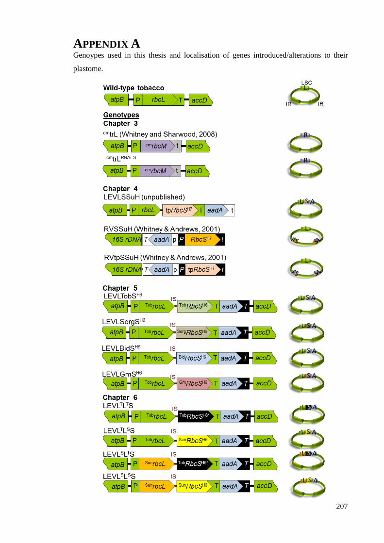
207
APPENDIX A Genoypes used in this thesis and localisation of genes introduced/alterations to their
plastome.
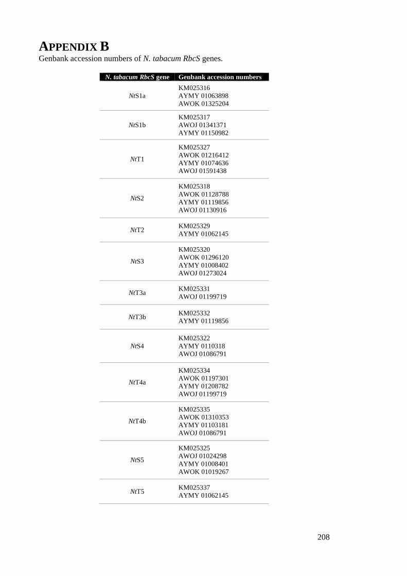
208
APPENDIX B Genbank accession numbers of N. tabacum RbcS genes.
N. tabacum RbcS gene Genbank accession numbers
NtS1a
KM025316
AYMY 01063898
AWOK 01325204
NtS1b
KM025317
AWOJ 01341371
AYMY 01150982
NtT1
KM025327
AWOK 01216412
AYMY 01074636
AWOJ 01591438
NtS2
KM025318
AWOK 01128788
AYMY 01119856
AWOJ 01130916
NtT2 KM025329
AYMY 01062145
NtS3
KM025320
AWOK 01296120
AYMY 01008402
AWOJ 01273024
NtT3a KM025331
AWOJ 01199719
NtT3b KM025332
AYMY 01119856
NtS4
KM025322
AYMY 0110318
AWOJ 01086791
NtT4a
KM025334
AWOK 01197301
AYMY 01208782
AWOJ 01199719
NtT4b
KM025335
AWOK 01310353
AYMY 01103181
AWOJ 01086791
NtS5
KM025325
AWOJ 01024298
AYMY 01008401
AWOK 01019267
NtT5 KM025337
AYMY 01062145