Embed Size (px)
Citation preview
Registered Office: 19 Old Mills Estate, Paulton, Bristol BS39 7SU Registered in England: 062 97451 1
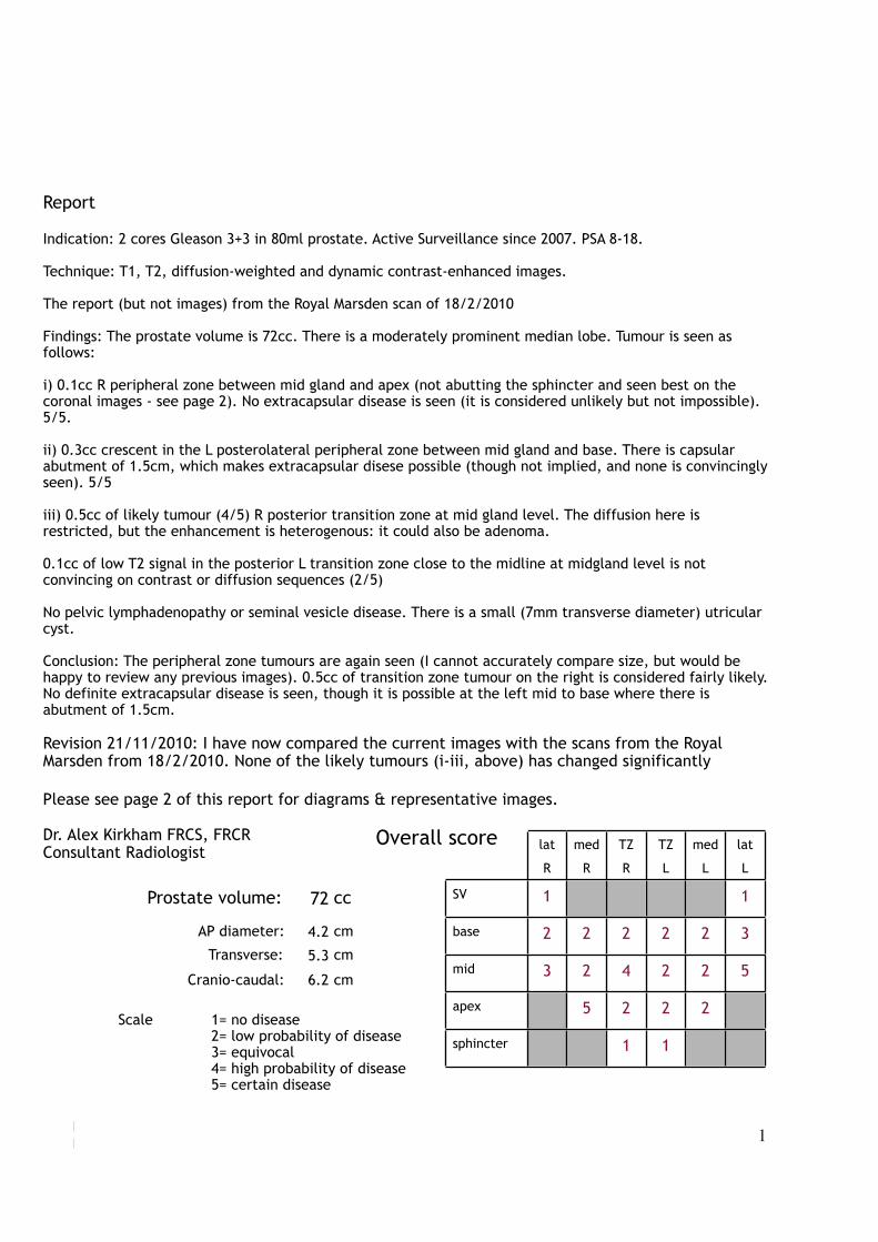
Prostate volume:
Cranio-caudal:
Transverse:
cc72
4.2
5.3
6.2
AP diameter: cm
cm
cm
Scale 1= no disease2= low probability of disease3= equivocal4= high probability of disease5= certain disease
lat
R
med
R
TZ
R
TZ
L
med
L
lat
L
SV 1 1
base 2 2 2 2 2 3
mid 3 2 4 2 2 5
apex 5 2 2 2
sphincter 1 1
Overall score
Report
Indication: 2 cores Gleason 3+3 in 80ml prostate. Active Surveillance since 2007. PSA 8-18.
Technique: T1, T2, diffusion-weighted and dynamic contrast-enhanced images.
The report (but not images) from the Royal Marsden scan of 18/2/2010
Findings: The prostate volume is 72cc. There is a moderately prominent median lobe. Tumour is seen as follows:
i) 0.1cc R peripheral zone between mid gland and apex (not abutting the sphincter and seen best on the coronal images - see page 2). No extracapsular disease is seen (it is considered unlikely but not impossible). 5/5.
ii) 0.3cc crescent in the L posterolateral peripheral zone between mid gland and base. There is capsular abutment of 1.5cm, which makes extracapsular disese possible (though not implied, and none is convincingly seen). 5/5
iii) 0.5cc of likely tumour (4/5) R posterior transition zone at mid gland level. The diffusion here is restricted, but the enhancement is heterogenous: it could also be adenoma.
0.1cc of low T2 signal in the posterior L transition zone close to the midline at midgland level is not convincing on contrast or diffusion sequences (2/5)
No pelvic lymphadenopathy or seminal vesicle disease. There is a small (7mm transverse diameter) utricular cyst.
Conclusion: The peripheral zone tumours are again seen (I cannot accurately compare size, but would be happy to review any previous images). 0.5cc of transition zone tumour on the right is considered fairly likely. No definite extracapsular disease is seen, though it is possible at the left mid to base where there is abutment of 1.5cm.
Revision 21/11/2010: I have now compared the current images with the scans from the Royal Marsden from 18/2/2010. None of the likely tumours (i-iii, above) has changed significantly
Please see page 2 of this report for diagrams & representative images.
Dr. Alex Kirkham FRCS, FRCRConsultant Radiologist

Position of disease and representative imagesR LVesicles
5/5, 0.3cc
Registered Office: 19 Old Mills Estate, Paulton, Bristol BS39 7SU Registered in England: 062 97451 2
T2 coronal
T2 coronal
T2 sagittal
diffusion (ADC) axiallong b diffusion axial
2/5
4/5, 0.5cc
5/5, 0.1cc
T2 coronal
T2 coronal
T2 axial
long b diffusion axial
contrast axial
contrast axial
T2 axial