Embed Size (px)
Citation preview
Over-the-Counter Monocyclic Non-SteroidalAnti-Inflammatory Drugs in Environment—Sources, Risks,Biodegradation
Ariel Marchlewicz & Urszula Guzik &
Danuta Wojcieszyńska
Received: 5 May 2015 /Accepted: 22 September 2015 /Published online: 30 September 2015# The Author(s) 2015. This article is published with open access at Springerlink.com
Abstract Recently, the increased use of monocyclicnon-steroidal anti-inflammatory drugs has resulted intheir presence in the environment. This may havepotential negative effects on living organisms. Thebiotransformation mechanisms of monocyclic non-steroidal anti-inflammatory drugs in the human bodyand in other mammals occur by hydroxylation andconjugation with glycine or glucuronic acid.Biotransformation/biodegradation of monocyclicnon-steroidal anti-inflammatory drugs in the environ-ment may be caused by fungal or bacterial microor-ganisms. Salicylic acid derivatives are degraded bycatechol or gentisate as intermediates which arecleaved by dioxygenases. The key intermediate ofthe paracetamol degradation pathways is hydroqui-none. Sometimes, after hydrolysis of this drug, 4-aminophenol is formed, which is a dead-end metabo-lite. Ibuprofen is metabolized by hydroxylation oractivation with CoA, resulting in the formation ofisobutylocatechol. The aim of this work is to attemptto summarize the knowledge about environmental riskconnected with the presence of over-the-counter anti-inflammatory drugs, their sources and the biotransfor-mation and/or biodegradation pathways of thesedrugs.
Keywords Monocyclic non-steroidal anti-inflammato-ry drugs . Toxicity . Biodegradation .Microorganisms
1 Introduction
In an age of high level care of human health, manypharmaceuticals are commonly used to cure or preventdiseases and other ailments, such as headache, musclepain, or inflammatory conditions. Presently, over-the-counter drugs are very popular, especially over-the-counter monocyclic and polycyclic non-steroidal anti-inflammatory drugs (NSAIDs). Among these drugs, themost popular and the most often used are monocyclicNSAIDs, such as ibuprofen, acetaminophen, andsalicylic acid (and its derivatives, like mesalazine), dueto their availability (Ziylan and Ince 2011). For exam-ple, the yearly intake of ibuprofen is up to 300 t inGermany, 162 t in England, and 58 t in Poland(Sosnowska et al. 2009; Guzik et al. 2013a). In theUSA during 2001–2005, about 29 billion doses of para-cetamol in all forms were sold (Li et al. 2014). The highintake of these widely available drugs may lead to theiror their metabolites’ presence in the environment. Inconnection with the presence of NSAIDs in the envi-ronment, there is a risk of long-term exposure, causingchronic toxic effect in organisms living there. This maycause negative effects for living creatures and the accu-mulation of drugs or their metabolites in the food chain(Carlsson et al. 2006; Sosnowska et al. 2009). Currentknowledge about the microbial metabolism of non-steroidal anti-inflammatory drugs is still very little, and
Water Air Soil Pollut (2015) 226: 355DOI 10.1007/s11270-015-2622-0
A. Marchlewicz :U. Guzik (*) :D. WojcieszyńskaDepartment of Biochemistry, Faculty of Biology andEnvironmental Protection, University of Silesia in Katowice,Jagiellonska 28, 40-032 Katowice, Polande-mail: [email protected]
the fact that we can find them in the environment sug-gests that sewage treatment plants are not currentlyadapted to completely remove these drugs before theyreach the environment. These drugs and their metabo-lites are found in wastewater influent and effluent fromwastewater treatment plants. For example, in Germany,acetylsalicylic acid was detected in the sewage effluentsat 0.22 μg/L (Heberer 2002) and paracetamol was de-tected in groundwater used as a source of public drink-ing water in California at 1.89 μg/L (Li et al. 2014).Observed metabolites are formed as a result of themetabolism by activated sludge microorganisms or en-ter the treatment plants as the human body biotransfor-mation products with municipal wastewater (Buser et al.1999; Zwiener et al. 2002; Marco-Urrea et al. 2009;Ziylan and Ince 2011). The fate of medicines, includingmonocyclic NSAIDs, in the natural environmental isstill less known. The main aim of this work is a compi-lation of the actual knowledge about sources and risksconnected with the presence of monocyclic non-steroidal anti-inflammatory drugs in the environment.Moreover, authors describe microbiological degradationof the three most widespread painkillers, antipyretic andanti-inflammatory and over-the-counter drugs—acetylsalicylic acid, paracetamol, and ibuprofen.
2 Sources of Pharmaceuticals in the Environment
The development of modern analytical methods makesit possible to detect NSAIDs in the environment. Con-sidering the high intake of drugs, it may be assumed thatthis has an impact on the presence of pharmaceuticals inwastewaters and surface waters. The main sources ofdrugs that reach the environment are excreted in non-metabolized form or slightly modified, i.e., hydroxylat-ed, conjugated, and disposed of through the toilet (Buseret al. 1999; Heberer 2002; Khan and Ongerth 2002;Zwiener et al. 2002; Metcalfe et al. 2004). Nearly halfof respondents declared the disposal of medications inthe household trash. That is why the presence of phar-maceuticals is expected in the landfill leachate orleachate-contaminated groundwater (Kuspis andKrenzelok 1996; Musson and Townsend 2009). Hospi-tals’ wastewaters and discharges from pharmaceuticalproduction also constitute a significant source of phar-maceuticals in the environment. In many countries,hospitals and pharmaceutical factories do not have sep-arate wastewater treatment plants (WWTP); therefore,
these contaminants pass into the general wastewatertreatment system (Metcalfe et al. 2004). The burdenedsewage flows into WWTPs, but not all NSAIDs areremoved in biological sewage treatment with activatedsludge. Drug detection in WWTP effluent confirmedinadequacy of wastewater treatment plants to complete-ly removing these pollutants from sewage (Ternes 1998;Buser et al. 1999; Tixier et al. 2003; Lee et al. 2005;Gómez et al. 2007; Salgado et al. 2010). Consequently,this leads to the detection of drugs even in surface waterssuch as lakes or rivers (Winkler et al. 2001; Dębska et al.2005; Roberts and Thomas 2006; Vieno et al. 2006;Togola and Budzinski 2008; Pailler et al. 2009). Addi-tionally, Kolpin et al. (2004) observed an increasedconcentration of drugs in downstream, above places ofoutflows from WWTP.
3 Environmental Risk of Monocyclic Non-SteroidalAnti-Inflammatory Drugs
Although NSAIDs are observed in the environment inlow concentrations, there is little known about the long-term effects of low concentrations of these drugs onliving organisms. The most data about toxicity of ibu-profen, paracetamol, and acetylsalicylic acid are basedon acute toxicity and short-term chronic toxicity tests(Table 1). In acute toxicity tests, high concentrations ofsubstances that may result in unrealistic effects are usu-ally used. In many cases, metabolites of drugs are nottaken into account in toxicity tests; therefore, it is diffi-cult to evaluate the real risk of NSAIDs and their me-tabolites on the environment (Webb 2004). Marco-Urrea et al. (2009), using Microtox toxicity test withPhotobacterium phosphoreum as a tested organism,proved that hydroxylated derivatives of ibuprofen(which was also found in sewage and surface waters)are more toxic than the original compound. Pomati et al.(2004) showed that even microgram per liter concentra-tion of ibuprofen can influence the growth of aquaticphototrophs. For example, Lemna minor exhibited inhi-bition of growth after 7-day exposure to low concentra-tion of ibuprofen. Under these conditions, the little effecton abscisic acid production was also observed (Pomatiet al. 2004; Brausch et al. 2012; Murdoch and Hay2013). High sensitivity to ibuprofen was also found forphytoplankton. Depending on tested organisms, EC50
value was between 1 and 315 mg/L after 72–120-hexposition to this drug (Brausch et al. 2012).
355 Page 2 of 13 Water Air Soil Pollut (2015) 226: 355
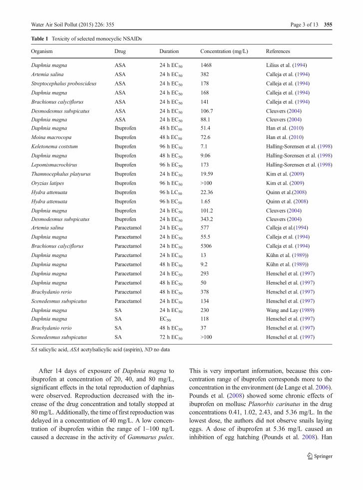
After 14 days of exposure of Daphnia magna toibuprofen at concentration of 20, 40, and 80 mg/L,significant effects in the total reproduction of daphniaswere observed. Reproduction decreased with the in-crease of the drug concentration and totally stopped at80mg/L. Additionally, the time of first reproductionwasdelayed in a concentration of 40 mg/L. A low concen-tration of ibuprofen within the range of 1–100 ng/Lcaused a decrease in the activity of Gammarus pulex.
This is very important information, because this con-centration range of ibuprofen corresponds more to theconcentration in the environment (de Lange et al. 2006).Pounds et al. (2008) showed some chronic effects ofibuprofen on mollusc Planorbis carinatus in the drugconcentrations 0.41, 1.02, 2.43, and 5.36 mg/L. In thelowest dose, the authors did not observe snails layingeggs. A dose of ibuprofen at 5.36 mg/L caused aninhibition of egg hatching (Pounds et al. 2008). Han
Table 1 Toxicity of selected monocyclic NSAIDs
Organism Drug Duration Concentration (mg/L) References
Daphnia magna ASA 24 h EC50 1468 Lilius et al. (1994)
Artemia salina ASA 24 h EC50 382 Calleja et al. (1994)
Streptocephalus proboscideus ASA 24 h EC50 178 Calleja et al. (1994)
Daphnia magna ASA 24 h EC50 168 Calleja et al. (1994)
Brachionus calyciflorus ASA 24 h EC50 141 Calleja et al. (1994)
Desmodesmus subspicatus ASA 24 h EC50 106.7 Cleuvers (2004)
Daphnia magna ASA 24 h EC50 88.1 Cleuvers (2004)
Daphnia magna Ibuprofen 48 h EC50 51.4 Han et al. (2010)
Moina macrocopa Ibuprofen 48 h EC50 72.6 Han et al. (2010)
Keletonema coststum Ibuprofen 96 h EC50 7.1 Halling-Sorensen et al. (1998)
Daphnia magna Ibuprofen 48 h EC50 9.06 Halling-Sorensen et al. (1998)
Lepomismacrochirus Ibuprofen 96 h EC50 173 Halling-Sorensen et al. (1998)
Thamnocephalus platyurus Ibuprofen 24 h EC50 19.59 Kim et al. (2009)
Oryzias latipes Ibuprofen 96 h EC50 >100 Kim et al. (2009)
Hydra attenuata Ibuprofen 96 h LC50 22.36 Quinn et al.(2008)
Hydra attenuata Ibuprofen 96 h EC50 1.65 Quinn et al. (2008)
Daphnia magna Ibuprofen 24 h EC50 101.2 Cleuvers (2004)
Desmodesmus subspicatus Ibuprofen 24 h EC50 343.2 Cleuvers (2004)
Artemia salina Paracetamol 24 h EC50 577 Calleja et al.(1994)
Daphnia magna Paracetamol 24 h EC50 55.5 Calleja et al. (1994)
Brachionus calyciflorus Paracetamol 24 h EC50 5306 Calleja et al. (1994)
Daphnia magna Paracetamol 24 h EC50 13 Kühn et al. (1989))
Daphnia magna Paracetamol 48 h EC50 9.2 Kühn et al. (1989))
Daphnia magna Paracetamol 24 h EC50 293 Henschel et al. (1997)
Daphnia magna Paracetamol 48 h EC50 50 Henschel et al. (1997)
Brachydanio rerio Paracetamol 48 h EC50 378 Henschel et al. (1997)
Scenedesmus subspicatus Paracetamol 24 h EC50 134 Henschel et al. (1997)
Daphnia magna SA 24 h EC50 230 Wang and Lay (1989)
Daphnia magna SA EC50 118 Henschel et al. (1997)
Brachydanio rerio SA 48 h EC50 37 Henschel et al. (1997)
Scenedesmus subspicatus SA 72 h EC50 >100 Henschel et al. (1997)
SA salicylic acid, ASA acetylsalicylic acid (aspirin), ND no data
Water Air Soil Pollut (2015) 226: 355 Page 3 of 13 355
et al. (2010) also observed a delay inOryzias latipes egghatching after exposure to 0.1 μg/L of ibuprofen. After120 days of ibuprofen exposure, the survival of fish wasalso significantly lower when compared to the controlpopulation (Han et al. 2010). The obtained results aresignificant, because the drug concentration used in theexperiment is observed in the environment (Dębskaet al. 2005; Pailler et al. 2009).
Wu et al. (2012) described p-aminophenol as a majormetabolite of paracetamol metabolism in microbes. 4-Aminophenol is one of the most toxic phenols, whichcauses the kidney or the liver damage (Newton et al.1982; Song and Chen 2001). Li et al. (2014) observedthe appearance of N-acetyl-p-benzoquinone imine(NAPQI) during biodegradation of acetaminophen insoil. Additionally, the increased sorption of paracetamolin soil biosolids was observed, which may cause adecrease of acetaminophen mineralization. Simulta-neously, it may affect the half-life of drugs in the envi-ronment (Li et al. 2014).
N-acetyl-p-benzoquinone imine is also one of thefirst phase metabolites of paracetamol detoxification inhumans, excreted as a glutathione conjugate with urine(Tsikas et al. 2011; Li et al. 2014). It is defined as highlyhepatotoxic (Bender andMacCrehan 2006; Hinson et al.2010; Tsikas et al. 2011). Bender et al. (2004) suggestthat NAPQI may be a potent inhibitor of human topo-isomerase IIα.
Toxicological research conducted in the presence ofhigh concentration of NSAIDs does not provide infor-mation about the influence of long-term exposure to lowconcentration of drugs. The answer to this question maybe provided only by long-term research of many gener-ations of aquatic organisms living in the presence of lowdrug concentration (Rzepa 2009).
4 Acetylsalicylic Acid Biodegradationby Microorganisms
In humans and animals, acetylsalicylic acid is immedi-ately hydrolyzed to salicylic acid which can be removedfrom the body unchanged or in the form of conjugateswith glycine (as a salicyluric acid) or with glucuronicacid, or hydroxylated at the C-5 position of the ring togentisate (de Gaetano et al. 1985; Ingelman-Sundberget al. 1991; Paterson et al. 2008). Apart from excretionfrom human and animal organisms, salicylic acid is awidespread molecule in plants. It plays a role in several
physiological processes, like stomatal closure, flowerinduction, heat production and, most of all, its mainfunction is defense against pathogen attack (Verberneet al. 2000). If we take this into consideration, it is notstrange that in nature, there are effective salicylate deg-radation mechanisms. Many bacterial strains, like Mi-crococcus, Sphingomonas, Amycolatopsis, Streptomy-ces, Pseudomonas, Alcaligenes, Pseudoramibacter,Rhodococcus (Chakrabarty 1972; Shamsuzzaman andBarnsley 1974; Haribabu et al. 1984; Grund et al. 1990;Grund et al. 1992; Civilini et al. 1999; Hintner et al.2001; Ishiyama et al. 2004; Deveryshetty et al. 2007;Jouanneau et al. 2007; Silva et al. 2007; Lanfranconiet al. 2009) and fungi, like Sclerotinia, Trichosporon,Aspergillus, Fusarium, Rhodotorula, Cryptococcus(Anderson and Dagley 1980; Kuswandi and Roberts1992; Middelhoven 1993; Iwasaki et al. 2009; Qi et al.2012; Penn and Daniel 2013) are capable of degradingsalicylate (Table 2) via a few catabolic pathways.
The strategy for degradation of aromatic structurecomprises hydroxylation and cleavage of the aromaticring. Hydroxylation into the dihydroxylated intermedi-ates, the first step in the oxidative degradation of aro-matic compounds, is catalyzed by oxygenases belong-ing to three groups: hydroxylating dioxygenases,activated-ring monooxygenases, or nonactivated-ringmonooxygenases. As a result of hydroxylation, the keyintermediates such as catechol, protocatechuic acid,hydroxyquinol, or gentisic acid are formed. These prod-ucts are substrates for ring-cleaving dioxygenases. Sa-licylates are mainly transformed to catechol andgentisate, which are cleaved in the next step bydioxygenases from two groups—intradiol or extradiol(Guzik et al. 2013b; Guzik et al. 2014).
Two of the most important enzymes involved insalicylate decomposition are salicylate 1-hydroxylaseand salicylate 5-hydroxylase (monooxygenases). Salic-ylate monooxygenases belong to one of the three groupsof hydroxylating oxygenases—activated-ringmonooxygenases (Wojcieszyńska et al. 2011). The gen-eral structure of these groups includes a three-component protein with separate functional units: reduc-tase with a flavin cofactor, ferrodoxin with a Rieskie-type iron-sulfur cluster [2Fe-2S] and hexamer-builtα3β3terminal oxygenase with [2Fe-2S] a cluster andone nonheme iron(II) per α subunit (Mason andCammack 1992; Bertini et al. 1996). These catalyticproteins are able to insert one atom of molecular oxygeninto the structure of the aromatic ring, simultaneously
355 Page 4 of 13 Water Air Soil Pollut (2015) 226: 355
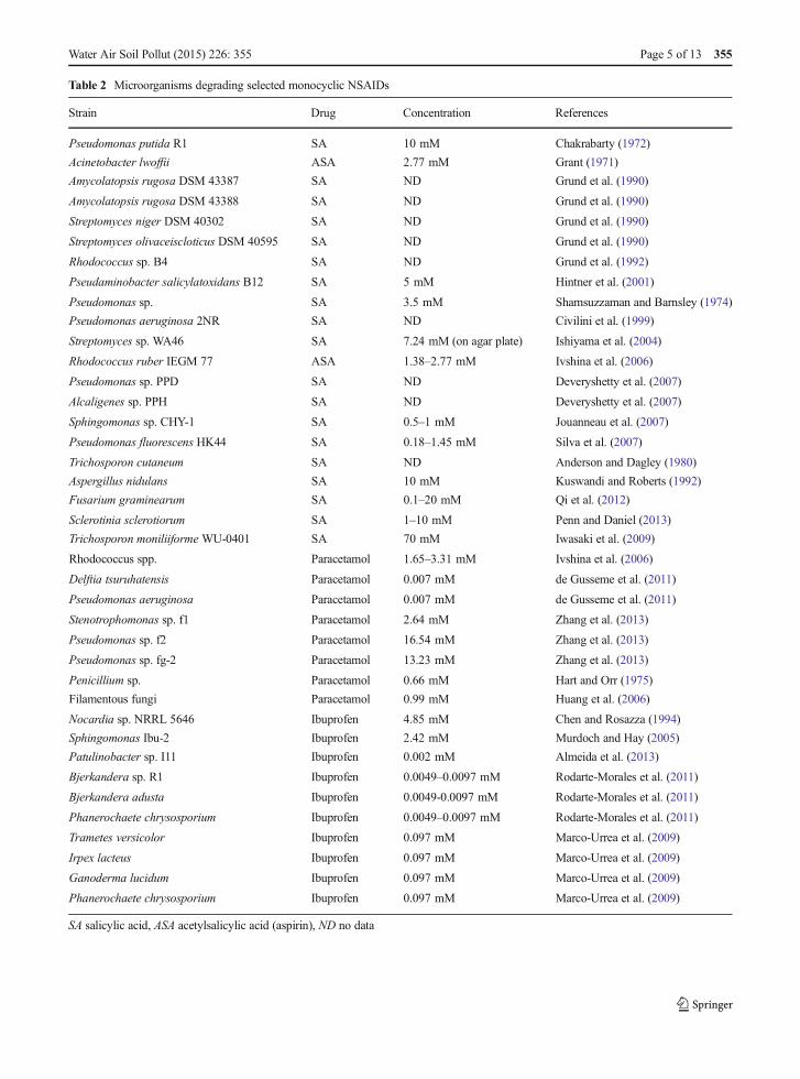
Table 2 Microorganisms degrading selected monocyclic NSAIDs
Strain Drug Concentration References
Pseudomonas putida R1 SA 10 mM Chakrabarty (1972)
Acinetobacter lwoffii ASA 2.77 mM Grant (1971)
Amycolatopsis rugosa DSM 43387 SA ND Grund et al. (1990)
Amycolatopsis rugosa DSM 43388 SA ND Grund et al. (1990)
Streptomyces niger DSM 40302 SA ND Grund et al. (1990)
Streptomyces olivaceiscloticus DSM 40595 SA ND Grund et al. (1990)
Rhodococcus sp. B4 SA ND Grund et al. (1992)
Pseudaminobacter salicylatoxidans B12 SA 5 mM Hintner et al. (2001)
Pseudomonas sp. SA 3.5 mM Shamsuzzaman and Barnsley (1974)
Pseudomonas aeruginosa 2NR SA ND Civilini et al. (1999)
Streptomyces sp. WA46 SA 7.24 mM (on agar plate) Ishiyama et al. (2004)
Rhodococcus ruber IEGM 77 ASA 1.38–2.77 mM Ivshina et al. (2006)
Pseudomonas sp. PPD SA ND Deveryshetty et al. (2007)
Alcaligenes sp. PPH SA ND Deveryshetty et al. (2007)
Sphingomonas sp. CHY-1 SA 0.5–1 mM Jouanneau et al. (2007)
Pseudomonas fluorescens HK44 SA 0.18–1.45 mM Silva et al. (2007)
Trichosporon cutaneum SA ND Anderson and Dagley (1980)
Aspergillus nidulans SA 10 mM Kuswandi and Roberts (1992)
Fusarium graminearum SA 0.1–20 mM Qi et al. (2012)
Sclerotinia sclerotiorum SA 1–10 mM Penn and Daniel (2013)
Trichosporon moniliiforme WU-0401 SA 70 mM Iwasaki et al. (2009)
Rhodococcus spp. Paracetamol 1.65–3.31 mM Ivshina et al. (2006)
Delftia tsuruhatensis Paracetamol 0.007 mM de Gusseme et al. (2011)
Pseudomonas aeruginosa Paracetamol 0.007 mM de Gusseme et al. (2011)
Stenotrophomonas sp. f1 Paracetamol 2.64 mM Zhang et al. (2013)
Pseudomonas sp. f2 Paracetamol 16.54 mM Zhang et al. (2013)
Pseudomonas sp. fg-2 Paracetamol 13.23 mM Zhang et al. (2013)
Penicillium sp. Paracetamol 0.66 mM Hart and Orr (1975)
Filamentous fungi Paracetamol 0.99 mM Huang et al. (2006)
Nocardia sp. NRRL 5646 Ibuprofen 4.85 mM Chen and Rosazza (1994)
Sphingomonas Ibu-2 Ibuprofen 2.42 mM Murdoch and Hay (2005)
Patulinobacter sp. I11 Ibuprofen 0.002 mM Almeida et al. (2013)
Bjerkandera sp. R1 Ibuprofen 0.0049–0.0097 mM Rodarte-Morales et al. (2011)
Bjerkandera adusta Ibuprofen 0.0049-0.0097 mM Rodarte-Morales et al. (2011)
Phanerochaete chrysosporium Ibuprofen 0.0049–0.0097 mM Rodarte-Morales et al. (2011)
Trametes versicolor Ibuprofen 0.097 mM Marco-Urrea et al. (2009)
Irpex lacteus Ibuprofen 0.097 mM Marco-Urrea et al. (2009)
Ganoderma lucidum Ibuprofen 0.097 mM Marco-Urrea et al. (2009)
Phanerochaete chrysosporium Ibuprofen 0.097 mM Marco-Urrea et al. (2009)
SA salicylic acid, ASA acetylsalicylic acid (aspirin), ND no data
Water Air Soil Pollut (2015) 226: 355 Page 5 of 13 355
reducing the second oxygen atom towater. All salicylatehydroxylases need NADH to remain active. The oxida-tion of NADH is directly connected with FAD reduction(Katagiri et al. 1965; Sze and Dagley 1984; Grund et al.1992; Suzuki et al. 1996; Fuenmayor et al. 1998;Balashova et al. 2001; Zhou et al. 2002; Jouanneauet al. 2007).
Chakrabarty (1972) and Deveryshetty et al. (2007)examined the ability of Pseudomonas putida R1 andAlcaligenes sp. PPH, respectively, to degrade salicylate.It was decomposed to catechol, the key intermediate,and further to 2-hydroxymuconic semialdehyde as aproduct of ring fission. Because of the significant activ-ity of catechol 2,3-dioxygenase the authors concludedthat P. putida R1 was capable of meta cleavage. On theother hand, decomposition of salicylate via catecholmay also run via ortho cleavage with cis,cis-muconicacid as an aromatic ring cleavage product. Not onlybacteria, such as Amycolatopsis, Streptomyces, or Pseu-domonas, but also fungi, like Fusarium, Rhodotorula,Trichosporon, and Sclerotinia show that kind of cate-chol ring fission (Shamsuzzaman and Barnsley 1974;Anderson and Dagley 1980; Sze and Dagley 1984;Grund et al. 1990; Fuenmayor et al. 1998; Civiliniet al. 1999; Ishiyama et al. 2004; Iwasaki et al. 2009;Qi et al. 2012; Penn and Daniel 2013).
Salicylate degradation may also lead via hydroxyl-ation to gentisate. For example, this pathway was re-ported for Rhodococcus sp. B4 or Streptomyces sp.WA46 strains (Grund et al. 1992; Ishiyama et al.2004). Enzymes engaged in this pathway need for theiractivity NADH, CoA and ATP as cofactors. Salicylate isconverted to gentisate via salicylyl-CoA and gentisyl-CoA. In the first step, salicylyl-AMP ligase and proba-bly salicylyl-CoA synthetase catalyze the formation ofthe thioester bond between salicylate and CoA andcreate salicylyl-CoA. The next step is the hydroxylationby salicylyl-CoA 5-hydroxylase with the formation ofgentisyl-CoA. Hydrolysis to gentisate opens the way toring cleavage by gentisate 1,2-dioxygenase tomaleylpyruvate which leads to central metabolism.Civilini et al. (1999) showed the ability of Pseudomonasaeruginosa 2NR to convert salicylate into both interme-diates, calechol, and gentisate.
Iwasaki et al. (2009) described a different model ofdecomposition of salicylate via catechol by yeastTrichosporon moniliiforme WU-0401. In their study, anon-oxidative way with phenol as an intermediate ofsalicylate degradation to catechol was presented.
Salicylate was immediately transformed to phenol with,simultaneously, decarboxylation, but without hydroxyl-ation. Before that, phenol was hydroxylated to catecholand further cleaved in the ortho position.
Most bacteria degrade salicylate via oxidative decar-boxylation to catechol or via hydroxylation to gentisate.Pseudaminobacter salicylatoxidans B12 is capable ofdirect ring cleavage using NADH-independent salicy-late 1,2-dioxygenase forming 2-oxohepta-3,5-dienedioic acid as an aliphatic product (Hintner et al.2001).
5 Microbial Degradation and Transformationof Paracetamol
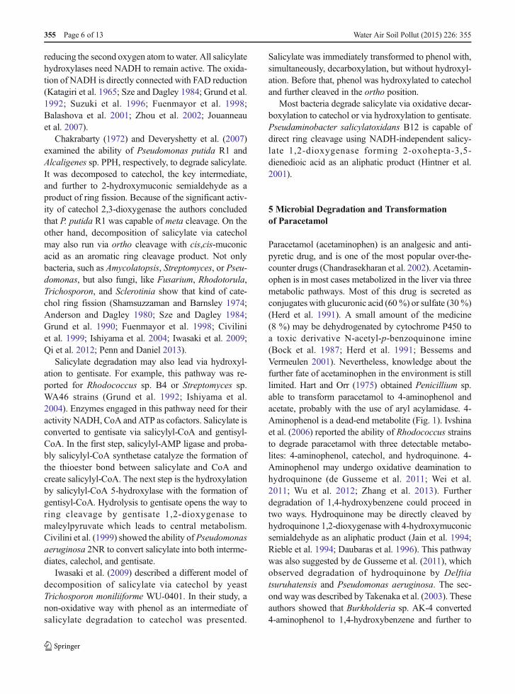
Paracetamol (acetaminophen) is an analgesic and anti-pyretic drug, and is one of the most popular over-the-counter drugs (Chandrasekharan et al. 2002). Acetamin-ophen is in most cases metabolized in the liver via threemetabolic pathways. Most of this drug is secreted asconjugates with glucuronic acid (60%) or sulfate (30%)(Herd et al. 1991). A small amount of the medicine(8 %) may be dehydrogenated by cytochrome P450 toa toxic derivative N-acetyl-p-benzoquinone imine(Bock et al. 1987; Herd et al. 1991; Bessems andVermeulen 2001). Nevertheless, knowledge about thefurther fate of acetaminophen in the environment is stilllimited. Hart and Orr (1975) obtained Penicillium sp.able to transform paracetamol to 4-aminophenol andacetate, probably with the use of aryl acylamidase. 4-Aminophenol is a dead-end metabolite (Fig. 1). Ivshinaet al. (2006) reported the ability of Rhodococcus strainsto degrade paracetamol with three detectable metabo-lites: 4-aminophenol, catechol, and hydroquinone. 4-Aminophenol may undergo oxidative deamination tohydroquinone (de Gusseme et al. 2011; Wei et al.2011; Wu et al. 2012; Zhang et al. 2013). Furtherdegradation of 1,4-hydroxybenzene could proceed intwo ways. Hydroquinone may be directly cleaved byhydroquinone 1,2-dioxygenase with 4-hydroxymuconicsemialdehyde as an aliphatic product (Jain et al. 1994;Rieble et al. 1994; Daubaras et al. 1996). This pathwaywas also suggested by de Gusseme et al. (2011), whichobserved degradation of hydroquinone by Delftiatsuruhatensis and Pseudomonas aeruginosa. The sec-ond way was described by Takenaka et al. (2003). Theseauthors showed that Burkholderia sp. AK-4 converted4-aminophenol to 1,4-hydroxybenzene and further to
355 Page 6 of 13 Water Air Soil Pollut (2015) 226: 355
1,2,4-trihydroxybenzene. Then 1,2,4-trihydroxybenzenewas cleaved by hydroxyhydroquinone 1,2-dioxygenaseto maleylacetic acid, which is introduced to the basicmetabolism (Mason and Cammack 1992; Chauhanet al. 2000; Miyauchi et al. 1999; Moonen et al. 2008;Kolvenbach et al. 2011) (Fig. 1). Zhang et al. (2013)described the conversion of acetaminophen to hydroqui-none, which was next transformed to an aliphatic producthexa-3-enedioic acid. Unfortunately, the authors did notdetermine the enzyme engaged in ring cleavage. How-ever, it seems that hexa-3-enedioic acid was a product ofaromatic ring fission or, if not, it that means some inter-mediate metabolites between aromatic and aliphatic com-pounds were passed over. Hexa-3-enedioic acid is similarto muconic acid—a product of ortho ring cleavage ofcatechol, but the authors did not find catechol in thestudied samples. Based on reported intermediates a pri-mary pathway of acetaminophen degradation could beproposed. The mechanism may be based on cutting offtwo carbon atoms in the form of formic acid (Fig. 1)(Zhang et al. 2013).
Furthermore, Huang et al. (2006) described the for-mation of glucoside conjugates with paracetamol by soilfilamentous fungi via O- and N-linkages. This is asimilar way to the human detoxication routes of xeno-biotics in phase II of detoxication (Halling-Sorensenet al. 1998).
In 2014, Li et al. described degradation pathwayof paracetamol in soil microorganisms. It was shownthat in the first step, aromatic ring of paracetamol ishydroxylated to 3-hydroxyacetaminophen, oxygen-ated to N-acetyl-p-benzoquinone imine, or methyl-ated to p-acetanisidide. It is suggested that cyto-chrome P-450 may be engaged in these processes.N-acetyl-p-benzoquinone imine is then metabolizedto 1,4-benzoquinone which is more stable andcritical toxic metabolite. p-Acetanisidide is trans-formed to 4-methoxyphenol and in the next step tothe 1,4-dimethoxybenzene. The presence of 2-hexenoic acid in the soil extract suggests thecleavage of the aromatic ring of paracetamol (Liet al. 2014).
Fig. 1 Biotransformation of paracetamol (Hart and Orr 1975; Takenaka et al. 2003; Kolvenbach et al. 2011; Wu et al. 2012; Zhang et al.2013; Guzik et al. 2014; Li et al. 2014)
Water Air Soil Pollut (2015) 226: 355 Page 7 of 13 355
6 Biodegradation/Biotransformation of Ibuprofen
Ibuprofen (2-(4-(2-methylpropyl)phenyl)propanoic ac-id) is one of the most popular and commonly usednon-steroidal anti-inflammatory drugs. This makes itthe third most popular drug in the world. Ibuprofen isalso one of the dominating medicines present in sewagebecause of its relatively high therapeutic dose (600–1200 mg per day), and significant levels are excretedfrom the human body (even 70–80 %). This drug maybe secreted as an unchanged molecule or as an un-changed molecule in conjugation with glucuronide(product of the second phase of detoxication that maybe hydrolyzed in the environment) or as a few metabo-lites: hydroxyibuprofen (two isomers), carboxyibuprofen,and carboxyhydratropic acid (Halling-Sorensen et al.1998; Buser et al. 1999; Zwiener et al. 2002). Nonetheless,little is still known about the environmental metabolism ofibuprofen, whose concentration in the environment rangesfrom nanograms per liter to micrograms per liter (Calamariet al. 2003; Bendz et al. 2005; Tauxe-Wuersch et al. 2005;Nakada et al. 2006; Roberts and Thomas 2006; Gómezet al. 2007; Lin et al. 2009; Pailler et al. 2009).
Many reports describe only the initial steps of ibu-profen transformation. Rodarte-Morales et al. (2011)used three species of ligninolytic fungi: Bjerkanderasp. R1, Bjerkandera adusta, and Phanerochaetechrysosporium to check their ability to degrade pharma-ceuticals, including ibuprofen (Table 2). They reported arapid decrease of ibuprofen in growth medium,explaining that fact by degradation of the drug. Howev-er, these authors did not search for intermediates
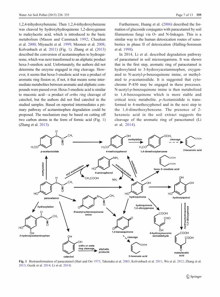
occurring in the degradation process, and they notedonly the loss of the parent compound. It may be sug-gested that ibuprofen was not completely mineralized.Trametes versicolor, Irpex lacteus, Ganodermalucidum, and P. chrysosporium are fungi capable ofdegrading ibuprofen (Marco-Urrea et al. 2009). All ofthem, excluding P. chrysosporium, degraded 10 mg/Libuprofen to non-detectable levels. P. chrysosporiumshowed the lowest degradation level, between 70 and88 %. Simultaneously, it should be noted that the au-thors tested the in vitro activity of laccase (also withlaccase mediators) and manganese peroxidase. More-over, they used inhibitors of the cytochrome P-450complex to analyze the participation of these enzymesin ibuprofen degradation. In all cases, the researchersdid not observe the contribution of the examined en-zymes in the metabolism of ibuprofen. It may be sug-gested that the metabolism of ibuprofen could runthrough another pathway. The major metabolites whichwere found were hydroxylated in the isopropyl chainfrom ibuprofen—1- and 2-hydroxyibuprofen after thef i r s t h o u r s o f t h e e x p e r imen t , a n d 1 , 2 -dihydroxyibuprofen as a final metabolite after 7 daysof cultivation (Fig. 2). Hydroxylated and carboxylatedderivatives are frequent in the microbial metabolism(Zwiener et al. 2002; Quintana et al. 2005). It is note-worthy that ibuprofen derivatives are more toxic thanthe parent compound and may accumulate in the envi-ronment (Marco-Urrea et al. 2009). Despite low con-centrations of these compounds in the ecosystem, theymay be hazardous. However, the long-term effects of theorganism exposure to this drug cannot be defined (Perry
Fig. 2 Microbiological transformation of ibuprofen (Chen and Rosazza 1994; Murdoch and Hay 2005; Quintana et al. 2005; Guzik et al.2014)
355 Page 8 of 13 Water Air Soil Pollut (2015) 226: 355
and Zylstra 2007). On the basis of European Union law,Cleuvers (2004) did not classify ibuprofen as toxic toaquatic organisms. However, a study with Daphniamagna and green algae showed that ibuprofen may bea toxic factor, especially in the presence of other drugs.This study informs about the effect of high-dose toxicityin short time exposure. In the environment, pharmaceu-ticals are not in high concentrations, so greater emphasisshould be put on studies on the chronic toxicity of drugs(Cleuvers 2004).
During the degradation of ibuprofen by the lignolyticbacterium Nocardia sp. NRRL 5646, two metabolites,ibuprofenol and ibuprofenol acetate, were observed(Fig. 2). These products underwent further mineraliza-tion (Chen and Rosazza 1994).
Murdoch and Hay (2005, 2013) characterized one ofthe most completed ibuprofen degradation pathways inSphingomonas Ibu-2 bacteria, capable of using ibupro-fen as a source of carbon and energy. Based on geneticanalyses, they proposed five-gene cluster (ipfABDEF),coding enzymes putatively involved in ibuprofen catab-olism. Two of these genes (ipfA, ipfB) were very similarto genes coding two subunits of dioxygenases; the thirdgene was identified as a gene coding enzyme for theremoval/addition of acyl groups—acyl-CoA synthetase(IpfD); the fourth one (ipfF) was described as a coen-zyme A ligase gene; and for the gene ipfE, no functionwas found. Two additional genes ipfH and ipfI encodeferredoxin reductase and ferredoxin components of anaromatic dioxygenase system, respectively (Murdochand Hay 2013). As the first step in the decompositionof ibuprofen by strain Ibu-2, thioesterification with co-enzyme A with the participation of coenzyme A ligasewas suggested. Removal of the propionic acid chain anddioxygenation reaction led to isobutylocatechol forma-tion. This compound undergoes oxygenolytic cleavage(Murdoch and Hay 2005; Murdoch and Hay 2013)(Fig. 2). Ibuprofen biotransformation by VariovoraxIbu-1 occurs with trihydroxyibuprofen as a metabolite.This compound may be a dead-end metabolite or issubstrate to the meta-ring fission reaction (Murdochand Hay 2015).
Patulinobacter sp. I11 did not grow with ibuprofen asthe only source of carbon and energy. However, degrada-tion of ibuprofen was observed in the presence of yeastextract and tryptone. This suggests that ibuprofen couldnot induce the expression of enzymes responsible for itsdecomposition. In the bacterial genome, homologousgenes were found coding enzymes potentially involved
in ibuprofen decomposition, such as acyl-CoA synthetase,a protein containing a Rieske-like (2Fe-2S) iron-sulfurcluster (dioxygenase-like protein), and enoyl-CoAhydratase/isomerase (Almeida et al. 2013).
7 Conclusion
The occurrence of micropollutants in the environment,such as non-steroidal anti-inflammatory drugs, is a rel-atively new problem. The presence of these drugs in theenvironment poses a risk of long-term exposure, causingchronic toxic effects for organisms. Although thesalicylic acid pathway is very well described, little isknown about the biotransformation/biodegradation ofother non-steroidal anti-inflammatory drugs such asibuprofen or paracetamol. Paracetamol degradationpathways lead through hydroquinone as a key interme-diate, whereas ibuprofen is metabolized by hydroxyl-ation or activation with CoA. However, sometimes bio-transformation of monocyclic NSAIDs leads to the ac-cumulation of intermediates more toxic than the parentcompounds. An understanding the drug mineralizationprocesses is key to creating commercially availablesolutions for this increasing problem.
Compliance with Ethical Standards
Funding This work was financed by the National Science Cen-tre (Poland), granted on the basis of decision DEC-2013/09/B/NZ9/00244.
Conflict of Interest The authors declare that they have noconflict of interest.
Open Access This article is distributed under the terms of theCreative Commons Attribution 4.0 International License (http://creativecommons.org/licenses/by/4.0/), which permits unrestrict-ed use, distribution, and reproduction in any medium, providedyou give appropriate credit to the original author(s) and the source,provide a link to the Creative Commons license, and indicate ifchanges were made.
References
Almeida, B., Kjeldal, H., Lolas, I., Knudsen, A. D., Carvalho, G.,Nielsen, K. L., Barreto Crespo, M. T., Stensballe, A., &Nielsen, J. L. (2013). Quantitative proteomic analysis ofibuprofen-degrading Patulibacter sp. strain I11.Biodegradation, 24, 615–630.
Water Air Soil Pollut (2015) 226: 355 Page 9 of 13 355
Anderson, J. J., & Dagley, S. (1980). Catabolism of aro-matic acids in Trichosporon cutaneum. Journal ofBacteriology, 141(2), 534–543.
Balashova, N. V., Stolz, A., Knackmuss, H. J., Kosheleva, I. A.,Naumov, A. V., & Boronin, A. M. (2001). Purification andcharacterization of a salicylate hydroxylase involved in 1-hydroxy-2-naphthoic acid hydroxylation from the naphtha-lene and phenanthrene-degrading bacterial strainPseudomonas putida BS202-P1. Biodegradation, 12, 179–188.
Bender, M., & MacCrehan, W. A. (2006). Transformation ofacetaminophen by chlorination produces the toxicants 1,4-benzoquinone and N-acetyl-p-benzoquinone imine.Environmental Science and Technology, 40, 516–522.
Bender, R. P., Lindsey, R. H., Burden, D. A., & Osheroff, N.(2004). N-Acetyl-p-benzoquinone imine, the toxic metabo-lite of acetaminophen, is a topoisomerase II poison.Biochemistry, 43, 3731–3739.
Bendz, D., Paxéus, N. A., Ginn, T. R., & Loge, F. J. (2005).Occurrence and fate of pharmaceutically active compoundsin the environment, a case study: Höje river in Sweden.Journal of Hazardous Materials, 122, 195–204.
Bertini, I., Cremonini, M. A., Ferretti, S., Lozzi, I., Luchinat, C., &Viezzoli, M. S. (1996). Arene hydroxylases: metalloenzymescatalysing dioxygenation of aromatic compounds.Coordination Chemistry Review, 151, 145–160.
Bessems, J. G. M., & Vermeulen, N. P. E. (2001). Paracetamol(acetaminophen)-induced toxicity: molecular and biochemi-cal mechanisms, analogues and protective approaches.Critical Review in Toxicology, 31(1), 55–138.
Bock, K. W., Wiltfang, J., Blume, R., Ullrich, D., & Bircher, J.(1987). Paracetamol as a test drug to determine glucuronideformation in man. Effects of inducers and of smoking.European Journal of Clinical Pharmacology, 31, 677–683.
Brausch, J. M., Connors, K. A., Brooks, B. W., & Rand, G. M.(2012). Human pharmaceuticals in the aquatic environment:a review of recent toxicological studies and consideration fortoxicity testing. Reviews of Environmental Contaminationand Toxicology, 218, 1–99.
Buser, H. R., Poiger, T., & Muller, M. D. (1999). Occurrence andenvironmental behavior of the chiral pharmaceutical drugibuprofen in surface waters and in wastewater.Environmental Science and Technology, 33, 2529–2535.
Calamari, D., Zuccato, E., Castiglioni, S., Bagnati, R., & Fanelli,R. (2003). Strategic survey of therapeutic drugs in the RiversPo and Lambro in Northern Italy. Environmental Science andTechnology, 37, 1241–1248.
Calleja, M. C., Personne, G., & Geladi, P. (1994). Comparativeacute toxicity of the first 50 multicentre evaluation of in vitrocytotoxicity chemicals to aquatic non vertebrates. Archives ofEnvironmental Contamination and Toxicology, 26, 69–78.
Carlsson, C., Johansson, A. K., Alvan, G., Bergman, K., &Kühler,T. (2006). Are pharmaceuticals potent environmental pollut-ants? Part I: Environmental risk assessments of selectedactive pharmaceutical ingredients. Science of the TotalEnvironment, 364, 67–87.
Chakrabarty, A. M. (1972). Genetic basis of the biodegradation ofsalicylate in Pseudomonas. Journal of Bacteriology, 112(2),815–822.
Chandrasekharan, N. V., Dai, H., Roos, K. L. T., Evanson, N. K.,Tomsik, J., Elton, T. S., & Simmons, D. L. (2002). COX-3, a
cyclooxygenase-1 variant inhibited by acetaminophen andother analgesic/antipyretic drugs: cloning, structure, and ex-pression. Proceedings of the National Academy of Sciences,99(21), 13926–13931.
Chauhan, A., Samanta, S. K., & Jain, R. K. (2000). Degradation of4-nitrocatechol by Burkholderia cepacia: a plasmid-encodednovel pathway. Journal of AppliedMicrobiology, 88(5), 764–772.
Chen, Y., & Rosazza, J. P. N. (1994). Microbial transformation ofibuprofen by aNocardia species. Applied and EnvironmentalMicrobiology, 60, 1292–1296.
Civilini, M., de Bertoldi, M., & Tell, G. (1999). Molecular char-acterization of Pseudomonas aeruginosa 2NR degradingnaphthalene. Letters in Applied Microbiology, 29, 181–186.
Cleuvers, M. (2004). Mixture toxicity of the anti-inflammatorydrugs diclofenac, ibuprofen, naproxen, and acetylsalicylicacid. Ecotoxicology and Environmental Safety, 59, 309–315.
Daubaras, D. L., Saido, K., & Chakrabarty, A. M. (1996).Purification of hydroxyquinol 1,2-dioxygenase andmaleylacetate reductase: the lower pathway of 2,4,5-trichlorophenoxyacetic acid metabolism by Burkholderiacepac ia AC1100. Appl ied and Envi ronmenta lMicrobiology, 62(11), 4276–4279.
de Gaetano, G., Cerletti, C., Dejana, E., & Latini, R. (1985).Pharmacology of platelet inhibition in humans: implicationsof the salicylate–aspirin interaction. Circulation, 72(6),1185–1193.
de Gusseme, B., Vanhaecke, L., Verstraete, W., & Boon, N.(2011). Degradation of acetaminophen by Delftiatsuruhatensis and Pseudomonas aeruginosa in a membranebioreactor. Water Research, 45, 1829–1837.
de Lange, H. J., Noordoven, W., Murk, A. J., Lurling, M., &Peeters, E. T. H. M. (2006). Behavioural responses ofGammarus pulex (Crustacea, Amphipoda) to low concentra-tions of pharmaceuticals. Aquatic Toxicology, 78, 209–216.
Dębska, J., Kot-Wasik, A., & Namiesnik, J. (2005). Determinationof nonsteroidal anti-inflammatory drugs in water samplesusing liquid chromatography coupled with diode–array de-tector and mass spectrometry. Journal of Separation Science,28, 2419–2426.
Deveryshetty, J., Suvekbala, V., Varadamshetty, G., & Phale, P. S.(2007). Metabolismof 2-, 3- and 4-hydroxybenzoates by soilisolates Alcaligenes sp. strain PPH and Pseudomonas sp.strain PPD. FEMS Microbiology Letters, 268, 59–66.
Fuenmayor, S. L., Wild, M., Boyes, A. L., & Williams, P. A.(1998). A gene cluster encoding steps in conversion of naph-thalene to gentisate in Pseudomonas sp. strain U2. Journal ofBacteriology, 180(9), 2522–2530.
Gómez, M. J., Martínez Bueno, M. J., Lacorte, S., Fernández-Alba, A. R., & Agüera, A. (2007). Pilot survey monitoringpharmaceuticals and related compounds in a sewage treat-ment plant located on the Mediterranean coast.Chemosphere, 66, 993–1002.
Grant, D. J. W. (1971). Degradation of acetylsalicylic acid by astrain of Acinetobacter lwoffii. Journal of AppliedBacteriology, 34, 689–698.
Grund, E., Denecke, B., & Eichenlaub, R. (1992).Naphthalene degradation via salicylate and gentisateby Rhodococcus sp . s t r a in B4. Appl ied andEnvironmental Microbiology, 58(6), 1874–1877.
355 Page 10 of 13 Water Air Soil Pollut (2015) 226: 355
Grund, E., Knorr, C., & Eichenlaub, R. (1990). Catabolism ofbenzoate and monohydroxyla ted benzoa tes byAmycolatopsis and Streptomyces spp. Applied andEnvironmental Microbiology, 56(5), 1459–1464.
Guzik, U., Hupert-Kocurek, K., Mazur, A., & Wojcieszyńska, D.(2013a). Biotransformacja wybranych niesteroidowychleków przeciwzapalnych w środowisku. Bromatologia iChemia Toksykologiczna, 1, 105–112 (in Polish).
Guzik, U., Hupert-Kocurek, K., & Wojcieszyńska, D. (2013b).Intradiol dioxygenases—the key enzymes in xenobioticsdegradation. In: Chamy, R., & Rosenkranz, F. (Ed.),Biodegradation of Hazardous and Special Products (pp.129–153). Rijeka
Guzik, U., Hupert-Kourek, K., & Wojcieszyńska, D. (2014).Microbial degradation non-steroidal anti-inflammatorydrugs. Postepy Mikrobiologii, 53, 61–69 (in Polish).
Halling-Sorensen, B., Nors Nielsen, S., Lanzky, P. F., Ingerslev, F.,Holten Lützhof, H. C., & Jorgensen, S. E. (1998).Occurrence, fate, and effects of pharmaceutical substancesin the environment—a review. Chemosphere, 36, 357–393.
Han, S., Choi, K., Kim, J., Ji, K., Kim, S., Aho, B., Yun, J., Choi,K., Khim, J. S., Zhang, X., & Giesy, J. P. (2010). Endocrinedisruption and consequences of chronic exposure to ibupro-fen in Japanese medaka Oryzias latipes and freshwater cla-docerans Daphnia magna and Moina macrocopa. AquaticToxicology, 98, 256–264.
Haribabu, B., Kamath, A. V., & Vaidyanathan, C. S. (1984).Degradation of substituted benzoic acids by a Micrococcusspecies. FEMS Microbiology Letters, 21(2), 197–200.
Hart, A., & Orr, D. L. J. (1975). The degradation of paracetamol(4-hydroxyacetanilide) and other substituted acetanilides by aPenicillium species.Antonie van Leeuwenhoek, 41, 239–247.
Heberer, T. (2002). Occurrence, fate, and removal of pharmaceu-ticals residues in the aquatic environment: a review of recentresearch data. Toxicology Letters, 131, 5–17.
Henschel, K. P., Wenzel, A., Diederich, M., & Fliedner, A. (1997).Environmental hazard assessment of pharmaceuticals.Regulatory Toxicology and Pharmacology, 25, 220–225.
Herd, B., Wynne, H., Wright, P., James, O., & Woodhouse, K.(1991). The effect of age on glucuronidation and sulphationof paracetamol by human liver fractions. British Journal ofClinical Pharmacology, 32, 768–770.
Hinson, A. H., Roberts, D.W., & James, L. P. (2010). Mechanismsof acetaminophen-induced liver necrosis. Handbook ofExperimental Pharmacology, 196, 369–405.
Hintner, J. P., Lechner, C., Riegert, U., Kuhm, A. E., Storm, T.,Reemtsma, T., & Stolz, A. (2001). Direct ring fission ofsalicylate by a salicylate 1,2-dioxygenase activity fromPseudaminobacter sal icylatoxidans. Journal ofBacteriology, 183(23), 6936–6942.
Huang, H. H., Lin, L. H., Zhang, P., Qi, X. L., & Zhong, D. F.(2006). Formation of glucoside conjugate of acetaminophenby fungi separated from soil. European Journal of DrugMetabolism and Pharmacokinetics, 31(2), 103–108.
Ingelman-Sundberg, M., Kaur, H., Terelius, Y., Persson, J. O., &Halliwell, B. (1991). Hydroxylation of salicylate by micro-somal fractions and cytochrome P-450.Biochemical Journal,276, 753–757.
Ishiyama, D., Vujaklija, D., & Davies, J. (2004). Novelpathway of salicylate degradation by Streptomyces sp.
strain WA46. Applied and Environmental Microbiology,70(3), 1297–1306.
Ivshina, I. B., Rychkova, M. I., Vikhareva, E. V., Chekryshkina, L.A., &Mishenina, I. I. (2006). Catalysis of the biodegradationof unusable medicines by alkanotrophic Rhodococci. AppliedBiochemistry and Microbiology, 42, 392–395.
Iwasaki, Y., Gunji, H., Kino, K., Hattori, T., Ishii, Y., & Kirimura,K. (2009). Novel metabolic pathway for salicylate biodegra-dation via phenol in yeast Trichosporon moniliiforme.Biodegradation, 21, 557–564.
Jain, R. K., Dreisbach, J. H., & Spain, J. C. (1994). Biodegradationof p-nitrophenol via 1-, 2-, 4-benzenetriol by an Arthrobactersp. Applied and Environmental Microbiology, 60(8), 3030–3032.
Jouanneau, Y., Micoud, J., & Meyer, C. (2007). Purification andcharacterization of a from Sphingomonas sp. strain CHY-1three-component salicylate 1-hydroxylase. Applied andEnvironmental Microbiology, 73(23), 7515–7521.
Katagiri, M., Maeno, H., Yamamoto, S., Hayaishi, O., Kitao, T., &Oae, S. (1965). Salicylate hydroxylase, a monooxygenaserequiring flavin adenine dinucleotide II. The mechanism ofsalicylate hydroxylation to catechol. The Journal ofBiological Chemistry, 240, 3414–3417.
Khan, S. J., & Ongerth, J. E. (2002). Estimation of pharmaceuticalresidues in primary and secondary sewage sludge based onquantities of use and fugacity modeling. Water Science andTechnology, 46, 105–113.
Kim, J. W., Ishibashi, H., Yamauchi, R., Ichikawa, N., Takao, Y.,Hirano, M., Koga, M., & Arizono, K. (2009). Acute toxicityof pharmaceutical and personal care products on freshwatercrustacean Thamnocephalus platyurus and fish Oryziaslatipes. Journal of Toxicological Sciences, 342, 227–232.
Kolpin, D. W., Skopiec, M., Meyer, M. T., Furlong, E. T., &Zaugg, S. D. (2004). Urban contribution of pharmaceuticalsand other organic wastewater contaminants to streams duringdiffering flow conditions. Science of the Total Environment,328, 119–130.
Kolvenbach, B. A., Lenz, M., Benndorf, D., Rapp, E., Fousek, J.,Vlcek, C., Schäffer, A., Gabriel, F. L. P., Kohler, H. P. E., &Corvini, P. F. X. (2011). Purification and characterization ofhydroquinone dioxygenase from Sphingomonas sp. strainTTNP3. AMB Express, 1(8), 2–11.
Kühn, R., Pattard, M., Pernak, K. D., &Winter, A. (1989). Resultsof the harmful effects of selected water pollutants (anilines,phenols, aliphatic compounds) to Daphnia magna. WaterResearch, 23(4), 495–499.
Kuspis, D. A., &Krenzelok, E. P. (1996).What happens to expiredmedications? A survey of community medication disposal.Veterinary and Human Toxicology, 38(1), 48–49.
Kuswandi, K., & Roberts, C. F. (1992). Genetic control of theprotocatechuic acid pathway inAspergillus nidulans. Journalof General Microbiology, 138, 817–823.
Lanfranconi, M. P., Christie-Oleza, J. A., Martin-Cardona, C.,Suárez-Suárez, L. Y., Lalucat, J., Nogales, B., & Bosch, R.(2009). Physiological role of NahW, the additional salicylatehydroxylase found in Pseudomonas stutzeri AN10. FEMSMicrobiology Letters, 300, 265–272.
Lee, H. B., Peart, T. E., & Svoboda, M. L. (2005). Determinationof endocrine-disrupting phenols, acidic pharmaceuticals, andpersonal-care products in sewage by solid-phase extraction
Water Air Soil Pollut (2015) 226: 355 Page 11 of 13 355
and gas chromatography–mass spectrometry. Journal ofChromatography. A, 1094, 122–129.
Li, J., Ye, Q., & Gan, J. (2014). Degradation and transformationproducts of acetaminophen in soil. Water Research, 49, 44–52.
Lilius, H., Isomaa, B., & Holmstrom, T. (1994). A comparison ofthe toxicity of 50 reference chemicals to freshly isolatedrainbow trout hepatocytes and Daphnia magna. AquaticToxicology, 30, 47–60.
Lin, A. Y. C., Yu, T. H., & Lateef, S. K. (2009). Removal ofpharmaceuticals in secondary wastewater treatment process-es in Taiwan. Journal of Hazardous Materials, 167, 1163–1169.
Marco-Urrea, E., Pérez-Trujillo, M., Vicent, T., & Caminal, G.(2009). Ability of white-rot fungi to remove selected phar-maceuticals and identification of degradation products ofibuprofen by Trametes versicolor. Chemosphere, 74, 765–772.
Mason, J. R., & Cammack, R. (1992). The electron transportproteins oh hydroxylating bacterial dioxygenases. AnnualReview of Microbiology, 46, 277–305.
Metcalfe, C., Miao, X. S., Hua, W., Letcher, R., & Servos, M.(2004). Pharmaceuticals in the Canadian environment. In K.Kümmerer (Ed.), Pharmaceuticals in the environment.Sources, fate, effects and risks (pp. 67–90). Berlin:Springer-Verlag.
Middelhoven,W. J. (1993). Catabolism of benzene compounds byascomycetous and basidiomycetous yeasts and yeast-likefungi. Antonie van Leeuwenhoek, 63, 125–144.
Miyauchi, K., Adachi, Y., Nagata, Y., & Takagi, M. (1999).Cloning and sequencing of a novel meta-cleavagedioxygenase gene whose product is involved in degradationof gamma-hexachlorocyclohexane in Sphingomonaspaucimobilis. Journal of Bacteriology, 181(21), 6712–6719.
Moonen, M. J. H., Synowsky, S. A., van den Berg, W. A. M.,Westphal, A. H., Heck, A. J. R., van den Heuvel, R. H. H.,Fraaije, M.W., & van Berkel,W. J. H. (2008). Hydroquinonedioxygenase from Pseudomonas fluorescens ACB: a novelmember of the family of nonheme-iron(II)-dependentdioxygenases. Journal of Bacteriology, 190(15), 5199–5209.
Murdoch, R. W., & Hay, A. G. (2005). Formation of catechols viaremoval of acid side chains from ibuprofen and related aro-matic acids. Applied and Environmental Microbiology,71(10), 6121–6125.
Murdoch, R. W., & Hay, A. G. (2013). Genetic and chemicalcharacterization of ibuprofen degradation by SphingomonasIbu-2. Microbiology, 159, 621–632.
Murdoch, R. W., & Hay, A. G. (2015). The biotransformation ofibuprofen to trihydroxyibuprofen in activated sludge and byVariovorax Ibu-1. Biodegradation, 26, 105–113.
Musson, S. E., & Townsend, T. G. (2009). Pharmaceutical com-pound content of municipal solid waste. Journal ofHazardous Materials, 162, 730–735.
Nakada, N., Tanishima, T., Shinohara, H., Kiri, K., & Takada, H.(2006). Pharmaceutical chemicals and endocrine disruptersin municipal wastewater in Tokyo and their removal duringactivated sludge treatment.Water Research, 40, 3297–3303.
Newton, J. F., Kuo, C. H., Gemborys, M. W., Mudge, G. H., &Hook, J. B. (1982). Nephrotoxicity of p-aminophenol, ametabolite of acetaminophen, in the Fischer 344 rat.Toxicolology and Applied Pharmacology, 65, 336–344.
Pailler, J. Y., Pfister, K. L., Hoffmann, L., & Guignard, C. (2009).Solid phase extraction coupled to liquid chromatography–tandem mass spectrometry analysis of sulfonamides, tetracy-clines, analgesics and hormones in surface water and waste-water in Luxembourg. Science of the Total Environment, 40,4736–4743.
Paterson, J. R., Baxter, G., Dreyer, J. S., Halket, J.M., Flynn, R., &Lawrence, J. R. (2008). Salicylic acid sans aspirin in animalsand man: persistence in fasting and biosynthesis frombenzoic acid. Journal of Agricultural and Food Chemistry,56, 11648–11652.
Penn, C. D., & Daniel, S. L. (2013). Salicylate degradation by thefungal plant pathogen Sclerotinia sclerotiorum. CurrentMicrobiology, 67(2), 218–225.
Perry, L. L., & Zylstra, J. (2007). Cloning of a gene clusterinvolved in the catabolism of p-nitrophenol by Arthrobactersp. strain JS443 and characterization of the p-nitrophenolmonooxygenase. Journal of Bacteriology, 189(21), 7563–7572.
Pomati, F., Netting, A. G., Calamari, D., & Neilan, B. A. (2004).Effects of erythromycin, tetracyclin and ibuprofen on thegrowth of Synechocystis sp. and Lemna minor. AquaticToxicology, 67, 387–396.
Pounds, N., Maclean, S., Webley, M., Pascoe, D., & Hutchinson,T. (2008). Acute and chronic effects of ibuprofen in themollusc Planorbis carinatus (Gastropoda: Planorbidae).Ecotoxicology and Environmental Safety, 70, 47–52.
Qi, P. F., Johnston, A., Balcerzak, M., Rocheleau, H., Harris, L. J.,Long, X. Y., Wei, Y. M., Zheng, Y. L., & Ouellet, T. (2012).Effect of salicylic acid on Fusarium graminearum, the majorcasual agent of fusarium head blight in wheat. FungalBiology, 116, 413–426.
Quinn, B., Gagne, F., & Blaise, C. (2008). An investigation intothe acute and chronic toxicity of eleven pharmaceuticals (andtheir solvents) found in wastewater effluent on the cnidarians,Hydra attenuate. Science of the Total Environment, 389,306–314.
Quintana, J. B., Weiss, S., & Reemtsma, T. (2005). Pathways andmetabolites of microbial degradation of selected acidic phar-maceutical and their occurrence in municipal wastewatertreated by a membrane bioreactor. Water Research, 39,2654–2664.
Rieble, S., Joshi, D. K., & Gold, M. H. (1994). Purification andcharacterization of a 1-, 2-, 4-trihydroxybenzene 1,2-dioxygenase from the basidiomycete Phanerochaetechrysosporium. Journal of Bacteriology, 176(16), 4838–4844.
Roberts, P. H., & Thomas, K. V. (2006). The occurrence ofselected pharmaceuticals in wastewater effluent and surfacewaters of the lower Tyne catchment. Science of the TotalEnvironment, 356, 143–153.
Rodarte-Morales, A. I., Feijoo, G., Moreira, M. T., & Lema, J. M.(2011). Degradation of selected pharmaceutical and personalcare products (PPCPs) by white-rot fungi. World Journal ofMicrobiology and Biotechnology, 27, 1839–1849.
Rzepa, J. (2009). Oznaczanie leków i pestycydów w wodachpowierzchniowych. In B. K. Głoda (Ed.), PostępyChromatografii (pp. 67–77). Siedlce: WydawnictwoAkademii Podlaskiej (in Polish).
Salgado, R., Noronha, J. P., Oehmen, A., Carvalho, G., & Reis, M.A. M. (2010). Analysis of 65 pharmaceuticals and personal
355 Page 12 of 13 Water Air Soil Pollut (2015) 226: 355
care products in 5 wastewater treatment plants in Portugalusing a simplified analytical methodology. Water Scienceand Technology, 6212, 2862–2871.
Shamsuzzaman, K., & Barnsley, E. A. (1974). The regulation ofnaphthalene oxygenase in Pseudomonads. Biochemical andBiophysical Research Communications, 60, 582–589.
Silva, T. R., Valdman, E., Valdman, B., & Leite, S. G. F. (2007).Salicylic acid degradation from aqueous solutions usingPseudomonas fluorescens HK44: parameters studies and ap-plication tools. Brazilian Journal of Microbiology, 38, 39–44.
Song, H., & Chen, T. S. (2001). p-Aminophenol-induced livertoxicity: tentative evidence of a role for acetaminophen.Journal of Biochemical and Molecular Toxicology, 15, 34–40.
Sosnowska, K., Styszko-Grochowiak, K., & Gołaś, J. (2009). Lekiw środowisku – źródła, przemiany, zagrożenia. IVKrakowska Konferencja Młodych Uczonych (pp. 395–404)(in Polish).
Suzuki, K., Mizuguchi, M., Ohnishi, K., & Itagaki, E. (1996).Structure of chromosomal DNA coding for Pseudomonasputida S-1 salicylate hydroxylase. Biochimica et BiophysicaActa, 1275, 154–156.
Sze, I. S., &Dagley, S. (1984). Properties of salicylate hydroxylaseand hydroxyquinol 1,2-dioxygenase purified fromTrichosporon cutaneum. Journal of Bacteriology, 159(1),353–359.
Takenaka, S., Okugawa, S., Kadowaki, M., Murakami, S., &Aoki, K. (2003). Metabolic pathway of 4-aminophenol inBurkholderia sp. strain AK-5 differs from that of aniline andaniline with C-4 substituents. Applied and EnvironmentalMicrobiology, 69(9), 5410–5413.
Tauxe-Wuersch, A., de Alencastro, L. F., Grandjean, D., &Tarradellas, J. (2005). Occurrence of several acidic drugs insewage treatment plants in Switzerland and risk assessment.Water Research, 39, 1761–1772.
Ternes, T. A. (1998). Occurrence of drugs in German sewagetreatment plants and rivers.Water Research, 32, 3245–3260.
Tixier, C., Singer, H. P., Oellers, S., & Müller, S. R. (2003).Occurrence and fate of carbamazepan, clofibric acid,diclofenac, ibuprofen, ketoprofen and naproxen in surfacewater. Environmental Science and Technology, 37, 1061–1068.
Togola, A., & Budzinski, H. (2008). Multi-residue analysis ofpharmaceutical compounds in aqueous samples. Journal ofChromatography A, 1177, 150–158.
Tsikas, D., Trettin, A., Zorner, A. A., & Gutzki, F. M. (2011). In-source formation of N-acetyl-p-benzoquinone imine(NAPQI), the putatively toxic acetaminophen (paracetamol)
metabolite, after derivatization with pentafluorobenzyl bro-mide and GC–ECNICI-MS analysis. Journal ofChromatography B, 879, 1476–1484.
Verberne, M. C., Verpoorte, R., Bol, J. F., Marcado-Blanco, J., &Linthorst, H. J.M. (2000). Overproduction of salicylic acid inplants by bacterial transgenes enhances pathogen resistance.Nature Biotechnology, 18, 779–783.
Vieno, N. M., Tuhkanen, T., & Kronberg, L. (2006). Analysis ofneutral and basic pharmaceuticals in sewage treatment plantsand in recipient rivers using solid phase extraction and liquidchromatography–tandem mass spectrometry detection.Journal of Chromatography A, 1134, 101–111.
Wang, W. H., & Lay, J. P. (1989). Fate and effects of salicylic acidcompounds in freshwater systems. Ecotoxicology andEnvironmental Safety, 17(3), 308–316.
Wei, F., Zhou, Q. W., Leng, S. Q., Zhang, L. L., & Chen, J. M.(2011). Isolation, identification and biodegradation charac-teristics of a new bacterial strain degrading paracetamol.Chinese Journal of Environmental Science, 32, 1812–1819.
Winkler, M., Lawrence, J. R., & Neu, T. R. (2001). Selectivedegradation of ibuprofen and clofibric acid in two modelriver biofilm systems. Water Research, 3513, 3197–3205.
Wojcieszyńska, D., Greń, I., Hupert-Kocurek, K., & Guzik, U.(2011). Modulation of FAD-dependent monooxygenase ac-t i v i t y f r om a r oma t i c c ompound s - d e g r a d i n gStenotrophomonas maltophilia strain KB2. Acta BiochimicaPolonica, 58, 421–426.
Wu, S., Zhang, L., & Chen, J. (2012). Paracetamol in the environ-ment and its degradation by microorganism. AppliedMicrobiology and Biotechnology, 96, 875–884.
Zhang, L., Hu, J., Zhu, R., Zhou, Q., & Chen, J. (2013).Degradation of paracetamol by pure bacterial cultures andtheir microbial consortium. Applied Microbiology andBiotechnology, 97, 3687–3698.
Zhou, N. Y., Al–Dulayymi, J., Baird, M. S., & Williams, P. A.(2002). Salicylate 5-hydroxylase from Ralstonia sp. strainU2: a monooxygenase with close relationships to and sharedelectron transport proteins with naphthalene dioxygenase.Journal of Bacteriology, 184(6), 1547–1555.
Ziylan, A., & Ince, N. H. (2011). The occurence and fate of anti-inflammatory and analgesic pharmaceuticals in sewage andfresh water: treatability by conventional and non-conventional processes. Journal of Hazardous Materials,187, 24–36.
Zwiener, C., Seeger, S., Glauner, T., & Frimmel, F. H. (2002).Metabolites from the biodegradation of pharmaceutical resi-dues of ibuprofen in biofilm reactors an batch experiments.Analytical and Bioanalytical Chemistry, 372, 569–575.
Water Air Soil Pollut (2015) 226: 355 Page 13 of 13 355