Embed Size (px)
Citation preview

Ovarian Cancer
The Case of Olivia Carson
You are a generalist in the community.
You have a new patient. She is a 65 year old with a large
pelvic mass arising from the right adnexum.
The Case of Olivia Carson
What is the likelihood that she has ovarian cancer?
The Case of Olivia Carson
What is the likelihood that she has ovarian cancer? Age Menopausal status Mass characteristics
Ovarian Neoplasms
90% benign 10% malignant
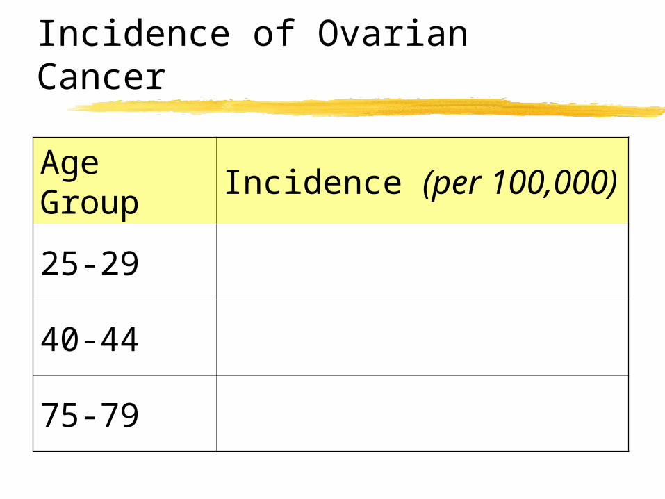
Incidence of Ovarian Cancer
Age Group
Incidence (per 100,000)
25-29
40-44
75-79
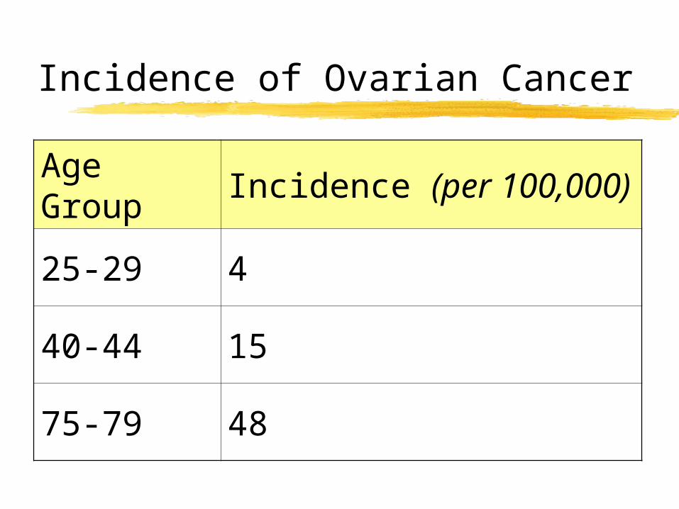
Incidence of Ovarian Cancer
Age Group
Incidence (per 100,000)
25-29 4
40-44 15
75-79 48
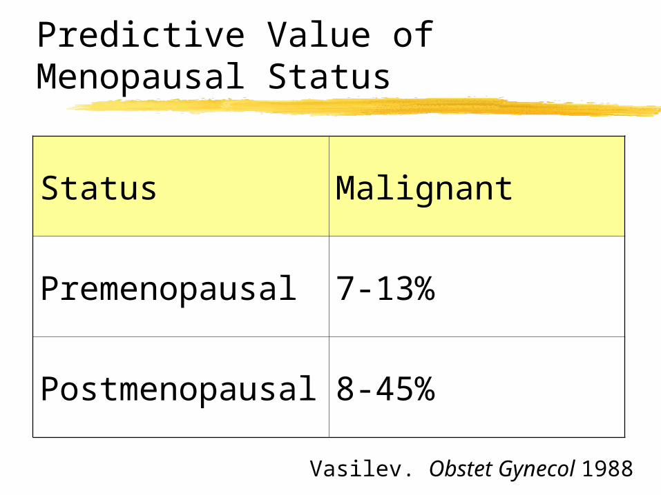
Predictive Value of Menopausal Status
Vasilev. Obstet Gynecol 1988
Status Malignant
Premenopausal 7-13%
Postmenopausal 8-45%
The Case of Olivia Carson
The only other data you have is that she underwent an imaging study.
Which study do you hope she has had?
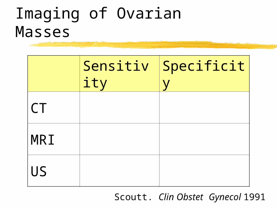
Imaging of Ovarian Masses
Scoutt. Clin Obstet Gynecol 1991
Sensitivity Specificity
CT
MRI
US
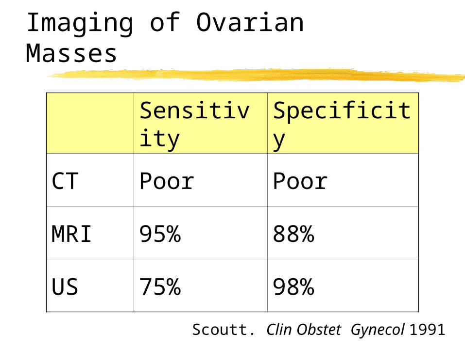
Imaging of Ovarian Masses
Scoutt. Clin Obstet Gynecol 1991
Sensitivity Specificity
CT Poor Poor
MRI 95% 88%
US 75% 98%
Transvaginal Ultrasound
Cost effective
High frequency
Improved resolution
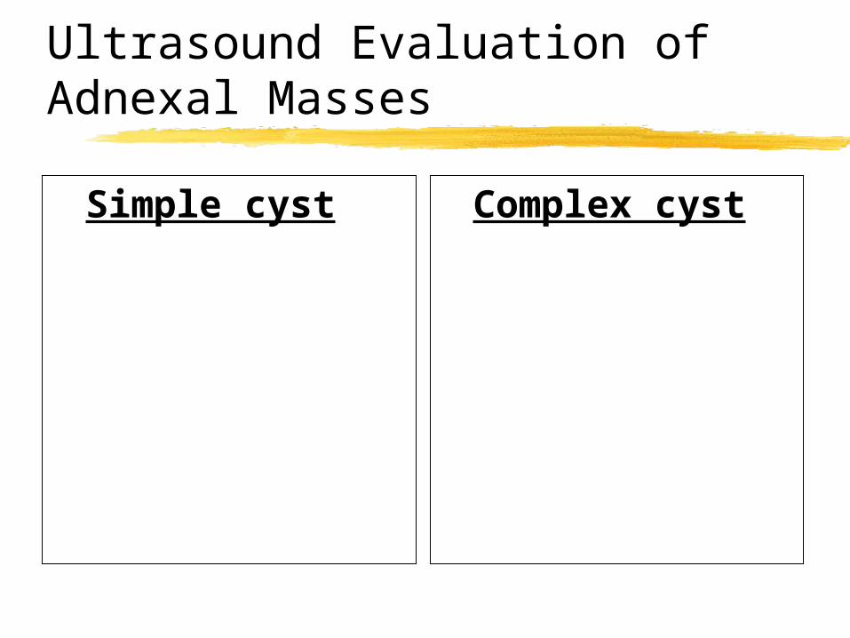
Ultrasound Evaluation of Adnexal Masses
Simple cyst Complex cyst
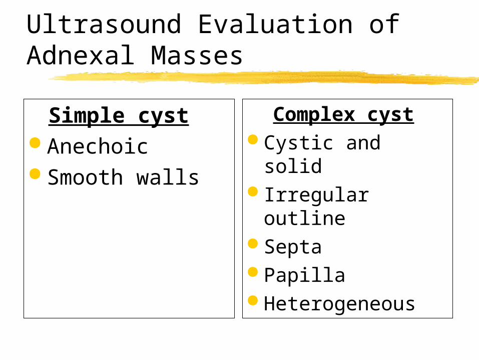
Ultrasound Evaluation of Adnexal Masses
Simple cyst Anechoic Smooth walls
Complex cyst Cystic and solid Irregular outline Septa Papilla Heterogeneous
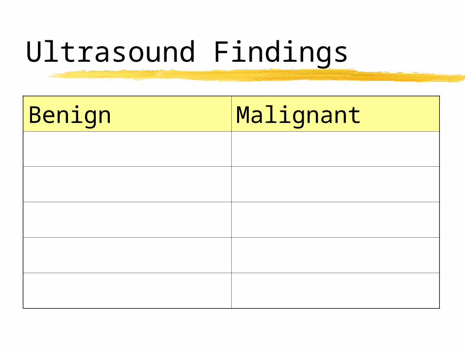
Ultrasound Findings
Benign Malignant
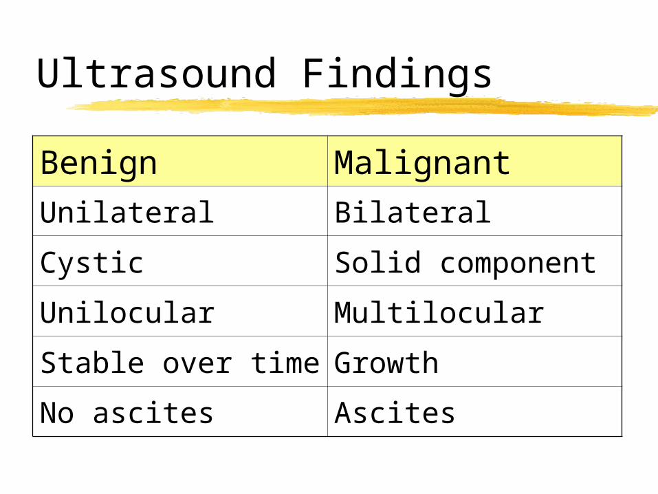
Ultrasound Findings
Benign Malignant
Unilateral Bilateral
Cystic Solid component
Unilocular Multilocular
Stable over time Growth
No ascites Ascites
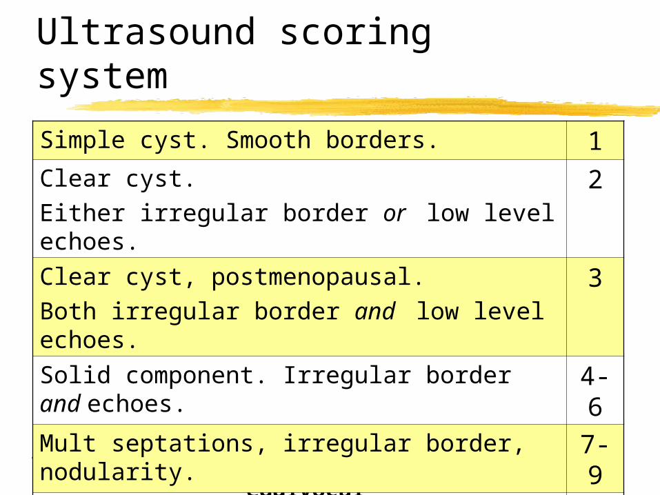
Ultrasound scoring system
1-3 = benign 4-6 = equivocal 7-10 = malig
Simple cyst. Smooth borders. 1Clear cyst. Either irregular border or low level echoes.
2
Clear cyst, postmenopausal. Both irregular border and low level echoes.
3
Solid component. Irregular border and echoes.
4-6
Mult septations, irregular border, nodularity.
7-9
As above plus ascites. 10
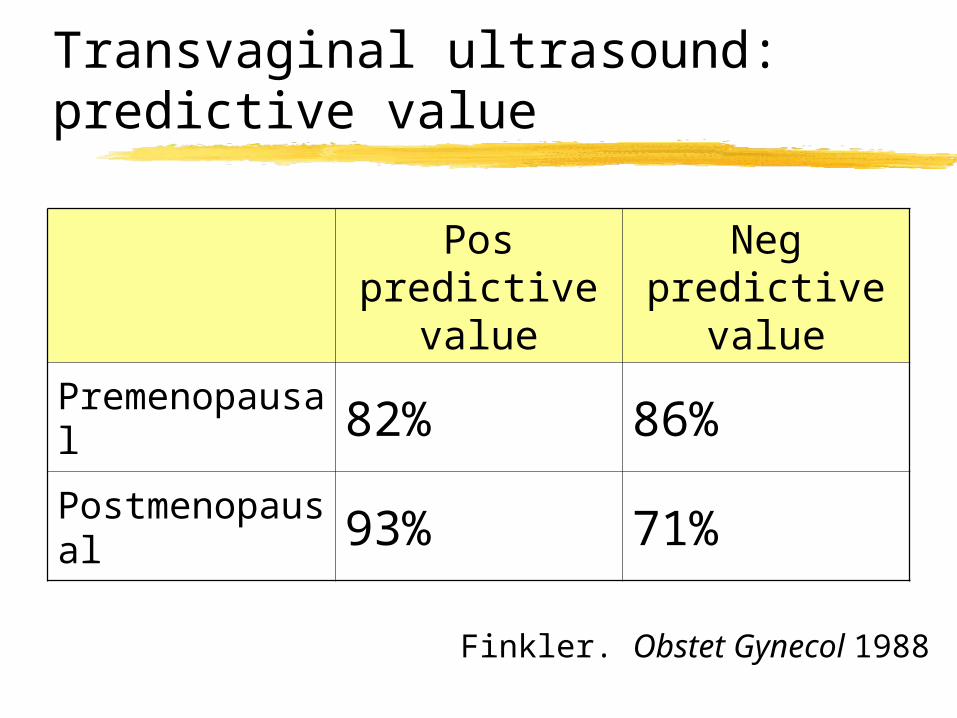
Transvaginal ultrasound: predictive value
Finkler. Obstet Gynecol 1988
Pos predictive
value
Neg predictive
valuePremenopausal 82% 86%
Postmenopausal 93% 71%
The Case of Olivia Carson
Her ultrasound shows a solid mass.
If this were cancer, predict the histology.
Cancer Rule of Thumb
Oncology recapitulates ontogeny
Embryology of the Ovary
3 cell types
Embryology of the Ovary
Coelomic epithelium
Mesenchyme
Germ cells
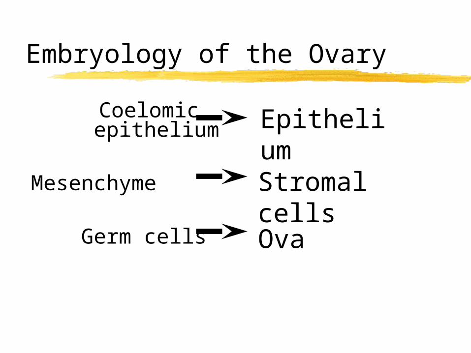
Embryology of the Ovary
Coelomic epithelium
Mesenchyme
Germ cells
EpitheliumStromal cells
Ova
Stromal Tumors: Histologic Subtypes
1. Fibroblasts
2. Granulosa cell tumors
3. Thecal cells
4. Sertoli-Leydig cells
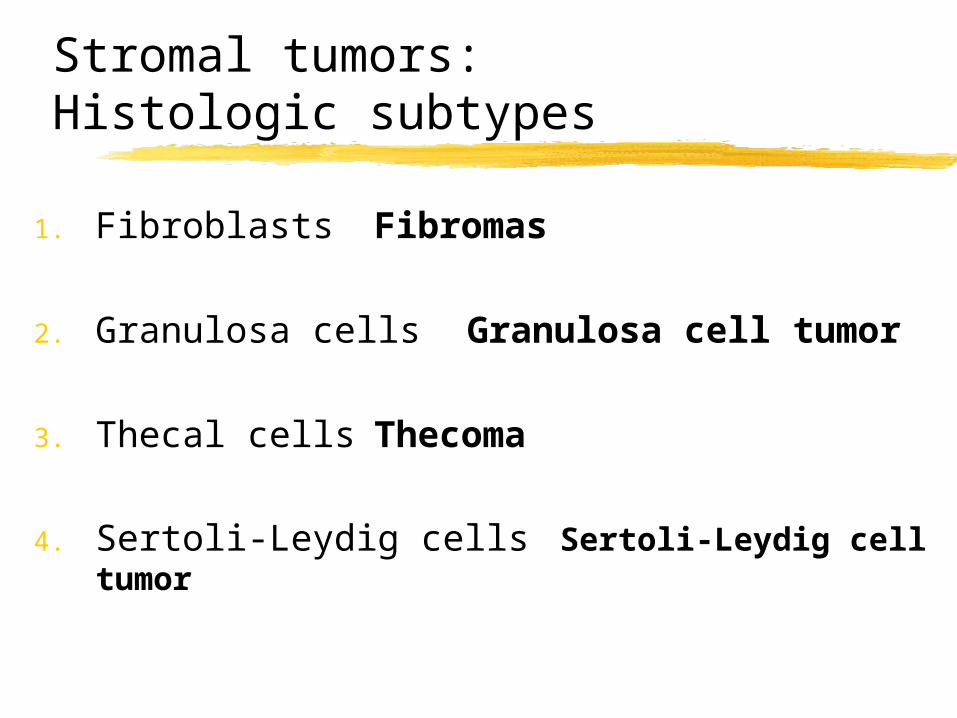
Stromal tumors: Histologic subtypes
1. Fibroblasts Fibromas
2. Granulosa cells Granulosa cell tumor
3. Thecal cells Thecoma
4. Sertoli-Leydig cells Sertoli-Leydig cell tumor
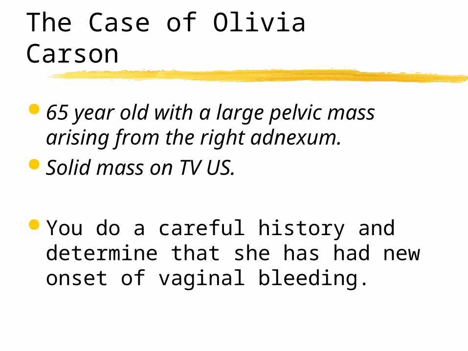
The Case of Olivia Carson
65 year old with a large pelvic mass arising from the right adnexum.
Solid mass on TV US.
You do a careful history and determine that she has had new onset of vaginal bleeding.
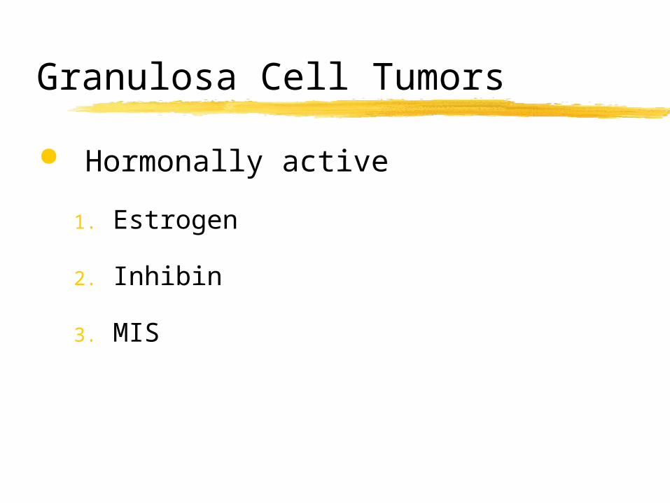
Granulosa Cell Tumors
Hormonally active
1. Estrogen
2. Inhibin
3. MIS
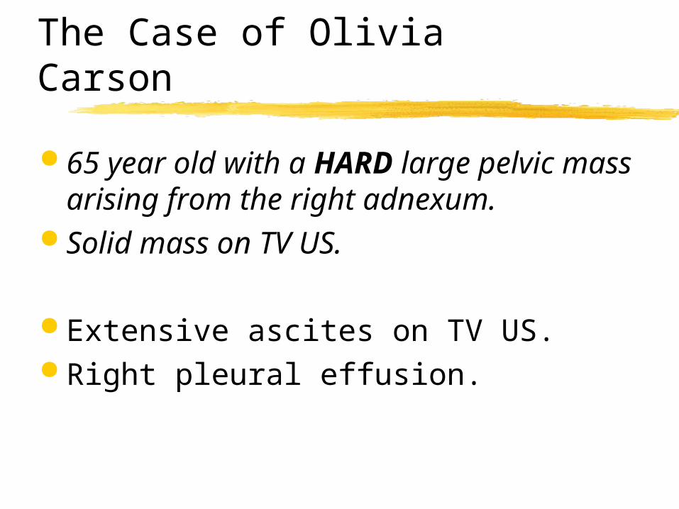
The Case of Olivia Carson
65 year old with a HARD large pelvic mass arising from the right adnexum.
Solid mass on TV US.
Extensive ascites on TV US. Right pleural effusion.
Meig’s syndrome
Fibroma Ascites (>200 ml) Hydrothorax
The Case of Olivia Carson
25 year old with acute pain and a HARD large pelvic mass arising from the right adnexum.
Solid mass on TV US.
Extensive ascites on TV US. Right pleural effusion.
You note multiple skin lesions.
Gorlin’s Syndrome
Ovarian fibromas Young women Multiple basal cell nevi and
carcinomas Dental cysts Skeletal abnormalities Autosomal dominant
The Case of Olivia Carson
65 year old with a large pelvic mass arising from the right adnexum.
Solid mass on TV US.
What will you do to treat her?
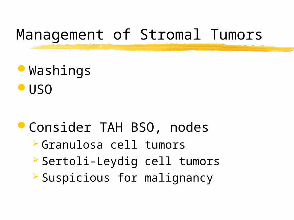
Management of Stromal Tumors
Washings USO
Consider TAH BSO, nodes Granulosa cell tumors Sertoli-Leydig cell tumors Suspicious for malignancy
The Case of Olivia Carson
You are a generalist in the community. You have a new patient.
She is a 15 year old with a large pelvic mass arising from the right adnexum.
What is the most likely tumor?
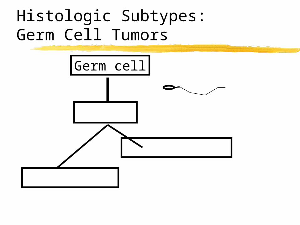
Histologic Subtypes: Germ Cell Tumors
Germ cell
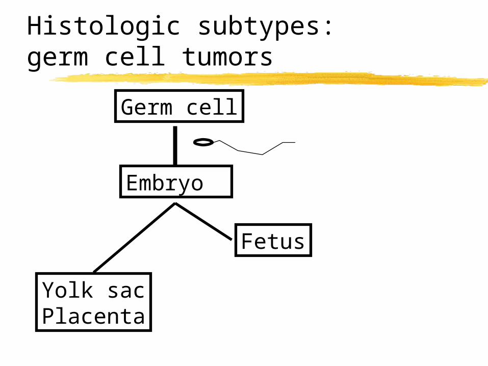
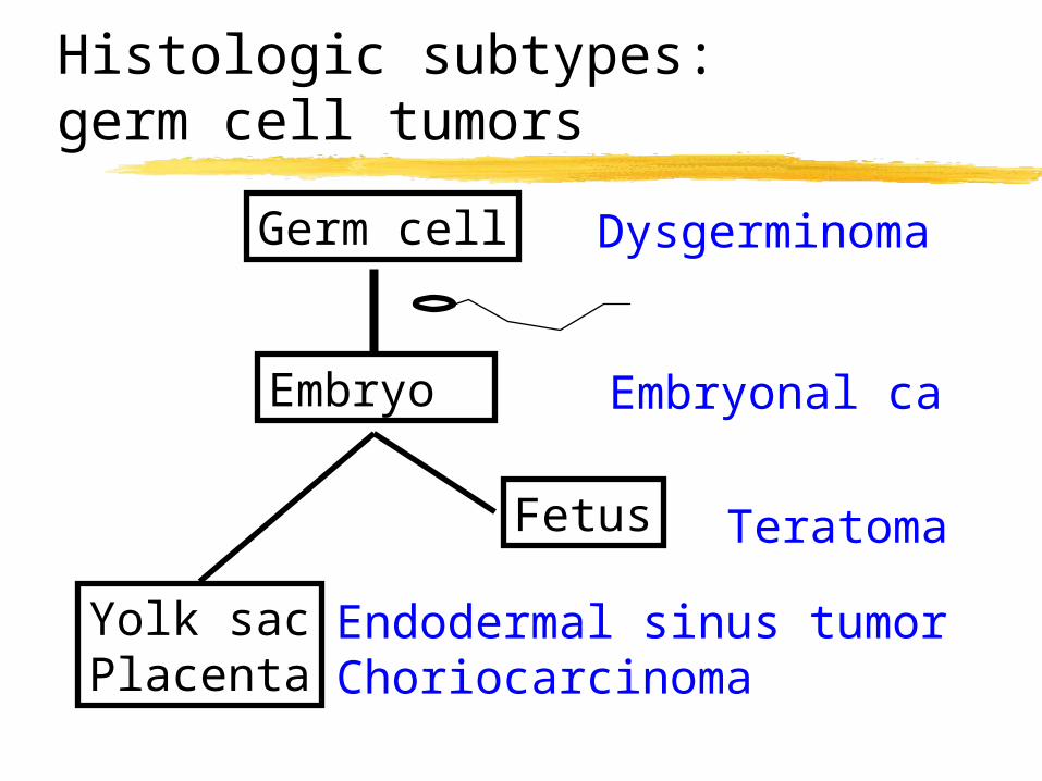
Histologic subtypes: germ cell tumors
Germ cell
Embryo
Fetus
Yolk sacPlacenta
Histologic subtypes: germ cell tumors
Germ cell
Embryo
Fetus
Yolk sacPlacenta
Dysgerminoma
Embryonal ca
Teratoma
Endodermal sinus tumorChoriocarcinoma
The Case of Olivia Carson
15 year old with a large pelvic mass arising from the right adnexum.
What pre-op labs do you want?
Tumor Markers: Germ Cell Tumors
LDH AFP hCG
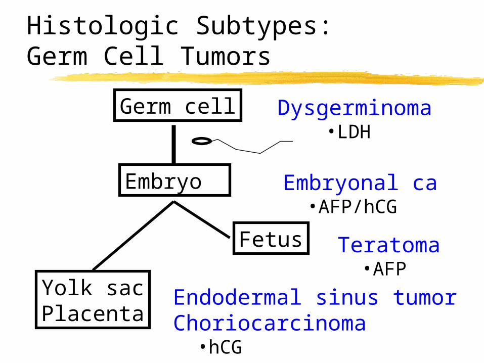
Histologic Subtypes: Germ Cell Tumors
Germ cell
Embryo
Fetus
Yolk sacPlacenta
Dysgerminoma•LDH
Embryonal ca•AFP/hCG
Teratoma•AFP
Endodermal sinus tumorChoriocarcinoma
•hCG

The Case of Olivia Carson
15 year old with a large pelvic mass arising from the right adnexum.
What will you do to treat her?
Germ Cell Tumors: Treatment
USO plus staging Chemotherapy
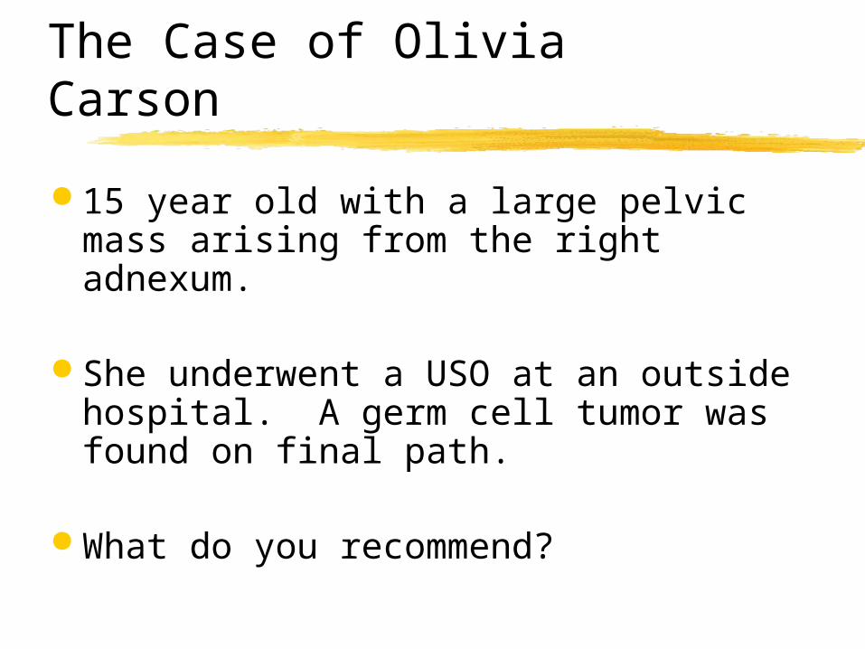
The Case of Olivia Carson
15 year old with a large pelvic mass arising from the right adnexum.
She underwent a USO at an outside hospital. A germ cell tumor was found on final path.
What do you recommend?
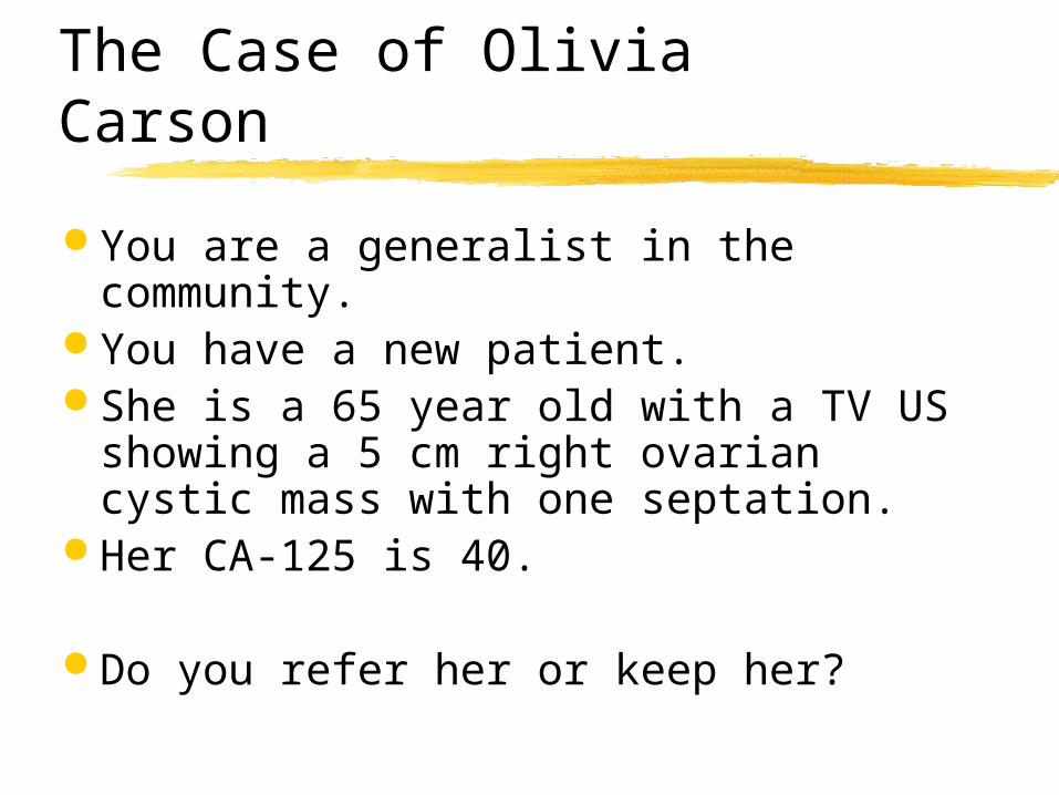
The Case of Olivia Carson
You are a generalist in the community. You have a new patient. She is a 65 year old with a TV US
showing a 5 cm right ovarian cystic mass with one septation.
Her CA-125 is 40.
Do you refer her or keep her?
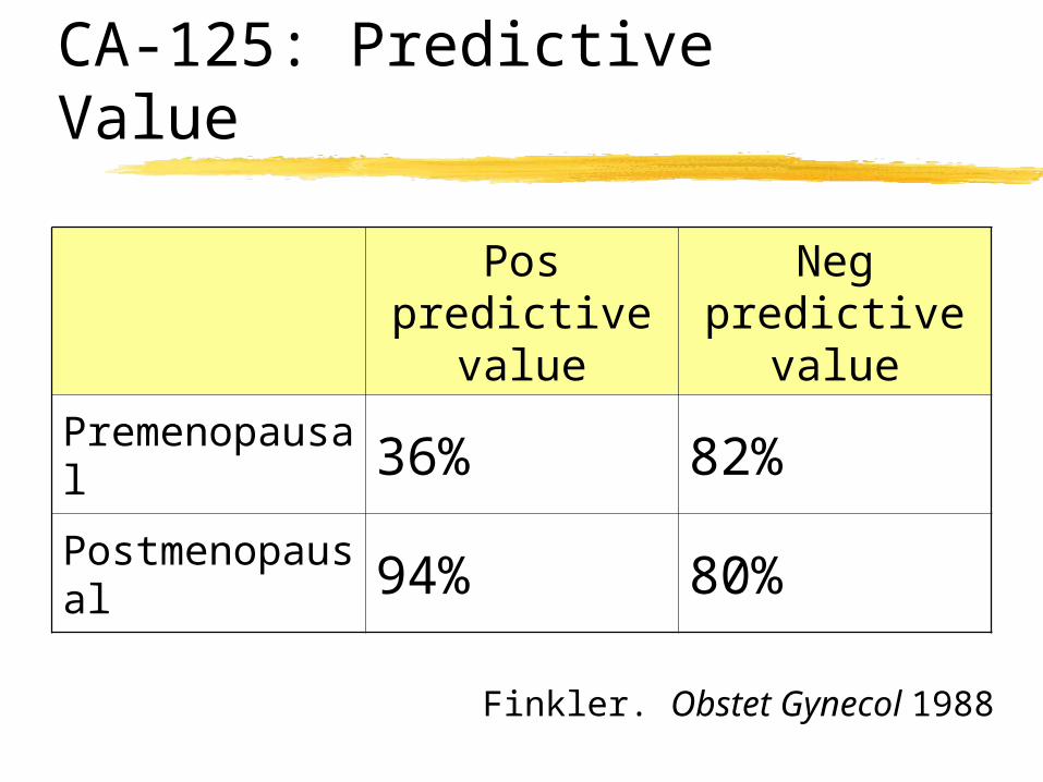
CA-125: Predictive Value
Finkler. Obstet Gynecol 1988
Pos predictive
value
Neg predictive
valuePremenopausal 36% 82%
Postmenopausal 94% 80%
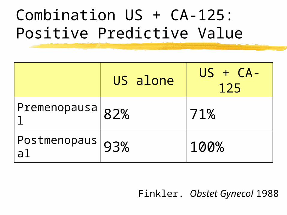
Combination US + CA-125: Positive Predictive Value
Finkler. Obstet Gynecol 1988
US aloneUS + CA-
125
Premenopausal 82% 71%
Postmenopausal 93% 100%
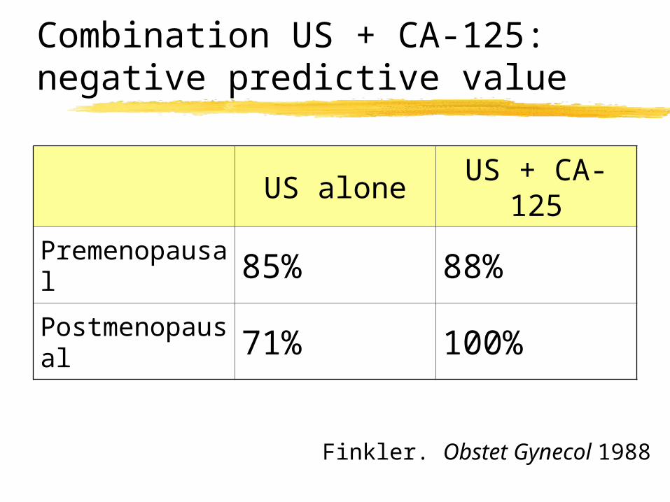
Combination US + CA-125: negative predictive value
Finkler. Obstet Gynecol 1988
US aloneUS + CA-
125
Premenopausal 85% 88%
Postmenopausal 71% 100%
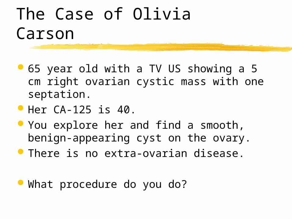
The Case of Olivia Carson
65 year old with a TV US showing a 5 cm right ovarian cystic mass with one septation.
Her CA-125 is 40. You explore her and find a smooth, benign-
appearing cyst on the ovary. There is no extra-ovarian disease.
What procedure do you do?
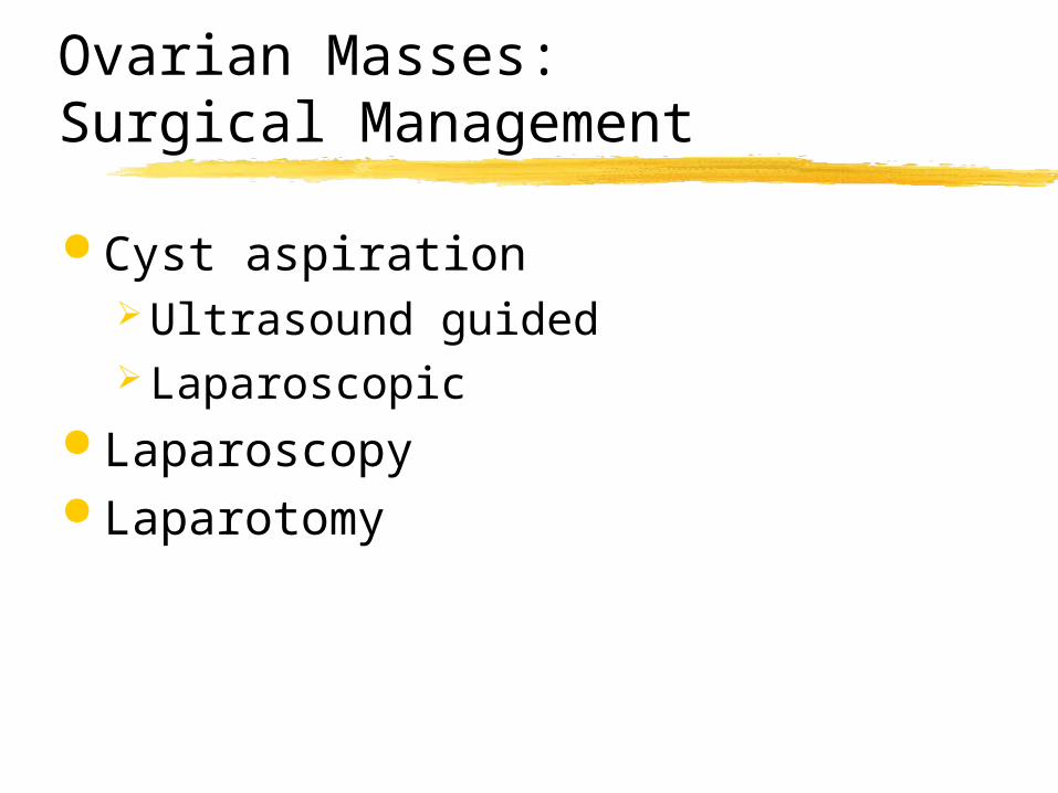
Ovarian Masses: Surgical Management
Cyst aspiration Ultrasound guided Laparoscopic
Laparoscopy Laparotomy
Ovarian Cyst Aspiration
Benign appearing cyst
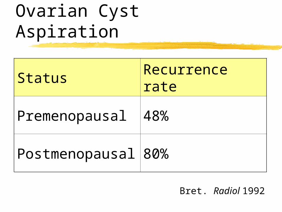
Ovarian Cyst Aspiration
Bret. Radiol 1992
Status Recurrence rate
Premenopausal 48%
Postmenopausal 80%
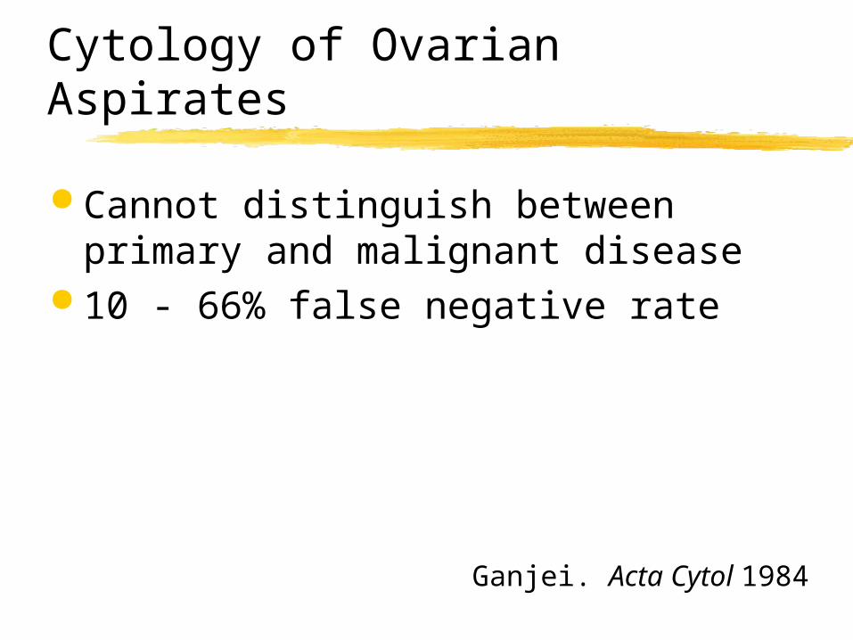
Cytology of Ovarian Aspirates
Cannot distinguish between primary and malignant disease
10 - 66% false negative rate
Ganjei. Acta Cytol 1984

Ovarian Cyst Aspiration
Limited value High recurrence rate Unreliable cytology ? Upstaging a malignant tumor
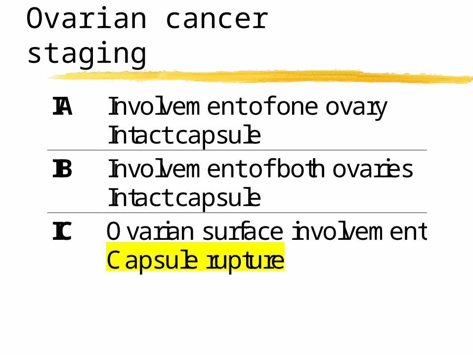
Ovarian cancer staging
IA Involvement of one ovaryIntact capsule
IB Involvement of both ovariesIntact capsule
IC Ovarian surface involvementCapsule rupture
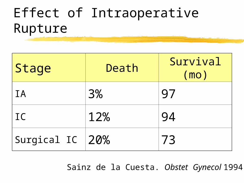
Effect of Intraoperative Rupture
Sainz de la Cuesta. Obstet Gynecol 1994
Stage DeathSurvival
(mo)
IA 3% 97
IC 12% 94
Surgical IC 20% 73
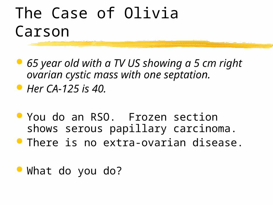
The Case of Olivia Carson
65 year old with a TV US showing a 5 cm right ovarian cystic mass with one septation.
Her CA-125 is 40.
You do an RSO. Frozen section shows serous papillary carcinoma.
There is no extra-ovarian disease.
What do you do?
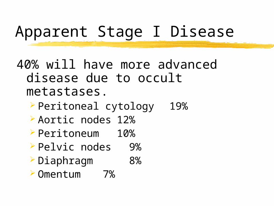
Apparent Stage I Disease
40% will have more advanced disease due to occult metastases. Peritoneal cytology 19% Aortic nodes 12% Peritoneum 10% Pelvic nodes 9% Diaphragm 8% Omentum 7%
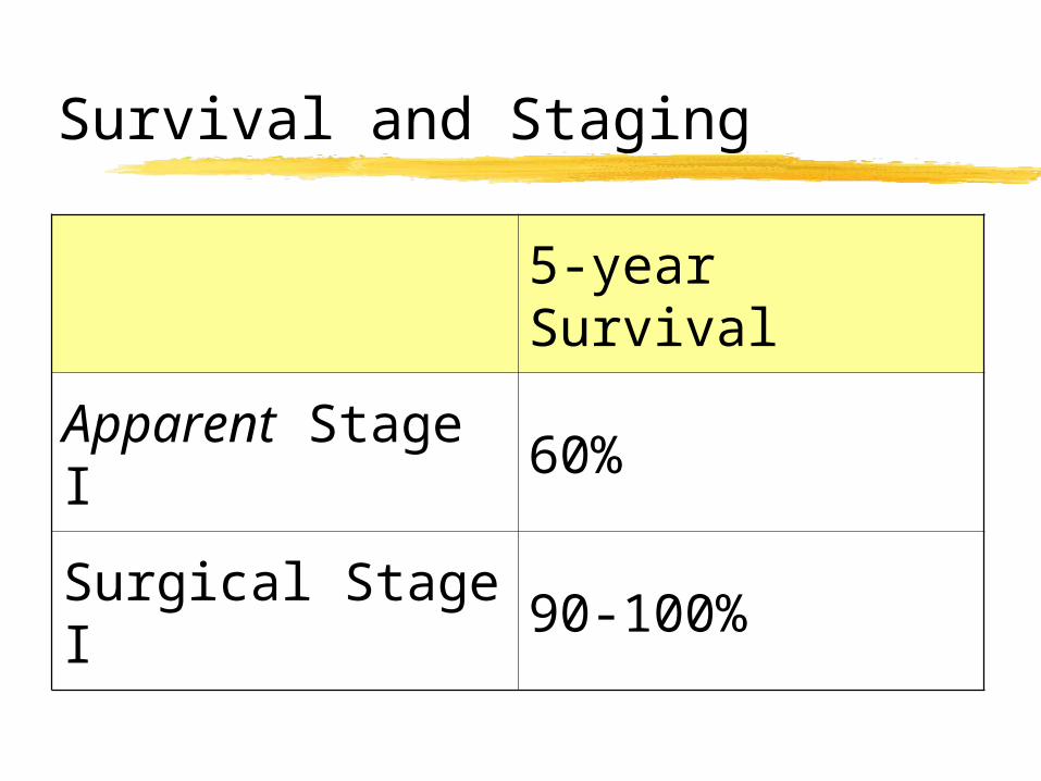
Survival and Staging
5-year Survival
Apparent Stage I 60%
Surgical Stage I 90-100%
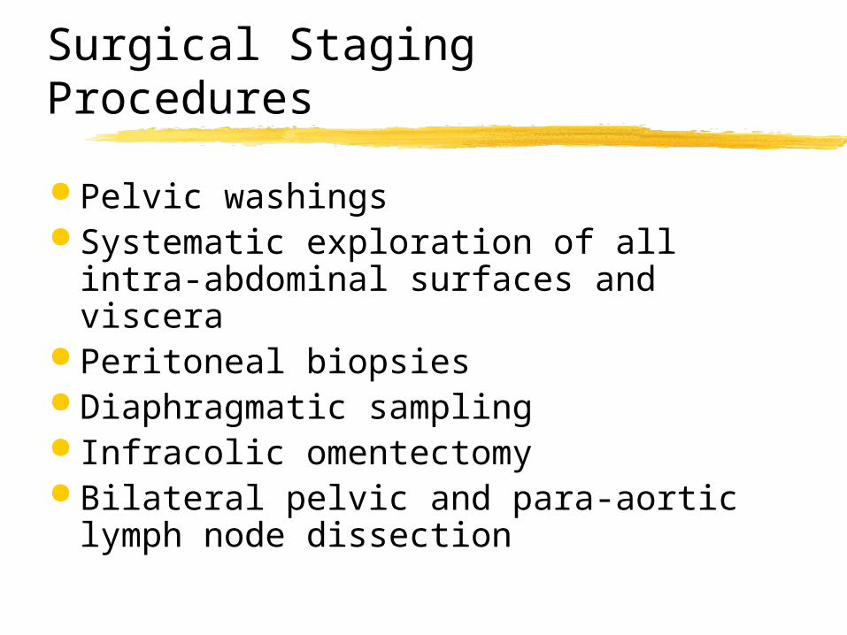
Surgical Staging Procedures
Pelvic washings Systematic exploration of all intra-
abdominal surfaces and viscera Peritoneal biopsies Diaphragmatic sampling Infracolic omentectomy Bilateral pelvic and para-aortic
lymph node dissection
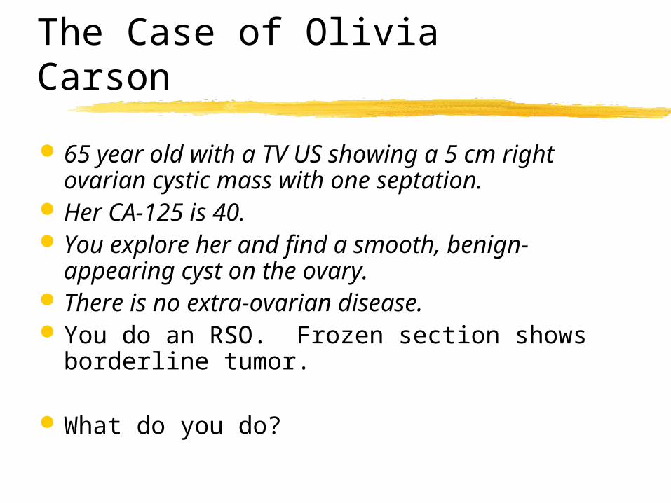
The Case of Olivia Carson
65 year old with a TV US showing a 5 cm right ovarian cystic mass with one septation.
Her CA-125 is 40. You explore her and find a smooth, benign-
appearing cyst on the ovary. There is no extra-ovarian disease. You do an RSO. Frozen section shows
borderline tumor.
What do you do?
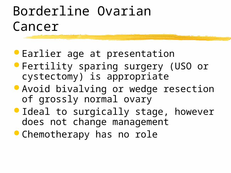
Borderline Ovarian Cancer
Earlier age at presentation Fertility sparing surgery (USO or
cystectomy) is appropriate Avoid bivalving or wedge resection
of grossly normal ovary Ideal to surgically stage, however
does not change management Chemotherapy has no role
Cytoreductive Surgery
Removal of the maximum amount of tumor possible.
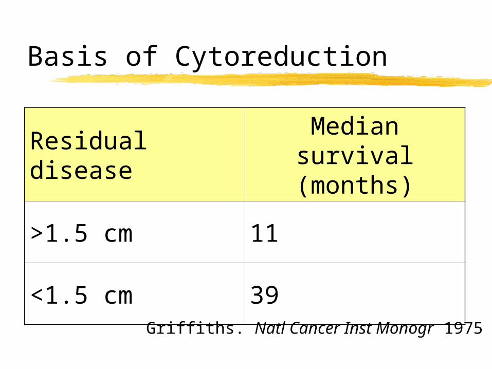
Basis of Cytoreduction
Griffiths. Natl Cancer Inst Monogr 1975
Residual diseaseMedian survival
(months)
>1.5 cm 11
<1.5 cm 39
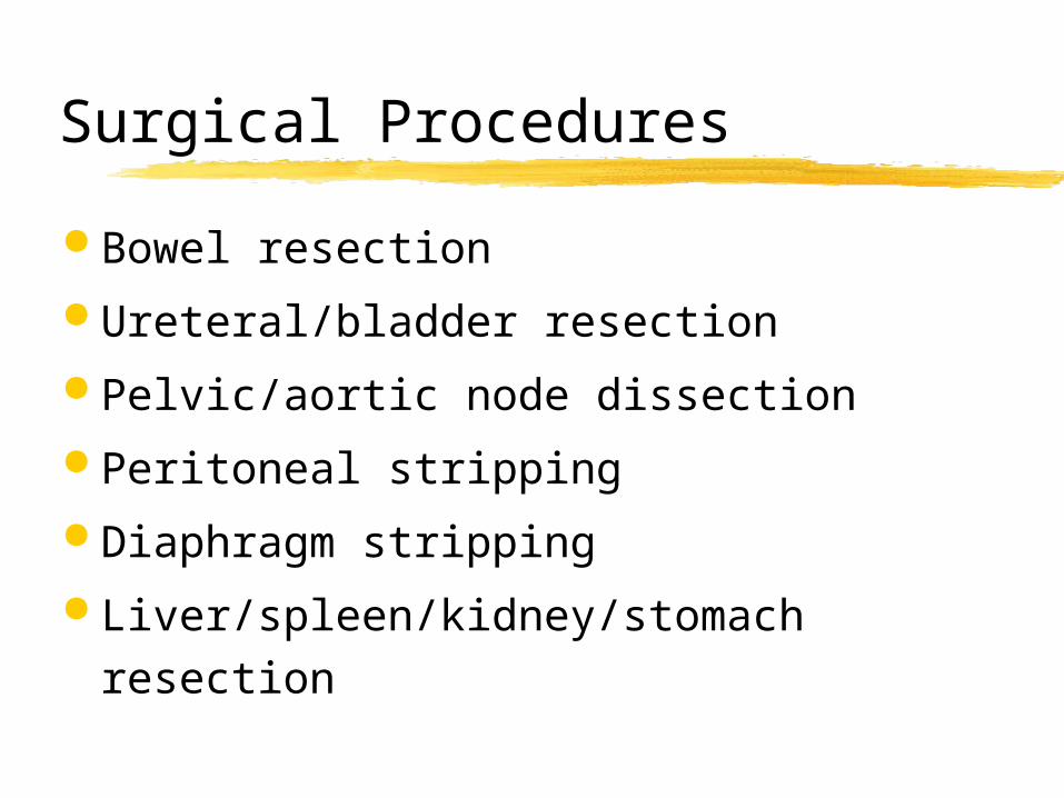
Surgical Procedures
Bowel resection
Ureteral/bladder resection
Pelvic/aortic node dissection
Peritoneal stripping
Diaphragm stripping
Liver/spleen/kidney/stomach resection
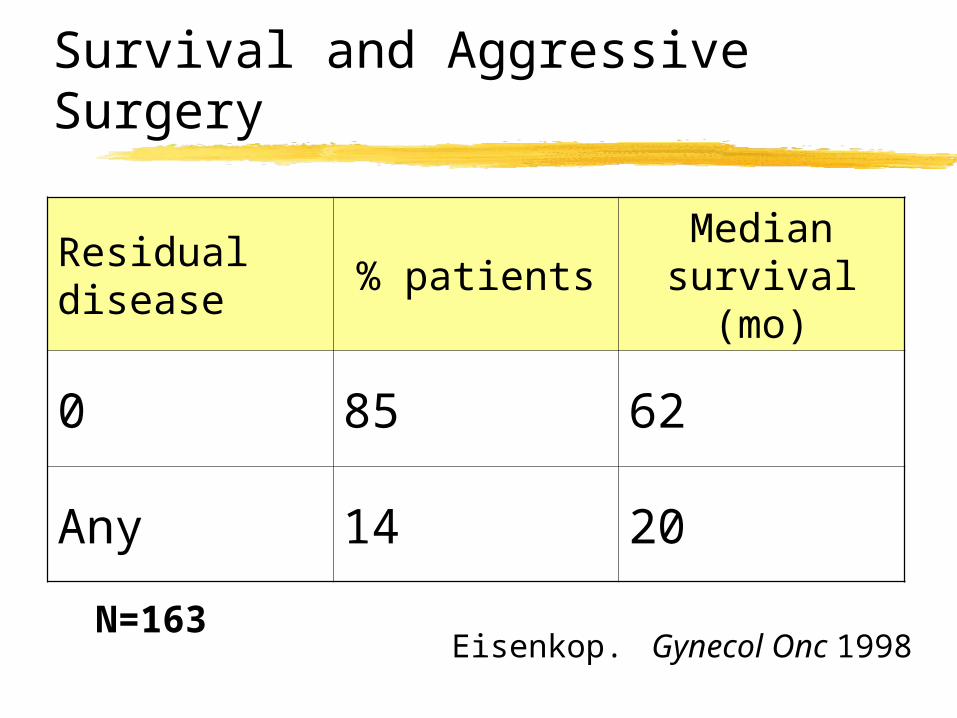
Survival and Aggressive Surgery
N=163Eisenkop. Gynecol Onc 1998
Residual disease
% patientsMedian
survival (mo)
0 85 62
Any 14 20
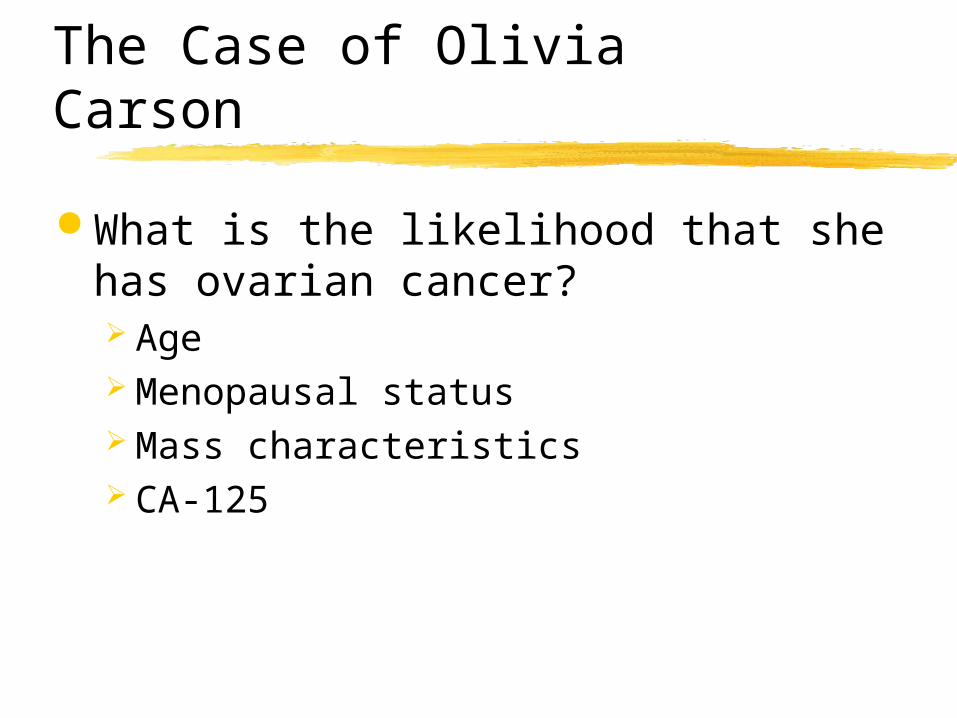
The Case of Olivia Carson
What is the likelihood that she has ovarian cancer? Age Menopausal status Mass characteristics CA-125
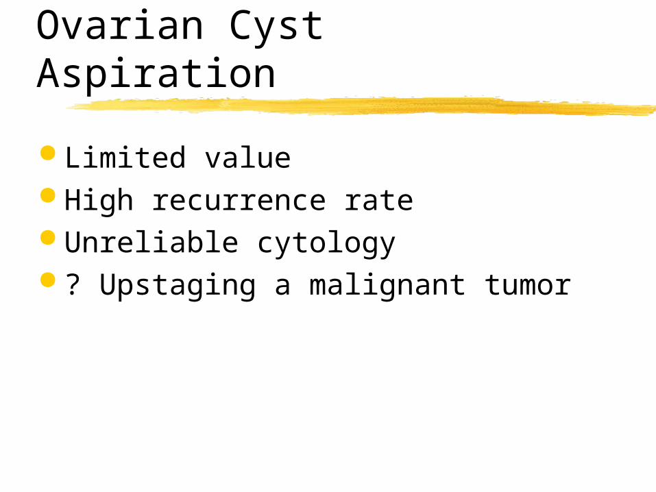
Ovarian Cyst Aspiration
Limited value High recurrence rate Unreliable cytology ? Upstaging a malignant tumor
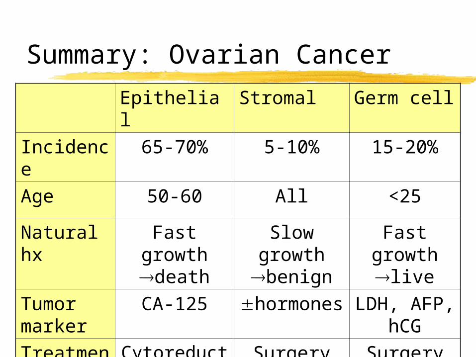
Summary: Ovarian Cancer
Epithelial Stromal Germ cell
Incidence 65-70% 5-10% 15-20%
Age 50-60 All <25
Natural hx
Fast growth death
Slow growth
benign
Fast growth live
Tumor marker
CA-125 hormones
LDH, AFP, hCG
Treatment
Cytoreduction
Chemo
Surgery Surgery Chemo
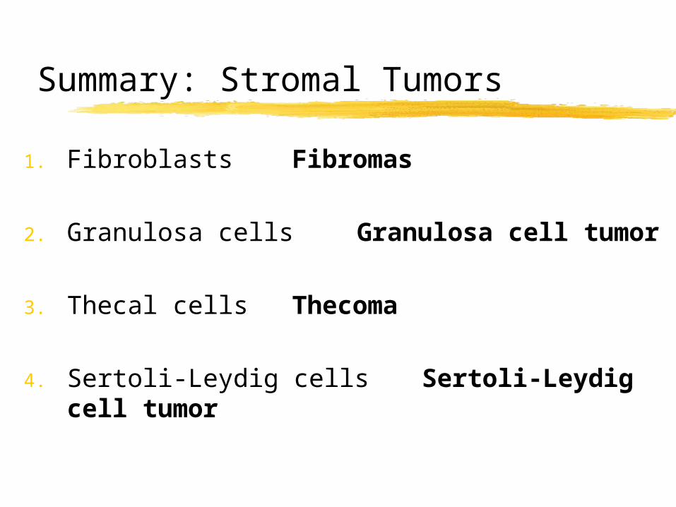
Summary: Stromal Tumors
1. Fibroblasts Fibromas
2. Granulosa cells Granulosa cell tumor
3. Thecal cells Thecoma
4. Sertoli-Leydig cells Sertoli-Leydig cell tumor
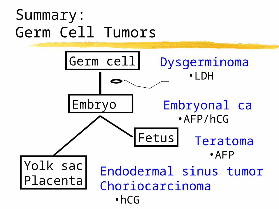
Summary: Germ Cell Tumors
Germ cell
Embryo
Fetus
Yolk sacPlacenta
Dysgerminoma•LDH
Embryonal ca•AFP/hCG
Teratoma•AFP
Endodermal sinus tumorChoriocarcinoma
•hCG
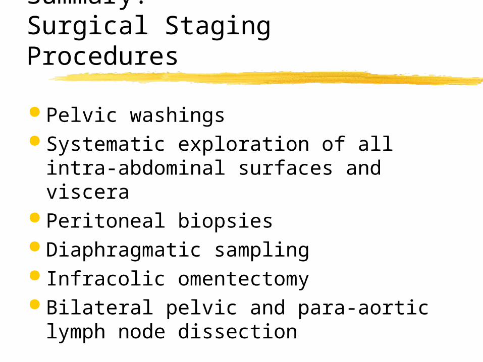
Summary:Surgical Staging Procedures
Pelvic washings Systematic exploration of all intra-
abdominal surfaces and viscera Peritoneal biopsies Diaphragmatic sampling Infracolic omentectomy Bilateral pelvic and para-aortic
lymph node dissection