Embed Size (px)
Citation preview

Ovarian Cancer, Renal Cancer, Urogenitary Tract Cancer, Urinary Bladder Cancer, Cervical Uterine Cancer, Skin Cancer, Leukemia, Multiple Myeloma and Sarcoma

Methods of Cancer Diagnosis, Therapy, and Prognosis
Volume 6
For other titles published in this series, go to www.springer.com/series/8172

Methods of Cancer Diagnosis, Therapy, and Prognosis
Volume 6
Ovarian Cancer, Renal Cancer, Urogenitary Tract Cancer, Urinary Bladder Cancer, Cervical Uterine Cancer, Skin Cancer, Leukemia, Multiple Myeloma and Sarcoma
Edited by
M.A. HayatDepartment of Biological Sciences,Kean University, Union, NJ, USA

ISBN 978-90-481-2917-1 e-ISBN 978-90-481-2918-8
Library of Congress Control Number: 2009929394
© Springer Science + Business Media B.V.No part of this work may be reproduced, stored in a retrieval system, or transmitted in any form or by any means, electronic, mechanical, photocopying, microfilming, recording or otherwise, without written permission from the Publisher, with the exception of any material supplied specifically for the purpose of being entered and executed on a computer system, for exclusive use by the purchaser of the work.
Printed on acid-free paper
springer.com
EditorM.A. HayatDepartment of Biological SciencesKean UniversityUnion, NJ, USA
2010

New technology, for better or for worse, will be used, as that is our nature.
Lewis Thomas
You have been given the key that opens the gates of heaven; the same key opens the gates of hell.
Writing at the entrance to a Buddhist temple

vii
Contributors
Laurent Alberti Centre Léon Bérard, 28, Rue Laennec, 69008 Lyon, France
Juan Luis Alcázar Department of Obstetrics and Gynecology, Clínica Universitaria de Navarra, University of Navarra, Avenida Pio XII, 36 Pamplona 31008, Spain
Damien Ambrosetti Service d’Anatomie Pathologique, Hôpital Pasteur, 30 Avenue de la Voie Romaine, 06000 Nice, France
Yaser Atlasi Department of Genetics, Faculty of Basic Science, Tarbiat Modares University, P.O. Box: 1411-175, Tehran, Iran
Allyson C. Baker University of Alabama at Birmingham, NP 3537, 619 19th Street South, Birmingham, AL 35249, USA
Surinder K. Batra Department of Biochemistry and Molecular Biology, College of Medicine, Eppley Cancer Institute 7052 Durham Research Center, University of Nebraska Medical Center, 985870 Nebraska Medical Center, Omaha, NE 68198-5870, USA
Vladimir Bilim Department of Urology, Yamagata University Faculty of Medicine, Iida-nishi 2-2-2, Yamagata 990-9585, Japan
Michael J. Birrer Cell and Cancer Biology, National Cancer Institute, 37 Convent Drive, Room 1068, Bethesda, MD 20892, USA
Jean-Yves Bla INSERM U590, Centre Léon Bérard, Rue Laennec, 69008 Lyon, France
Malte Böhm Department of Urology, Otto-von-Guericke Universität, Leipziger Str. 44, D-39120 Magdeburg, Germany
Kristin L.M. Boylan Department of Laboratory Medicine and Pathology, The University of Minnesota Medical School, Minneapolis, MN 55455, USA
Laura Brousseau Centre Léon Bérard, 28, rue Laennec, 69008 Lyon, France
Fanny Burel-Vandenbos Service d’Anatomie Pathologique, Hôpital Pasteur, 30 Avenue de la Voie Romaine, 06000 Nice, France
vii

viii Contributors
Gabriel Caponetti Department of Pathology, Baystate Medical Center, Tufts School of Medicine, 759 Chestnut Street, Springfield, MA 01109, USA
Nathalie Cardot-Leccia Service d’Anatomie Pathologique, Hôpital Pasteur, 30 Avenue de la Voie Romaine, 06000 Nice, France
Philippe Cassier Hôpital Edouard Herriot, Service d’Oncologie Médicale, Pavillon E, 5 Place d’Arsonval, 69003 Lyon, France
Tridib Chakraborty Division of Biochemistry, Department of Pharmaceutical Technology, Jadavpur University, P.O. Box 17028, Calcutta 700032, West Bengal, India
Malay Chatterjee Division of Biochemistry, Department of Pharmaceutical Technology, Jadavpur University, P.O. Box 17028, Calcutta 700032, West Bengal, India
David Chhieng University of Alabama at Birmingham, 619 19th Street South, Birmingham, AL 35249, USA
William A. Cliby Division of Gynecologic Surgery, Department of Obstetrics and Gynecology, Mayo Clinic, 200 First Street SW, Rochester, MN 55905, USA
Joseph P. Connor University of Wisconsin-Madison, CSC H4/656, 600 Highland Avenue, Madison, WI 53792, USA
Isabelle Ray Coquard Centre Léon Bérard, 28, Rue Laennec, 69008 Lyon, France
Dean Daya Department of Pathology and Nuclear Medicine, Henderson General Hospital, McMaster University, 711 Concession Street, Hamilton, Ontario L8V IC3, Canada
Anne-Valérie Decouvelaere Centre Léon Bérard, 28, Rue Laennec, 69008 Lyon, France
Bruce J. Dezube Department of Medicine, Beth Israel Deaconess Medical Center, Harvard Medical School, Boston, MA 02215, USA
Sean C. Dowdy Division of Gynecologic Surgery, Department of Obstetrics and Gynecology, Mayo Clinic, 200 First Street SW, Rochester, MN 55905, USA
Armelle Dufresne Hôpital Edouard Herriot, Service d’Oncologie médicale, Pavillon E, 5 Place d’Arsonval, 69003 Lyon, France
Jérôme Fayette Centre Léon Bérard, 28, Rue Laennec, 69008 Lyon, France
Michael Fiegl Department of Internal Medicine V, Hemato-Oncology, Innsbruck Medical University, Anichstrasse 35, A-6020 Innsbruck, Austria
Olivier Gheysens Department of Nuclear Medicine, University Hospital KU Leuven, Herestraat 49, B-3000 Leuven, Belgium
Contributors ix
Elfriede Greimel Department of Obstetrics and Gynecology, Medical University Graz, Auenbruggerplatz 14, A-8036 Graz, Austria
Perry W. Grigsby Department of Radiation Oncology, Mallinckrodt Institute of Radiology, Washington University School of Medicine, Box 8224, 4921 Parkview Place, Lower Level, St. Louis, MO 63110, USA
William E. Grizzle University of Alabama at Birmingham, 619 19th Street South, Birmingham, AL 35249, USA
Jennifer A. A. Gubbels University of Wisconsin-Madison, CSC H4/656, 600 Highland Avenue, Madison, WI 53792, USA
Christian Hafner Department of Dermatology, University of Regensburg, Franz-Josefstrauss-Allee 11, Regensburg 93042, Germany
Hiroshi Hashimoto Department of Pathology and Oncology, School of Medicine, University of Occupational and Environmental Health, 1-1 Iseigaoka, Yahatanishi-ku, Kitakyushu 807-8555, Japan
Juliette Haudebourg Service d’Anatomie Pathologique, Hôpital Pasteur, 30 Avenue de la Voie Romaine, 06000 Nice, France
Masanori Hisaoka Department of Pathology and Oncology, School of Medicine, University of Occupational and Environmental Health, 1-1 Iseigaoka, Yahatanishi-ku, Kitakyushu 807-8555, Japan
Andrew Horvai University of California, San Francisco, 1600 Divisadero Drive, B220, San Francisco, CA 94115, USA
Kazuhiko Ino Department of Obstetrics and Gynecology, Nagoya University Graduate School of Medicine, 65 Tsurumai-cho, Showa-ku, Nagoya 466-8550, Japan
Toshiyuki Itoi Department of Urology, Yamagata University Faculty of Medicine, Iida-nishi 2-2-2, Yamagata 990-9585, Japan
Seyed Mehdi Jafarnejad, Department of Genetics, Faculty of Basic Science, Tarbiat Modares University, P.O. Box: 1411-175, Tehran, Iran
Hiroaki Kajiyama Department of Obstetrics and Gynecology, Nagoya University Graduate School of Medicine, 65 Tsurumai-cho, Showa-ku, Nagoya 466-8550, Japan
Shingo Kato Research Central Hospital for Charged Particle Therapy, National Institute of Radiological Sciences, 4-9-1 Anagawe, Inage-ku, Chiba 263-8555, Japan
Fumitaka Kikkawa Department of Obstetrics and Gynecology, Nagoya University Graduate School of Medicine, 65 Tsurumai-cho, Showa-ku, Nagoya 466-8550, Japan
Seung Hyup Kim Department of Radiology, Seoul National University Bundang Hospital 300, Gumi-dong, Bundang-Gu, Songnam-si, Gyeonggi-do, 463-707, Korea

x Contributors
Tobias Klatte Department of Urology, Otto-von-Guericke Universität, Leipziger Str. 44, D-39120 Magdeburg, Germany
Michael Landthaler Department of Dermatology, Regensburg University Medical Center, Franz-Josef-Strauss-Allee 11, Regensburg 93053, Germany
Hak Jong Lee Department of Radiology, Seoul National University Bundang Hospital 300, Gumi-dong, Bundang-Gu, Songnam-si, Gyeonggi-do, 463-707, Korea
Subodh M. Lele Department of Biochemistry and Molecular Biology, College of Medicine, Eppley Cancer Institute, 7052 Durham Research Center, University of Nebraska Medical Center, 985870 Nebraska Medical Center, Omaha, NE 68198-5870, USA
Jonathan S. Lewin Department of Radiology, Duke University Medical Center, Duke North-Room 1417, Erwin Road, Durham, NC 27710, USA
Lilie L. Lin Department of Radiation Oncology, Mallinckrodt Institute of Radiology, Washington University School of Medicine, 4921 Parkview Place, Lower Level, St. Louis, MO 63110, USA
Yair Lotan Department of Urology, The University of Texas Southwestern Medical Center, 5323 Harry Hines Boulevard, Dallas, TX 75390, USA
Ryo Maruyama Department of Urology, Yamagata University Faculty of Medicine, Iida-nishi 2-2-2, Yamagata 990-9585, Japan
Atsuji Matsuyama Department of Pathology and Oncology, School of Medicine, University of Occupational and Environmental Health, 1-1 Iseigaoka, Yahatanishi-ku, Kitakyushu 807-8555, Japan
Milena M. Maule Cancer Epidemiology Unit, CeRMS and CPO-Piemonte, University of Turin, 10126 Torino, Italy
Jiri Mayer Department of Internal Medicine V, Hemato-Oncology, Masaryk University Hospital, Jihlavska 20, CZ 62500 Brno, Czech Republik
Pierre Méeus Centre Léon Bérard, 28, Rue Laennec, 69008 Lyon, France
Elmar M. Merkle Department of Radiology, Duke University Medical Center, Duke North-Room 1417, Erwin Road, Durham, NC 27710, USA
Jean-Francois Michiels Service d’Anatomie Pathologique, Hôpital Pasteur, 30 Avenue de la Voie Romaine, 06000 Nice, France
Samuel C. Mok Department of Gynecologic Oncology, M.D. Anderson Cancer Center, T403908, 1515 Holcombe Boulevard, Houston, TX 77030, USA

Contributors xi
Felix M. Mottaghy Department of Nuclear Medicine, University Hospital KU Leuven, Herestraat 49, B-3000 Leuven, Belgium
Seyed Javad Mowla Department of Genetics, Faculty of Basic Science, Tarbiat Modares University, P.O. Box: 1411-175, Tehran, Iran
Akihiro Nawa Department of Obstetrics and Gynecology, Nagoya University Graduate School of Medicine, 65 Tsurumai-cho, Showa-ku, Nagoya 466-8550, Japan
Rendon C. Nelson Department of Radiology, Duke University Medical Center, Duke North-Room 1417, Erwin Road, Durham, NC 27710, USA
Torsten O. Nielsen PGY3 Anatomical Pathology, 1500 JPPN Vancouver General Hospital, 899 12th Avenue W, Vancouver, BC V5Z 1M9, Canada
Tatsuya Ohno Research Central Hospital for Charged Particle Therapy, National Institute of Radiological Sciences, 4-9-1 Anagawe, Inage-ku, Chiba 263-8555, Japan
Yasemin Ozluk Department of Pathology, Istanbul University, Faculty of Medicine, Capa, Topkapi 34390, Istanbul
Manish S. Patankar University of Wisconsin-Madison, CSC H4/656, 600 Highland Avenue, Madison, WI 53792, USA
Liron Pantanowitz Department of Pathology, Baystate Medical Center, Tufts School of Medicine, 759 Chestnut Street, Springfield, MA 01109, USA
Louis L. Pisters Department of Urology, The University of Texas M.D. Anderson Cancer Center, 1515 Holcombe Boulevard, Houston, TX 77030, USA
Moorthy P. Ponnusamy Department of Biochemistry and Molecular Biology, College of Medicine, Eppley Cancer Institute, 7052 Durham Research Center, University of Nebraska Medical Center, 985870 Nebraska Medical Center, Omaha, NE 68198-5870, USA
Francine M. Quan Division of Hematology/Oncology, East Carolina University Brody School of Medicine, 600 Moyle Boulevard, Greenville, NC 27858, USA
Walter D.Y. Quan Division of Hematology/Oncology, East Carolina University Brody School of Medicine, Brody 3E-127, 600 Moyle Boulevard, Greenville, NC 27858, USA
Ajay Rana Division of Biochemistry, Department of Pharmaceutical Technology, Jadavpur University, P.O. Box 17028, Calcutta 700032, West Bengal, India
Basabi Rana Division of Biochemistry, Department of Pharmaceutical Technology, Jadavpur University, P.O. Box 17028, Calcutta 700032, West Bengal, India

xii Contributors
Dominique Ranchère Centre Léon Bérard, 28, Rue Laennec, 69008 Lyon, France
Lorenzo Richiardi Cancer Epidemiology Unit, University of Turin, Via Santena 7, 10126 Torino, Italy
Alexander Roesch Department of Dermatology, Regensburg University Medical Center, Franz-Josef-Strauss-Allee 11, Regensburg 93053, Germany
Albert Rübben Department of Dermatology, University Hospital RWTH Aachen, Pauwelsstrasse 30, D-52074 Aachen, Germany
Naoki Sasaki Department of Obstetrics and Gynecology, National Defense Medical College, 3-2 Namiki, Tokorozawa, Saitama 359-8513, Japan
Kiyosumi Shibata Department of Obstetrics and Gynecology, Nagoya University Graduate School of Medicine, 65 Tsurumai-cho, Showa-ku, Nagoya 466-8550, Japan
Ajay P. Singh Department of Biochemistry and Molecular Biology, College of Medicine, Eppley Cancer Institute, 7052 Durham Research Center, University of Nebraska Medical Center, 985870 Nebraska Medical Center, Omaha, NE 68198-5870, USA
Amy P.N. Skubitz Department of Laboratory Medicine and Pathology, The University of Minnesota Medical School, MMC 609, 420 Delaware Street, SE, Minneapolis, MN 55455, USA
Keith M. Skubitz Department of Medicine, The Masonic Cancer Center, Minneapolis, MN 55455, USA
Philippe E. Spiess Department of Urology, The University of Texas M.D. Anderson Cancer Center, 1515 Holcombe Boulevard, Houston, TX 77030, USA
Michael P. Stanley Cell and Cancer Biology, National Cancer Institute, 37 Convent Drive, Room 1068, Bethesda, MD 20892, USA
Toru Sugiyama Department of Obstetrics and Gynecology, National Defense Medical College, 3-2 Namiki, Tokorozawa, Saitama 359-8513, Japan
Marie-Pierre Sunyach Centre Léon Bérard, 28, Rue Laennec, 69008 Lyon, France
Monalisa Sur Department of Pathology and Nuclear Medicine, Henderson General Hospital, McMaster University, 711 Concession Street, Hamilton, Ontario L8V IC3, Canada
Robert S. Svatek Department of Urology, The University of Texas Southwestern Medical Center, 5323 Harry Hines Boulevard, Dallas, TX 75390, USA
Masashi Takano Department of Obstetrics and Gynecology, National Defense Medical College, 3-2 Namiki, Tokorozawa, Saitama 359-8513, Japan

Contributors xiii
Nizar M. Tannir Department of Urology, The University of Texas M.D. Anderson Cancer Center, 1515 Holcombe Boulevard, Houston, TX 77030, USA
Jefferson Terry PGY3 Anatomical Pathology, 1500 JPPN Vancouver General Hospital, 899 12th Avenue W, Vancouver, BC V5Z 1M9, Canada
Yoshihiko Tomita Department of Urology, Yamagata University Faculty of Medicine, Iida-nishi 2-2-2, Yamagata 990-9585, Japan
Hiroshi Tsuda Department of Obstetrics and Gynecology, Osaka City General Hospital, 2-13-22 Miyakojimahondori, Miyakojima, Osaka 5340021, Japan
Thomas Vogt Department of Dermatology, Regensburg University Medical Center, Franz-Josef-Strauss-Allee 11, Regensburg 93053, Germany
Eiko Yamamoto Department of Obstetrics and Gynecology, Nagoya University Graduate School of Medicine, 65 Tsurumai-cho, Showa-ku, Nagoya 466-8550, Japan
Preface
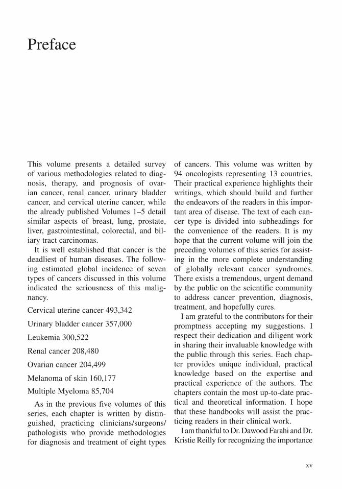
This volume presents a detailed survey of various methodologies related to diag-nosis, therapy, and prognosis of ovar-ian cancer, renal cancer, urinary bladder cancer, and cervical uterine cancer, while the already published Volumes 1–5 detail similar aspects of breast, lung, prostate, liver, gastrointestinal, colorectal, and bil-iary tract carcinomas.
It is well established that cancer is the deadliest of human diseases. The follow-ing estimated global incidence of seven types of cancers discussed in this volume indicated the seriousness of this malig-nancy.
Cervical uterine cancer 493,342
Urinary bladder cancer 357,000
Leukemia 300,522
Renal cancer 208,480
Ovarian cancer 204,499
Melanoma of skin 160,177
Multiple Myeloma 85,704
As in the previous five volumes of this series, each chapter is written by distin-guished, practicing clinicians/surgeons/pathologists who provide methodologies for diagnosis and treatment of eight types
of cancers. This volume was written by 94 oncologists representing 13 countries. Their practical experience highlights their writings, which should build and further the endeavors of the readers in this impor-tant area of disease. The text of each can-cer type is divided into subheadings for the convenience of the readers. It is my hope that the current volume will join the preceding volumes of this series for assist-ing in the more complete understanding of globally relevant cancer syndromes. There exists a tremendous, urgent demand by the public on the scientific community to address cancer prevention, diagnosis, treatment, and hopefully cures.
I am grateful to the contributors for their promptness accepting my suggestions. I respect their dedication and diligent work in sharing their invaluable knowledge with the public through this series. Each chap-ter provides unique individual, practical knowledge based on the expertise and practical experience of the authors. The chapters contain the most up-to-date prac-tical and theoretical information. I hope that these handbooks will assist the prac-ticing readers in their clinical work.
I am thankful to Dr. Dawood Farahi and Dr. Kristie Reilly for recognizing the importance
xv

of scholarship (research, writing, and pub-lishing) in an institution of higher education and for providing the resources for complet-ing this project. I appreciate receiving expert
help from Myrna Ortiz and Erin McNally in preparing this volume.
M.A. HayatMarch 2009
xvi Preface
The enormous burden of liver cancer on society becomes clear by considering the fact that approximately 625,000 new cases of this cancer are diagnosed globally each year. Distressingly, the number of deaths is approximately the same at 598,000 per year. Liver cancer, therefore, is the third most common cause of death from cancer. Survival rates for liver cancer are only 3–5% globally. In the United States, 19,160 new cases of liver cancer and 16,780 deaths were reported for 2007. The major risk factors for this cancer include prior infection with hepatitis B and C viruses, with the former more prevalent. Dietary exposure to fungus Aspergillus fumigatus (aflatoxins) also contributes to the incidence of liver cancer in many parts of the world. Tobacco use is the most serious preventable cause of cancer, as its use causes cancer of the lung, throat, mouth, liver, pancreas, urinary bladder, stomach, kidney, as well as other types. Alcohol-induced liver injury is another major risk factor for hepatocellular carcinoma (HCC).
In view of these devastating statistics, the urgency of deciphering the molecular mech-anism underlying this disease, perfecting reliable diagnostic methods, understanding risk factors, developing effective targeted drugs, improving other treatments, assessing the effectiveness of therapies, and providing improved care for post-treatment patients, becomes apparent. This volume provides up-to-date information on the above-mentioned aspects of liver cancer; specifically, details of the methodologies used are included. The other seven volumes in this series provide similar information on other types of can-cers.
This series of handbooks has taken the unique approach of discussing cancer diagno-sis, treatment, and prognosis in the same volume. It is pointed out that this vast subject cannot be fully discussed by only one author. This is the primary reason for inviting a large number of oncologists/clinicians/surgeons to write each of the eight volumes of this series of handbooks. Another advantage of involving more than one author is to present different points of view on a specific controversial aspect of cancer. I hope these goals were accomplished in this and other published volumes of this series.
IntroductionM.A. Hayat
xvii
Contents of Volumes 1, 2, 3, 4 and 5
Volume 1
1. Breast Cancer: An Introduction
2. Breast Cancer: Computer-Aided Detection
3. Sebaceous Carcinoma of the Breast: Clinicopathologic Features
4. Breast Cancer: Detection by In-Vivo Imaging of Angiogenesis
5. Breast and Prostate Biopsies: Use of Optimized High-Throughput MicroRNA Expression for Diagnosis (Methodology)
6. Familial Breast Cancer: Detection of Prevalent High-Risk Epithelial Lesions
7. Differentiation Between Benign and Malignant Papillary Lesions of Breast: Excisional Biopsy or Stereotactic Vacuum-Assisted Biopsy (Methodology)
8. Multicentric Breast Cancer: Sentinel Node Biopsy as a Diagnostic Tool
9. Breast Cancer Recurrence: Role of Serum Tumor Markers CEA and CA 15-3
10. Breast Cancer Patients Before, During or After Treatment: Circulating Tumor Cells in Peripheral Blood Detected by Multigene Real-Time Reverse Transcriptase-Polymerase Chain Reaction
11. Breast Cancer Patients: Diagnostic Epigenetic Markers in Blood
xix
xx Contents of Volumes 1, 2, 3, 4 and 5
12. Breast Cancer Patients: Detection of Circulating Cancer Cell-Related mRNA Markers with Membrane Array Method
13. Prediction of Metastasis and Recurrence of Breast Carcinoma: Detection of Survivin-Expressing Circulating Cancer Cells
14. Node-Negative Breast Cancer: Predictive and Prognostic Value of Peripheral Blood Cytokeratin-19 mRNA-Positive Cells
15. Breast and Colon Carcinomas: Detection with Plasma CRIPTO-1
16. Breast Cancer Risk in Women with Abnormal Cytology in Nipple Aspirate Fluid
17. Tissue Microarrays: Construction and Utilization for Biomarker Studies
18. Systematic Validation of Breast Cancer Biomarkers Using Tissue Microarrays: From Construction to Image Analysis
19. Phyllodes Tumors of the Breast: The Role of Immunohistochemistry in Diagnosis
20. Phyllodes Tumor of the Breast: Prognostic Assessment Using Immunohistochemistry
21. Metaplastic Breast Carcinoma: Detection Using Histology and Immunohistochemistry
22. Invasive Breast Cancer: Overexpression of HER-2 Determined by Immunohistochemistry and Multiplex Ligation-Dependent Probe Amplification
23. Operable Breast Cancer: Neoadjuvant Treatment (Methodology)
24. Chemotherapy for Breast Cancer
25. Locally Advanced Breast Cancer: Role of Chemotherapy in Improving Prognosis
26. Relevance of Dose-Intensity for Adjuvant Treatment of Breast Cancer

Contents of Volumes 1, 2, 3, 4 and 5 xxi
27. Advanced Breast Cancer: Treatment with Docetaxel/Epirubicin
28. Systemic Therapy for Breast Cancer: Using Toxicity Data to Inform Decisions
29. Chemotherapy for Metastatic Breast Cancer Patients Who Received Adjuvant Anthracyclines (An Overview)
30. Estrogen Receptor-Negative and HER-2/neu-Positive Locally Advanced Breast Carcinoma: Therapy with Paclitaxel and Granulocyte-Colony Stimulating Factor
31. Breast Cancer: Side Effects of Tamoxifen and Anastrozole
32. Breast Cancer: Expression of HER-2 and Epidermal Growth Factor Receptor as Clinical Markers for Response to Targeted Therapy
33. Young Breast Cancer Patients Undergoing Breast-Conserving Therapy: Role of BRCA1 and BRCA2
34. Radiation Therapy for Older Women with Early Breast Cancer
35. Acute Side Effects of Radiotherapy in Breast Cancer Patients: Role of DNA-Repair and Cell Cycle Control Genes
36. 18F-Fluorodeoxyglucose/Positron Emission Tomography in Primary Breast Cancer: Factors Responsible for False-Negative Results
37. Sentinel Lymph Node Surgery During Prophylactic Mastectomy (Methodology)
38. Breast Conservation Surgery: Methods
39. Lymph Node-Negative Breast Carcinoma: Assessment of HER-2/neu Gene Status as Prognostic Value
40. Multifocal or Multicentric Breast Cancer: Understanding Its Impact on Management and Treatment Outcomes
41. Are Breast Cancer Survivors at Risk for Developing Other Cancers?
xxii Contents of Volumes 1, 2, 3, 4 and 5
42. Distant Metastasis in Elderly Patients with Breast Cancer: Prognosis with Nodal Status
43. Concomitant Use of Tamoxifen with Radiotherapy Enhances Subcutaneous Breast Fibrosis in Hypersensitive Patients
44. Malignant Phyllodes Tumor of the Breast: Is Adjuvant Radiotherapy Necessary?
45. Locally Advanced Breast Cancer: Multidrug Resistance
46. Breast Cancer: Diagnosis of Recurrence Using 18 F-Fluorodeoxyglucose-Positron Emission Tomography/Computed Tomography
47. Role of Sentinel Lymph Node Biopsy in Ductal Carcinoma In Situ: Diagnosis and Methodology
48. Breast Conservation Treatment of Early Stage Breast Carcinoma: Risk of Cardiac Mortality
Volume 2
Part I General Methods and Overviews
1. Metabolic Transformations of Malignant Cells: An Overview
2. Detection of Recurrent Cancer by Radiological Imaging
3. Tumor Gene Therapy: Magnetic Resonance Imaging and Magnetic Resonance Spectroscopy
4. Assessment of Gene Transfer: Magnetic Resonance Imaging and Nuclear Medicine Techniques
5. Role of Mutations in TP53 in Cancer (An Overview)
6. Personalized Medicine for Cancer
7. Radiation Doses to Patients Using Computed Radiography, Direct Digital Radiography and Screen-Film Radiography
Contents of Volumes 1, 2, 3, 4 and 5 xxiii
8. Cancer Vaccines and Immune Monitoring (An Overview)
9. New Insights into the Role of Infection, Immunity, and Apoptosis in the Genesis of the Cancer Stem Cell
10. Successful Cancer Treatment: Eradication of Cancer Stem Cells
11. Overexposure of Patients to Ionizing Radiation: An Overview
Part II Lung Cancer
12. Lung Carcinoma
13. Extra-Pulmonary Small Cell Cancer: Diagnosis, Treatment, and Prognosis
14. Magnetic Resonance Imaging of the Lung: Automated Segmentation Methods
15. Peripheral Lung Lesions: Diagnosis Using Transcutaneous Contrast-Enhanced Sonography
16. Small Pulmonary Nodules: Detection Using Multidetector-Row Computed Tomography
17. Secondary Primary Cancer Following Chemoradiation for Non-Small-Cell Lung Cancer
18. Advanced Non-Small Cell Lung Cancer: Second-Line Treatment with Docetaxel
19. Non-Small Cell Lung Cancer with Brain Metastases: Platinum-Based Chemotherapy
20. Non-Small Cell Lung Carcinoma: EGFR Gene Mutations and Response to Gefitinib
21. Advanced Non-Small Cell Lung Carcinoma: Acquired Resistance to Gefitinib
22. Prognostic Significance of [18F]-Fluorodeoxyglucose Uptake on Positron Emission Tomography in Patients with Pathological Stage I Lung Adenocarcinoma

xxiv Contents of Volumes 1, 2, 3, 4 and 5
23. Non-Small Cell Lung Cancer: Prognosis Using the TNM Staging System
24. Differentiation Between Malignant and Benign Pleural Effusions: Methylation Specific Polymerase Chain Reaction Analysis
25. Pathological Distinction of Pulmonary Large Cell Neuroendocrine Carcinoma from Small-Cell Lung Carcinoma Using Immunohistochemistry
26. Differentiating Between Pleuropulmonary Desmoid Tumors and Solitary Fibrous Tumors: Role of Histology and Immunohistochemistry
27. Non-Small Cell Lung Cancer with Brain Metastasis: Role of Epidermal Growth Factor Receptor Gene Mutation
Part III Prostate Cancer
28. Prostate Carcinoma
29. The Role of Intermediary Metabolism and Molecular Genetics in Prostate Cancer
30. Array-Based Comparative Genomic Hybridization in Prostate Cancer: Research and Clinical Applications
31. Prostate Cancer: Role of Vav3 Overexpression in Development and Progression
32. Prostate Cancer: Detection and Monitoring Using Mitochondrial Mutations as a Biomarker
33. Prognostic Markers in Prostate Carcinoma
34. Prostate Cancer: Detection of Free Tumor-Specific DNA in Blood and Bone Marrow
35. Prostate Carcinoma: Evaluation Using Transrectal Sonography
36. Prostate Cancer: 16b-[18F]Fluoro-5α-Dihydrotesterone(FDHT) Whole-Body Positron Emission Tomography
37. Effects of Standard Treatments on the Immune Response to Prostate Cancer
Contents of Volumes 1, 2, 3, 4 and 5 xxv
38. Vinorelbine, Doxorubicin, and Prednisone in Hormone Refractory Prostate Cancer
39. Locally Advanced Prostate Cancer Biochemical Recurrence After Radiotherapy: Use of Cyclic Androgen Withdrawal Therapy
Volume 3
Part I Gastrointestinal Cancers
1. Introduction: Gastrointestinal Cancer
2. Metastatic Gastrointestinal Cancer: Safety of Cisplatin Combined with Continuous 5-FU Versus Bolus 5-FU and Leucovorin (Methodology)
3. Gastrointestinal Cancer: Endoscopic Submucosal Dissection (Methodology)
4. Gastrointestinal Epithelial Neoplasms: Endoscopic Submucosal Dissection (Methodology)
5. Inoperable Abdomino-Pelvic Tumors: Treatment with Radio-Frequency Ablation and Surgical Debulking
6. Gastrointestinal Neuroendocrine Tumors: Diagnosis Using Gastrin Receptor Scintigraphy
Part II Esophageal Cancer
7. Distal Esophagus: Evaluation with 18F-FDG PET/CT Fusion Imaging
8. Endoscopic Ultrasound and Staging of Esophageal Cancer
9. Esophageal Cancer: Role of RNASEN Protein and microRNA in Prognosis
10. Esophageal Cancer: Initial Staging
xxvi Contents of Volumes 1, 2, 3, 4 and 5
Part III Gastric Cancer
11. Automated Disease Classification of Colon and Gastric Histological Samples Based on Digital Microscopy and Advanced Image Analysis
12. Early Gastric Cancer: Prediction of Metachronous Recurrence Using Endoscopic Submucosal Dissection (Methodology)
13. Helicobacter pylori-Infected Neoplastic Gastric Epithelium: Expression of MUC2 as a Biomarker
14. Gastric Cancer: Role of Intestinal Metaplasia by Histochemical Detection Using Biopsy Specimens
15. Gastric Cancer: Antitumor Activity of RUNX3
16. Early Gastric Cancer: Laparoscopic Gastrectomy (Methodology)
17. Gastric Cancer: Overexpression of Hypoxia-Inducible Factor 1 as a Prognostic Factor
Part IV Pancreatic Cancer
18. Pancreatic Cancer: Hepatoma-Derived Growth Factor as a Prognostic Factor
19. Pancreatic Cancer: 18F-Fluorodeoxyglucose Positron Emission Tomography as a Prognostic Parameter
20. Imaging and Pathologic Findings of Peculiar Histologic Variants of Pancreatic Endocrine Tumors
21. Periampullary Adenocarcinoma: Diagnosis and Survival After Pancreaticoduodenectomy
22. Unresectable Locally Advanced Pancreatic Cancer: Concurrent Chemotherapy
Index
Contents of Volumes 1, 2, 3, 4 and 5 xxvii
Volume 4
Part I Colorectal Cancer
1. Introduction: Colorectal Cancer
2. Poorly Differentiated Colorectal Adenocarcinoma: (Methodology)
3. Colorectal Cancer: Immunohistochemical Diagnosis with Heterogenous Nuclear Ribonucleoprotein K
4. Metastases and Recurrence of Colorectal Cancer: Diagnostic Role of Immunoscintigraphy
5. Colorectal Cancer Diagnosis Using DNA Levels in Blood and Stool
6. Colorectal Carcinoma: Identification of MicroRNAs Using Real-Time Polymerase Chain Reaction
7. Colorectal Cancer: Optimization of the Combination of 5-Flouroracil and Irinotecan
8. Detection of Abdominal Abscesses After Colorectal Surgery: Ultrasonography, Computed Tomography, and Gallium Scan
9. Antimetastatic Therapy in Colorectal Cancer: Role of Tumor Cell Matrix Metalloproteinase 9 (Methodology)
10. Endoscopic Resection of Early Colorectal Tumours: Novel Diagnostic and Therapeutic Techniques
11. Role of Stromal Variables in Development and Progression of Colorectal Cancer
12. Quantitative Assessment of Colorectal Cancer Perfusion: Perfusion Computed Tomography and Dynamic Contrast-Enhanced Magnetic Resonance Imaging
13. Colorectal Cancer: Positron Emission Tomography
14. Prognostic Significance of Protein Markers in Colorectal Cancer Stratified by Mismatch Repair Status
15. Colorectal Cancer: Lactate Dehydrogenase (LDH) Activity as a Prognostic Marker
xxviii Contents of Volumes 1, 2, 3, 4 and 5
Part II Colon Cancer
16. Detection of Tumor Cells in Lymph Nodes of Colon Cancer Patients Using Real-Time Quantitative Reverse Transcription-Polymerase Chain Reaction
17. Colon Cancer: Laparoscopic Surgery
18. Sentinal Node-Based Immunotherapy of Colon Cancer
Part III Rectal Cancer
19. Rectal Cancer: Preoperative Staging Using Endorectal Ultrasonography (Methodology)
20. Rectal Cancer: Spectral Imaging and Immunohistochemistry of Thymidylate Synthase
21. Cancer of the Rectum: Abdominoperineal and Sphincter-Saving Resections
22. Chemoradiation for Rectal Cancer
23. Resectable Rectal Cancer: Preoperative Short-Course Radiation
24. Preoperative Chemoradiotherapy Allows for Local Control in Rectal Cancer, but Distant Metastases Remain an Unsolved Problem
25. Locally Advanced Rectal Cancer: Combined Chemotherapy During Preoperative Radiation Therapy
Part IV Colorectal Liver Metastases
26. Colorectal Cancer Liver Metastases: Neoadjuvant Therapy with Bevacizumab
27. Colorectal Liver Metastases: Radiofrequency Ablation
Part V Anal Cancer
28. Anal Squamous Cell Carcinomas: Diagnosis Using p63 Immunohistochemistry
29. Anorectal Melanoma: Prediction of Outcome Based on Molecular and Clinicopathologic Features

Contents of Volumes 1, 2, 3, 4 and 5 xxix
Volume 5
Part I Liver Cancer
A. Diagnosis
1. Applications of Positron Emission Tomography in Liver Imaging: An Overview
2. Localized Fibrous Tumor of the Liver: Imaging Features
3. A Radial Magnetic Resonance Imaging Method for Imaging Abdominal Neoplasms
4. Liver: Helical Computed Tomography and Magnetic Resonance Imaging
Part II Resectable Liver Cancer
A. Diagnosis
5. Selection of Patients for Resection of Hepatic Colorectal Metastases: 18F-Fluorodeoxyglucose/Positron Emission Tomography
B. Treatment
6. Ultrasonography During Liver Surgery
Part III Unresectable Liver Cancer
A. Treatment
7. Intraoperative Magnetic Resonance Imaging for Radiofrequency Ablation of Hepatic Tumors
8. Surgically Unresectable and Chemotherapy-Refractory Metastatic Liver Carcinoma: Treatment with Yttrium-90 Microsphere Followed by Assessment with Positron Emission Tomography
B. Prognosis
9. Unresectable Liver Metastases from Colorectal Cancer: Methodology and Prognosis with Radiofrequency Ablation

xxx Contents of Volumes 1, 2, 3, 4 and 5
Part IV Hepatocellular Carcinoma
A. Diagnosis
10. Screening with Ultrasonography of Patients at High-Risk for Hepatocellular Carcinoma: Thrombocytopenia as a Valid Surrogate of Cirrhosis
11. Hepatocellular Carcinoma: Contrast-Enhanced Sonography
12. Focal Liver Lesion: Nonlinear Contrast-Enhanced Ultrasound Imaging
13. Hepatocellular Carcinoma: Magnetic Resonance Imaging
14. Expression of Vascular Endothelial Growth Factor in Hepatocellular Carcinoma: Correlation with Radiologic Findings
15. Detection of Small Hepatic Lesions: Superparamagnetic Oxide-Enhanced Diffusion-Weighted T2 FSE Imaging
16. Diagnosis of Hepatocellular Carcinoma: Multidetector-Row Computed Tomography and Magnetic Resonance Imaging
17. Hepatocellular Carcinoma: Effect of Injection Rate/Injection Duration of Contrast Material on Computed Tomography
18. Detection of Combined Hepatocellular and Cholangiocarcinomas: Enhanced Computed Tomography
19. Hepatocellular Carcinoma and Adenomatous Hyperplasia (Dysplastic Nodules): Dynamic Computed Tomography and a Combination of Computed Tomography and Angiography
20. Hepatocellular Cancer in Cirrhotic Patients: Radiological Imaging
B. Treatment
21. Treatment of Hepatocellular Carcinoma with Thalidomide: Assessment with Power Doppler Ultrasound
22. Perfusion Scintigraphy with Integrated Single Photon Emission Computed Tomography/Computed Tomography in the Management of Transarterial Treatment of Hepatic Malignancies
23. Postoperative Interferon Alpha Treatment of Patients with Hepatocellular Carcinoma: Expression of p48 Using Tissue Microarray
Contents of Volumes 1, 2, 3, 4 and 5 xxxi
C. Prognosis
24. Hepatocellular Carcinoma: Overexpression of Homeoprotein Six 1 as a Marker for Predicting Survival
25. Hepatocellular Carcinoma: KiSS-1 Overexpression as a Prognostic Factor
26. Hepatocellular Carcinoma: Prognosis Using Hepatoma-Derived Growth Factor Immunohistochemistry
27. Hepatitis C Virus-Related Human Hepatocellular Carcinoma: Predictive Markers Using Proteomic Analysis (Methodology)
Part V Metastases
A. Diagnosis
28. Liver Metastases from Colorectal Cancer: Ultrasound Imaging
29. Preclinical Liver Metastases: Three-Dimensional High-Frequency Ultrasound Imaging
30. Colorectal Liver Metastases: 18F-Fluorodeoxyglucose-Positron Emission Tomography
Part VI Biliary Cancer
A. Diagnosis
31. Biliary Cystic Tumors: Clinicopathological Features
32. Cholangiocarcinoma: Intraductal Sonography
B. Prognosis
33. Extrahepatic Bile Duct Carcinoma: Role of the p53 Protein Family
34. Extrahepatic Bile Duct Carcinoma: Mucin 4, a Poor Prognostic Factor
C. Treatment
35. Hilar Cholangiocarcinoma: Photodynamic Therapy and Stenting

xxxii Contents of Volumes 1, 2, 3, 4 and 5
Part VII Splenic Cancer
A. Diagnosis
36. Splenic Metastases: Diagnostic Methods
Index

xxxiii
Contributors ........................................................................................................... vii
Preface ..................................................................................................................... xv
Introduction ............................................................................................................ xvii
Contents of Volumes 1, 2, 3, 4 and 5 .................................................................... xix
Part I Ovarian Cancer
A. Diagnosis
1. Identification of Biomarkers for Clear Cell Ovarian Adenocarcinoma ............................................................................................. 5Samuel C. Mok, Michael P. Stanley, Hiroshi Tsuda, and Michael J. Birrer
Introduction ................................................................................................... 5Genetic Alterations in Clear Cell Ovarian Cancer ........................................ 6Clear Cell Ovarian Cancer Has Distinct Transcription Profiles .................... 8Differential Gene Expression in Clear Cell Adenocarcinoma
of Different Organs ................................................................................... 9
2. Ovarian Carcinoma: Diagnostic Immunohistochemistry of MUCIN4 (MUC4) ....................................................................................... 13Moorthy P. Ponnusamy, Ajay P. Singh, Subodh M. Lele, and Surinder K. Batra
Introduction ................................................................................................... 13Histopathology of Ovarian Cancer ............................................................... 14Stages and Prognosis of Ovarian Cancer ...................................................... 14Biomarkers and Screening of Ovarian Cancer .............................................. 14
Contents
xxxiv Contents
Aberrant Mucin Expression in Ovarian Cancer: A Novel Class of Biomarkers ................................................................... 15MUCIN4: Structure and Biology .............................................................. 16MUCIN4 in Ovarian Cancer ..................................................................... 16
Methodology for MUCIN4 Immunohistochemistry ..................................... 18Tissue Sectioning ...................................................................................... 18Immunolabeling ........................................................................................ 18Assessment of MUCIN4 Staining ............................................................. 19
3. Distinguishing Benign from Malignant Complex Adnexal Masses in Ovarian Cancer: Two-Dimensional Power-Doppler Imaging ................................................................................. 23Juan Luis Alcázar
Introduction ................................................................................................... 23Patients and Methods .................................................................................... 24Results ........................................................................................................... 27Discussion ..................................................................................................... 28
4. Subgroups of Ovarian Carinoma: Identification Using Differential Gene Expression ......................................................................... 35Kristin L. M. Boylan, Keith M. Skubitz, and Amy P. N. Skubitz
Introduction ................................................................................................... 35Ovarian Cancer Heterogeneity ...................................................................... 35Selection of Samples for Gene Microarray Analysis ................................................... 36Contamination of Gene Expression Profiles by Other Cells
in Tissues ................................................................................................... 37Number of Samples to Analyze for Gene Profiling ...................................... 38Tissue Processing Protocols .......................................................................... 38Importance of Pathological Quality Control ................................................. 38Clinical Correlations ..................................................................................... 39Gene Microarray Platforms ........................................................................... 39RNA Isolation for Generating Gene Expression Data .................................. 39Analysis of Gene Microarray Data ............................................................... 40Need for Secondary Validation of Data ........................................................ 40Goals for Gene Microarray Analysis ............................................................ 41Gene Expression Analysis Used to Determine Ovarian
Cancer Subgroups ..................................................................................... 41Gene Expression Analysis Used to Compare Different
Stages or Grades of Ovarian Cancer ......................................................... 43Gene Expression Profiles Based on Metastasis ............................................ 46Correlation of Gene Expression Profiles to Chemotherapeutic
Response ................................................................................................... 48

Correlation of Gene Expression Profiles to Surgical Debulking .................. 51Correlation of Gene Expression Profiles to Patients’ Survival ..................... 52Summary ....................................................................................................... 54
5. Sertoliform Endometrioid Carcinoma of the Ovary: Diagnosis and Prognosis ................................................................................. 59Monalisa Sur and Dean Daya
Introduction ................................................................................................... 59Diagnosis ....................................................................................................... 59
Clinical Features ....................................................................................... 59Gross Findings .......................................................................................... 60Microscopic Findings ................................................................................ 60
Differential Diagnosis ................................................................................... 61Immunohistochemistry ................................................................................. 62
Cytokeratins .............................................................................................. 62Epithelial Membrane Antigen ................................................................... 63Inhibin ....................................................................................................... 63Calretinin ................................................................................................... 63Neural Cell Adhesion Molecule (N-CAM/CD56) .................................... 64Estrogen and Progesterone Receptors ....................................................... 64Other Makers ............................................................................................ 64
Prognosis ....................................................................................................... 66
B. Prognosis
6. Role of MUC16 (CA125) in the Pathogenesis of Epithelial Ovarian Cancer ............................................................................................... 71Jennifer A. A. Gubbels, Joseph P. Connor, and Manish S. Patankar
Introduction ................................................................................................... 71CA125 and MUC16 .................................................................................. 71MUC16 in Epithelial Ovarian Cancer ....................................................... 73
Mesothelin and MUC16 Binding: A Model for Metastasis .......................... 73Mesothelin ................................................................................................. 73Mesothelin and MUC16 Binding .............................................................. 74Kinetics of Mesothelin–MUC16 Binding ................................................. 75Mesothelin Binds to N-Linked Oligosaccharides
Present on MUC16 ................................................................................ 76MUC16 Binding to Natural Killer Cells:
Immunosuppressive Effects ...................................................................... 79A Phenotypic Shift .................................................................................... 79NK Cell Differentiation ............................................................................ 81Tumor Cell Layers of Protection .............................................................. 82
Contents xxxv

xxxvi Contents
7. Clear Cell Carcinoma of the Ovary: Prognosis Using Cytoreductive Surgery .................................................................................. 85Masashi Takano, Naoki Sasaki, and Toru Sugiyama
Introduction ................................................................................................. 85Clinical Characteristics ............................................................................... 85
Presentation at Early Stages and Association with Endometriosis .............................................................................. 85
Molecular Characteristics ........................................................................... 86Clinical Outcome ........................................................................................ 87
Resistance to Platinum-Based Chemotherapy ......................................... 87Retroperitoneal Involvement .................................................................... 87Prognosis After Cytoreductive Surgery ................................................... 87
8. Advanced Ovarian Cancer: Prediction of Surgical Outcomes Using Computed Tomography ..................................................................... 93Sean C. Dowdy and William A. Cliby
Introduction ................................................................................................. 93Value of Cytoreduction ............................................................................... 93
Ability of Computed Tomography to Predict Optimal Cytoreduction ....................................................................................... 96
Other Techniques for Predicting Surgical Outcomes .................................. 99Conclusion .................................................................................................. 101
Part II Renal Cancer
A. Treatment
9. Renal Cell Carcinoma: Follow-Up with Magnetic Resonance Imaging After Percutaneous Radiofrequency Ablation ............................ 109Elmar M. Merkle, Rendon C. Nelson, and Jonathan S. Lewin
Introduction ................................................................................................. 109Involution of the Radiofrequency Induced Thermal Ablation Zone ........... 110Magnetic Resonance Signal Characteristics of Radiofrequency
Induced Thermal Ablation Zones ........................................................... 110Residual or Recurrent Tumor ...................................................................... 112
10. Metastatic Kidney Cancer: Treatment with Infusional Interleukin-2 Plus Famotidine ..................................................................... 115Walter D.Y. Quan, JR and Francine M. Quan
Introduction ................................................................................................. 115Patients and Methods .................................................................................. 115Results ......................................................................................................... 117Discussion ................................................................................................... 117
Contents xxxvii
11. Renal Cell Carcinoma: Preoperative Treatment with Cytokines Followed by Surgery .......................................................... 121Tobias Klatte and Malte Böhm
Introduction ................................................................................................. 121Cytokines for Immunomodulation .............................................................. 122
Interleukin-2 (IL-2) .................................................................................. 122Interferon-a (IFN-a) ................................................................................ 122
Methodological Aspects of Perioperative Immunomonitoring ................... 123Flow Cytometry ....................................................................................... 124Enzyme-Linked Immunosorbent Assay ................................................... 126
Perioperative Immunomodulation with Interleukin-2 ................................. 127Perioperative Immunomodulation with Interferon-Alpha .......................... 129Other Agents ............................................................................................... 131Conclusions and Future Directions ............................................................. 132
12. Metastatic Renal Cell Carcinoma: Use of Bcl-2 and Fas to Predict Responses to Immunotherapy ...................................... 137Yoshihiko Tomita, Ryo Maruyama, Toshiyuki Itoi, and Vladimir Bilim
Introduction ................................................................................................. 137Apoptotic Machinery and Tumor Cells ...................................................... 138Fas-Driven Apoptosis and Bcl-2 in Renal Cell Cancer Cells ..................... 138Bcl-2 or Fas and Prognosis of Renal Cell Cancer Patients ......................... 139Absence of Bcl-2 and Fas/CD95/Apo-1 Predicts the Response
to Immunotherapy in Metastatic Renal Cell Carcinoma ......................... 140Clinical Course of the Patients ................................................................. 141Expression of Bcl-2 .................................................................................. 141Expression of Fas ..................................................................................... 142
Detection of Cell Proliferation and Apoptosis ............................................ 142Conclusion .................................................................................................. 143
13. Wilms Tumor: Prognosis Using Microvessel Density ................................ 147Yasemin Ozluk
Introduction ................................................................................................. 147Prognostic Factors in Wilms Tumor ........................................................... 147
Stage I ...................................................................................................... 147Stage II ..................................................................................................... 147Stage III .................................................................................................... 148Stage IV ................................................................................................... 148Stage V ..................................................................................................... 148
Angiogenesis ............................................................................................... 148Quantification Methods ............................................................................ 148Angiogenesis and Wilms Tumor .............................................................. 150
Part III Urogenitary Tract Cancer
A. Adrenal
14. Adenomatoid Tumor of the Adrenal Gland: Differential Diagnosis Using Immunohistochemistry..................................................... 161Fanny Burel-Vandenbos, Nathalie Cardot-Leccia, Juliette Haudebourg, Damien Ambrosetti, and Jean-Francois Michiels
Introduction ................................................................................................. 161General Features ......................................................................................... 161Histology and Differential Diagnosis ......................................................... 162Immunophenotype ...................................................................................... 163
15. Testicular Cancer: Post-Chemotherapy Retroperitoneal Lymph Node Dissection ................................................................................ 167Philippe E. Spiess, Nizar M. Tannir, and Louis L. Pisters
Introduction ................................................................................................. 167Indications for PC-RPLND ......................................................................... 167Preoperative Considerations ....................................................................... 169Technical Considerations ............................................................................ 170Treatment-Related Outcomes ..................................................................... 173Potential Complications .............................................................................. 176Postoperative Follow-Up............................................................................. 176Conclusions ................................................................................................. 177
16. Survivors of Germ-Cell Testicular Cancer: Increased Risk of Second Primary Tumors .................................................................. 181Lorenzo Richiardi and Milena M. Maule
Introduction ................................................................................................. 181Methods to Investigate Second Primary Cancers ........................................ 181
Cohort Studies .......................................................................................... 181Nested Case-Control Studies ................................................................... 183Methodological Limitations ..................................................................... 184
Second Primary Cancers Among Survivors of Testicular Cancer .............. 185All Testicular Cancers .............................................................................. 185Seminomas and Nonseminomas .............................................................. 186Chemotherapy and Radiotherapy ............................................................. 186
xxxviii Contents

Part IV Urinary Bladder Cancer
Diagnosis
17. Urothelial Bladder Cancer: Screening with Urine-Based Tumor Markers ............................................................................................. 197Robert S. Svatek and Yair Lotan
Rationale ..................................................................................................... 197Previous Screening Programs ..................................................................... 198Screening in People with Occupational Exposure ...................................... 198Hematuria Screening ................................................................................... 199Urine-Based Tumor Markers ...................................................................... 200Methodological Aspects of Marker Evaluation .......................................... 201Specific Urine-Based Tumor Markers ........................................................ 203Bladder Tumor Associated Antigen Test .................................................... 203Nuclear Matrix Protein-22 .......................................................................... 203Urovysion .................................................................................................... 204ImmunoCyt/uCyt ........................................................................................ 204Cost-Effectiveness ....................................................................................... 205Biases and Pitfalls in Bladder Cancer Screening ........................................ 206Future Considerations ................................................................................. 207Conclusions ................................................................................................. 207
18. Detection of OCT-4 in Bladder Cancer: Role of Cancer Stem Cell ......... 211Seyed Javad Mowla, Seyed Mehdi Jafarnejad, and Yaser Atlasi
Introduction ................................................................................................. 211Materials ..................................................................................................... 212Methods ....................................................................................................... 215
Human Clinical Samples .......................................................................... 215Total RNA Extraction .............................................................................. 215Analyzing the Quality of Extracted Total RNA ....................................... 216Determining the Concentration of Extracted RNA .................................. 216Semi-Quantitative Reverse Transcription-Polymerase
Chain Reaction (RT-PCR) .................................................................... 216Agarose Gel Electrophoresis .................................................................... 217
Western Blotting ......................................................................................... 217Total Protein Extraction ........................................................................... 217Quantification of the Concentration of Extracted Protein ........................ 217
Contents xxxix
xl Contents
SDS-PAGE .................................................................................................. 217Transfer .................................................................................................... 217Blotting .................................................................................................... 218Stripping and Reprobing the Membrane .................................................. 218Immunohistochemistry ............................................................................ 219Statistical Analyses .................................................................................. 219
Results ......................................................................................................... 220Expression of OCT-4 in Tumor and Non-Tumor Tissues
of Human Bladder ................................................................................ 220Tissue Distribution and Intracellular Localization of OCT-4
Protein in Bladder Tumors ................................................................... 221Discussion ................................................................................................... 223
Part V Cervical Uterine Cancer
Diagnosis
19. Uterine Cervical Glandular Lesions: Differentiation Using Immunohistochemistry of Mucins .................................................... 231Allyson C. Baker, William E. Grizzle, and David Chhieng
Introduction ................................................................................................. 231Materials ..................................................................................................... 232
Solvents, Media, and Solutions ................................................................ 232Other Materials and Equipment ............................................................... 233
Methods ....................................................................................................... 233Sectioning of Tissues and Slide Preparation ............................................ 234Antigen Retrieval ..................................................................................... 234Delineating Tissue Sections ..................................................................... 234Inactivation of Endogenous Peroxidase ................................................... 234Blocking Non-specific Binding of Proteins ............................................. 235Primary Antibody Step ............................................................................ 235Amplification of Primary Antibody ......................................................... 235Develop Color with Peroxidase Substrate ............................................... 235Counterstaining ........................................................................................ 235Mounting the Tissue Specimens .............................................................. 236
Results ......................................................................................................... 236Discussion ................................................................................................... 238
20. Uterine Cervical Carcinoma: Preoperative Magnetic Resonance Imaging Staging ......................................................................... 243Hak Jong Lee and Seung Hyup Kim
Introduction ................................................................................................. 243Normal Anatomy of Uterine Cervix ........................................................... 243
Contents xli
General Consideration of Uterine Cervical Cancer .................................... 244Magnetic Resonance Imaging Technique for Uterine
Cervical Cancer ....................................................................................... 245Magnetic Resonance Findings of Uterine Cervical Cancer ..................... 246Magnetic Resonance Staging of Uterine Cervical Cancer ....................... 247
Pelvic Computed Tomography Versus Magnetic Resonance ..................... 250Evaluation of Pelvic Lymph Nodes ......................................................... 251
Treatment
21. Cancer Imaging and Intracavitary Brachytherapy for Cervical Cancer ....................................................................................... 257Shingo Kato and Tatsuya Ohno
Introduction ................................................................................................. 257Intracavitary Brachytherapy for Cervical Cancer ....................................... 258
Applicator Insertion ................................................................................. 258Dose Specification ................................................................................... 258
Magnetic Resonance Imaging for Cervical Cancer Brachytherapy ............ 259Image-Based Brachytherapy ....................................................................... 260
22. Cervical Cancer: Methods for Assessing the Quality of Life .................... 263Elfriede Greimel
Introduction ................................................................................................. 263Concept of Quality of Life .......................................................................... 263Selecting Appropriate Quality of Life Measurements ................................ 264
First Step: Questions to Be Asked When Selecting a Quality of Life Instrument ................................................................ 264
Second Step: Introducing a Quality of Life Instrument in Clinical Practice ............................................................................... 265
Psychometric Properties of a Quality of Life Instrument ........................... 265Reliability ................................................................................................. 265Validity ..................................................................................................... 265Responsiveness to Change ....................................................................... 266
Types of Qualty of Life Measurments ........................................................ 266Development and Cross-Cultural Validation of Quality
of Life Instruments .................................................................................. 268EORTC Modular Approach to Quality of Life Assessment ....................... 268Development of the Cervical Cancer Module
(EORTC QLQ-CX24) ............................................................................. 269Phase I: Generation of QoL Issues ........................................................... 269Phase II: Construction of Items and Translation ...................................... 269Phase III: Pretesting ................................................................................. 269Phase IV: Testing the Psychometric Properties ........................................ 270
xlii Contents
23. Cervical Cancer: Positron Emission Tomography and Positron Emission Tomography/Computed Tomography.................. 275Lilie L. Lin and Perry W. Grigsby
Introduction ................................................................................................. 275Background and Staging .......................................................................... 275Directing Therapy .................................................................................... 279Prognosis .................................................................................................. 280Posttherapy Monitoring ........................................................................... 281
24. Endometrial Cancer: Indoleamine 2,3-Dioxygenase as a Prognostic Indicator .............................................................................. 285Kazuhiko Ino, Eiko Yamamoto, Kiyosumi Shibata, Hiroaki Kajiyama, Akihiro Nawa, and Fumitaka Kikkawa
Introduction ................................................................................................. 285Materials and Methods ................................................................................ 287
Antibodies ................................................................................................ 287Patients ..................................................................................................... 287Immunohistochemical Staining ............................................................... 288Evaluation of Indoleamine 2,3-Dioxygenase Expression ........................ 288Statistical Analysis ................................................................................... 288
Results ......................................................................................................... 289Immunohistochemical Expression of IDO in Endometrial
Cancer Tissues ..................................................................................... 289Association of IDO Expression with the Patient Survival ....................... 290Multivariate Analysis of Prognostic Variables in Endometrial
Cancer Patients ..................................................................................... 290Discussion ................................................................................................... 291
Part VI Skin Cancer
Melanoma
25. Neurofibromatosis Type 1-Associated Malignant Melanoma: Molecular Evidence of Inactivation of the NF1 Gene ............................... 301Albert Rübben
Introduction ................................................................................................. 301Methodology ............................................................................................... 302
Definition of Cancer Genes ...................................................................... 302Identification of Genes Implicated in Oncogenesis ................................. 302
Role of NF1 Gene Mutations in NF1-Associated Melanoma ..................... 304Melanoma Incidence in NF1 .................................................................... 304Biologic Role of Neurofibromin in Melanocytes ..................................... 304Mutations of the NF1 Gene in NF1-Associated
Malignant Melanoma ........................................................................... 305
Contents xliii
Inactivation of the NF1 Gene in NF1-Associated Malignant Melanoma ........................................................................... 306
Conclusion .................................................................................................. 307
26. Malignant Melanoma: Localisation and Characterization Using Fluorodeoxyglucose-Positron Emission Tomography/Computed Tomography ......................................................... 311Olivier Gheysens and Felix M. Mottaghy
Introduction and Clinical Background ........................................................ 311Potential Indications of Fluorodeoxyglucose Positron Emission
Tomography Imaging in the Management of Malignant Melanoma .......................................................................... 312
Detection of Locoregional Lymph Node Invasion ...................................... 313Detection of Distant Metastases .................................................................. 314Pitfalls and Additional Value of Integrated PET/CT Imaging .................... 314Role of FDG-PET in Monitoring Response to Therapy ............................. 317Role of FDG-PET in Patient Management ................................................. 318Alternative Tracers for Diagnosing MM and Monitoring
Therapy Response ................................................................................... 318
27. Malignant Melanoma Versus Deep Penetrating Nevus: Diagnostic and Prognostic Immunohistochemistry of Dipeptidyl Peptidase IV (Methodology) ................................................. 323Alexander Roesch, Michael Landthaler, and Thomas Vogt
Introduction ................................................................................................. 323The Deep Penetrating Nevus as a Model of Paradoxical
Melanocytic Invasion .............................................................................. 323Common Melanoma Markers Fail to Separate Between
Melanocytic Invasion and True Melanocytic Malignancy ...................... 324Immunostaining of Dipeptidyl Peptidase IV Discriminates
Metastatic Malignant Melanoma from Deep Penetrating Nevus – Application of a New Histomorphologic Expression Algorithm (Methodology) ....................................................................... 324Tissue Sample Collection and Immunohistochemistry ............................ 325Immunohistochemical Evaluation............................................................ 325
Discussion and Biologic Background ......................................................... 327
28. Nonmelanoma Skin Cancer: Use of Epha1 Receptor as a Prognostic Marker ................................................................................. 333Christian Hafner
The Eph/Ephrin Family ............................................................................... 333Eph/Ephrin Expression in Adult Human Tissues ....................................... 334Eph/Ephrin Expression in Human Skin ...................................................... 334Epha1 and Nonmelanoma Skin Cancer ...................................................... 336
xliv Contents
Part VII Leukemia
29. Pretreated Chronic Lymphocytic Leukemia: Use of Alemtuzumab ..................................................................................... 343Michael Fiegl and Jiri Mayer
Introduction ................................................................................................. 343Evolution of Treatments for Chronic Lypmpho cytic Leukemia ................. 344Alemtuzumab as Monotherapy in Pretreated Chronic
Lymphocytic Leukemia........................................................................... 344Combination Therapy ................................................................................. 350Consolidation Therapy with Alemtuzumab ................................................ 352
Part VIII Multiple Myeloma
30. Immunotherapeutic Strategies, Radiotherapy, and Targeted Radionuclide Therapy Approaches for the Treatment of Multiple Myeloma ..................................................................................... 361
Malay Chatterjee, Rangasamy Manivannan, Amalendu Pande, Tridib Chakraborty, and Ajay Rana
Introduction ................................................................................................. 361Therapeutic Strategies in Multiple Myeloma ............................................. 362
Immunotherapy ........................................................................................ 362Radiotherapy ............................................................................................ 371Targeted Radiotherapy ............................................................................. 373
Conclusion and Perspectives ....................................................................... 378
Part IX Sarcoma
Diagnosis
31. Low Grade Fibromyxoid Sarcoma: Diagnosis by Detecting FUS-CREB3L2 Fusion Gene Using Reverse Transcription–Polymerase Chain Reaction ................................................ 387Atsuji Matsuyama, Masanori Hisaoka, and Hiroshi Hashimoto
Introduction ................................................................................................. 387Detection of the FUS-CREB3l2 Fusion Transcripts Using
Formalin-Fixed, Paraffin-Embedded Tumor Tissue ............................... 388Primers ..................................................................................................... 388RNA Extraction ........................................................................................ 389RT-PCR .................................................................................................... 389Sequence Analysis ................................................................................... 390

Contents xlv
Results ......................................................................................................... 390Evaluation of the RT-PCR Results .............................................................. 391
32. Synovial Sarcoma: Role of TLE1 as a Diagnostic Immunohistochemical Marker .................................................................... 393Jefferson Terry and Torsten O. Nielsen
Introduction ................................................................................................. 393Materials ..................................................................................................... 395Methods ....................................................................................................... 396
Manual Immunostaining .......................................................................... 396Automated Immunostaining ..................................................................... 397Interpretation of TLE Staining ................................................................. 398
Results and Discussion ............................................................................... 398
33. The Immunohistochemistry of Kaposi’s Sarcoma ..................................... 405Liron Pantanowitz, Gabriel Caponetti, and Bruce J. Dezube
Introduction ................................................................................................. 405Materials ..................................................................................................... 407Methods ....................................................................................................... 408Interpretation ............................................................................................... 417Histogenesis ................................................................................................ 418Pathogenesis ................................................................................................ 421
HHV8 Infection ....................................................................................... 421Angiogenesis ............................................................................................ 422Chemokines .............................................................................................. 423Apoptosis ................................................................................................. 423
Diagnosis ..................................................................................................... 424Therapy ....................................................................................................... 424Conclusion .................................................................................................. 425
34. Synovial Sarcoma: Role of Immunohistochemistry and Molecular Genetics in Diagnosis and Prognosis ................................. 433Andrew E. Horvai
Introduction ................................................................................................. 433Diagnosis ..................................................................................................... 434
Histology .................................................................................................. 434Ultrastructure ........................................................................................... 436Immunohistochemistry ............................................................................ 436Cytogenetics and Molecular Genetics ..................................................... 438
Prognostic Markers for Synovial Sarcoma ................................................. 442
xlvi Contents
Treatment
35. Sarcoma: Treatment with Ecteinascidin-743 .............................................. 451Jérôme Fayette, Philippe Cassier, Laura Brousseau, Armelle Dufresne, Isabelle Ray Coquard, Laurent Alberti, Pierre Méeus, Anne-Valérie Decouvelaere, Marie-Pierre Sunyach, Dominique Ranchère, and Jean-Yves Bla
Introduction ................................................................................................. 451Mechanisms of Action and Resistance ....................................................... 452
Binding to DNA ....................................................................................... 452Inhibition of Transcription Factors .......................................................... 452Inhibition of DNA Repair Machinery ...................................................... 453Other Effects ............................................................................................ 454
Metabolism and Toxicity of ET-743 ........................................................... 454Clinical Activity of ET-743 in Patients with Sarcoma ................................ 456ET-743 in Combination with Other Drugs .................................................. 458References ................................................................................................... 458
Index ........................................................................................................................ 461