Embed Size (px)
Citation preview

PET/CT simulation for radiation therapy applications
Tinsu Pan, Ph.D.
Depts. of Imaging Physics and Radiation PhysicsU.T.M.D. Anderson Cancer Center
Outlines• Challenges in PET/CT for RT
• Dose efficiency of single slice CT and multi-slice CT
• Radiation dose in PET/CT
• Spatial and temporal resolution
• Free breathing vs breath hold protocol
• Potential misregistration in the thorax
• 4DCT, MIP CT and Average CT
• Clinical examples
• Summary
Evolution of PET/CT
Townsend et al, JNM 2004;45(1)
CT Gantry Add Table Add Carriage/Base Plate
Major PET/CT components
Add PET Ring and ElectronicsAdd Source Loader Assembly & Cal Ring
GE Health Care

PET/CT systems• GE
– Discovery LS (BGO, 2D/3D, CT: 4/8/16-slice, 0.5 sec)– Discovery ST (BGO, 2D/3D, CT: 4/8/16-slice, 0.5 sec)– Discovery DSTE (BGO/LYSO, 2D/3D, CT:16, 0.5 sec)– Discovery DVCT (BGO/LYSO, 2D/3D, CT:64, 0.35 sec)
• Philips– Gemini (GSO, 3D, CT: 6-slice, 0.4 sec)– Gemini (GSO, 3D, CT:10-slice, 0.4 sec)Gemini (GSO, 3D, CT:10 slice, 0.4 sec)– Gemini (GSO, 3D, CT:16-slice, 0.4 sec)– Germini-TF (LYSO, 3D, CT:16/64-slice, 0.4 sec)
• Siemens– Biograph 2 (LSO, 3D, CT: 2-slice 0.8 sec)– Biograph 6 (LSO, 3D, CT: 6-slice, 0.6 sec)– Biograph 16 (LSO, 3D, CT: 16-slice, 0.42 sec)– Biograph 40 (LSO, 3D, CT: 40-slice, 0.37 sec)– Biography 64 (LSO, 3D, CT: 64-slice, 0.33 sec)
Differences btw PET and CT• PET or PET/CT (functional)
– PET spatial resolution ~ 5-8 mm– PET temporal resolution ~ 3 mins or breathing cycle(s) 4-6 s– Wall motion assessment and ejection fraction with cardiac gating (4D-PET)– Tumor motion imaging with respiratory gating (4D-PET)
• CT (anatomical)– CT spatial resolution in Z ~ 0.6 to 2.5 mm– CT spatial resolution in plane ~ 0.4 to 0.6 mm– CT temporal resolution ~ 125 ms to 4 s– Coronary artery imaging with cardiac gating (Cardiac-CT)– Tumor motion imaging with respiratory gating (4D-CT)
Challenges of PET/CT in RT• Reimbursement
• PET/CT is mostly in Nuclear Medicine
• Diagnosis and staging in Nuclear Medicine
• Treatment planning in Radiation Therapy
• No standard for GTV delineation of PET volume
• Training of personnel
• More charge for diagnostic PET/CT than for treatment planning CT
• SUV not reproducible from Nuc. Med. to RT
• Location, location and location (not perfect in the thorax)
Tool for tumor contouring with SUV
Max uptakeMax SUV
Uptake unitSUV unit
GE Advantage Windows
Max SUV
Pinnacle Planning System

Multi-slice CT: detector
ScintilatorConverts X-rays to light
Photo DiodePhoto Diode ArrayConverts light to electrical signal
ConnectorCarries signal to DAS
Multi-slice CThigh temporal resolution
Advances in CT• Step-and-shoot (S &S) CT
• Helical CT (1989)
• 4-slice CT (1998) plus 0.5 s rotation
• 8-slice CT (2000)
1 cm
2 cm
S & S
Helical
• 16-slice CT (2002)
• 64-slice CT (2004)
• Dual detector & x-ray tube CT (2005)
• 256-slice CT (2006)
• 320-slice CT (2007)
Cardiac CT4 cm
16 cm S & S
Helical
Selection of PET/CT• 16-slice PET/CT
– oncology application– If not 16-slice, then go for 8-slice
• 16-slice PET/CTdi l li ti ith t t CT i i– cardiology application without coronary artery CT imaging
• 64-slice PET/CT– cardiology application with coronary artery imaging– perfusion CT imaging

Dose efficiency: SSCT > MSCT
Unused X-ray penumbra
Single Slice Detector
100% of beamutilized
66% of beamUtilized (4x1.25)
97 % of beamUtilized (4x5)
Larger collimation is more dose efficient!
Coronary artery imaging
Univ. of Wales - Cardiff
Radiation dose• FDG dose: 10.73±3.48 mSv with 10 mCi injection and 70 kg body weight
(Deloar et al, EJNM 1998; 25:565-674)– 1.1 mSv/mCi
(http://www.internaldosimetry.com/freedosestimates/adult/linkedpages/f18FDG.html)
• CT dose: 16.2 mSv @120 kV, 300 mA, 0.5 s, 16x1.25 mm, pitch 1.375,100 cm – CT dose ~ (mA x s) / Pitch with same x-ray collimation
• Technique 1Technique 1– 300 mA, 0.5 s, 1.375 pitch (300*0.5/1.375)=109 mAs
• Technique 2 (2.4 times the technique 1)– 300 mA, 0.8 s, 0.938 pitch (300*0.8/0.938)=256 mAs
– Dose efficiency:• 66% for 4x1.25 mm• 81% for 8x1.25 mm• 97% for 16x1.25 mm
• CT dose: 13 mSv @120 kV, 300 mA, 0.5 s, 16x1.25 mm, pitch 1.375, and noise index=20
Pediatric CT Protocol for PET/CT(0.5 sec, 8 x 1.25 mm collimation and 1.35 pitch)
(0.5 sec, 16 x 1.25 mm collimation and 1.375 pitch)
Age kVp mA % adult dose (11.3 mGy)
0-18 mos 100 60 14%
1.5-4 yrs 100 70 16%
5-7 yrs 100 90 21%
8-12 yrs 100 120 28%
12-17 yrs 120 110 37%
Adult protocol uses 120 kVp and 300 mAAdult protocol uses 120 kVp and 300 mA

2nd scan is 30% dose of 1st scan
Pediatric CT scans5 yr old 1st scan
13 yr old 1st scan 2nd scan is 25% dose of 1st scan
Infrared Reflector
Infrared Camera
Radiation Oncology PET/CT
Flat Tabletop
Courtesy of Peter BalterCourtesy of Peter Balter
4D-CT Protocol
• Cine scan• 8 slices of 2.5 mm per rotation• 0.5 sec sec/revolution• 0.1 to 0.5 sec interval between reconstructions• Scan duration = breathing cycle + 1 sec• Synchronized with RPM (Real-time Position Management
Gating System)• Dose is 50 mGy for chest and < 75-100 mGy for abdomen
Cine 4DCT for 4/8/16/64-slice, and helical 4DCT for 16/64-slice
Cine CT user interface

Detector configuration for 4D-CT
X-ray Tube Focal Spot
X-ray Beam Collimator
8x2 50 mm Detector8x2 50 mm Detector8x2.50 mm Detector 8x2.50 mm Detector ConfigurationConfiguration
Scaleable MultiScaleable Multi--Slice XSlice X--Ray DetectionRay DetectionScaleable MultiScaleable Multi--Slice XSlice X--Ray DetectionRay Detection
1616--row Mosaic Detectorrow Mosaic Detector
DiodeDiode FET Switching ArrayFET Switching Array
Flex Connector AFlex Connector A Flex Connector Flex Connector
X-ray tube PET det.
4D-CT Data Acquisition
PET det.CT det.
X-ray tube PET det.
4D-CT Data Acquisition
PET det.CT det.
X-ray tube PET det.
4D-CT Data Acquisition
PET det.CT det.
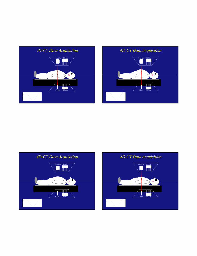
X-ray tube PET det.
4D-CT Data Acquisition
PET det.CT det.
X-ray tube PET det.
4D-CT Data Acquisition
PET det.CT det.
X-ray tube PET det.
4D-CT Data Acquisition
PET det.CT det.
X-ray tube PET det.
4D-CT Data Acquisition
PET det.CT det.

X-ray tube PET det.
4D-CT Data Acquisition
PET det.CT det.
X-ray tube PET det.
4D-CT Data Acquisition
PET det.CT det.
X-ray tube PET det.
4D-CT Data Acquisition
PET det.CT det.
X-ray tube PET det.
4D-CT Data Acquisition
PET det.CT det.

4D-CT
Signal from RPM system
• Respiratory tracking with Varian RPM optical monitor
• CT images acquired over complete respiratory cycles
Pan et al, Med Phys, 2004
X-ray on First couch position Second couch position Third couch
1st clinical case from MGH
55 sec Cine scan (200 mAs sec per step) , with free breathing of 3.72 sec average breathing cycle
moviemovie moviemoviemoviemovie
MGHMGH
4D-CT patient study (1)
Axial Coronal Sagittal
4D-CT patient study (2)
Axial Coronal Sagittal

4D-CT patient study (3)
Axial Coronal Sagittal
Average and MIP from 4D-CT
Current PET/CT only matches spatial not temporal resolutionspatial not temporal resolution
between PET and CT
High contrast resolution
Standard, 7.5 lp/cm, 0.67 mm PETAC, 2.5 lp/cm, 2 mm

Ring Artifactsin 120 kV not in 140 kVin 120 kV not in 140 kV
Ring artifacts from CT
Ring artifacts at different window & levels
WW=2000, WL=0 WW=400, WL=0 WW=400, WL= -100
These are what PET sees
WW=2000, WL=0 WW=400, WL=0 WW=400, WL= -100

Ring artifacts from CT
Ring artifact at 120 kV No ring artifact at 140 kV
From PET-Transmission to CT• Advance NXi PET scanner (Wu et al, EJNM 2004)
– Two rotating 68Ge (511 keV, T1/2=287 days) rod sources
– 20 rotations per min
– 15.3 cm long and 4.0 mm in diameter
– Maximum activity 370 MBq/rod (740 MBq total)
– Acquisition time 5-15 min/bed
– High noise and time consuming
– 40 observations per location per minute in PET-Transmission
• CT– 1 observation per location in CT (0.5 to 1 s)
– Low noise and quick, yet sometimes causing problems
Differences between PET and CTCT – 0.5 sec rotationPET
• scan of 90 cm < 20 sec• spatial resolution < 0.5 mm• temporal resolution < 1 sec
• scan of 15 cm for 3 to 6 mins,• spatial resolution ~ 5-8 mm• temporal resolution ~ breathing cycle
Potential misalignment between PET and CT images
Mis-matched PET-CT data sets
Mismatch 1:
CT diaphgram position lower than PET
Mismatch 1:
CT diaphgram position lower than PET
Mismatch 2:
CT diaphgram position higher than PET
Mismatch 2:
CT diaphgram position higher than PET

PET/CT protocol
• CT scout 80 kV 10 mA• CT (helical)
– 16 x 1.25 mm collimation, 120 kV, 300 mA, 0.5 sec, pitch 1.35 and auto dose
– Dose ~7.5 mGy
• PET (2D) 3 min/bed (15 to 20 mCi) and 6 beds (15 cm/bed)
30 mins per patient
Breath-hold or free-breathing in CT ?f g
Scout Helical CT
X-ray on
Misalignment in breathing states
End-inspiration (FB)
End-expiration (FB)
Mid-expiration(BH)
0 20 40 60 80 100 120 (sec)
Pan et al, JNM, 2005
Freq. of misalignment in 100 patients @ BH
Pan et al, JNM, 2005

Breathing Artifacts
Protocol: 16x0.625 mm, 0.8 s gantry rotation, pitch 1.375:1
Speed: 13.75 mm/0.8 s or 17.2 mm/s
Breathing artifacts to physiological info
Breath cycle= 80.3/(13.75/0.8)= 4.67 s
Heart rate= (21/(13.75/0.8)) -1*60= 49 bpm
Sl CT ≠ A CT
Average CT (ACT)
Slow CT ≠ Average CT
Long slow scan ≠ Long fast scan
Average CT for dose calculation, proton plan and IGRT
Axial (Step and shoot)
one rotation (<= 4 s)one rot.(<= 2 s for 64-slice)
Basic CT scan modes
Helical
pitch 0.5 to 1.5
Pitch =table translation per rotation
X-ray beam width

Axial (Step and shoot) Axial (step and shoot)$ (low dose cardiac, CACS) $
Cine$ (4DCT) $
one rotation (<= 4 s)one rot.(<= 2 s for 64-slice)
2/3 rotation multiple rotations
Basic CT scan modes
Helical $ Helical $$ (cardiac) $
$ Helical $$ (4DCT) $
pitch 0.5 to 1.5 pitch 0.2 to 0.3 pitch < 0.1
Pitch =table translation per rotation
X-ray beam width
Slow scan CT artifacts
0.5 sec rotation 4 sec rotation
Average CT (4 sec) Slow CT (4 sec)
Average CT is better than slow CT(2 adjacent CT slices of 2.5 mm apart)
4-s slow CT 4-s slow CT
Average CTAverage CT
876
4321
5
High radiation dose with cine CT ?
131211109
ACT
60 mAs & 1.2 mSv
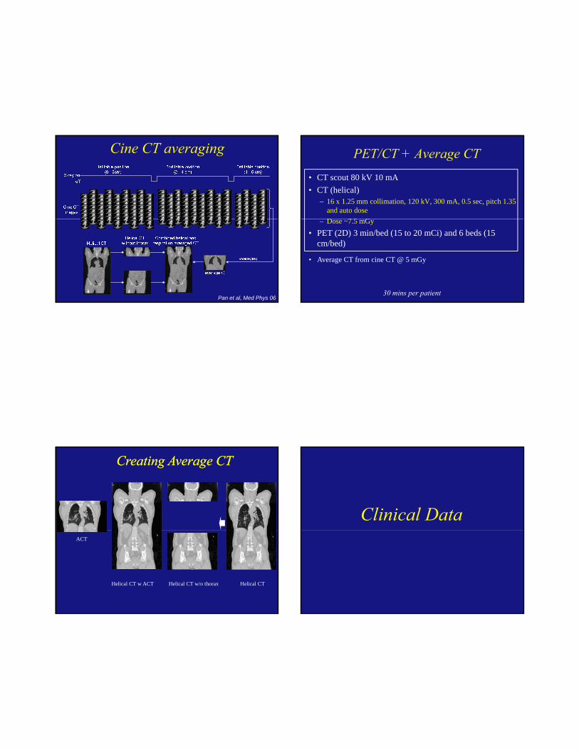
Cine CT averaging
Pan et al, Med Phys 06
PET/CT + Average CT• CT scout 80 kV 10 mA• CT (helical)
– 16 x 1.25 mm collimation, 120 kV, 300 mA, 0.5 sec, pitch 1.35 and auto doseD 7 5 G– Dose ~7.5 mGy
• PET (2D) 3 min/bed (15 to 20 mCi) and 6 beds (15 cm/bed)
30 mins per patient
• Average CT from cine CT @ 5 mGy
Creating Average CTCreating Average CT
Helical CT
ACT
Helical CT w/o thoraxHelical CT w ACT
Clinical Data

Clinical Studies
Mismatch 1:
CT diaphgram position lower
than PET
Mismatch 1:
CT diaphgram position lower
than PET
+57%
Mismatch 2:
CT diaphgram position higher
than PET
Mismatch 2:
CT diaphgram position higher
than PET
Mismatch 3:
CT diaphgram position lower
than PET
Mismatch 3:
CT diaphgram position lower
than PET
+56%
Clinical Studies
Mismatch 4:
CT diaphgram position lower
than PET
Mismatch 4:
CT diaphgram position lower
than PET
+100%
Patient study #1
SUV=10.8 SUV=13.7 (+27%) SUV=3.9
Patient study #2
SUV=3.7 (- 5%)

SUV=4.6
Patient study #3
SUV=7.5(+62%)
HCT
FDG uptake in the liver?
ACT
FDG uptake in the kidney Lung lesion or liver lesion?

Lung lesion or liver lesion?
Average CT
Example from CGMH (Taipei)
ACTHCT
PET/CT scan indicated a positive response to induction chemo with HCT
Improve the restaging after chemo
PET/CT scan indicated a positive response to induction chemo with HCT.
The patient had a negative response to the chemo with ACT.
Impact on treatment planning
Previous GTV was outlined based on CT and clinical PET without motion correction. New GTV was redefined based on the correct information from PET with ACT.
Old GTV
New GTV
Provided by Helen Liu

Average CT in cardiac PET
HCT- misregistration
Pan et al, Med. Phys. 2006
ACT
Heart motion in breath-hold
The heart does not seem to move in space during the breath-hold
250 msec temporal resolution
Cardiac and respiratory motion
500 msec temporal resolution and 100 msec interval between reconstructions
Table shifted between HCT and ACT
HCT (correct table) HCT (incorrect table)

Table shift between HCT and ACT
ACT (correct table) ACT (incorrect table)
Summary
• Challenges for PET/CT in RT
• PET/CT = PET/MSCT
• Cardiac CT is the main driving force for new MSCT
• 4D CT is important in PET/CT for radiation therapy
• Misregistration between CT and PET may be brought under control by average CT