Embed Size (px)
Citation preview
1
Hip and Pelvis Imaging
www.warhols.com/ colored%20shoe%20and%20leg.JPG
Joel Fallano, PT, DPT, MS, OCSAimee Klein, PT, DPT, DSc, OCSCSM 2013Imaging SIGJanuary 24th, 2013
Outline
Imaging TechniquesPlain filmsMRIMRASonography
Cases
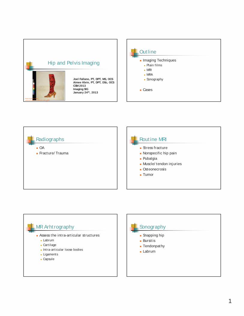
Radiographs
OAFracture/Trauma
Routine MRI
Stress fracture Nonspecific hip painPubalgiaMuscle/tendon injuriesOsteonecrosisTumor
MR Arhtrography
Assess the intra-articular structuresLabrumCartilageIntra-articular loose bodiesLigamentsCapsule
Sonography
Snapping hipBursitisTendonpathyLabrum
2
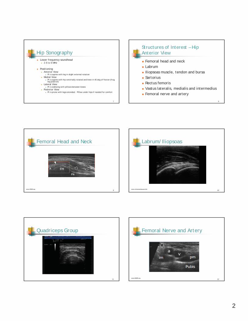
Hip SonographyLower frequency soundhead
2.5 to 5 MHz
PositioningAnterior View
Pt is supine with leg in slight external rotationMedial View
Pt is supine with hip externally rotated and knee in 45 deg of flexion (frog leg position)
Lateral ViewPt is sidelying with pillows between knees
Posterior ViewPt is prone with legs extended. Pillow under hips if needed for comfort
7
Structures of Interest – HipAnterior View
Femoral head and neckLabrumIliopsoas muscle, tendon and bursaSartoriusRectus femorisVastus lateralis, medialis and intermediusFemoral nerve and artery
8
Femoral Head and Neck
9www.ESSR.org
Labrum/Iliopsoas
10www.ultrasoundcases.info
Quadriceps Group
11
Femoral Nerve and Artery
12www.ESSR.org
3
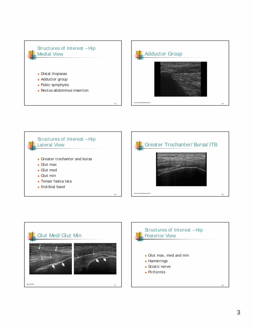
Structures of Interest – HipMedial View
Distal iliopsoasAdductor groupPubic symphysisRectus abdominus insertion
13
Adductor Group
14www.ultrasoundcases.info
Structures of Interest – HipLateral View
Greater trochanter and bursaGlut maxGlut medGlut minTensor fasica lataIliotibial band
15
Greater Trochanter/Bursa/ITB
16www.ultrasoundcases.info
Glut Med/Glut Min
17Garcia 2010
Structures of Interest – HipPosterior View
Glut max, med and minHamstringsSciatic nervePiriformis
18
4
Hamstring
19
Longitudinal Transverse
HIP PATHOLOGY
20
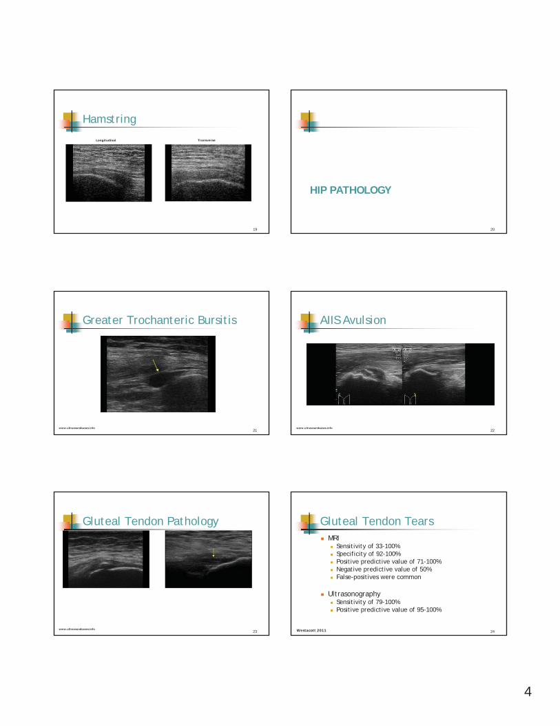
Greater Trochanteric Bursitis
21www.ultrasoundcases.info
AIIS Avulsion
22www.ultrasoundcases.info
Gluteal Tendon Pathology
23www.ultrasoundcases.info
Gluteal Tendon TearsMRI
Sensitivity of 33-100%Specificity of 92-100%Positive predictive value of 71-100% Negative predictive value of 50%False-positives were common
UltrasonographySensitivity of 79-100%Positive predictive value of 95-100%
24Westacott 2011
5
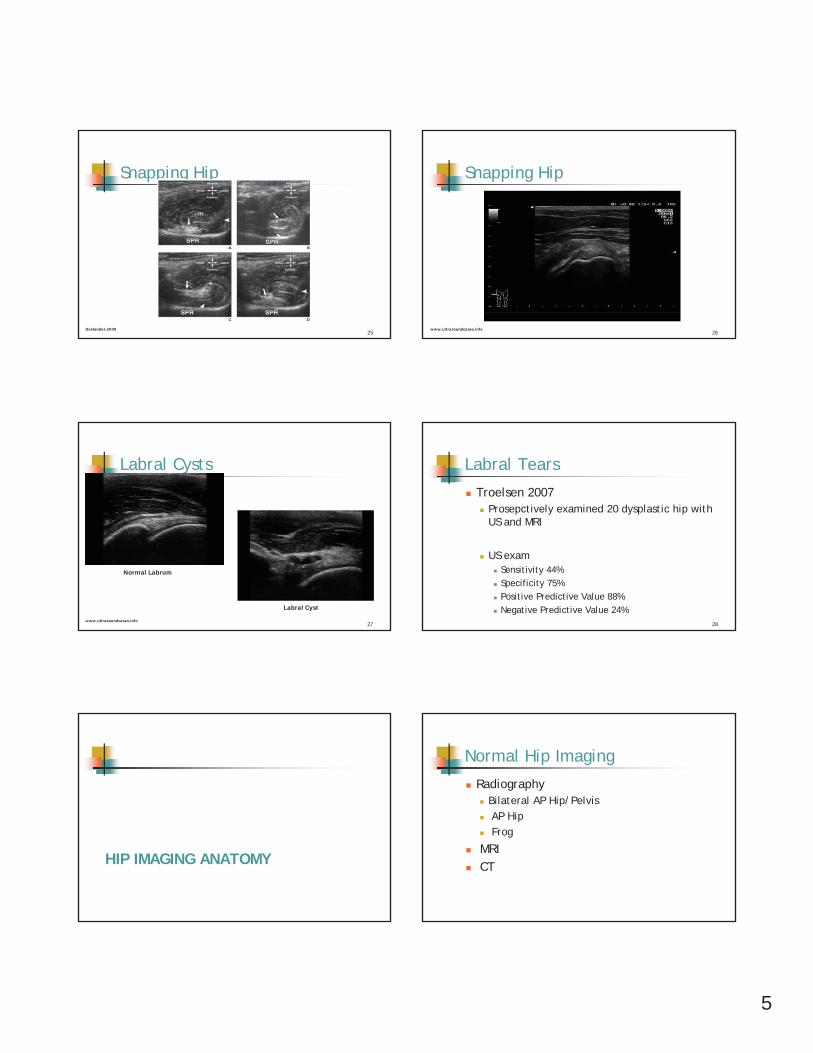
Snapping Hip
25Deslandes 2008
Snapping Hip
26www.ultrasoundcases.info
Labral Cysts
27www.ultrasoundcases.info
Normal Labrum
Labral Cyst
Labral Tears
Troelsen 2007Prosepctively examined 20 dysplastic hip with US and MRI
US examSensitivity 44%Specificity 75%Positive Predictive Value 88%Negative Predictive Value 24%
28
HIP IMAGING ANATOMY
Normal Hip Imaging
RadiographyBilateral AP Hip/PelvisAP HipFrog
MRICT
6
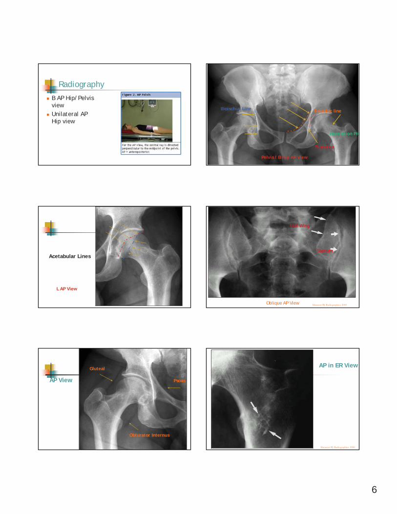
Radiography
B AP Hip/Pelvis viewUnilateral AP Hip view
Iliopubic lineIlioischial Line
Teardrop
Herniation Pit
Pelvis / B Hip AP View
Acetabular Lines
L AP View
Manaster BJ. Radiographics. 2000Oblique AP View
Sacrum
Ilial Wing
AP View
Obturator Internus
Gluteal
Psoas
Manaster BJ. Radiographics. 2000
AP in ER View
7
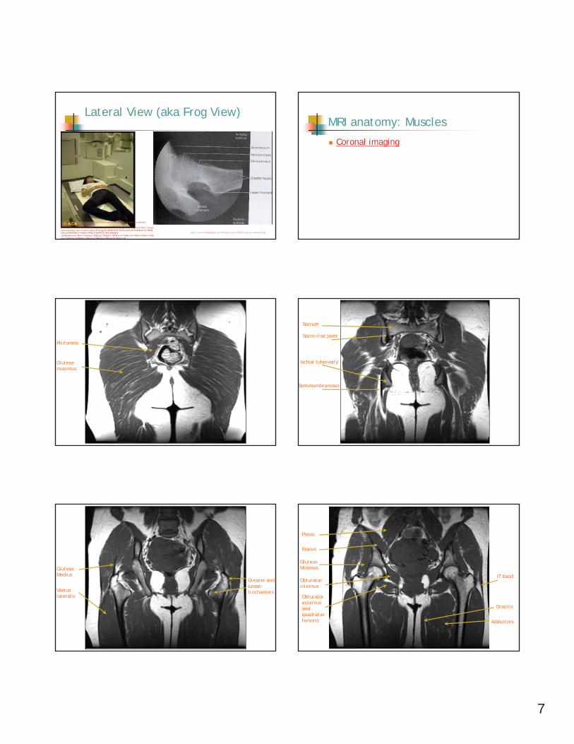
Lateral View (aka Frog View)
http://www.google.com/imgres?q=lateral+hip+radiograph&um=1&hl=en&client=firefox-a&sa=N&rls=org.mozilla:en-US:official&biw=1280&bih=870&tbm=isch&tbnid=h1HaeCKEyKG2DM:&imgrefurl=http://images.rheumatology.org/viewphoto.php%3FimageId%3D2861938%26albumId%3D75682&docid=RMQLGimvPZBa4M&w=366&h=549&ei=EDWGTr-MLcHk0QH-oaDsDw&zoom=1&iact=hc&vpx=384&vpy=396&dur=287&hovh=143&hovw=95&tx=101&ty=154&page=4&tbnh=143&tbnw=95&start=70&ndsp=24&ved=1t:429,r:1,s:70
http://www.e-radiography.net/technique/pelvis/Pelvis_hip_lat_anatomy2.jpg
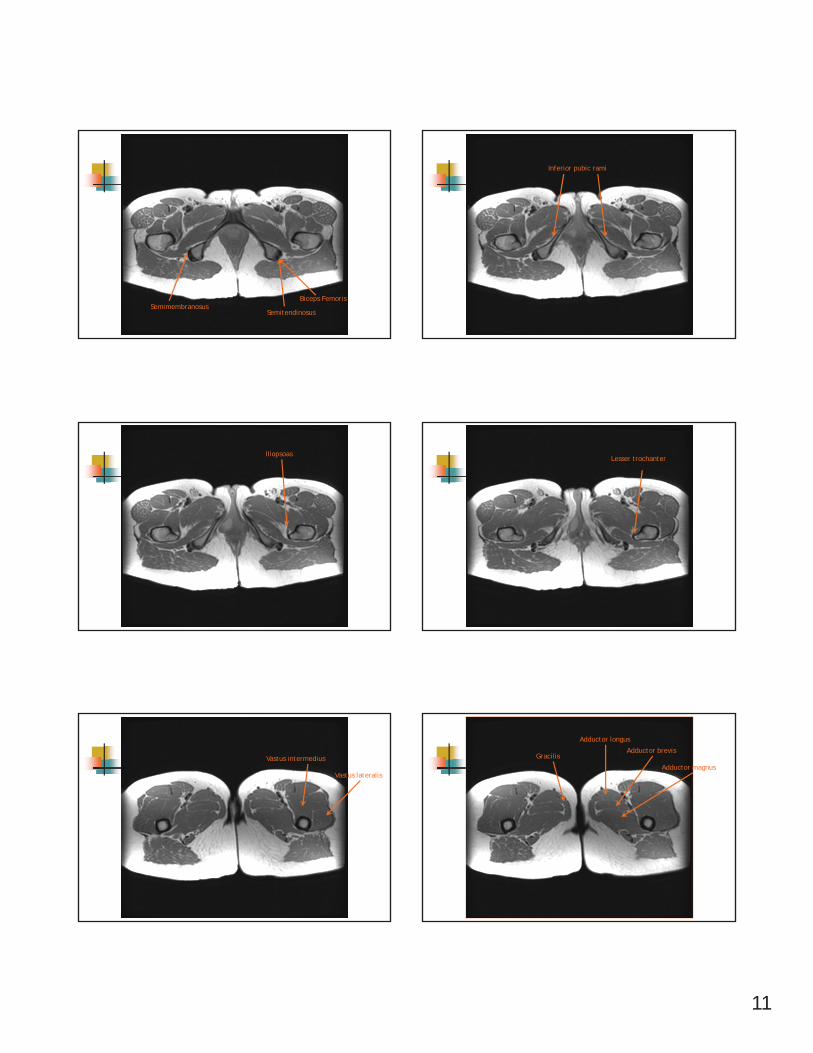
MRI anatomy: Muscles
Coronal imaging
Gluteus maximus
Piriformis
Semimembranosus
Sacrum
Sacro-iliac joint
Ischial tuberosity
Gluteus Medius
Vastus lateralis
Greater andLessertrochanters
Psoas
Iliacus
Gluteus Minimus
Obturatorinternus
Obturatorexternus andquadratusfemoris
IT band
Gracilis
Adductors
8
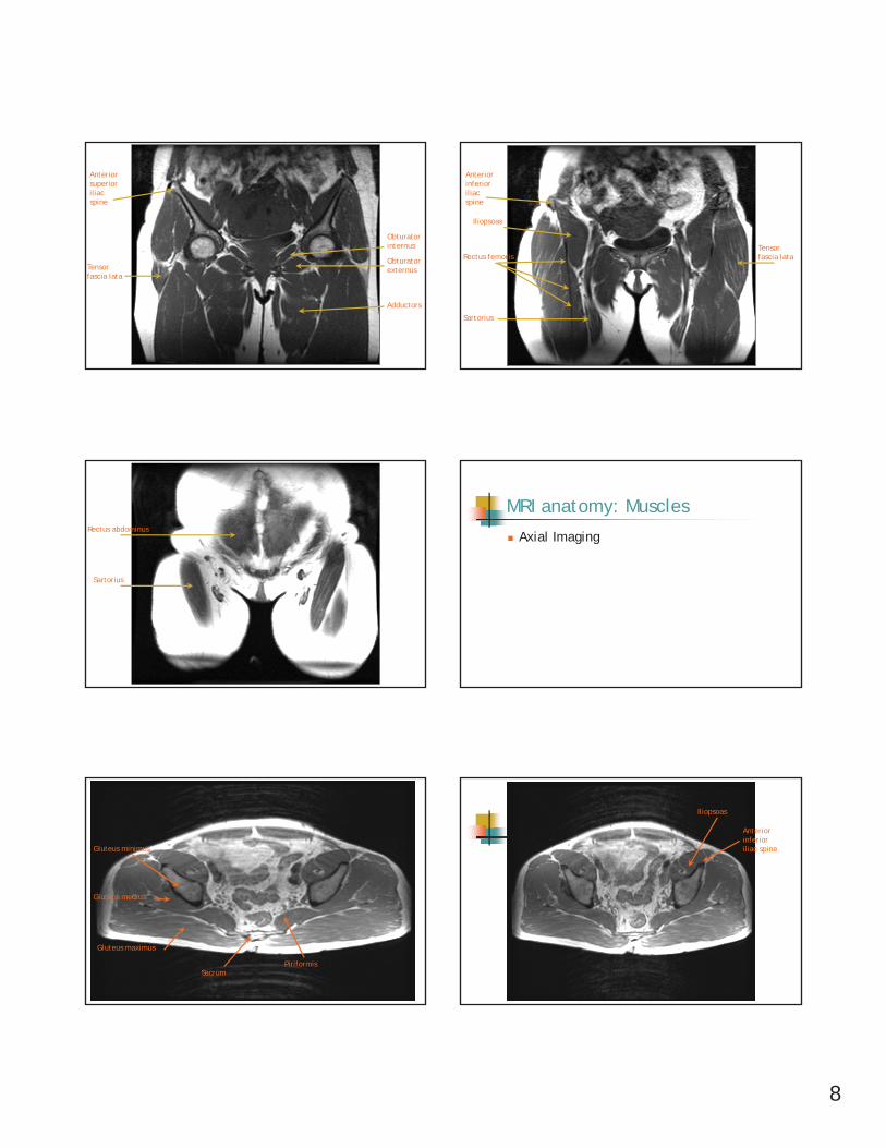
Adductors
Obturatorinternus
ObturatorexternusTensor
fascia lata
Anteriorsuperior iliac spine
Tensor fascia lata
Sartorius
Iliopsoas
Anteriorinferior iliac spine
Rectus femoris
Sartorius
Rectus abdominus
MRI anatomy: Muscles
Axial Imaging
Piriformis
Gluteus maximus
Gluteus medius
Gluteus minimus
Sacrum
Iliopsoas
Anteriorinferior iliac spine
9
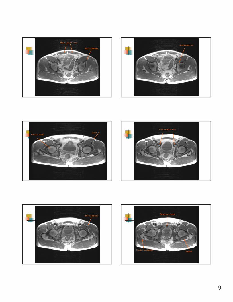
Rectus abdominus
Rectus femoris
Acetabular roof
SartoriusFemoral head
Superior pubic rami
Rectus femoris
Gemelli
Symphysis pubis
Greater trochanter
10
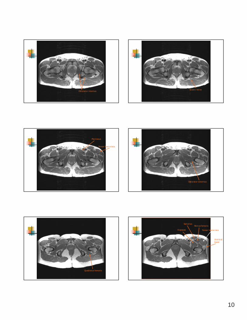
Obturator internusSciatic nerve
Pectineus
Tensor fascia lata
Obturator externus
Quadratus femoris
Tensor fascia lata
Iliotibialband
Iliopsoas
SartoriusRectus femoris
11
SemimembranosusSemitendinosus
Biceps Femoris
Inferior pubic rami
IliopsoasLesser trochanter
Vastus lateralis
Vastus intermedius GracilisAdductor brevis
Adductor magnus
Adductor longus
12
CT Imaging
Axial Osteoarthritis
Patient Profile60 yo femaleHPI
10/10 walking at conference, sat on bench, went to get up and has severe pain x 4 hours, then resolvedMid 11/10, rolled over in bed and felt sharp pain in L hip
PMH: OsteoporosisReferred by PCP to address L hip pain and decreasing functional statusFunctionally
Increased pain with walkingAM stiffnessInability to play golf or exercise
PT Examination
R/i L Hip OA Cluster for the Identification of Hip OA
Cluster 2Painful hip with IR> 50 yoMorning stiffness < 60 min
Diagnostic AccuracyAll 3 component of cluster are present: + LR = 3.4
Evaluation/Plan of Care
Differential Pathologic Diagnosis:OA L hipStress Fx due to underlying h/o osteoporosis
Referred to Orthopedist for medical work-upRadiographyMRI
Non-traumaHip Pain ImagingPathway
www.imagingpathways.health.wa.gov.au
13
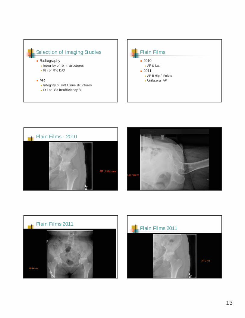
Selection of Imaging Studies
RadiographyIntegrity of joint structuresR/i or R/o DJD
MRIIntegrity of soft tissue structuresR/i or R/o insufficiency fx
Plain Films
2010AP & Lat
2011AP B Hip / PelvisUnilateral AP
Plain Films - 2010
AP UnilateralLat View
Plain Films 2011
AP Pelvis
Plain Films 2011
AP L Hip
14
Lat View 2011
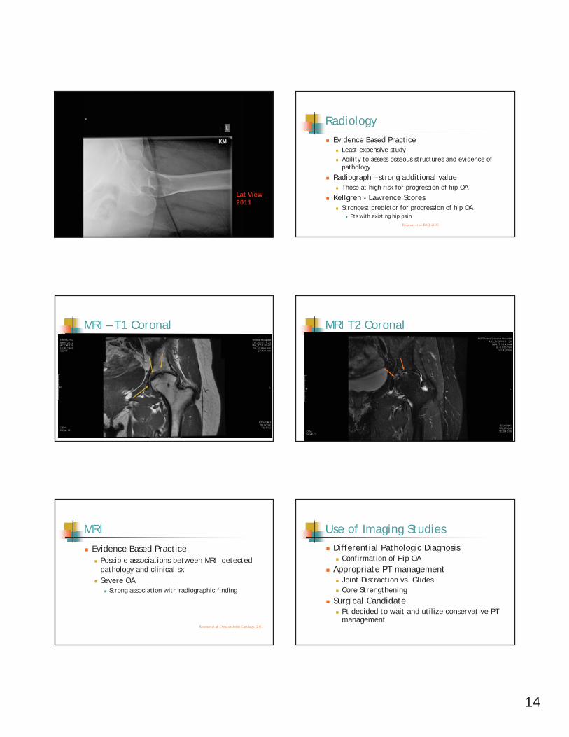
RadiologyEvidence Based Practice
Least expensive studyAbility to assess osseous structures and evidence of pathology
Radiograph – strong additional valueThose at high risk for progression of hip OA
Kellgren - Lawrence ScoresStrongest predictor for progression of hip OA
Pts with existing hip painReijman et al: BMJ, 2005
MRI – T1 Coronal MRI T2 Coronal
MRI
Evidence Based PracticePossible associations between MRI –detected pathology and clinical sx Severe OA
Strong association with radiographic finding
Roemer et al: Osteoarthritis Cartilage. 2011
Use of Imaging StudiesDifferential Pathologic Diagnosis
Confirmation of Hip OAAppropriate PT management
Joint Distraction vs. GlidesCore Strengthening
Surgical Candidate Pt decided to wait and utilize conservative PT management
15
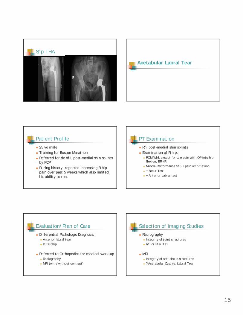
S/p THA
Acetabular Labral Tear
Patient Profile
25 yo male Training for Boston MarathonReferred for dx of L post-medial shin splints by PCPDuring history, reported increasing R hip pain over past 5 weeks which also limited his ability to run.
PT Examination
R/i post-medial shin splintsExamination of R hip:
ROM WNL except for c/o pain with OP into hip flexion, ER>IRMuscle Performance 5/5 + pain with flexion+ Scour Test+ Anterior Labral test
Evaluation/Plan of Care
Differential Pathologic Diagnosis:Anterior labral tearDJD R hip
Referred to Orthopedist for medical work-upRadiographyMRI (with/without contrast)
Selection of Imaging Studies
RadiographyIntegrity of joint structuresR/i or R/o DJD
MRIIntegrity of soft tissue structures? Acetabular Cyst vs. Labral Tear
16
Radiography
AP View Bilateral AP View
Radiography
Evidence Based PracticeLeast expensive studyAbility to assess osseous structures and evidence of pathology
MRI – T2 Weighted
Axial View
MRI
Evidence Based PracticeStrong correlation between MR imaging and pathology
Holder et al: Am J Roetgenol, 1995
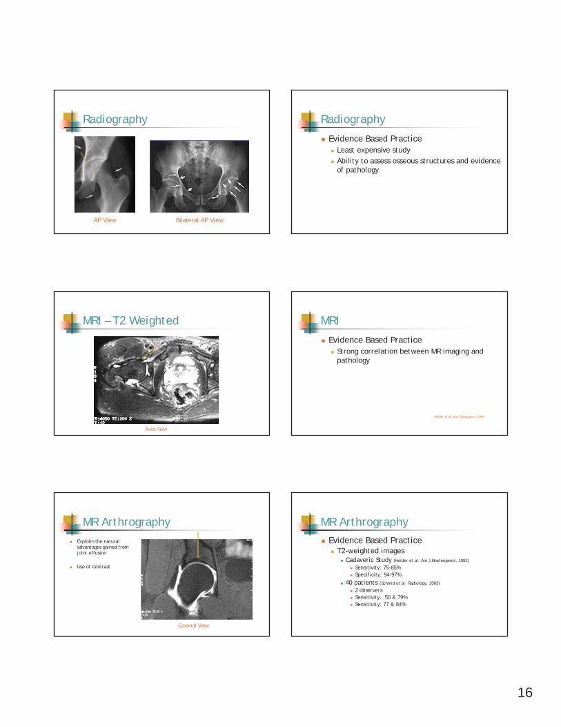
MR ArthrographyExploits the natural advantages gained from joint effusion
Use of Contrast
Coronal View
MR ArthrographyEvidence Based Practice
T2-weighted imagesCadaveric Study (Holder et al: Am J Roetengenol, 1992)
Sensitivity: 75-85%Specificity: 94-97%
40 patients (Schmid et al: Radiology, 2003)
2 observersSensitivity: 50 & 79%Sensitivity: 77 & 84%
17
Use of Imaging Studies
Differential Pathologic DiagnosisConfirmation of Anterior Labral Tear
Not appropriate PT managementSurgical Candidate
Osseous Injuries
Stress Reaction ResponseStress (Fatigue) FxInsufficiency Fx
Hip Fx Imaging Pathway
www.imagingpathways.health.wa.gov.au
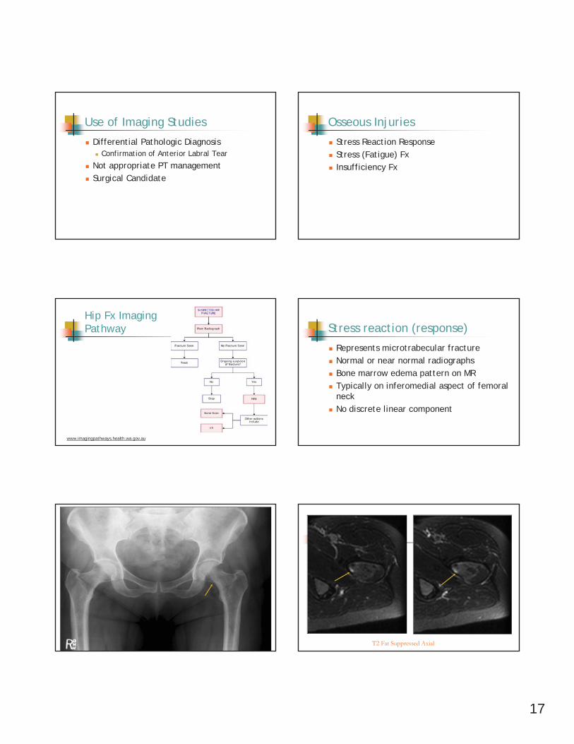
Stress reaction (response)
Represents microtrabecular fractureNormal or near normal radiographsBone marrow edema pattern on MRTypically on inferomedial aspect of femoral neckNo discrete linear component
T2 Fat Suppressed Axial
18
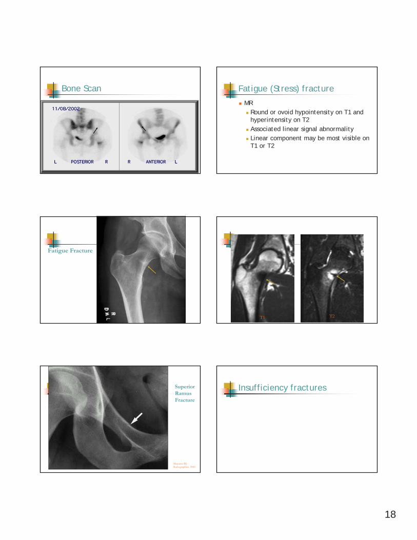
Bone Scan Fatigue (Stress) fracture
MRRound or ovoid hypointensity on T1 and hyperintensity on T2Associated linear signal abnormalityLinear component may be most visible on T1 or T2
Fatigue Fracture
T1 T2
Manaster BJ. Radiographics. 2000
SuperiorRamus Fracture
Insufficiency fractures
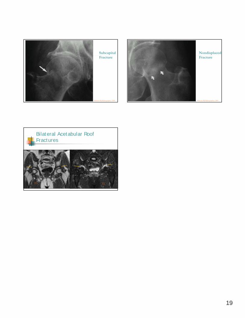
19
Manaster BJ. Radiographics. 2000
SubcapitalFracture
Manaster BJ. Radiographics. 2000
NondisplacedFracture
Bilateral Acetabular Roof Fractures
T1 T2