Embed Size (px)
Citation preview
1
Out of Hours Multidetector Computed Tomography Pulmonary Angiography:
Are Specialist Registrar Reports Reliable?
George C. Jakanani, FRCR; Rajesh Botchu, FRCR; Sumit Gupta, MRCP;
James Entwisle, FRCR; Amrita Bajaj, FRCR
ABSTRACT
Aim The purpose of this study was to assess the accuracy of the trainee radiologist’s
report for CTPA, and determine agreement or discrepancy with the final verified
consultant report.
Materials and Methods: We prospectively analysed 100 consecutive out of hours
CTPA examinations. Fifty one male and 49 female subjects were included in the
study. Mean (range) age of patients scanned was 63.7 (17 – 98) years.
Results: 18 of the 100 subjects (18%) had findings positive for PE. The interobserver
agreement for PE between on-call radiology registrars and consultant radiologists was
almost perfect [Kappa = 0.932 (p<0.0001; 95% CI, 0.84 – 1.0)]. There was one false
negative CTPA report. Eighty two CTPA scans (82%) were reported as negative for
PE by consultant radiologists. In this group, there was a single false positive
interpretation by the on call specialist registrar. The interobserver agreement for all
findings between registrar and consultant reports was almost perfect [weighted Kappa
= 0.87 (p<0.0001; 95% CI, 0.79 – 0.96)]. The overall discrepancy rate, including both
false positive and false negative findings, between the on-call radiology registrar and
consultant radiologist was 8% (8 of 100).
Conclusion: CTPA reports by radiology registrars can be relied and acted upon
without any major discrepancies. There is a relatively much higher proportion of

2
patients with alternative diagnoses mainly infective consolidation and heart failure
presenting with similar symptoms and signs of pulmonary emboli. It is imperative for
the trainee to be systematic and review all images if observational omissions are to be
reduced.
3
Out of Hours Multidetector Computed Tomography Pulmonary Angiography:
Are Specialist Registrar Reports Reliable?
INTRODUCTION
Multidetector computed tomography pulmonary angiography (CTPA) is now the most
common imaging modality in the evaluation of suspected pulmonary embolism (PE).
A large number of CTPAs are performed out of hours, and within teaching hospitals,
the initial provisional reports are issued by the trainee radiologist and not checked
until the following morning by the consultant radiologist. These trainee radiologist are
referred to as Specialist registrar or SpR who undergo structured specialist training in
their choose field of medicine. This is at least over a 5 year period in Radiology at the
end of which the registrar is considered trained, ready to be a consultant. (Appendix
1) The SpR’s do on site training out of normal working hours on a rotational basis
which is referred to as “on call rota” These provisional reports are crucial as they
provide the basis for out of hours clinical decisions. The purpose of this study was to
assess the accuracy of the trainee radiologist’s report for CTPA, and determine
agreement or discrepancy with the final verified consultant report. To the best of the
authors’ knowledge, this is the first study of its kind performed in a UK teaching
hospital.
METHOD AND MATERIALS
We prospectively analysed 100 consecutive out of hours CTPA examinations. These
were performed during a 28 day period from August to September 2008. 64 scans
were performed on a 16 slice MDCT sytem (Siemens Somatom Sensation, Siemens
AG, Munich, Germany; Technical parameters – 120 Kv, effective mAs 140, rotation
4
time 0.5, 0.75 collimation with a reconstruction slice thickness of 1mm,
reconstruction interval of 0.5 mm.). 36 scans were performed on a 64 slice MDCT
system (Aquilion, Toshiba, Tokyo, Japan; Technical parameters- 120 Kv, effective
mAs 182, rotation time 0.5, pitch of 0.828 and reconstruction slice thickness of 1
mm, reconstruction interval of 0.5 mm).
Images were acquired after injection of 100ml of iohexol 350 (350 mg iodine/ml, GE
Healthcare, Oslo, Norway) using bolus trigger set at 100 HU on the pulmonary
trunk. Images were reviewed on a patient archive and communication system (PACS)
workstation (AGFA Impax 5.1, Morstel, Belgium). Analysis of CT images was
performed on axial, coronal and sagittal reformatted images (1mm multiplanar
reconstructions). Both soft-tissue and lung windows were used to identify
subsegmental bronchi and arteries.
Acute PE was diagnosed when there was filling defect within the vessel or when
vessel truncation implied the presence of occlusion. The level of PE was categorized
as central, lobar, segmental and subsegmental.
The initial provisional reports issued by the on call specialist registrar were
prospectively collected and findings documented. All trainees on the on call rota had
completed at least 2 years of specialist radiology training and had been signed off to
at least level 3 to report CTPA according to the Royal College of Radiology trainee
portfolio (Appendix 2).
5
The provisional reports were verified by a consultant radiologist within 24 hours of
the examination and the consultant report was used as the reference standard. Both
the trainee registrar and consultant groups were unaware of the study in progress at
the time of their reports in order to avoid bias.
Two cardiothoracic radiologists (JJE and AB), who were blinded to both the initial
registrar and final verified consultant reports reviewed the cases with discrepancy and
issued a final report by consensus.
Statistical analysis was performed using using SPSS for Windows, Rel. 16.0.1.2008.
Chicago: SPSS Inc and an online statistical computation website
(http://faculty.vassar.edu/lowry/kappa.html). Indices of agreement were calculated as
described previously [1]. Kappa (unweighted and weighted) statistic was used for an
inter-observer reliability analysis. Kappa statistic were interpreted as indicating
poor(κ<0.2), fair(0.21<κ<0.4), moderate(0.41<κ<0.6), substantial(0.61<κ<0.8) and
almost perfect(0.81<κ<1.0) observer agreement.[1,2] A p value of <0.05 was taken as
statistically significant.
RESULTS
One hundred consecutive out of hours CTPA examinations performed at a university
teaching hospital over 16 day period were prospectively included in this study. These
were reported by 16 different SpR and the final reports were verified by 6
Consultants, three of whom were subspecialist Consultants in Cardiothoracic
Radiology. Fifty one male and 49 female subjects had CTPA examinations. Mean
(range) age of patients scanned was 63.7 (17 – 98) years.
6
18 of the 100 subjects (18%) had findings positive for PE. The interobserver
agreement for PE between on-call radiology registrars and consultant radiologists was
almost perfect [Kappa = 0.932 (p<0.0001; 95% CI, 0.84 – 1.0)]. There was one false
negative CTPA report in the specialist registrar group. This was for a PE in a single
segmental pulmonary artery branch. 6 of the 18 subjects with PE had other concurrent
chest findings. These were all correctly reported by the registrar. A summary of
positive and negative interpretation of CTPA scans for PE by on-call radiology
registrars and consultant radiologists is presented in Table 1. Table 2 is a summary of
any additional chest findings in the 18 subjects with PE.
Eighty two CTPA scans (82%) were reported as negative for PE by consultant
radiologists. In this group, there was a single false positive interpretation by the on
call specialist registrar. The consultant opinion in the false positive case was that the
finding was artefactual, and caused by suboptimal pulmonary opacification in a
technically inadequate examination. The expert panel agreed with this assessment.
44 of the 82 subjects with no PE had significant other chest findings on CTPA
sufficient to explain the clinical symptoms. Six of the 44 were unreported by the
registrar. The six cases included two subjects with CT evidence of heart failure, a
subject with CT features of hypertrophic obstructive cardiomyopathy (HOCM)
(Figure 1), a missed small pneumothorax, a subject with bronchiectasis, and another
with subtle early interstitial lung disease. One subject had an incidental inter-atrial
septum aneurysm which was not felt to have been the cause of symptoms (Figure 2).
The remaining 37 of the 82 subjects with no PE had no other additional chest findings
and had completely normal CTPA examinations. Table 3 is a summary of the
7
additional chest findings in patients with no PE highlighting the findings missed by
SpR.
The interobserver agreement for all findings between registrar and consultant reports
was almost perfect [weighted Kappa = 0.87 (p<0.0001; 95% CI, 0.79 – 0.96)]. The
overall discrepancy rate, including both false positive and false negative findings,
between the on-call radiology registrar and consultant radiologist was 8% (8 of 100)
(Table 4).
The important missed finding of HOCM was asymptomatic prior to scan and is now
under the care of a cardiologist. The other missed findings of bronchiectasis and early
ILD were unlikely to explain the symptomatology but were in any case referred to
chest consultant. The patients with heart failure were managed accordingly by the
Medical team. Patient with small pneumothorax did not need any intervention.
Discussion
CTPA is a quick and reliable way of diagnosing pulmonary embolism. A prompt
diagnosis is essential to decrease the morbidity and mortality arising from this
condition [3]. In UK teaching hospitals, the on call radiology registrar discusses,
authorises and supervises all out of hours CT examinations, and thereafter issues a
provisional report to the referring clinical team. This provisional report is the basis
upon which clinical management decisions are made, making it vitally important that
it is accurate and reliable.
8
The accuracy of out of hours radiology resident reports has previously been reported
in a number of studies in the American literature with respect to CTPA, CT of the
head, trauma imaging and in suspected appendicitis [4,5,6,7,8,9]. Radiology
departments within the NHS have different clinical audit and clinical governance
mechanisms to ensure that out of hours patient safety is not compromised. The current
study was developed after an initial small audit of out of hours specialist registrar
activity, and to the best of the authors’ knowledge, is the first to report on the
accuracy of out of hours specialist registrar CTPA reports within a UK teaching
institution.
The study demonstrates excellent agreement between specialist registrar and
consultant reports in the diagnosis or exclusion of PE. This is in keeping with a
number of studies which have shown that appropriately trained radiology residents
can provide a safe CTPA service out of hours [10, 11, 12]. Shaham at al have reported
good interobserver agreement between on call resident and specialist staff in reporting
CTPA [10]. In a similar study, Safriel et al have reported that the provisional report
can be relied upon without any significant descrepancy [11].
In common to our study, another report shows evidence that exposure to 2 years of
radiology training enables a resident to provide a useful and reliable provsional report
for CTPA [12]. In this respect, our results validate the accuracy of the training
assessments done by our local educational supervisors and consultant trainers with
regards to signing off trainees appropriately for on call reporting. The high accuracy
of these trainee reports also shows that individual trainees could potentially be signed
off as completely independent (Level 4 competence) earlier on, possibly in the fourth
9
year of training, freeing up valuable consultant time and allowing for more efficient
use of consultant resources.
The single case of false positive interpretation due to inadequate pulmonary tree
opacification highlights the need for adoption of robust scanning protocols, and re-
enforces the need for meticulous attention to detail with regards to exam technique
[13, 14]. It is important that both trainees and radiographers are aware of how various
technical parameters can affect the accuracy of what is otherwise an excellent
diagnostic tool. With the new generation of multi-detector scanners, our own
experience shows that technically inadequate examinations lead to suboptimal
opacification in the subsegmental branches of the pulmonary vasculature. At the same
time, a properly performed MDCT allows increased detection of PEs in the
subsegmental arteries, as opposed to earlier single slice scanners and conventional
pulmonary angiography [15]. Whether a missed sub segmental PE in this location is
clinically significant however continues to be a matter of considerable debate with a
recent study showing that despite multidetector CTPA increasing the detection rate of
subsegmental PEs, follow up at 3 months in these patients suggests that untreated PEs
in this location may not affect clinical outcome [16].
We showed a high proportion of patients without PE but with significant other
findings sufficient to explain the patient’s presenting symptoms. This is not surprising
given the non specific clinical presentation of PE. This ability to provide an
alternative diagnosis is one of the main reasons why CTPA has replaced conventional
pulmonary angiography as the gold standard test in imaging suspected pulmonary
embolism. The commonest findings in our group of patients were pneumonia,
10
atelectasis and heart failure (Table 2). This is in keeping with previous studies in the
literature. Richman et al in a multicentre study involving 1025 patients showed that
pneumonia was the most common non thrombotic finding in patients with no PE [17].
In another study, Tsai et al reported atelectasis and pneumonia as being the most
common findings [18]. Within the context of the current study, what our results and
these aforementioned studies demonstrate is the importance of CTPA in providing an
alternative explanation for a patient’s acute symptoms. In an out hours setting, the
radiology registrar therefore becomes the first person to point out the presence of for
example pneumonia or heart failure, allowing the clinical team on the wards, who
nowadays are often quite junior and inexperienced, to treat these conditions
expeditiously. The excellent agreement in our study on these findings is testament to
the fact that this is a message that is continuously emphasized to our radiology
registrars during their training.
It is important to point out that there are some additional findings that will be entirely
incidental but will require follow up, for example the subjects with inter atrial septum
aneurysm and HOCM from our series. The need to have appropriate mechanisms for
follow up is also highlighted in a recent review of 589 examinations by Hall et al
which showed that CTPAs were more than twice as likely to reveal an incidental
pulmonary nodule or adenopathy than PE [19].
What is clear on review of the 6 subjects with significant unreported findings is that in
5 of these subjects, the findings were not reported due to what we believe is the
inherent satisfaction of search that plagues the early years of radiology training.
[20]The subject with HOCM highlights how the heart is often a blind spot for most
11
non cardiothoracic radiologists. The case is a good example of how the exquisite
detail provided on MDCT images has made it mandatory for every radiologist to be
conversant not only with the normal cross sectional imaging anatomy of the whole
body , but in this case the CT manifestations of cardiac conditions which have
traditionally been imaged by other modalities in the past.
We do accept that this study had a few limitations. Firstly the study had a relatively
small cohort of patients. Also the Consultants who verified the reports were not
blinded to the SpR’s report raising the possibility of reporting bias. Our institute does
have high throughput of CTPA’s and all the consultants in the study have sufficient
exposure to cardiothoracic cases on a regular basis.
CTPA reports by radiology registrars can be relied and acted upon without any major
discrepancies. There is a relatively much higher proportion of patients with alternative
diagnoses mainly infective consolidation and heart failure presenting with similar
symptoms and signs of pulmonary emboli. It is imperative for the trainee to be
systematic and review all images if observational omissions are to be reduced.
References
1. Kundell HL, Polansky M. Measurement of observer agreement. Radiology
2003; 228:303-308
2. Landis JR, Koch GG. The measurement of observer agreement for categorical
data. Biometrics. 1977;33:159-174
3. Barritt DW, Jordan SC. Anticoagulant drugs in the treatment of pulmonary
embolism. A controlled trial. Lancet. 1960; 18;1(7138):1309-12.
12
4. Murray UM, Eldevik OP, Desmond JS. Clinical consequences of
misinterpretations of neuroradiologic CT scans by on-call radiology
residents.AJNR Am J Neuroradiol. 2000 ;21(1):43-4.
5. Wysoki MG, Nassar CJ, Koenigsberg RA, Novelline RA, Faro SH, Faerber EN.
Head trauma: CT scan interpretation by radiology residents versus staff
radiologists. Radiology. 1998 ;208(1):125-8.
6. Albano MC, Ross GW, Ditchek JJ, Duke GL, Teeger S, Sostman HD,
Flomenbaum N, Seifert C, Brill PW. Resident interpretation of emergency CT
scans in the evaluation of acute appendicitis. Acad Radiol. 2001;8(9):915-8.
7. Wechsler RJ, Spettell CM, Kurtz AB, Lev-Toaff AS, Halpern EJ, Nazarian LN,
Feld RI, Needleman L, Alexander AA. Effects of training and experience in
interpretation of emergency body CT scans. Radiology. 1996 ;199(3):717-20.
8. RuchmanRB, Jaeger J, Wiggins EF, et al. Preliminary radiology resident
interpretations versus final attending radiologist interpretations and the impact
on patient care in a community hospital. AJR Am J Roentgenol 2007; 189:523–
526.
9. Tieng N, Grinberg D, Li SF. Discrepancies in interpretation of ED body
computed tomographic scans by radiology residents. Am J Emerg Med. 2007
;25(1):45-8
10. Shaham D, Heffez R, Bogot NR, Libson E, Brezis M. CT pulmonary
angiography for the detection of pulmonary embolism: interobserver agreement
between on-call radiology residents and specialists (CTPA interobserver
agreement). Clin Imaging. 2006;30(4):266-70
11. Safriel Y, Sclafani S, Gale B, Patel D, Gordon D. Comparing the interpretations
of CT pulmonary angiograms by attending and resident radiologists: can
13
residents identify life-threatening pulmonary emboli in hospitalized patients?
Emerg Radiol. 2002 ;9(1):55-9.
12. Ghanima W, Nielssen BE, Holmen LO, Witwit A, Al-Ashtari A, Sandset PM.
Multidetector computed tomography (MDCT) in the diagnosis of pulmonary
embolism: interobserver agreement among radiologists with varied levels of
experience. Acta Radiol. 2007 ;48(2):165-70.
13. Uysal Ramadan S, Kosar P, Sonmez I, Karahan S, Kosar U. Optimisation of
contrast medium volume and injection-related factors in CT pulmonary
angiography: 64-slice CT study. Eur Radiol 2010; 20(9): 2100-7.
14. Patel S, Kazerooni EA, Cascade PN. Pulmonary embolism: optimization of
small pulmonary artery visualization at multi-detector row CT. Radiology
2003;227 : 455-460
15. Schoepf UJ, Holzknecht N, Helmberger TK, et al. Subsegmental pulmonary
emboli: improved detection with thin-collimation multi-detector row spiral CT.
Radiology 2002;222 : 483-490
16. Carrier M, Righini M, Wells PS, Perrier A, Anderson DR, Rodger MA,
Pleasance S, Le Gal G. Subsegmental pulmonary embolism diagnosed by
computed tomography: incidence and clinical implications. A systematic review
and meta-analysis of the management outcome studies. J Thromb Haemost.
2010; 8(8):1716-22.
14
17. Richman PB, Courtney DM, Friese J, Matthews J, Field A, Petri R, Kline JA. Prevalence and significance of nonthromboembolic findings on chest computed
tomography angiography performed to rule out pulmonary embolism: a
multicenter study of 1,025 emergency department patients. Acad Emerg Med.
2004 Jun;11(6):642-7.
18. Tsai KL, Gupta E, Haramati LB. Pulmonary atelectasis: a frequent alternative
diagnosis in patients undergoing CT-PA for suspected pulmonary embolism.
Emerg Radiol 2004;10 : 282-286
19. Hall WB, Truitt SG, Scheunemann LP, Shah SA, Rivera MP, Parker LA,
Carson SS. The prevalence of clinically relevant incidental findings on chest
computed tomographic angiograms ordered to diagnose pulmonary embolism.
Arch Intern Med. 2009 Nov 23;169(21):1961-5
20. Ashman CJ, Yu JS, Wolfman D. Satisfaction of search in osteoradiology. AJR
2000; 175: 541–544
15
Appendix 1
The Royal college of Radiologists. Clinical Radiology/ Training and qualifications /
Specialty Training / Becoming a Clinical Radiologist
Appendix 2: Royal College of Radiologists Trainee Portfolio Competencies
Level 1 – The radiology trainee has a comprehensive understanding of the principles
of the procedure including, where applicable, complications and interpretation of
results and has witnessed the procedure being performed.
Level 2 – The radiology trainee is able to carry out the procedure under direct
supervision of a Consultant
Level 3 – The radiology trainee is able to carry out the procedure under indirect
supervision i.e. Consultant is available for advice but is not physically present during
the investigation
Level 4 – The radiology trainee is able to carry out the procedure competently and
independently (independent competence)
Figures and Legends
16
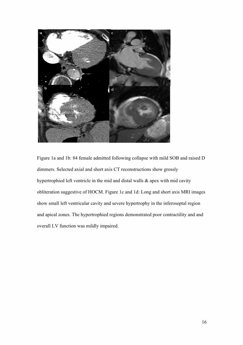
Figure 1a and 1b: 84 female admitted following collapse with mild SOB and raised D
dimmers. Selected axial and short axis CT reconstructions show grossly
hypertrophied left ventricle in the mid and distal walls & apex with mid cavity
obliteration suggestive of HOCM. Figure 1c and 1d: Long and short axis MRI images
show small left ventricular cavity and severe hypertrophy in the inferoseptal region
and apical zones. The hypertrophied regions demonstrated poor contractility and and
overall LV function was mildly impaired.
17
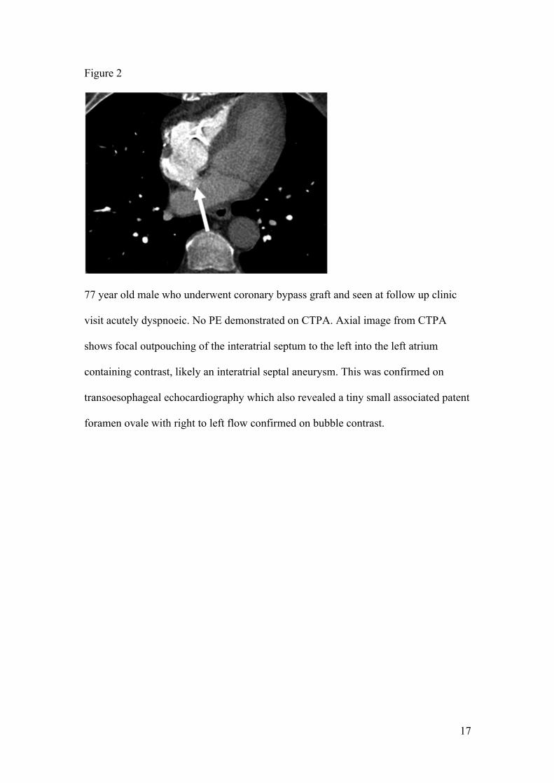
Figure 2
77 year old male who underwent coronary bypass graft and seen at follow up clinic
visit acutely dyspnoeic. No PE demonstrated on CTPA. Axial image from CTPA
shows focal outpouching of the interatrial septum to the left into the left atrium
containing contrast, likely an interatrial septal aneurysm. This was confirmed on
transoesophageal echocardiography which also revealed a tiny small associated patent
foramen ovale with right to left flow confirmed on bubble contrast.
18
Tables
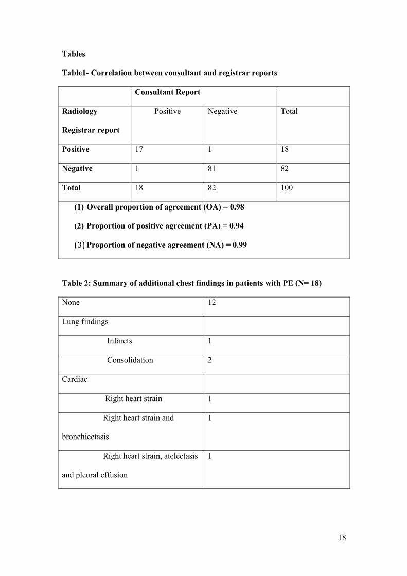
Table1- Correlation between consultant and registrar reports
Consultant Report
Radiology
Registrar report
Positive Negative Total
Positive 17 1 18
Negative 1 81 82
Total 18 82 100
(1) Overall proportion of agreement (OA) = 0.98
(2) Proportion of positive agreement (PA) = 0.94
(3) Proportion of negative agreement (NA) = 0.99
Table 2: Summary of additional chest findings in patients with PE (N= 18)
None 12
Lung findings
Infarcts 1
Consolidation 2
Cardiac
Right heart strain 1
Right heart strain and
bronchiectasis
1
Right heart strain, atelectasis
and pleural effusion
1
19
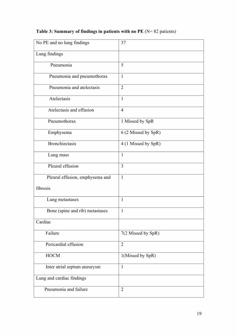
Table 3: Summary of findings in patients with no PE (N= 82 patients)
No PE and no lung findings 37
Lung findings
Pneumonia 5
Pneumonia and pneumothorax 1
Pneumonia and atelectasis 2
Atelectasis 1
Atelectasis and effusion 4
Pneumothorax 1 Missed by SpR
Emphysema 6 (2 Missed by SpR)
Bronchiectasis 4 (1 Missed by SpR)
Lung mass 1
Pleural effusion 3
Pleural effusion, emphysema and
fibrosis
1
Lung metastases 1
Bone (spine and rib) metastases 1
Cardiac
Failure 7(2 Missed by SpR)
Pericardial effusion 2
HOCM 1(Missed by SpR)
Inter atrial septum aneurysm 1
Lung and cardiac findings
Pneumonia and failure 2
20
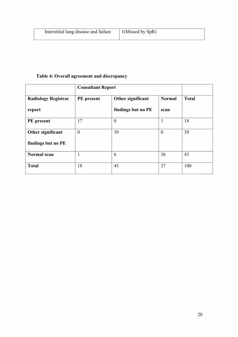
Interstitial lung disease and failure 1(Missed by SpR)
Table 4: Overall agreement and discrepancy
Consultant Report
Radiology Registrar
report
PE present Other significant
findings but no PE
Normal
scan
Total
PE present 17 0 1 18
Other significant
findings but no PE
0 39 0 39
Normal scan 1 6 36 43
Total 18 45 37 100