Embed Size (px)
Citation preview
Volume 5 • Issue 5 • 1000237J Neurol NeurophysiolISSN: 2155-9562 JNN, an open access journal
Research Article Open Access
Cerf and Barnett, J Neurol Neurophysiol 2014, 5:5 DOI: 10.4172/2155-9562.1000237
Review Article Open Access
Journal of Neurology & NeurophysiologyJo
urna
l of N
eurology & Neurophysiology
ISSN: 2155-9562
Epilepsy ― Eavesdropping on the Conversations of Rebellious NeuronsMoran Cerf1,2* and Samuel B Barnett1,2
1Kellogg School of Management, Northwestern University, Evanston IL, USA2Interdepartmental Neuroscience Program, Northwestern University, Evanston IL, USA
*Corresponding author: Moran Cerf, Kellogg School of Management,Northwestern University, Evanston IL, USA, E-mail: [email protected]
Received August 20, 2014; Accepted October 10, 2014; Published October 15, 2014
Citation: Cerf M, Barnett SB (2014) Epilepsy – Eavesdropping on the Conversations of Rebellious Neurons. J Neurol Neurophysiol 5: 237. doi:10.4172/2155-9562.1000237
Copyright: © 2014 Cerf M, et al. This is an open-access article distributed under the terms of the Creative Commons Attribution License, which permits unrestricted use, distribution, and reproduction in any medium, provided the original author and source are credited.
IntroductionWhenever we think or act, billions of neurons send messages to
precise destinations throughout the brain. Without conscious effort, our neurons call one another on a network of trillions of connections. In epilepsy, we observe entire populations of neurons initiating a conversation without provocation, resulting in what we term a seizure, often causing a violent loss of bodily control and consciousness. Epileptic seizures have been historically viewed as signs of demonic possession, divine intervention, and even artistic genius, but now they are powerful and remarkable reminders that our own neurons can unite and turn against us. Advances in medicine can help many patients achieve seizure freedom, but others are candidates for invasive brain surgery. Not only has surgery given hope to medically intractable patients, the practice has led to some of the most profound discoveries about the brain. We showcase the evolving understanding of epilepsy as well as landmark neuroscience research uniquely made possible by neurosurgery.
We like to feel in control of our lives. We sometimes take for granted the freedom to think and move however we please. However, we have taken up arms and fought wars when our autonomy has been threatened. We immediately notice and urgently respond when some force restricts our freedom.
Epilepsy is one of these forces, unexpectedly wresting control of its sufferers’ bodies and minds. Its symptoms, which can include violent convulsions of extremities, forced contraction of muscles, and loss of consciousness, force us to face the unpleasant reality that our autonomy is not guaranteed. The word “epilepsy” itself, derived from the Greek epilambanein (“to seize”), reflects the ancient belief that its sufferers were becoming possessed by supernatural beings, stripped of all personal control [1].
History of EpilepsyThe overwhelming nature of epilepsy has challenged the scientific
community for thousands of years, all the while contributing to critical scientific advances [2]. Despite epilepsy’s uncanny characteristics, [3] Hippocrates (400 BCE) claimed that this phenomenon was “no more divine than others.” This perspective arguably paved the way to our modern, naturalistic approach to medicine. We now define epilepsy generally as a susceptibility to recurrent, spontaneous seizures [4]. For decades, many world leaders, scientists, and artists have been living healthy, productive lives with epilepsy [1]. Nowadays, one of the only immediate concerns for epileptic patients is the possibility of a spontaneous seizure occurring at a moment that will endanger their lives or those around them. Such moments include driving a car, holding a baby, or being in another critical situation that requires high alertness. If those are controlled, life with recurring seizures can be seemingly normal.
Nonetheless, despite millennia of progress, epilepsy remains one of the most common neurological conditions. Epilepsy occurs in over 50 million people, almost 1% of the world’s population, and it affects individuals of all backgrounds [5]. Regional studies of its economic impact have been conducted around the world; in Europe alone, the
estimated costs related to epilepsy exceed $18.8 billion per year [6,7]. These calculations cannot capture the complete economic impact, which also includes substantial losses in earnings and productivity.
Here, we review the latest understanding of epilepsy itself and also the extraordinary research that has arisen tangentially.
Potential Causes of EpilepsyOne of the challenges that epilepsy poses to clinicians and
researchers is that there are many possible causes, often impossible to determine in a given case.
Epilepsy may be caused by an underlying neurological condition or brain damage, which causes unprovoked sequences of activity that quickly propagate throughout the brain site. Some epileptic conditions are present at birth, while others develop later in life [8]. A patch of scar tissue in some part of the brain, a head injury, stroke, cerebral palsy, genetic syndromes, growths or tumors of the brain, and previous infections of the brain such as meningitis or encephalitis could all lead to an irritation of the surrounding brain cells, which could trigger the recurrent seizures that characterize epilepsy [9]. Accordingly, clinicians tend to classify cases of epilepsy not by their potential causes, but by the types of seizures experienced.
Signs of EpilepsyClinicians have documented many types of seizures, but despite these
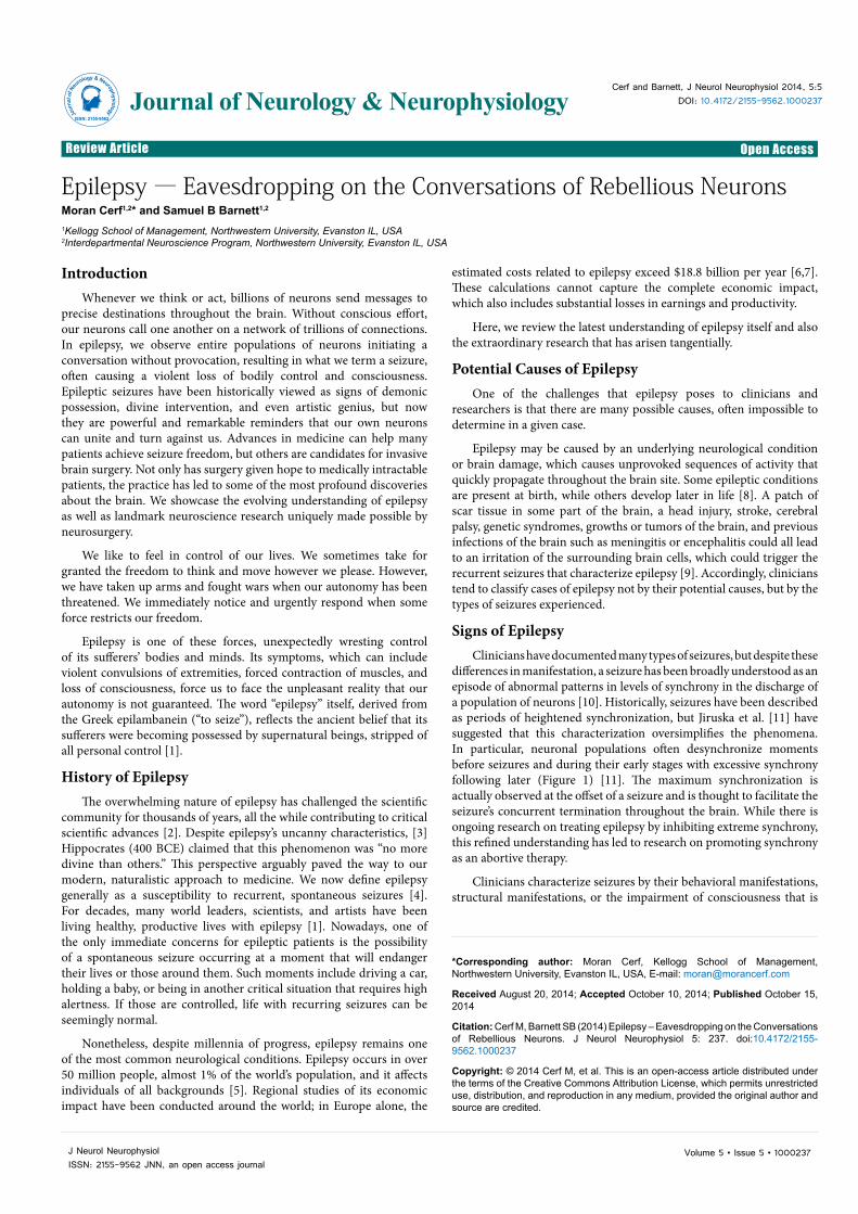
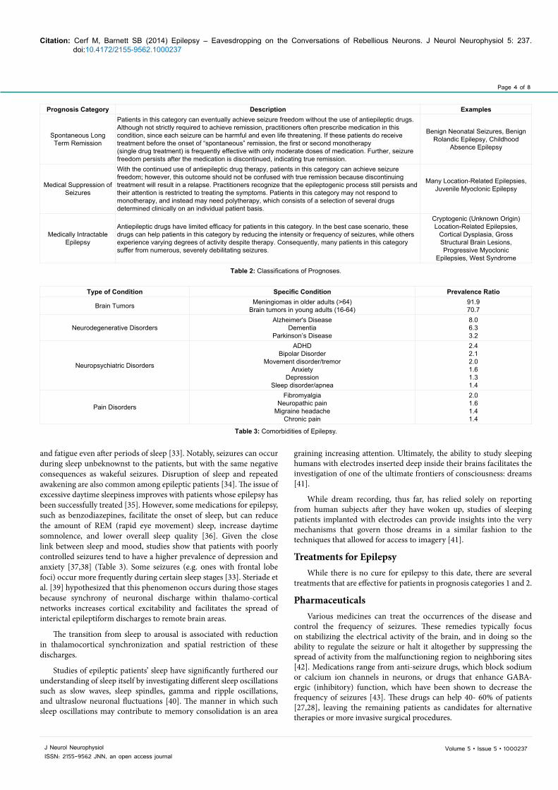
differences in manifestation, a seizure has been broadly understood as an episode of abnormal patterns in levels of synchrony in the discharge of a population of neurons [10]. Historically, seizures have been described as periods of heightened synchronization, but Jiruska et al. [11] have suggested that this characterization oversimplifies the phenomena. In particular, neuronal populations often desynchronize moments before seizures and during their early stages with excessive synchrony following later (Figure 1) [11]. The maximum synchronization is actually observed at the offset of a seizure and is thought to facilitate the seizure’s concurrent termination throughout the brain. While there is ongoing research on treating epilepsy by inhibiting extreme synchrony, this refined understanding has led to research on promoting synchrony as an abortive therapy.
Clinicians characterize seizures by their behavioral manifestations, structural manifestations, or the impairment of consciousness that is
Page 2 of 8
Volume 5 • Issue 5 • 1000237J Neurol NeurophysiolISSN: 2155-9562 JNN, an open access journal
Citation: Cerf M, Barnett SB (2014) Epilepsy – Eavesdropping on the Conversations of Rebellious Neurons. J Neurol Neurophysiol 5: 237. doi:10.4172/2155-9562.1000237
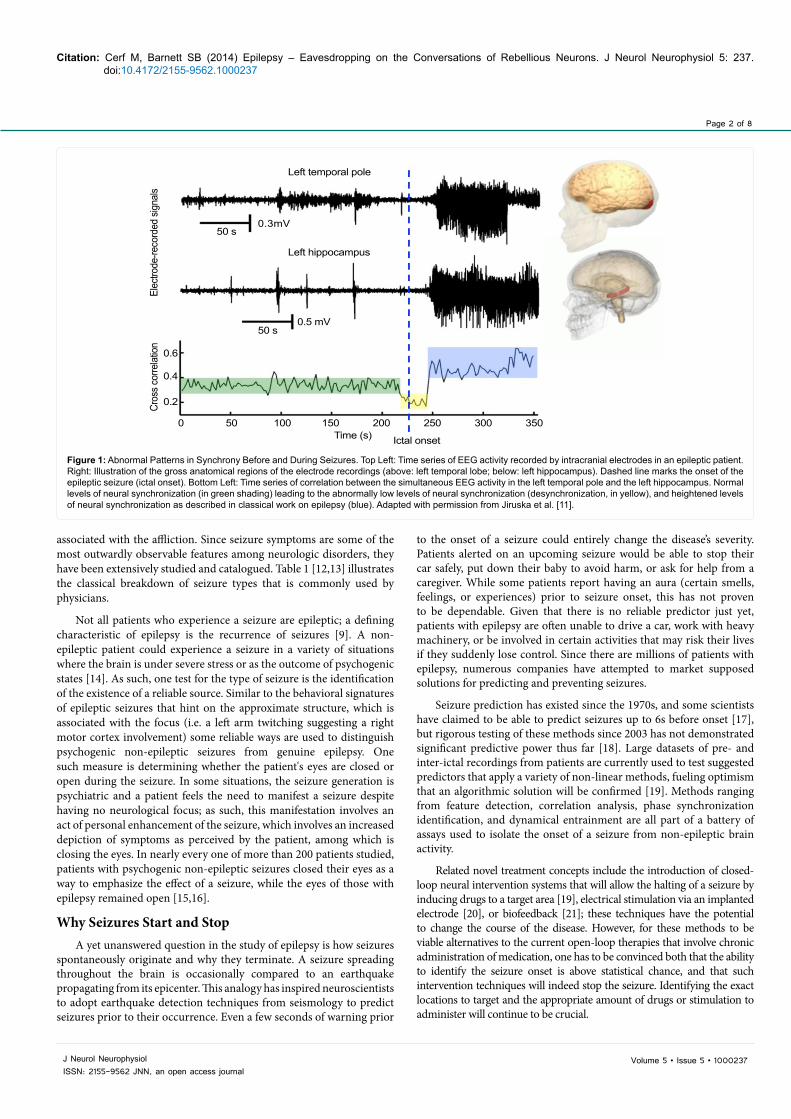
associated with the affliction. Since seizure symptoms are some of the most outwardly observable features among neurologic disorders, they have been extensively studied and catalogued. Table 1 [12,13] illustrates the classical breakdown of seizure types that is commonly used by physicians.
Not all patients who experience a seizure are epileptic; a defining characteristic of epilepsy is the recurrence of seizures [9]. A non-epileptic patient could experience a seizure in a variety of situations where the brain is under severe stress or as the outcome of psychogenic states [14]. As such, one test for the type of seizure is the identification of the existence of a reliable source. Similar to the behavioral signatures of epileptic seizures that hint on the approximate structure, which is associated with the focus (i.e. a left arm twitching suggesting a right motor cortex involvement) some reliable ways are used to distinguish psychogenic non-epileptic seizures from genuine epilepsy. One such measure is determining whether the patient's eyes are closed or open during the seizure. In some situations, the seizure generation is psychiatric and a patient feels the need to manifest a seizure despite having no neurological focus; as such, this manifestation involves an act of personal enhancement of the seizure, which involves an increased depiction of symptoms as perceived by the patient, among which is closing the eyes. In nearly every one of more than 200 patients studied, patients with psychogenic non-epileptic seizures closed their eyes as a way to emphasize the effect of a seizure, while the eyes of those with epilepsy remained open [15,16].
Why Seizures Start and StopA yet unanswered question in the study of epilepsy is how seizures
spontaneously originate and why they terminate. A seizure spreading throughout the brain is occasionally compared to an earthquake propagating from its epicenter. This analogy has inspired neuroscientists to adopt earthquake detection techniques from seismology to predict seizures prior to their occurrence. Even a few seconds of warning prior
to the onset of a seizure could entirely change the disease’s severity. Patients alerted on an upcoming seizure would be able to stop their car safely, put down their baby to avoid harm, or ask for help from a caregiver. While some patients report having an aura (certain smells, feelings, or experiences) prior to seizure onset, this has not proven to be dependable. Given that there is no reliable predictor just yet, patients with epilepsy are often unable to drive a car, work with heavy machinery, or be involved in certain activities that may risk their lives if they suddenly lose control. Since there are millions of patients with epilepsy, numerous companies have attempted to market supposed solutions for predicting and preventing seizures.
Seizure prediction has existed since the 1970s, and some scientists have claimed to be able to predict seizures up to 6s before onset [17], but rigorous testing of these methods since 2003 has not demonstrated significant predictive power thus far [18]. Large datasets of pre- and inter-ictal recordings from patients are currently used to test suggested predictors that apply a variety of non-linear methods, fueling optimism that an algorithmic solution will be confirmed [19]. Methods ranging from feature detection, correlation analysis, phase synchronization identification, and dynamical entrainment are all part of a battery of assays used to isolate the onset of a seizure from non-epileptic brain activity.
Related novel treatment concepts include the introduction of closed-loop neural intervention systems that will allow the halting of a seizure by inducing drugs to a target area [19], electrical stimulation via an implanted electrode [20], or biofeedback [21]; these techniques have the potential to change the course of the disease. However, for these methods to be viable alternatives to the current open-loop therapies that involve chronic administration of medication, one has to be convinced both that the ability to identify the seizure onset is above statistical chance, and that such intervention techniques will indeed stop the seizure. Identifying the exact locations to target and the appropriate amount of drugs or stimulation to administer will continue to be crucial.
Left temporal pole
Elec
trode
-reco
rded
sign
alsCr
oss c
orre
lation
Left hippocampus
50 s
50 s
Time (s) Ictal onset
0.6
0.4
0.2
0.5 mV
0.3mV
0 50 100 150 200 250 300 350
Figure 1: Abnormal Patterns in Synchrony Before and During Seizures. Top Left: Time series of EEG activity recorded by intracranial electrodes in an epileptic patient. Right: Illustration of the gross anatomical regions of the electrode recordings (above: left temporal lobe; below: left hippocampus). Dashed line marks the onset of the epileptic seizure (ictal onset). Bottom Left: Time series of correlation between the simultaneous EEG activity in the left temporal pole and the left hippocampus. Normal levels of neural synchronization (in green shading) leading to the abnormally low levels of neural synchronization (desynchronization, in yellow), and heightened levels of neural synchronization as described in classical work on epilepsy (blue). Adapted with permission from Jiruska et al. [11].
Page 3 of 8
Volume 5 • Issue 5 • 1000237J Neurol NeurophysiolISSN: 2155-9562 JNN, an open access journal
Citation: Cerf M, Barnett SB (2014) Epilepsy – Eavesdropping on the Conversations of Rebellious Neurons. J Neurol Neurophysiol 5: 237. doi:10.4172/2155-9562.1000237
Additionally, in order to use any of these techniques, one must understand not only why seizures originate and how to identify their onset, but also why they stop. Fortunately, most seizures stop on their own after a few minutes [22]. Given that the length of a seizure is correlated with higher damage to the brain it is essential to be able to stop seizures rapidly [23]. The median seizure durations are 78s (complex partial) and 130s (secondarily generalized), with termination caused by multiple reasons, ranging from loss of ionic gradient at the single neuron level, (which effectively is equivalent to depletion of the cell’s energy substrates locally, resulting in the cessation of neuronal firing), lack of amplification at the network level, which stops the excitation and synchronicity across sites, external mechanisms that limit the seizure spread, or specific intervention in the form of activation of structures that inhibit the seizure [22]. One of the most studied mechanisms of seizure onset and offset is the effect of inhibitory (e.g. GABA) and excitatory (e.g. glutamate) neurotransmitters.
Electrophysiological studies suggest that reduced GABA (gamma-aminobutyric acid) inhibition contributes to the neuronal hyperexcitability that is characteristic of epilepsy [24]. Additionally, microdialysis studies have found heightened levels of extracellular glutamate the main excitatory neurotransmitter immediately preceding or during seizure onset [25]. In general, perturbing the balance of the excitatory and inhibitory neurotransmitters has been implicated in seizure generation [26].
These understandings of how seizures start as well as stop, and the fact that even among severe epileptic patients, the tendency for seizures to terminate within minutes suggests that better understanding of the brain’s own control mechanisms and the way they put an end to a rebellion of
specific cells can prove to be essential in unraveling in novel treatments for epilepsy, and potentially even help identify fundamental control mechanisms in the brain which will have implications for other diseases.
Epilepsy PrognosisThree typical prognoses are commonly used to determine the
treatability of the disease (in increasing order of difficulty): (1) “spontaneous long term remission,” seizure freedom after different amounts of activity and time, (2)“medical suppression of seizures,” requiring ongoing antiepileptic drug therapy, and (3) “medically intractable epilepsy,” which may require surgery to achieve seizure freedom (see the Neurosurgery section later in this paper). The first two prognoses account for a combined 40-60% of epileptic patients, while the third type occurs in the remaining proportion [27,28]. Table 2 provides descriptions and examples of each prognosis category [27].
ComorbidityAnother important component to the prognosis of an epilepsy
patient is that the disorder co-occurs with numerous other diseases, especially brain tumors, neurodegenerative disorders [29], neuropsychiatric disorders, and pain disorders [30]. Other kinds of conditions have also been shown to have statistically significant comorbidity; for example, asthma has a prevalence ratio of 1.25 with epileptic patients (Table 3) [29,30].
Effects on SleepThe comorbidity of epilepsy and sleep disorders has been particularly
well studied amongst researchers and clinicians as patients with epilepsy are twice as likely to suffer from daytime sleepiness, insomnia [31,32],
Table 1: Classifications of Seizures.
Type of Seizure General Manifestation Onset Signs Consciousness Routine (Non-Seizure)EEG Observations
Simple Partial (focal motor)
Localized (depending on affected brain region) jerking
beginning in a small anatomical area (e.g. hand) and progressing
to the surrounding region (e.g. arm)
Sensorimotor, autonomic, orpsychic symptoms depending on
the location of abnormalneuronal synchrony
Not Impaired Focal slowing and/or sharp-wave activity
Complex Partial (temporal lobe or psychomotor) Same as Simple Partial
None or with sensorimotor, autonomic, or psychic
symptoms
Impaired, often followed by temporary confusion
Focal slowing and/or sharp-wave activity
Secondary Generalized Partial (tonic-clonic or grand mal)
Same as Simple Partial,except that the seizure
progresses further,causing patients to fall,
rigidly extend extremities(“tonic phase”), and thenconvulse (“clonic phase”)
Sensorimotor, autonomic, orpsychic symptoms
Lost, often followed by coma and slow recovery
Focal slowing and/or sharp-wave activity
(Generalized) Absence (petit mal)
All motor activity stops,with minor exceptions
such as eye blinking, butdo not fall nor exhibit
convulsive (“tonic-clonic”)movements
None or hyperventilation (which can precipitate seizure)
Brief period of unresponsiveness(often 10 seconds)
3 Hz spike-wave pattern
Primarily Generalized Tonic-Clonic (grand mal)
Forced contraction ofmuscles for 30s (“tonicphase”), often causing
patients to fall andbecome cyanotic (blue).
Jerking of extremitiesfollows for 1-2 min (“clonicphase”). Sleepiness and
soreness followconvulsions
None or myoclonic jerks Lost, often followed by coma and slow recovery 3-5 Hz spike-wave pattern
Page 4 of 8
Volume 5 • Issue 5 • 1000237J Neurol NeurophysiolISSN: 2155-9562 JNN, an open access journal
Citation: Cerf M, Barnett SB (2014) Epilepsy – Eavesdropping on the Conversations of Rebellious Neurons. J Neurol Neurophysiol 5: 237. doi:10.4172/2155-9562.1000237
and fatigue even after periods of sleep [33]. Notably, seizures can occur during sleep unbeknownst to the patients, but with the same negative consequences as wakeful seizures. Disruption of sleep and repeated awakening are also common among epileptic patients [34]. The issue of excessive daytime sleepiness improves with patients whose epilepsy has been successfully treated [35]. However, some medications for epilepsy, such as benzodiazepines, facilitate the onset of sleep, but can reduce the amount of REM (rapid eye movement) sleep, increase daytime somnolence, and lower overall sleep quality [36]. Given the close link between sleep and mood, studies show that patients with poorly controlled seizures tend to have a higher prevalence of depression and anxiety [37,38] (Table 3). Some seizures (e.g. ones with frontal lobe foci) occur more frequently during certain sleep stages [33]. Steriade et al. [39] hypothesized that this phenomenon occurs during those stages because synchrony of neuronal discharge within thalamo-cortical networks increases cortical excitability and facilitates the spread of interictal epileptiform discharges to remote brain areas.
The transition from sleep to arousal is associated with reduction in thalamocortical synchronization and spatial restriction of these discharges.
Studies of epileptic patients’ sleep have significantly furthered our understanding of sleep itself by investigating different sleep oscillations such as slow waves, sleep spindles, gamma and ripple oscillations, and ultraslow neuronal fluctuations [40]. The manner in which such sleep oscillations may contribute to memory consolidation is an area
graining increasing attention. Ultimately, the ability to study sleeping humans with electrodes inserted deep inside their brains facilitates the investigation of one of the ultimate frontiers of consciousness: dreams [41].
While dream recording, thus far, has relied solely on reporting from human subjects after they have woken up, studies of sleeping patients implanted with electrodes can provide insights into the very mechanisms that govern those dreams in a similar fashion to the techniques that allowed for access to imagery [41].
Treatments for EpilepsyWhile there is no cure for epilepsy to this date, there are several
treatments that are effective for patients in prognosis categories 1 and 2.
PharmaceuticalsVarious medicines can treat the occurrences of the disease and
control the frequency of seizures. These remedies typically focus on stabilizing the electrical activity of the brain, and in doing so the ability to regulate the seizure or halt it altogether by suppressing the spread of activity from the malfunctioning region to neighboring sites [42]. Medications range from anti-seizure drugs, which block sodium or calcium ion channels in neurons, or drugs that enhance GABA-ergic (inhibitory) function, which have been shown to decrease the frequency of seizures [43]. These drugs can help 40- 60% of patients [27,28], leaving the remaining patients as candidates for alternative therapies or more invasive surgical procedures.
Table 2: Classifications of Prognoses.
Prognosis Category Description Examples
Spontaneous LongTerm Remission
Patients in this category can eventually achieve seizure freedom without the use of antiepileptic drugs. Although not strictly required to achieve remission, practitioners often prescribe medication in this condition, since each seizure can be harmful and even life threatening. If these patients do receive treatment before the onset of “spontaneous” remission, the first or second monotherapy(single drug treatment) is frequently effective with only moderate doses of medication. Further, seizure freedom persists after the medication is discontinued, indicating true remission.
Benign Neonatal Seizures, Benign Rolandic Epilepsy, Childhood
Absence Epilepsy
Medical Suppression ofSeizures
With the continued use of antiepileptic drug therapy, patients in this category can achieve seizure freedom; however, this outcome should not be confused with true remission because discontinuing treatment will result in a relapse. Practitioners recognize that the epileptogenic process still persists and their attention is restricted to treating the symptoms. Patients in this category may not respond tomonotherapy, and instead may need polytherapy, which consists of a selection of several drugsdetermined clinically on an individual patient basis.
Many Location-Related Epilepsies, Juvenile Myoclonic Epilepsy
Medically IntractableEpilepsy
Antiepileptic drugs have limited efficacy for patients in this category. In the best case scenario, these drugs can help patients in this category by reducing the intensity or frequency of seizures, while othersexperience varying degrees of activity despite therapy. Consequently, many patients in this category suffer from numerous, severely debilitating seizures.
Cryptogenic (Unknown Origin) Location-Related Epilepsies,
Cortical Dysplasia, GrossStructural Brain Lesions,Progressive Myoclonic
Epilepsies, West Syndrome
Type of Condition Specific Condition Prevalence Ratio
Brain Tumors Meningiomas in older adults (>64)Brain tumors in young adults (16-64)
91.970.7
Neurodegenerative DisordersAlzheimer's Disease
DementiaParkinson’s Disease
8.06.33.2
Neuropsychiatric Disorders
ADHDBipolar Disorder
Movement disorder/tremorAnxiety
DepressionSleep disorder/apnea
2.42.12.01.61.31.4
Pain Disorders
FibromyalgiaNeuropathic pain
Migraine headacheChronic pain
2.01.61.41.4
Table 3: Comorbidities of Epilepsy.
Page 5 of 8
Volume 5 • Issue 5 • 1000237J Neurol NeurophysiolISSN: 2155-9562 JNN, an open access journal
Citation: Cerf M, Barnett SB (2014) Epilepsy – Eavesdropping on the Conversations of Rebellious Neurons. J Neurol Neurophysiol 5: 237. doi:10.4172/2155-9562.1000237
Non-Pharmaceutical SolutionsAdditional treatments include brain stimulation of the target area or
of nerves and of sites such as the thalamus or the hippocampus to drive seizure reduction [44]. Thalamic stimulation as well as intracranial stimulating devices to predict and prevent seizures offer an alternative route of medication, yet their success rate is limited [45]. Given the genetic aspects of epilepsy and cell mutations that are linked to the syndrome, more gene-directed therapies are now tested to control those mutations. Currently, a number of animal studies focusing on transfection use viral components to introduce new genes into brain cells, which are aimed at producing therapeutic proteins in the brain. Additionally, the rise of the new field of optogenetics, which allows for specific targeting of brain cells driven by light-sensitive proteins, can be used to inhibit or stimulate cells to help regulate an excited network [46,47]. While these are currently being tested, they are not yet offered as a potential treatment for humans due to the risky nature of injection viral genetic material, which is part of the optogenetic solution. However, a number of scientists and clinicians have proposed starting clinical trials of gene therapy for epilepsy [48].
Finally, given the clear metabolic aspect of genetic modification and protein altering in epilepsy, certain dietary and nutrition changes tested show an effect on the frequency of seizures, especially with children. Currently, more than half of the tests involving the ketogenic (high fat, low carbohydrates) diet have shown an improvement in seizure control. Some of the patients achieve full seizure freedom [49]. To this date, the nature of the diet and the reasons it decreases the frequency is not fully understood.
NeurosurgeryFor patients in whom pharmacological treatment and alternative
therapies do not yield seizure control (i.e. prognosis category 3: medically intractable epilepsy), an additional treatment option is invasive brain surgery. In some of these patients, the neuronal network causing the seizures can be identified in sufficient detail to enable focal surgical intervention in the form of resection, disconnection, or chronic stimulation to neutralize the seizures [50]. These surgical procedures, and in particular precise focal surgical resections, can often produce a complete cure or considerable improvement in seizure frequency or severity with very few, if any, consequences in terms of neurological or cognitive function.
Epilepsy surgery for focal seizures began more than a century ago and progressed with the technical innovations of EEG and neuroimaging. In the1860s and 1870s, the pioneering clinical work of the epileptologist John Hughlings Jackson laid the groundwork for understanding the cortical localization of focal epilepsies, while the animal experiments of the neurophysiologists. Gustav Theodor Fritsch, Eduard Hitzig, and David Ferrier gave parallel confirmation of Jackson's conclusions [51].
After pre-surgical evaluation has identified the epileptogenic zone, clinicians plan the operative procedure most likely to benefit the patient. The most common procedures involve the epileptic focus being resected or the corpus callosum being severed. In rare instances, an entire hemisphere will be removed, although it is more common for clinicians to perform a “functional hemispherectomy,” which is less invasive but still severs major connections between lobes to separate the healthy portions of the brain from the epileptogenic zones. This is done in order to contain the seizure and prevent a spread and takeover of the entire brain by the synchronous activity.
The majority of resective procedures performed for epilepsy are
variants of anteromesial temporal resections since mesial temporal lobe epilepsy is both the most common and the most medically refractory focal epilepsy [52].
In addition, a disconnective procedure for temporal lobe epilepsy referred to as temporal lobotomy has been reported in a small case series with preliminary outcome data [53].
Cognitive Studies of Epileptic PatientsFinally, the advancement of surgical techniques for epilepsy
treatment has pushed the boundaries of neuroscience. Some of the most remarkable discoveries about the brain were made possible via direct recording of neural activity during surgery [54]. This kind of surgery requires the temporary removal of a section of the patient’s skull (craniotomy) to expose the brain and strategically implant electrodes for real-time monitoring and analysis.
Since the electrodes remain implanted and continue to record neural activity while the patient is awake, neuroscientists have been able to study some of the most profound aspects of our psyche. Discoveries such as the split-brain phenomenon and direct recordings from memory areas (e.g. hippocampus, entorhinal cortex) and emotion areas (e.g. amygdala) have spurred a branch of neuroscience that eavesdrops on the communications of individual brain cells.
Split-Brain PhenomenonOne of the most intriguing phenomena that unraveled much of
today’s understanding on the brain and consciousness is that of split-brain. The split-brain phenomenon arises from the severing of the corpus callosum—the bridge connecting the two hemispheres in the brain—and as such the main highway of communication between the two parts. This last resort in treating epilepsy limits seizure propagation to a single hemisphere, leaving the other hemisphere intact and as such, in control of the opposite side of the body. Patients are able to live relatively normal lives, albeit with the unique situation in which the two halves of the brain are unable to communicate with each other. This, in turn, leads to interesting documented cases of patients unable to verbally communicate (as language is typically controlled by the left hemisphere) what they are seeing in the left half of the visual field (as the left hemifield is controlled by the right hemisphere), or unable to solve puzzles (right hemisphere) with their right arm (left hemisphere), and other outcomes rising from the severed communication between the two hemispheres. Accordingly, this helped explain the interesting internal vying for dominance over our single consciousness by multiple entities residing inside our brain, which is an essential part of our understanding of choice, decisions and our psyche [55].
Neural MappingThe localized nature of epileptic foci resection additionally gives
rise to an opportunity to use the invasive procedures to study the human brain. For nearly a century, neurosurgeons have used this rare exposure of the brain to probe individual sites for cognitive research. This groundbreaking research has lead to extraordinary understanding of human cognition [40]. One of the earliest and most notable examples of this research was the work of neurosurgeon Wilder Penfield who pioneered the “Montreal procedure” of resecting the epileptic foci to eliminate seizures [56].
While trying to locate seizure foci, Penfield probed his patient’s brain and observed responses. This neural stimulation procedure enabled Penfield to map the sensory and motor cortices of the brain [57]. Following this line of research, there are now multiple labs that use
Page 6 of 8
Volume 5 • Issue 5 • 1000237J Neurol NeurophysiolISSN: 2155-9562 JNN, an open access journal
Citation: Cerf M, Barnett SB (2014) Epilepsy – Eavesdropping on the Conversations of Rebellious Neurons. J Neurol Neurophysiol 5: 237. doi:10.4172/2155-9562.1000237
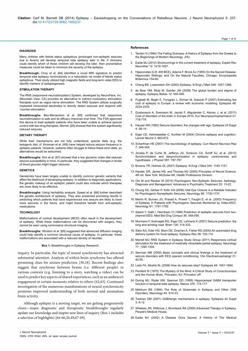
a version of the same technique to study the brain during the resection process. Neuroscientists carefully monitor the exposed part of the brain while the patient resides in the epileptic ward by placing a set of electrodes either on the surface of the cortex or deep inside the brain. The clinical requirement is that following placement of these electrodes performed in the operating room, the patients need to be monitored for several days and sometimes for up to three weeks until a sufficient number of spontaneous seizures are captured. This protocol enables the correlation of brain activity with the actual seizures to be captured on a video monitor in the hope of then identifying the network underlying the seizures. If this monitoring is successful, later surgery may be offered for resection, which is likely to prove curative. These electrodes continuously record the activity of a local field or individual neurons (single- or multi-units). Clinically, these electrodes are used to locate the seizure foci; patients wait for a spontaneous seizure to occur, and once it does, the electrodes are used to triangulate the origin of the seizure and accordingly guide the resection of the focus (Figure 2). Independently, there are countless pure research applications of these electrodes, either targeting the disease itself [58], or investigating other cognitive functions altogether (Figure 2) [40].
Human brain studies using these techniques can be classified by data type, electrode type, and the functions targeted. The electrodes are typically either “grid electrodes,” sheets of dozens (or even hundreds) of electrodes that cover a large surface on the cortex and usually record local field potentials (Figure 2), or “depth electrodes,” used in much smaller quantities, located in close proximity within a larger macro-electrode implanted in the brain that targets deeper regions and accesses the spiking activity of individual neurons.
Neurosurgical procedures for treatment of severe intractable epilepsy have provided the most opportunities to record single neuron activity over relatively long periods of time in awake, conscious patients [59].
Anecdotally, this field of research was born when Penfield noted that one of his patients claimed to have an “aura” of a burnt toast. That is, prior to having a seizure she reported experiencing the smell of burnt toast. This story [60] suggests that the patient had an olfactory precursor that, upon resection, led to having no seizures. Such auras as precursors for seizures are common among the patients and often indicative of the brain area that afflicted with the damage causing the seizure (e.g. a feeling of fear prior to the seizure could indicate a pre-activation in the amygdala as a precursor to the seizure). Today, clinicians commonly ask the patient’s relatives prior to the surgery to describe any phenomena arising prior to the seizure as a way to identify signatures of brain areas for further investigation or future resection.
ConsciousnessGiven the unique nature of epilepsy and the fact that consciousness
is impaired during a majority of seizures, the disease has become a platform for hypotheses, tests and studies of the nature of consciousness. Studies of varying degrees of consciousness in epilepsy patients during surgery under local anesthesia have captured the imaginations of generations of neuroscientists [61] (Penfield, in the famous 1936 Harvey Lectures, suggested that such studies with epilepsy patients could pave the road to a robust theory of consciousness).
ConclusionWas Hippocrates right when he said epilepsy was “no more divine
than others?”
After all, epilepsy has inspired, taught, challenged, and enlightened us more than any other phenomenon. When we think of usual diseases, we imagine the archetypal battle of “good versus evil,” our bodies versus pathogens. However, with epilepsy, these lines are blurred as civil wars spontaneously erupt within our brains. Friendly neurons can become rebellious ones in an instant. Our understanding of the condition, and by extension the entire brain, is constantly evolving. Just when we think we are regaining our freedom from the “rebellious neurons” that characterize epilepsy, we are challenged to rethink our models and build new ones. Every discovery marks progress, but also inevitably leads to the realization that the phenomenon is even more complex than originally imagined.
Sweeping advances in medicine and surgery have transformed an utterly debilitating disorder into a manageable condition for millions of people. Epilepsy has offered us a way to learn more about ourselves and tackle the mechanisms underlying memory, cognition, consciousness, and personal identity.
By passionately pursuing this kind of research, we might save the world from some of its most frightening forces—neurodegenerative diseases like Alzheimer’s, neuropsychiatric disorders like depression, brain tumors, and chronic pain. Epilepsy need not be viewed as a threat to our identity, but rather as an opportunity to understand ourselves more deeply and shape our future as individuals and as a society.
For example, the association of epileptic seizures with heightened synchrony between neurons was followed by the observation that it is actually a dynamic process that includes desynchronization in addition to excessive synchronization [11]. Additionally, findings and models in epilepsy research have led to new lines of scientific
Figure 2: Craniotomy and Schematic of Seizure Focus Localization by Triangulation. Left: Image of an exposed portion of the brain of an epileptic patient undergoing a craniotomy. A 5x4 array of grid electrodes (white) is pictured. Right: Magnification of a section of the grid electrodes annotated with a schematic of seizure focus localization by triangulation. Each electrode is circumscribed with a radius corresponding to the distance between that electrode and the mock seizure focus. The point at which the three circles intersect marks the location of the estimated seizure focus.
Page 7 of 8
Volume 5 • Issue 5 • 1000237J Neurol NeurophysiolISSN: 2155-9562 JNN, an open access journal
Citation: Cerf M, Barnett SB (2014) Epilepsy – Eavesdropping on the Conversations of Rebellious Neurons. J Neurol Neurophysiol 5: 237. doi:10.4172/2155-9562.1000237
inquiry. In particular, the topic of neural synchronicity has attracted substantial attention. Analysis of within-brain synchrony has offered promising clues for seizure prediction [30,18]. Recent findings also suggest that synchrony between brains (i.e. different people) in various contexts (e.g. listening to a story, watching a video) can be used to predict key aspects of shared experiences, such as an audience’s engagement at certain moments relative to others [62,63]. Continued investigation of the numerous manifestations of neural synchronicity promises improved understanding of both normal and anomalous brain activity.
Although epilepsy is a moving target, we are getting progressively closer—major diagnostic and therapeutic breakthroughs regularly update our knowledge and inspire new lines of inquiry (Box 1 includes a selection of highlights) [64-66,20,49,67-69].
References1. Temkin O (1994) The Falling Sickness: A History of Epilepsy from the Greeks to
the Beginnings of Modern Neurology. JHU.
2. Eadie MJ (2012) Shortcomings in the current treatment of epilepsy. Expert Rev Neurother 12: 1419-1427.
3. Hippocrates G (fl. 400 BCE), Adams F, Brock AJ (1955) On the Sacred Disease. Hippocratic Writings; and On the Natural Faculties. Chicago: Encyclopedia Britannica 154-60.
4. Chang BS, Lowenstein DH (2003) Epilepsy. N Engl J Med 349: 1257-1266.
5. de Boer HM, Mula M, Sander JW (2008) The global burden and stigma of epilepsy. Epilepsy Behav 12: 540-546.
6. Pugliatti M, Beghi E, Forsgren L, Ekman M, Sobocki P (2007) Estimating the cost of epilepsy in Europe: a review with economic modeling. Epilepsia 48: 2224-2233.
7. Gustavsson A, Svensson M, Jacobi F, Allgulander C, Alonso J, et al. (2011) Cost of disorders of the brain in Europe 2010. Eur Neuropsychopharmacol 21: 718-779.
8. Hauser WA (1992) Seizure disorders: the changes with age. Epilepsia 33 Suppl 4: S6-14.
9. Elger CE, Helmstaedter C, Kurthen M (2004) Chronic epilepsy and cognition. Lancet Neurol 3: 663-672.
10. Scharfman HE (2007) The neurobiology of epilepsy. Curr Neurol Neurosci Rep 7: 348-354.
11. Jiruska P, de Curtis M, Jefferys JG, Schevon CA, Schiff SJ, et al. (2013) Synchronization and desynchronization in epilepsy: controversies and hypotheses. J Physiol 591: 787-797.
12. Browne TR, Holmes GL (2001) Epilepsy. N Engl J Med 344: 1145-1151.
13. Kandel, ER, James HS, and Thomas MJ (2000) Principles of Neural Science. 4th ed. New York: McGraw-Hill, Health Professions Division.
14. Oto M and Reuber M (2014) Psychogenic Non-Epileptic Seizures: Aetiology, Diagnosis and Management. Advances in Psychiatric Treatment 20: 13-22.
15. Chung SS, Gerber P, Kirlin KA (2006) Ictal Eye Closure is a Reliable Indicator for Psychogenic Nonepileptic Seizures. Neurology 66: 1730-1731.
16. Martin R, Burneo JG, Prasad A, Powell T, Faught E, et al. (2003) Frequency of Epilepsy in Patients with Psychogenic Seizures Monitored by Video-EEG. Neurology 61: 1791-1792.
17. Salant Y, Gath I, Henriksen O (1998) Prediction of epileptic seizures from two-channel EEG. Med Biol Eng Comput 36: 549-556.
18. Mormann F, Andrzejak RG, Elger CE, Lehnertz K (2007) Seizure prediction: the long and winding road. Brain 130: 314-333.
19. Stein AG, Eder HG, Blum DE, Drachev A, Fisher RS (2000) An automated drug delivery system for focal epilepsy. Epilepsy Res 39: 103-114.
20. Morrell MJ; RNS System in Epilepsy Study Group (2011) Responsive cortical stimulation for the treatment of medically intractable partial epilepsy. Neurology 77: 1295-1304.
21. Sterman MB (2000) Basic concepts and clinical findings in the treatment of seizure disorders with EEG operant conditioning. Clin Electroencephalogr 31: 45-55.
22. Lado FA, Moshé SL (2008) How do seizures stop? Epilepsia 49: 1651-1664.
23. Penfield W (1975) The Mystery of the Mind: A Critical Study of Consciousness and the Human Brain. Princeton, NJ: Princeton UP.
24. During MJ, Ryder KM, Spencer DD (1995) Hippocampal GABA transporter function in temporal-lobe epilepsy. Nature 376: 174-177.
25. Meldrum BS (1994) The Role of Glutamate in Epilepsy and Other CNS Disorders. Neurology 44: S14-23.
26. Treiman DM (2001) GABAergic mechanisms in epilepsy. Epilepsia 42 Suppl 3: 8-12.
27. Wheless JW, Willmore J, Brumback RA (2009) Advanced Therapy in Epilepsy. People's Medical House.
28. Eadie MJ (2002) A Disease Once Sacred. A History of The Medical
Box 1: Breakthroughs in Epilepsy Research.
DIAGNOSIS
Many children with febrile status epilepticus (prolonged non-epileptic seizures due to fevers) will develop temporal lobe epilepsy later in life. If clinicians could identify which of these children will develop the later, then preventative measures could be taken to minimize the severity of the epilepsy.
Breakthrough: Choy et al. [64] identified a novel MRI signature to predict temporal lobe epilepsy noninvasively in a naturalistic rat model of febrile status epilepticus. Their study utilized high magnetic fields and long-term video EEG to identify markers of epileptogenesis.
STIMULATION THERAPY
The RNS (responsive neurostimulation) System, developed by NeuroPace, Inc. (Mountain View, CA) provides an alternative to indirect modulatory stimulation therapies such as vagus nerve stimulation. The RNS System utilizes surgically implanted intracranial electrodes to directly detect seizures and respond with ‘counter-stimulation’.
Breakthroughs: Ben-Menachem et al. [65] confirmed that responsive neurostimulation is safe and its efficacy improves over time. The FDA approved this device to treat epileptic patients who have been unable to achieve seizure control with two drug therapies. Morrell [20] showed that this system significantly reduced seizures.
DIETARY THERAPY
While their mechanisms are not fully understood, special diets (e.g. the ketogenic diet; cf. Kinsman et al. [49]) have helped reduce seizure frequency in epileptic patients. However, patients often struggle to follow these strict diets, so alternatives would be welcomed.
Breakthroughs: Kim et al. [67] showed that a low glycemic index diet reduces seizure susceptibility in mice. In particular, they suggested that changes in levels of blood glucose might trigger seizures.
GENETICS
Geneticists have been largely unable to identify common genetic variants that affect the likelihood of developing epilepsy. In addition to diagnostic applications, the genetic makeup of an epileptic patient could also indicate which therapies are more likely to be effective.
Breakthroughs: Using heritability analysis, Speed et al. [68] further described the genetic architecture of epilepsy. They also presented promising results in predicting which patients that have experienced one seizure are likely to have more seizures in the future, and might therefore benefit from anti-epileptic medications.
TECHNOLOGY
Malformations of cortical development (MCD) often result in the development of epilepsy. While these malformations can be discovered with surgery, they cannot be seen using noninvasive structural imaging.
Breakthroughs: Winston et al. [69] suggested that advanced diffusion imaging could help identify a common structural cause of epilepsy. In particular, these malformations are associated with a reduced density of neurites.
Page 8 of 8
Volume 5 • Issue 5 • 1000237J Neurol NeurophysiolISSN: 2155-9562 JNN, an open access journal
Citation: Cerf M, Barnett SB (2014) Epilepsy – Eavesdropping on the Conversations of Rebellious Neurons. J Neurol Neurophysiol 5: 237. doi:10.4172/2155-9562.1000237
Understanding of Epilepsy. Brain: A Journal of Neurology 2nd ser 125: 441-442. Oxford Journals. Oxford University Press.
29. Gaitatzis A, Carroll K, Majeed A, W Sander J (2004) The epidemiology of thecomorbidity of epilepsy in the general population. Epilepsia 45: 1613-1622.
30. Ottman R, Lipton RB, Ettinger AB, Cramer JA, Reed ML, et al. (2011)Comorbidities of epilepsy: results from the Epilepsy Comorbidities and Health(EPIC) survey. Epilepsia 52: 308-315.
31. de Weerd A, de Haas S, Otte A, Trenité DK, van Erp G, et al. (2004) Subjective sleep disturbance in patients with partial epilepsy: a questionnaire-based study on prevalence and impact on quality of life. Epilepsia 45: 1397-1404.
32. Piperidou C, Karlovasitou A, Triantafyllou N, Terzoudi A, et al. (2008) Influence of Sleep Disturbance on Quality of Life of Patients with Epilepsy. Seizure 17:588-594.
33. Crespel A, Coubes P, Baldy-Moulinier M (2000) Sleep influence on seizures and epilepsy effects on sleep in partial frontal and temporal lobe epilepsies. Clin Neurophysiol 111 Suppl 2: S54-59.
34. Vignatelli L, Bisulli F, Naldi I, Ferioli S, Pittau F, et al. (2006) Excessive daytime sleepiness and subjective sleep quality in patients with nocturnal frontal lobeepilepsy: a case-control study. Epilepsia 47 Suppl 5: 73-77.
35. Carrion MJ, Nunes ML, Martinez JV, Portuguez MW, da Costa JC (2010)Evaluation of sleep quality in patients with refractory seizures who undergoepilepsy surgery. Epilepsy Behav 17: 120-123.
36. Bazil CW (2003) Epilepsy and sleep disturbance. Epilepsy Behav 4 Suppl 2:S39-45.
37. Stefanello S, Marín-Léon L, Fernandes PT, Li LM, Botega NJ (2010) Psychiatric Comorbidity and Suicidal Behavior in Epilepsy: A Community-Based Case-Control Study. Epilepsia 51: 1120-1125.
38. Van Mill, JG, Witte JG, Nicole V, Richard VD, et al. (2010) Insomnia and Sleep Duration in a Large Cohort of Patients With Major Depressive Disorder andAnxiety Disorders. The Journal of Clinical Psychiatry 71: 239-246.
39. Steriade M, Contreras D, Amzica F (1994) Synchronized sleep oscillations and their paroxysmal developments. Trends Neurosci 17: 199-208.
40. Fried I, Rutishauser U, Cerf M, and Kreiman G (2014) Single Neuron Studies of the Human Brain: Probing Cognition. MIT Press.
41. Cerf M, Gelbard-Sagiv H, Fried I (2014) Studying Thoughts and DeliberationsUsing Single Neuron Recordings in Humans. In: Single Neuron Studies of theHuman Brain: Probing Cognition. Eds: Fried I, Rutishauser U, Cerf M, Kreiman G. MIT Press (2014).
42. Karceski S, Morrell M, Carpenter D (2001) The Expert Consensus GuidelineSeries. Epilepsy & Behavior 2: A1-A50.
43. Macdonald RL, Kelly KM (1995) Antiepileptic drug mechanisms of action.Epilepsia 36 Suppl 2: S2-12.
44. Loddenkemper T, Pan A, Neme S, Baker KB, Rezai AR, et al. (2001) Deepbrain stimulation in epilepsy. J Clin Neurophysiol 18: 514-532.
45. Mormann F, Kreuz T, Rieke C, Andrzejak RG, Kraskov A, et al. (2005) On thepredictability of epileptic seizures. Clin Neurophysiol 116: 569-587.
46. Krook-Magnuson E, Armstrong C, Oijala M, Soltesz I (2013) On-demandoptogenetic control of spontaneous seizures in temporal lobe epilepsy. NatCommun 4: 1376.
47. Bentley JN, Chestek C, Stacey WC, Patil PG (2013) Optogenetics in epilepsy.Neurosurg Focus 34: E4.
48. Kullmann DM, Schorge S, Walker MC, Wykes RC (2014) Gene therapy inepilepsy-is it time for clinical trials? Nat Rev Neurol 10: 300-304.
49. Kinsman SL, Vining EP, Quaskey SA, Mellits D, Freeman JM (1992) Efficacy of the ketogenic diet for intractable seizure disorders: review of 58 cases.Epilepsia 33: 1132-1136.
50. Cerf M, Thiruvengadam N, Mormann F, Kraskov A, Quiroga RQ, et al. (2010)On-line, voluntary control of human temporal lobe neurons. Nature 467: 1104-1108.
51. Rosenow F, Lüders H (2001) Presurgical evaluation of epilepsy. Brain 124:1683-1700.
52. Dupont S1, Semah F, Baulac M, Samson Y (1998) The underlying
pathophysiology of ictal dystonia in temporal lobe epilepsy: an FDG-PET study. Neurology 51: 1289-1292.
53. Smith JR, VanderGriff A, Fountas K (2004) Temporal lobotomy in the surgicalmanagement of epilepsy: technical report. Neurosurgery 54: 1531-1534.
54. Sperry RW (1961) Cerebral Organization and Behavior: The Split BrainBehaves in Many Respects like Two Separate Brains, Providing New Research Possibilities. Science 133: 1749-1757.
55. Cerf M, Mackay M (2011) Studying Consciousness Using Direct Recordingfrom Single Neurons in the Human Brain. In: Characterizing Consciousness:From Cognition to the Clinic. Research and Perspectives in Neurosciences.Springer 133-146.
56. Olivier A, Boling WB, and Taniverdi T (2012) Techniques in Epilepsy Surgery:The MNI Approach. Cambridge: Cambridge UP.
57. PENFIELD W (1950) The supplementary motor area in the cerebral cortex ofman. Arch Psychiatr Nervenkr Z Gesamte Neurol Psychiatr 185: 670-674.
58. Sun FT, Morrell MJ, Wharen RE Jr (2008) Responsive cortical stimulation forthe treatment of epilepsy. Neurotherapeutics 5: 68-74.
59. Truccolo W, Donoghue JA, Hochberg LR, Eskandar EN, Madsen JR, et al.(2011) Single-neuron dynamics in human focal epilepsy. Nat Neurosci 14: 635-641.
60. Penfield W (1997) No Man Alone: A Neurosurgeon's Life. Boston: Little, Brown.
61. Crick F, Koch C, Kreiman G, Fried I (2004) Consciousness and neurosurgery.Neurosurgery 55: 273-281.
62. Barnett SB, Cerf M (2014) Method for Measuring Engagement. US Provisional Patent No. 61930574.
63. Barnett SB, Cerf M (2014) Engaged Minds Think Alike: Measures of NeuralSimilarity Predict Content Engagement. Working Paper.
64. Choy M, Dubé CM, Patterson K, Barnes SR, Maras P, et al. (2014) A novel,noninvasive, predictive epilepsy biomarker with clinical potential. J Neurosci34: 8672-8684.
65. Ben-Menachem, E, Krauss GL (2014) Epilepsy: Responsive Neurostimulation-Modulating the Epileptic Brain. Nat Rev Neurol 10: 247-248.
66. Proffitt, A (2014) Epilepsy Neurodevice Approved. Nature Biotechnology 32.
67. Kim TH, Petrou S, Reid CA (2014) Low Glycaemic Index Diet Reduces Seizure Susceptibility in a Syndrome-Specific Mouse Model of Generalized Epilepsy. Epilepsy Research 108: 139-143.
68. Speed D, O'Brien TJ, Palotie A, Shkura K, Marson AG, et al. (2014) Describing the genetic architecture of epilepsy through heritability analysis. Brain 137:2680-2689.
69. Winston GP, Micallef C, Symms M, Alexander DC, Duncan JS, et al. (2013)Advanced Diffusion Imaging Sequences Could Aid Assessing Patients withFocal Cortical Dysplasia and Epilepsy. Epilepsy Res 108: 336-339.


![Silent Sufferers (Frederick)(Final) - Mississippi Sports Law Reviewmssportslaw.olemiss.edu/files/2012/05/Saving-Silent... · 2012-05-19 · 2012] Saving Silent Sufferers 413 The result](https://img.pdfslide.us/doc/110x75/5f0d98467e708231d43b21d3/silent-sufferers-frederickfinal-mississippi-sports-law-2012-05-19-2012.jpg)


![What Should Eczema Sufferers Eat? [Must Read]](https://img.pdfslide.us/doc/110x75/5555e937d8b42ad3548b4ae4/what-should-eczema-sufferers-eat-must-read.jpg)