Embed Size (px)
Citation preview
Our vision is to save your vision
www.scienceofamd.org
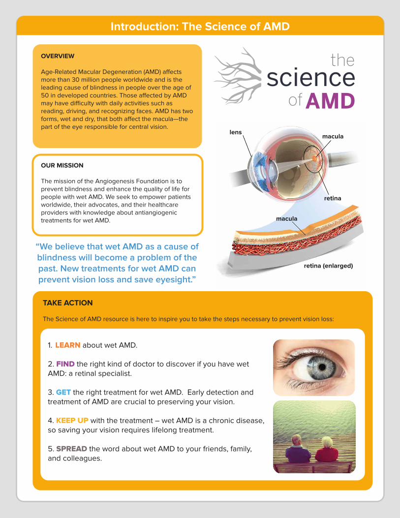
OUR MISSION
The mission of the Angiogenesis Foundation is to prevent blindness and enhance the quality of life for people with wet AMD. We seek to empower patients worldwide, their advocates, and their healthcare providers with knowledge about antiangiogenic treatments for wet AMD.
TAKE ACTION
The Science of AMD resource is here to inspire you to take the steps necessary to prevent vision loss:
1. LEARN about wet AMD.
2. FIND the right kind of doctor to discover if you have wet AMD: a retinal specialist.
3. GET the right treatment for wet AMD. Early detection and treatment of AMD are crucial to preserving your vision.
4. KEEP UP with the treatment – wet AMD is a chronic disease, so saving your vision requires lifelong treatment.
5. SPREAD the word about wet AMD to your friends, family, and colleagues.
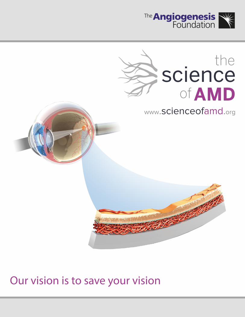
OVERVIEW
Age-Related Macular Degeneration (AMD) a�ects more than 30 million people worldwide and is the leading cause of blindness in people over the age of 50 in developed countries. Those a�ected by AMD may have di�culty with daily activities such as reading, driving, and recognizing faces. AMD has two forms, wet and dry, that both a�ect the macula—the part of the eye responsible for central vision.
“We believe that wet AMD as a cause of blindness will become a problem of the past. New treatments for wet AMD can prevent vision loss and save eyesight.”
lensmacula
retina
macula
retina (enlarged)
Introduction: The Science of AMD
WET AMD
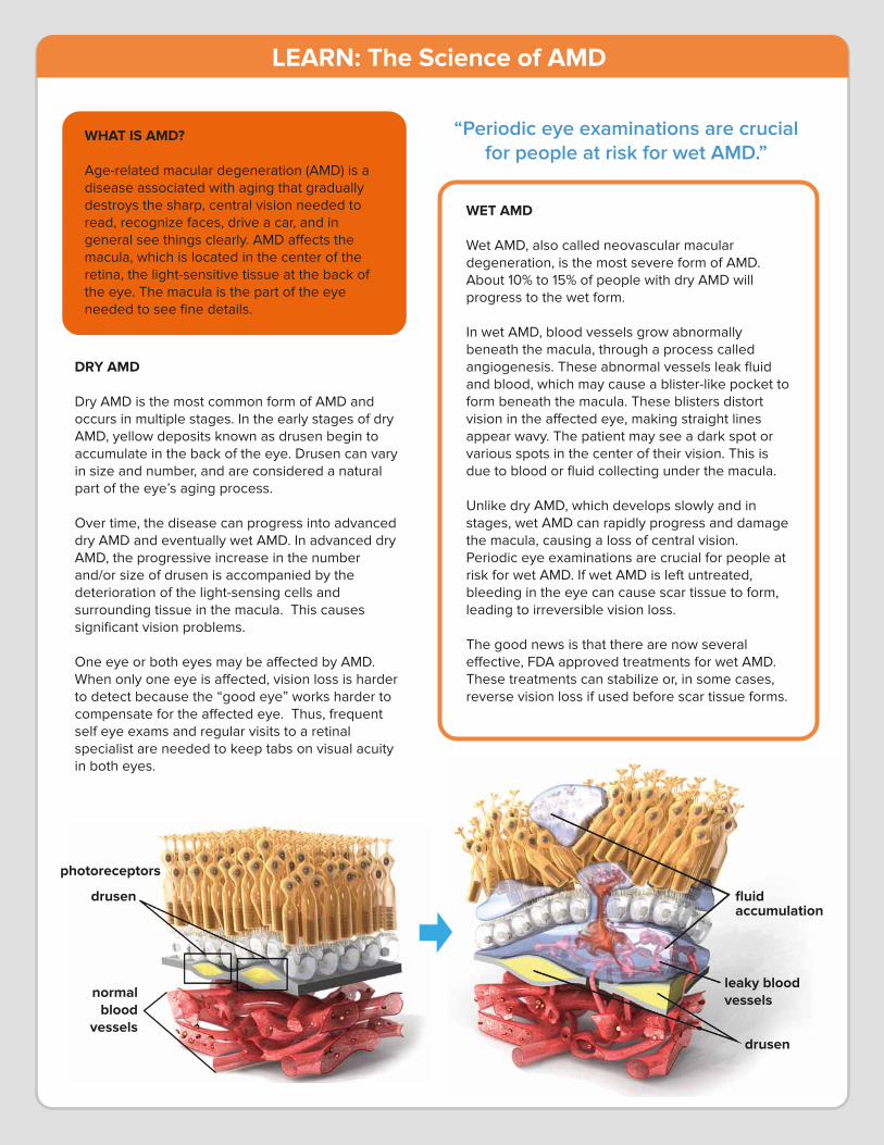
Wet AMD, also called neovascular macular degeneration, is the most severe form of AMD. About 10% to 15% of people with dry AMD will progress to the wet form.
In wet AMD, blood vessels grow abnormally beneath the macula, through a process called angiogenesis. These abnormal vessels leak fluid and blood, which may cause a blister-like pocket to form beneath the macula. These blisters distort vision in the a�ected eye, making straight lines appear wavy. The patient may see a dark spot or various spots in the center of their vision. This is due to blood or fluid collecting under the macula.
Unlike dry AMD, which develops slowly and in stages, wet AMD can rapidly progress and damage the macula, causing a loss of central vision. Periodic eye examinations are crucial for people at risk for wet AMD. If wet AMD is left untreated, bleeding in the eye can cause scar tissue to form, leading to irreversible vision loss.
The good news is that there are now several e�ective, FDA approved treatments for wet AMD. These treatments can stabilize or, in some cases, reverse vision loss if used before scar tissue forms.
“Periodic eye examinations are crucial for people at risk for wet AMD.”
fluidaccumulation
leaky blood vessels
drusen
drusen
photoreceptors
normalblood
vessels
DRY AMD
Dry AMD is the most common form of AMD and occurs in multiple stages. In the early stages of dry AMD, yellow deposits known as drusen begin to accumulate in the back of the eye. Drusen can vary in size and number, and are considered a natural part of the eye’s aging process.
Over time, the disease can progress into advanced dry AMD and eventually wet AMD. In advanced dry AMD, the progressive increase in the number and/or size of drusen is accompanied by the deterioration of the light-sensing cells and surrounding tissue in the macula. This causes significant vision problems.
One eye or both eyes may be a�ected by AMD. When only one eye is a�ected, vision loss is harder to detect because the “good eye” works harder to compensate for the a�ected eye. Thus, frequent self eye exams and regular visits to a retinal specialist are needed to keep tabs on visual acuity in both eyes.
WHAT IS AMD?
Age-related macular degeneration (AMD) is a disease associated with aging that gradually destroys the sharp, central vision needed to read, recognize faces, drive a car, and in general see things clearly. AMD a�ects the macula, which is located in the center of the retina, the light-sensitive tissue at the back of the eye. The macula is the part of the eye needed to see fine details.
LEARN: The Science of AMD
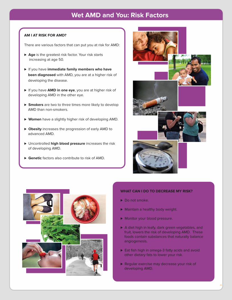
AM I AT RISK FOR AMD?
There are various factors that can put you at risk for AMD:
� Age is the greatest risk factor. Your risk starts increasing at age 50.
� If you have immediate family members who have been diagnosed with AMD, you are at a higher risk of
developing the disease.
� If you have AMD in one eye, you are at higher risk of developing AMD in the other eye.
� Smokers are two to three times more likely to develop AMD than non-smokers.
� Women have a slightly higher risk of developing AMD.
� Obesity increases the progression of early AMD to advanced AMD.
� Uncontrolled high blood pressure increases the risk of developing AMD.
� Genetic factors also contribute to risk of AMD.
WHAT CAN I DO TO DECREASE MY RISK?
� Do not smoke.
� Maintain a healthy body weight.
� Monitor your blood pressure.
� A diet high in leafy, dark green vegetables, and fruit, lowers the risk of developing AMD. These foods contain substances that naturally balance angiogenesis.
� Eat fish high in omega-3 fatty acids and avoid other dietary fats to lower your risk.
� Regular exercise may decrease your risk of developing AMD.
Wet AMD and You: Risk Factors
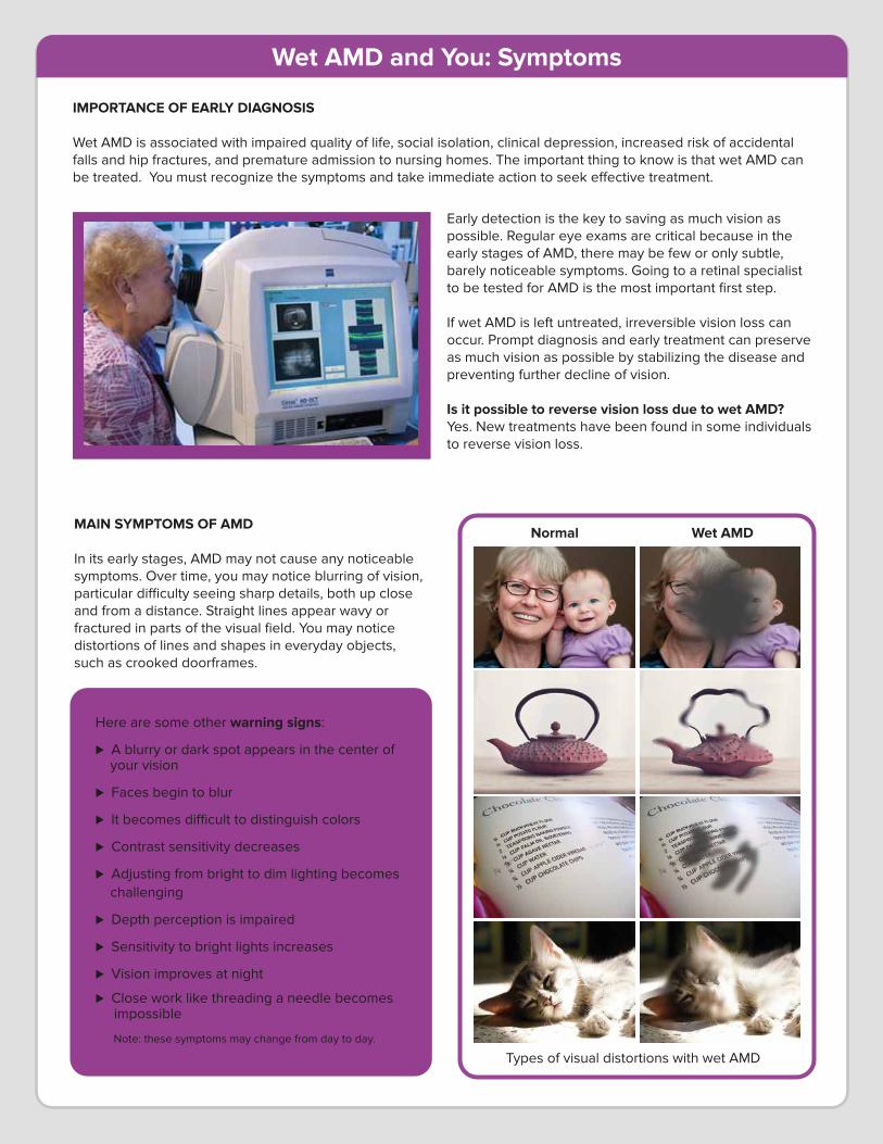
MAIN SYMPTOMS OF AMD
In its early stages, AMD may not cause any noticeable symptoms. Over time, you may notice blurring of vision, particular di�culty seeing sharp details, both up close and from a distance. Straight lines appear wavy or fractured in parts of the visual field. You may notice distortions of lines and shapes in everyday objects, such as crooked doorframes.
Early detection is the key to saving as much vision as possible. Regular eye exams are critical because in the early stages of AMD, there may be few or only subtle, barely noticeable symptoms. Going to a retinal specialist to be tested for AMD is the most important first step.
If wet AMD is left untreated, irreversible vision loss can occur. Prompt diagnosis and early treatment can preserve as much vision as possible by stabilizing the disease and preventing further decline of vision.
Is it possible to reverse vision loss due to wet AMD?Yes. New treatments have been found in some individuals to reverse vision loss.
Here are some other warning signs:
� A blurry or dark spot appears in the center of your vision
� Faces begin to blur
� It becomes di�cult to distinguish colors
� Contrast sensitivity decreases
� Adjusting from bright to dim lighting becomes challenging
� Depth perception is impaired
� Sensitivity to bright lights increases
� Vision improves at night
� Close work like threading a needle becomes impossible
Note: these symptoms may change from day to day.
IMPORTANCE OF EARLY DIAGNOSIS
Wet AMD is associated with impaired quality of life, social isolation, clinical depression, increased risk of accidental falls and hip fractures, and premature admission to nursing homes. The important thing to know is that wet AMD can be treated. You must recognize the symptoms and take immediate action to seek e�ective treatment.
Normal Wet AMD
Types of visual distortions with wet AMD
Wet AMD and You: Symptoms
HOW DOES A DOCTOR TEST FOR AMD?
WET AMD TREATMENTS
There are a growing number of e�ective treatment options available for wet AMD. Treatments aimed at angiogenesis (new blood vessel growth) in the eye are called “antiangiogenic” therapies or "anti-VEGF” therapies. The VEGF (Vascular Endothelial Growth Factor) family of proteins stimulates the growth of new blood vessels. Anti-VEGF therapies can slow the progression of wet AMD and in some cases improve vision. The treatments are especially beneficial when they are used before scar tissue develops, when therapy can prevent irreversible vision loss. Anti-VEGF Therapies
There are four di�erent VEGF inhibitors that have been shown to be e�ective for wet AMD:
Macugen (Pegaptanib) – first VEGF inhibitor to be approved for treating wet AMD
Lucentis (Ranibizumab) – highly e�ective treatment for wet AMD
Eylea (Aflibercept) – highly e�ective treatment for wet AMD, prescribed with a lower dosing frequency
Avastin (Bevacizumab) – cancer drug with anti-VEGF activity that has been prescribed as an o�-label therapy by retinal specialists to treat wet AMD
All anti-VEGF therapies for wet AMD are injected into the eye by a retinal specialist. Retinal specialists are trained to perform this simple procedure in a safe and painless way. The frequency of treatment is determined by a retinal specialist based on your condition.
KEEP UP WITH TREATMENTS
It is important to remember that wet AMD is a chronic disease that requires lifelong monitoring and treatment. With the current anti-VEGF therapies, regular treatments are required to maintain control over angiogenesis and to preserve vision. If treatments are not followed according to the retinal specialist’s orders, vision may still deteriorate and blindness may result.
TREATMENT RISKS
Anti-VEGF therapies are all safe treatments when given by trained retinal specialists. All drugs, however, have risks that need to be balanced with their benefits.
For anti-VEGF therapies, those risks include:
� Eye infection
� Increased eye pressure
� Retinal detachment
� Vitreous floaters: tiny clumps of gel or cells moving around the fluid in the back of the eye.
� Other commonly reported adverse events include: inflammation, blurred vision, conjunctival hemorrhage, eye irritation, and eye pain.
“Anti-VEGF therapies can slow the progression of AMD and in some cases improve vision.”
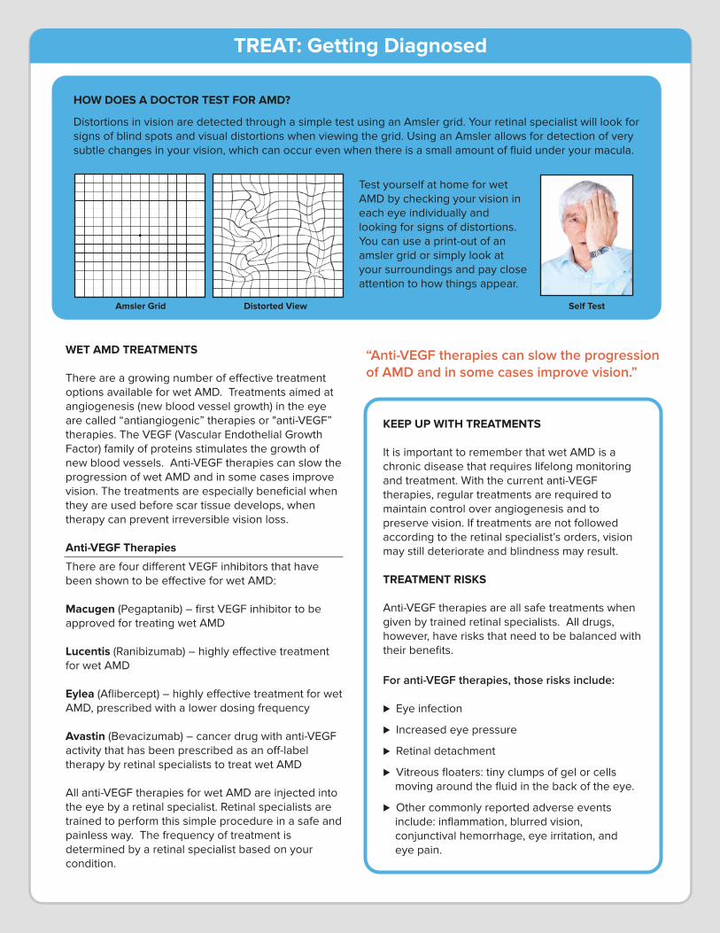
Distortions in vision are detected through a simple test using an Amsler grid. Your retinal specialist will look for signs of blind spots and visual distortions when viewing the grid. Using an Amsler allows for detection of very subtle changes in your vision, which can occur even when there is a small amount of fluid under your macula.
Test yourself at home for wet AMD by checking your vision in each eye individually and looking for signs of distortions. You can use a print-out of an amsler grid or simply look at your surroundings and pay close attention to how things appear.
Amsler Grid Distorted View Self Test
TREAT: Getting Diagnosed
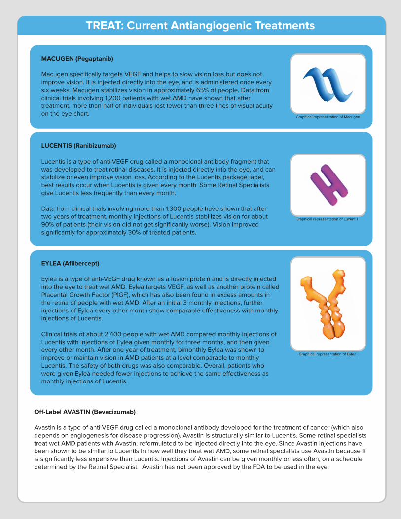
EYLEA (Aflibercept)
Eylea is a type of anti-VEGF drug known as a fusion protein and is directly injected into the eye to treat wet AMD. Eylea targets VEGF, as well as another protein called Placental Growth Factor (PlGF), which has also been found in excess amounts in the retina of people with wet AMD. After an initial 3 monthly injections, further injections of Eylea every other month show comparable e�ectiveness with monthly injections of Lucentis. Clinical trials of about 2,400 people with wet AMD compared monthly injections of Lucentis with injections of Eylea given monthly for three months, and then given every other month. After one year of treatment, bimonthly Eylea was shown to improve or maintain vision in AMD patients at a level comparable to monthly Lucentis. The safety of both drugs was also comparable. Overall, patients who were given Eylea needed fewer injections to achieve the same e�ectiveness as monthly injections of Lucentis.
O�-Label AVASTIN (Bevacizumab)
Avastin is a type of anti-VEGF drug called a monoclonal antibody developed for the treatment of cancer (which also depends on angiogenesis for disease progression). Avastin is structurally similar to Lucentis. Some retinal specialists treat wet AMD patients with Avastin, reformulated to be injected directly into the eye. Since Avastin injections have been shown to be similar to Lucentis in how well they treat wet AMD, some retinal specialists use Avastin because it is significantly less expensive than Lucentis. Injections of Avastin can be given monthly or less often, on a schedule determined by the Retinal Specialist. Avastin has not been approved by the FDA to be used in the eye.
MACUGEN (Pegaptanib)
Macugen specifically targets VEGF and helps to slow vision loss but does not improve vision. It is injected directly into the eye, and is administered once every six weeks. Macugen stabilizes vision in approximately 65% of people. Data from clinical trials involving 1,200 patients with wet AMD have shown that after treatment, more than half of individuals lost fewer than three lines of visual acuity on the eye chart.
LUCENTIS (Ranibizumab)
Lucentis is a type of anti-VEGF drug called a monoclonal antibody fragment that was developed to treat retinal diseases. It is injected directly into the eye, and can stabilize or even improve vision loss. According to the Lucentis package label, best results occur when Lucentis is given every month. Some Retinal Specialists give Lucentis less frequently than every month.
Data from clinical trials involving more than 1,300 people have shown that after two years of treatment, monthly injections of Lucentis stabilizes vision for about 90% of patients (their vision did not get significantly worse). Vision improved significantly for approximately 30% of treated patients.
Graphical representation of Macugen
Graphical representation of Lucentis
Graphical representation of Eylea
TREAT: Current Antiangiogenic Treatments
Summary
This resource – Science of AMD -- is for general health information only. This brochure is not to be used as a substitute for medical advice, diagnosis, or treatment of any health condition or problem. The Angiogenesis Foundation does not assume any risk for your use of this brochure or the information contained herein. Health-related information changes frequently and therefore information contained in this brochure may be outdated, incomplete or incorrect.
The Angiogenesis Foundation is the world’s first and leading nonprofit organization dedicated to conquering diseases using a groundbreaking approach based on angiogenesis, the growth
of new blood vessels in the body. Angiogenesis is the “common denominator” in society’s most feared diseases, including wet AMD.
Understanding the science behind wet AMD and its treatment guides patients, their advocates, and ophthalmologists to understand the consequences of complacency and the evidence-based need for proactive care and regularized treatments. We believe everyone
a�ected by wet AMD should be empowered with knowledge of available treatments and an understanding of the practical steps they can take to protect their vision.
Acknowledgements
The Angiogenesis Foundation thanks the following sponsors for their generosity in helping to make this resource possible.
Bayer Healthcare PharmaceuticalsAJA Foundation
John and Vicky Miller
www.scienceofamd.org
© 2014 by The Angiogenesis Foundation. All Rights Reserved.