Embed Size (px)
Citation preview
Volume 70, Number 2Printed in the 11. 5.A.
(ISSN 0148-916X)
INTERNATIONAL JOURNAL 01. LEPROSY
Study of HLA-DR Expression on Skin Lesions of Leprosy
Before and During Multiple Drug Therapy'
Amany M. Abdel Latif and Eman A. Essa'
Leprosy is a chronic inflammatory dis-ease caused by Mycobacterium lepae. Thehuman response to this pathogen exhibitsintriguing aspects which are until now notwell understood ('h). The disease is charac-terized by a broad spectrum of clinicalforms depending on the patient immune sta-tus. T-helper 1 (Thl) cells are associatedwith tuberculoid leprosy patients, who havestrong cell-mediated immune response,while Th2 cells are expressed in leproma-tous leprosy patients who have strong hu-moral response with a lack of T-cell re-sponse ( 5 ). However, patients often sufferfrom immunologically mediated reactionseither spontaneously or during treatment.Two major reactions are recognized, thefirst is the reversal reaction in which skinbecomes red and swollen with tenderness ofperipheral nerves, which may lead to addi-tional disability. Such reaction seemed to bedue to increased CMI response followingincreased release of antigen after startingtreatment of either tuberculoid or leproma-tous leprosy. The second reaction is ery-thema nodosum leprosum in which painfulinflamed nodules occur in the skin of lepro-matous leprosy patients which seemed to beimmune complex mediated reaction (4).
Fortunately, multiple drug therapy(MDT) has revolutionized the treatment ofleprosy where many patients are now curedwithin six months to two years dependingon their bacterial load ( 7 ).
' Received for publication on 1 December 2000. Ac-cepted for publication on 29 April 2002.
2 Amany M. Abdel Latif, Departments of Dermatol-ogy and Venereology, Faculty of Medicine, Tanta Uni-versity, Egypt; Eman A. Essa, Department of Microbi-ology, Faculty of Medicine, Tanta University, Egypt.
Reprint requests to Dr. Amany M. Abdel Latif,Saudi German Hospital, P.O. Box 2550, Jeddah, SaudiArabia. Telephone: 966-2-6394000, extension 6458;Fax: 966-2-6835874; e-mail: hashemgamal@hotmai I .com
There is an increasing interest in the im-munomodulatory role of MDT because itsbeneficial effect may he accompanied byimportant changes in the immune cell pro-file which have a great role in overcomingsuch infection. In some studies, it was sug-gested that the CMI was reactivated in mostpatients under MDT, which is not restrictedto those who developed immunologicallymediated adverse reactions during the ther-apeutic course, such as reversal reaction orerythema nodosum leprosum ( 5 ).
In the present study, we tried to investi-gate the immunomodulatory effect of MDTOn the skin lesions of patients with leprosyby studying the HLA-DR display beforeand a few weeks after starting MDT usingimmunofluorescent staining. In addition,new cases who did not receive any treat-ment for 2-4 weeks were included for com-parison. Patients who developed reversalreaction during MDT were studied for theeffect of prednisolone administration on theHLA-DR expression in their granuloma.
PATIENTS AND METHODSThis study included 35 patients with lep-
rosy from the Outpatient Clinic of the De-partment of Dermatology and Venereology,Tanta University Hospital. There were 24males and 11 females with an age range of18 years to 50 years (mean = 35.5 years).They included 30 newly-diagnosed patientsand five patients who developed reversal re-action during MDT.
Skin punch biopsy specimens (4 mm)were taken before and at least once at 2-4weeks after starting MDT in 20 cases whowere newly-diagnosed clinically and whowere confirmed in the first biopsy byhistopathology. Two biopsies, 2-4 weeksapart, were also taken from each of 10newly-diagnosed patients who did not yetreceive any treatment for comparison (con-trol group). In addition, two or more Hop-
104
70, 2^
Lull: et al.: HLA-DR before and during MDT^105
TABLE 1. Chwilication of the studied patients based on Ridley and lopling scaleaccording to bacterial load.
Paticibacillary(PBL)
(MBL) Total
TT.' BT^B13`^BL('^LLe
Newly diagnosed 5 7 3 7 8 30Reversal reaction 0 5'rota! 5 9 4 8 9 35
'Borderline tuk_.rculoid.'Border'Borderline lepromatous.Lepromatous.
sies were taken before and during cortico-steroid therapy of reversal reaction in thefive patients who were clinically diagnosedas being in reversal reaction. Slit-skinsmear was done for all patients to determinethe bacteriological index in order to classifythe patients into paucibacillary leprosy(PBL) and multibacillary leprosy (MBL).In all cases, biopsies were taken from theedge of the same skin lesion to ensure re-producibility of histology (). Biopsies werefixed in 10% formol saline then routinelyprocessed. Paraffin-embedded sectionswere cut at 5 11M, stained with hematoxylinand eosin (to confirm the histologic diagno-sis) and with immunofluorescent staining inorder to detect the HLA-DR display.
Procedure of indirect immunofluores-cence. Sections of biopsies from the samepatient both before and after MDT werestained on parallel, on the same day. Thiswas performed according to manufacturer'sinstructions as follows: The paraffin sec-tions of skin biopsies were dewaxed (by xy-lol for 1-2 hrs) and rehydrated (by alcohol00%-70%-50% and 30%), then dried
overnight at 37°C. The sections were rinsedin Tris saline buffer, pH 7.8, and then put ina solution of Tris saline buffer containing0.1% calcium chloride (w/v) and 0.05%trypsin for 40 minutes. After rinsing in irissaline buffer, the sections were left in thisbuffer overnight at 4°C. The sections werethen incubated at room temperature (RT)for 30 minutes with monoclonal antibodiesspecific for HLA-DR (BioGenex, AbuDhabi, United Arab Emirates). After rinsingwith PBS, the sections were incubated atRT for 30 minutes with antimouse IgG flu-orescein conjugate (Ortho Diagnostics,
Rochester, New York, U.S.A.). Lastly thesections were rinsed with PBS and exam-ined with a fluorescent microscope in adarkened room using UVR light of350-400 pm wavelength. An excitation fil-ter was used to produce a wavelength capa-ble of causing fluorescent activation and aharrier filter was also used to removed theinterfering waves of light. An isotype con-trol was included. i.e., sections stained onlywith antimouse Ig,G fluorescein conjugate.
Positively stained material (HLA-DR)had bright yellowish-green fluorescence,while the negatively stained one appeareddull green in color. The degree of HLA-DRexpression was determined by the degree ofbrightness in the yellowish-green fluores-cence, which was scored as faint (+), mod-erate (++), and strong (+++) expression.
RESULTSThis study included 30 cases of leprosy
who were newly diagnosed and 5 caseswho were clinically diagnosed as being inreversal reaction during MDT. Table 1shows the classification of the patientsbased on the Ridley and Jopling scale ac-cording to their bacterial load which wasdetected after making slit-skin smears.
The newly diagnosed patients included12 with PBL and 18 with MBL, while the 5patients with reversal reaction included 2with PBL and 3 with MBL. Indirect im-munofluorescence showed an increased ex-pression of HLA-DR in the second skinbiopsy of 7 out of 8 PBL patients (87.5%)and 1() out of 12 MBL patients (83.3%)within 2-4 weeks after starting MDT, for atotal number of 17 out of 20 (85%) (Table2). On the other hand, none of the 10
106 la tiOna l JO H rilal LeprOSy^ 2002
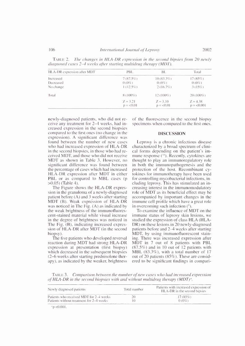
TABLE 2. The changes in IILA-I)!? expression in the .s.econd bipsies .from 20 newlydiagnosed cases 2-1 vveek.s• alter^multidrug therapy (441)1').
HI .A-DR expression a1tcr MI)]' Bl. Tot a I
Increased 7 (87.5%) 10 (83.3%) 17 (85%)Decreased () (0(4;) (0%) 0 (0%)No change I^(12.5%) 2 ( 16.74 I 3 ( 15%)
rFotal 8^(^I) 12 ( I 00e/c) 20 ( I OW)
z^3.21p = <0.01
= 3.10p <0.01
Z = 4.38-= <0.001
newly-diagnosed patients, who did not re-ceive any treatment for 2-4 weeks, had in-creased expression in the second biopsiescompared to the first ones (no change in theexpression). A significant difference wasfound between the number of new caseswho had increased expression of HLA-DRin the second biopsies, in those who had re-ceived MDT, and those who did not receiveMDT as shown in Table 3. However, nosignificant difference was found betweenthe percentage of cases which had increasedHLA-DR expression after MDT in eitherPBL or as compared to MBL cases (p>0.05) (Table 4).
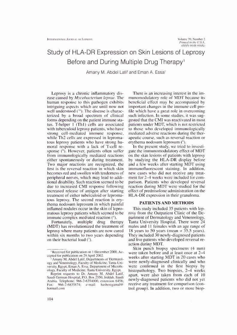
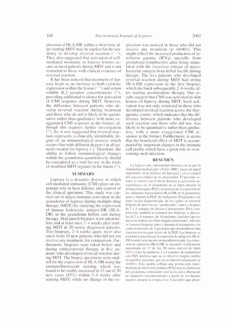
The Figure shows the HLA-DR expres-sion in the granuloma of a newly-diagnosedpatient before (A) and 3 weeks after startingMDT (B). Weak expression of HLA-DRwas noticed in The Fig. (A) as indicated bythe weak brightness of the immunofluores-cent-stained material while visual increasein the degree of brightness was noticed inThe Fig. (B), indicating increased expres-sion of HLA-DR after MDT (in the secondbiopsy).
The five patients who developed reversalreaction during MDT had strong HLA-DRexpression at presentation (first biopsy)which decreased in the subsequent biopsies(2-6 weeks after starting prednisolone ther-apy), as indicated by the weaker, brightness
of the fluorescence in the second biopsyspecimens when compared to the first ones.
DISCUSSION
Leprosy is a chronic infectious diseasecharacterized by a broad spectrum of clini-cal fm-ms depending on the patient's im-mune response ( " ). Recently, cytokines arethought to play an immunoregulatory rolein both the immunopathogenesis and theprotection of the host. Recombinant cy-tokines for immunotherapy have been usedfor controlling, mycohacterial infections, in-cluding leprosy. This has stimulated an in-creasing interest in the immunomodulatoryrole of MDT as its beneficial effect may heaccompanied by important changes in theimmune cell profile which have a great rolein overcoming such infection (8).
To examine the influence of MDT on theimmune status of leprosy skin lesions, westudied the expression of class HLA (HLA-DR) on these lesions in 20 newly-diagnosedpatients before and 2-4 weeks after startingMDT, by using immunofluorescent stain-ing. There was increased expression afterMDT in 7 out of 8 patients with PBL(87.5%) and in I() out of 12 patients withMBL (83.3%), with a total number of 17out of 20 patients (85%). These are consid-ered to be significant findings in compari-
TABLE 3. Comparison between the 'lumber of new cases who had increased expressionof HLA - DR in the second biopsies with and without multi/rug therapy (MDT).`.
Newly diagnosed patients^ Total numberPatients with increased expression of
I-ILA-DR in the second hipsies
Patients who received MDT for 2-4 weeks^
17 (85°4)Patients without treatment for 2-4 weeks
^0 (0%)
'p <0.001.

70, 2^La/if et al.: HLA-DR before and during MDT^107
TABLE 4. Comparison between thenumber olcase.s . which /1(1(1 increased HLA-1)1? di.splav after multidrug therapy (MDT)ill both palrcibacill(1ry (1)BL) and multi-bacillary (MBI.)".
'Total^Cases with increasedIllllllhel^expression after MI)T
PBL^ 7 (87.5%)MBL^
12^
10 (83.3%)
"p >0.05.
son to the results obtained from the 10 newcases who did not receive any treatment for2-4 weeks (during the period between thefirst and second biopsies), in whom nochanges were noticed in HLA-DR expres-sion. This is strong evidence of an increasein macrophage/epithelioid cell activationwith more enhanced CMI response in suchlesions. This process may involved CD4-Tlymphocytes of Th 1 type, NK cells, alphadelta cells or a combination of them all ( N ).
In some studies, increased HLA-DR ex-pression was noticed following the injec-tion of IFN-y into lesions (()), or exposure oflepromatous macrophages in culture toIFN-y ( 6 ). Therefore, our results mightreflect the increased local production of
A
Before MDT
IFN-y by lymphocytes within the granu-loma after starting MDT. This is possiblydue to the stimulation of lymphocytes by thelarge quantities of mycobacterial antigensreleased from killed bacilli ( '"). Similarly,Mouhasher, et al. (") mentioned that thechange in the antigenic stimulation of theimmune system might have an effect on cy-tokine production and HLA-DR expression.
In the present study, the five cases of lep-rosy that developed reversal reaction duringMDT had strong HLA-DR expression inthe first biopsy specimens, which declined,subsequently, after prednisolone therapy.This indicates an exaggerated CMI re-sponse in such patients which is widely be-lieved to he the cause of this reaction ( 4 ).The excessive stimulation of lymphocytesby antigen released during treatment leadsto more influx of lymphocytes with in-creased macrophage activation and giantcell formation ("). So it is evident that re-versal reaction is cell-mediated, whereaserythema nodosum leprosuiu is essentiallyall immune complex diseases ( " ).
Our results go well with those of Cree. etal. ( 3 ) who studied HLA-DR display in thegranulonia of leprosy before and duringMDT using immunohistochemical tech-nique. They stated that the increased ex-
B
3 weeks after MDTFRitiRE. HLA-DR expression m the granuloma of a newly diagnosed patient hefore and during mul-
iidrug therapy (MDT). A: Before MDT: B: 3 weeks after MDT. Increased expression of HLA-DR is more no-ticeable in B than in A as indicated by the visual increase in the brightness of the immum)tluorescent stained ma-terial as shown by the arrows.
108^ hiternatiomil .lournal of Lepravy^ 2002
pression of !ILA-DR within a short time af-ter starting MDT may he marker for the ten-dency to develop reversal reaction (2' 15).They also suggested that.tictivation of cell-mediated immunity in leprosy lesions oc-CUIS 111 1110St patients during MDT and is notrestricted to those with clinic:al evidence ofreversal reaction.
It has been noticed that treatment of lep-rosy leads to an increase in both cytokincexpression within the lesion ( '• '') and serumsoluble IL2 receptor concentrations ('').providing additional evidence fOr activationof CMI response during MDT. However,the difference between patients who de-velop reversal reaction during treatmentand those who do not is likely to he quanti-tative rather than qualitative with more ex-aggerated CMI response in the former. al-though this requires further investigation
'). So, it was suggested that reversal reac-tion represents a clinically identifiable de-gree of an immunological process whichoccurs (hut with different degrees) in all pa-tients treated for leprosy (). Therefore, theability to follow immunological changeswithin the granuloma quantitatively shouldbe considered as a tool for use in the trialsof modified MDT regimes in the future (").
SUMMARYLeprosy is a dynamic disease in which
cell mediated immunity (CMI) plays an im-portant role in host defense and control ofthe clinical spectrum. This study was car-ried out to detect immune activation in thegranuloma of leprosy during multiple drugtherapy (MDT) by studying the expressionof human leukocytic antigen-DR (HLA-DR) in the granuloma before and duringtherapy. Skin punch biopsies were taken be-fore and at least once 2-4 weeks after start-ing MDT in 20 newly diagnosed patients.Two biopsies, 2-4 weeks apart, were alsotaken from 10 new patients who did not yetreceive any treatment, for comparison. Fur-thermore, biopsies were taken before andduring corticosteroid therapy in five pa-tients who developed reversal reaction dur-ing MDT. The biopsy specimens were stud-ied for the expression of HLA-DR using theimmunofluorescent staining which wasfound to be visibly increased in 17 out of 20new cases (85%) within 2-4 weeks afterstarting MDT, while no change in the ex-
pression was noticed in those who did notreceive any treatment (p <0.001 ). Thismight reflect the increased production of in-terleron gamma ( IFN7) specially fromgranuloma lymphocytes alter being stimu-kited with the excessive release of myco-bacterial antigen from killed bacilli duringtherapy. The five patients who developedreversal reaction during MDT had strongHLA-DR expression in the first biopsieswhich declined subsequently 2-6 weeks af-ter starting prednisolone therapy. OW le-Stilts suggest that CM I was activated in skinlesions of leprosy during MDT. Such acti-vation vv'as not only restricted to those whodeveloped reversal reaction across the ther-apeutic course, which indicates that the dif-ference between patients who developedsuch reactionind those who did not, waslikely to be quantitative rather than qualita-tive, with a more exaggerated CMI re-sponse in the former. Furthermore, it seemsthat the beneficial effect of MDT is accom-panied by important changes in the immunecell profile which have a great role in over-coming such infection.
RESUMENLa lepra es una entermedad dittimica en la que la
inmunidad mediada por celulas (1MC') juega un impelimportante en la delensa del huesped y in el controldel espectro clinico de la enlermedad. El presente es-tudio se realizO con el 1in de detectar la activaciOn in-munolOgica en el granuloma de la lepra durante lapoliquimioterapia (PQT) en funcian de la expresiOn delos antigenos lcucocitanos HLA-DR en el 21"allalorna.antes y durante la PQT. Se incluyeron 20 pacientes conlepra reck'n diagnosticada, de los cuales se tomaronhiopsias de piel con un "sacabocado-, antes y despuesde 2 a 4 semanas de iniciar el tratamiento. Para com-paraci6n. tambk'n se tomaron dos hiopsias, a interva-los de 2 a 4 semanas, de 10 pacientes similares que to-davia no habian recibido ningLin tratamiento. Ademdsse tomaron biopsias antes y durante el tratamiento concorticoesteroides de 5 pacientes que desarrollaronreacciOn reversa por efecto de la PQT. Las biopsias seestudiaron pant buscar la expresiOn de antigenos I1LA-DR usando una tinciOn inmunofluorescente. La expre-sicin de antigenos HLA-DR se encontrO visiblementeaumentada en 17 de los 20 casos nuevos de lepra(85%) entre las primeras 2 a 4 semanas de tratamientocon PQT mientras que no se observO ningtin cambioen aquellos pacientes que no recibieron tratamiento (p<0.001 ). Esto podria reflejar una producciOn incre-mentada de interferon gamma (11-'1\17) por los linfocitosdel granuloma estimulados por la excesiva liberaciOnde antigenos micobacteritnos a 1-)triir de los haci losmuertos durante la terapia. Los 5 pack:111es clue desar-

70, 2^/AO: et al.: HLA -DR before mid during MDT^109
rollaron reacciOn reversa durante la 1)()1 - mostraron
una marcada expresicin de •,ilit11.:,,enos^en las
primeras^per() sll CxpicsiOn (ICCIII16 2 a 6 sm-
arms despues cle el traiamiento con pre('
nisolona. Nuestr)s resultaclos sugieren clue la 11\/1(' se
activ6 durante eI tratailiks ilt0 con POT. Sin embargo.
tal activation no estuvo solo restringida a los pacientes
que desarrollaron la reaction rcversa (lurante la PQT.
lo clue indica clue la diferencia entre los pacientes Line
desarrollaron reaction y aquel los que desarrol-
laron Ilie prol)a1)1(.111cilte CliantitzttiVa tine cua lita-
tiva, con Litra 11\1(' 111',.1S exageracla en los primeros.
Ade et CICCIO hcnCliC0 (IC la PQ:I . se acompana de
cainl)ios importantes el et peril! de as cclulas inimmi-
tarias yue participan en et control de la infeccicin.
RESUMELa liTre est une maladie evolutive inimunite
mediation cellulaire (1MC) joue un role important dans
la defense de I' Noteet dans le caractere spectral des
manifestations cliniques. Cette etude Im mise en (uuvre
dans le but de detector les signes une activation i111-
111lIllitaire au sein des granuloines de la lepre pendant
WIC polychimiotherapie (PCT). Cette activation Int
etudiee en evaluant lc niveau d - expression des
antigenes leucocytaires humains de type 1)1: ( HLA-
DR) present dans les granuloines avant et ipi -es la
prise en charge therapeutique. Des biopsies de peat'
furent prelevees a partly de lesions de 20 patients
recemment cliagnostiques avant et an moins 2 it -t se-
maines apres la mise en cruvre de la PCT. Pour coin-
paraison, 11) nouveaux patients hanseniensfurent 1(1551
biopsies a 2-4 semaines d - intervalle avant la inise en
cruvre de tout traitement. De plus. des biopsies furent
ohtenues a part i r de 5 patients qui developperent des
reactions reverses pendant la PCT, avant et apres la
rinse en cruvre de la corticothOrapie. 1:expression (IC
HLA-DR au sein des biopsie hit etudiee par immuno-
fluorescence. Elle etait visiblement .(niginentee chez 17
parmi 20 nouveaux eas (85(4) dans les 2 a 4 semaines
suivant le debut de la PCT. tandis qu'auciin change-
ment de niveaux cl - expression de HLA-DR ne furent
detectes chez les patients sans traitement (p <0.0(11 ).
Cette augmentation pourrait etre la consequence d'uneproduction .taigniente d'interfron gamma (IFN7),
particulier par les lymphocytes prsents dans le granii-
lome, apres ete stimules par tin liheration accrued - .(intigenes inycohacteriens provenant de hacilles teespal - le traitement. I.es 5 patients qui ont cleveloppes une
reaction reverse durant la PCT lint montre line forteexpression HLA-I)R aiI sein des premieres biopsies.
Cette expression rut ensuite plus faible 2 a 6 semaines
apres le debut du traitement par la prednisolone. Nos
resultats semblent done indiquer quel'IMC est activee
all sein des lesions cutanees des patients hanseniens
pendant la PCT. line telle activation n'etait pas seule-
merit restreinte aux patients montrant une reaction re-
verse pendant la mise en (cuvre du traitement. cc qui
indique que la difference entre les individus qui
cleveloppent et ceux ciiii tic developpent pas Line tclle
reaction est plus probahlement quantitative que quali-
tative, avec notamment une IMC exageree chez les
premiers. De plus, il semhle que les effets benefiques
de Ia PCT soient iccollipa.pies par des modifications
importantes du profit des celltiles immunitaires, qui out
Lin role eminent pour_jugulcr unc infection de cc type.
AcktioNs lektitent. We hope to thank Pr. 1)r.
Karima El-Desouky, the Professor of Pathology, Fac-
ulty of Nledicine, Tanta Um% ersity, for her kind help in
preparing the tissue sections for staining and in con-
firming the histopatholo ,..ic diagnosis.
REFERENCESI. ARN01.1). .1.. GFRI)F., J. and HAD. D. Inuntino-
lol!ic assessment of ('ytokine production from in-
filtrating cells in various forms ol leprosy. Am. .1.
Pathol. 137 (1990) 749-753.c.,m1.1,1mis, L. A.. TIDNIAN. N. and Pout.TER, L. W.Quantitation of HLA-[)R expression by cells in-
volved in the skin lesions of Iiiherculoid and lep-
romatous leprosy. ( lin. Exp. Immunol. 61 (1985)
58-66.CREF, I. A., CoGilli.i., A. M.. Sti-IF.D1, A. M., AR-
not N. C., By] I IN, S. R.. SArosoN, P. I). andS\\ \NSON. BECK. .1. 1-111CCt of treatment on the
histopathology of leprosy. J. Chit. Palhol. 48( I 995 ) 3114 307.
4. CRFE, I. A.. SNII1 . 11. W. ('.. RITS, RI. andS. The influence of antimycohacteriiilcheinother-
.tipy on delayed hypersensitivity skin reaction in
leprosy patients. Lepr. Rev. 59 (1988) 145-151.
CRIT, I. A.. SkINIVASAN, -r.. KRISIINIAN, S. A.,
GARDINFR, C. A.. MEHT.A..1. and FISHFR. C. A. Re-
producibility of histology in leprosy lesions. Int..1.
Lepr. 56 (1988) 296-301.
6. DEs.m, S. D., Bum. T. J. and ANTIA, N. 1-1. Corre-lation between macrophage activation .,ind
hauerium Icproe antigen presentation in macro-
phage of leprosy patients and normal indi\ !duals.
Infect. Immun. 57 (1989) 1311-1317.
7. F.I.LARD, G.^Rationale of multidrug regimes rec-ommended by a W1-1() study -group on chemother-
apy of leprosy for control programmes. Int. J.Lepr. 52 ( 1984 ) 394-401.
M. 1-1ANsENBY0, N. and ZAssi II, G. Immunoregulation
of cytokines in infectious diseases ( leprosy), Fu-
ture strategies. Japan 67 ( 1999 ) 263-268.KAPLAN, G., Ntisttxr, A., SARNo, F.^C. K..
Poiao..1. A. Cellular respon sesto II1C^injection of rceonlI)iliaill hum a n
gamma interferon In leprolllatMls leprosy patients.
Am..1. Pathol. 128 (1987) 345-353.M wo. P.. A RRAms. sANTos. S ..
BRFNNAN.
P.. BARRAI., A. and NETT°. M. Production of host
protecti\ c (IFN-gamma) host impairing (11,-10.
IL-13) and inflammatory ("FFN-alpha) cytokines by
PBMC from leprosy patients stimulated With my-
cobacterial antigens. Euro..1. Dermatol. (France) 811998)98-103.
3.
5.
1).
II()^ International Jew-mil el Lepres^ 2002
I I. MotiBAstimz, A. D., KAMEI., N. A., lt-DAN, H. ii)(1RAMAN, D. D. Cytokines in leprosy, 1. Scrumcytokine prolile in leprosy. Int. .1. Dermatol. .37(1998) 733-740.
12. Mot;RAstulz, A. D., KAMM., N. A., irDAN, II. indRAiiffm, D. D. Cytokines in leprosy, II. Effect oftreatment on serum cyt()kinc in leprosy. Int. .1.Dermatol. 37 (1998) 741-746.
13. MlikilkR.IFF, A., CRFEK. I. A., ABALos, R.,CHArK(), C. J., DFSIKAN, K. V. and En-71.1)s, J. I).14th International Leprosy Congress. PathologyWorkshop Report. Intl. Lepr. 61 (1994) 737-739.
14. 01.1.1Ati()H-7, T. H., CoNvEksk, P. .1., lillINE, G. andDF VI:IFS, R. R. 11LA antigens and neural reversalreactions in Ethiopian borderline tuberculoid lep-rosy patients. Int.^Lepr. 55 (1987) 261-266.
15. RIDLFY, M. J. and RIDLFY. D. S. Unique expres-sion of HLA-DR antigen in the lesions of polar tu-berculoid leprosy. Lepr. Rev. 53 ( 1982) 249-252.
16. SANIPAR), E. R. and SARNo, E. N. Expression ofcytokine secretion in the Stalk: of immune reactiva-
^
tion in leprosy.^Med. Biol. les. 31 (1998)69-76.
17. TRA(), V. T., I ItioNci, P. L.. TtniAN, A. T., ANII,
D., TRACI', I). D., ROOK, G. A. ;tnd WRIcarr, E. P.Changes in cellular response to mycobacterialantigensmil cytokinc production pattern Ill lep-rosy patients during MDT. Immunology 94 (1998)197-206.
18. TuNG, K. S., UmtAND, E., MATZNFIZ, P., NELSON,
K.. ScifAuF, V..,ind IiIitIN, L. Soluble serum inter-leukin 2 receptor levels in leprosy patients. ('hi).Exp. Immunol. (8 (1987) 10-15.
19. Vot.c-PIA17/1..1<, KRFMSNER, P., STI.MBER(;OR,
and W111)11tMAN, G. ReStOraliOil of defectivecylokine activity within lepromatous leprosy le-sions. Int..I. Med. Microbiol. 272 (1990)458-466.