Embed Size (px)
Citation preview

UNIVERSITY OF OULU P .O. Box 8000 F I -90014 UNIVERSITY OF OULU FINLAND
A C T A U N I V E R S I T A T I S O U L U E N S I S
Professor Esa Hohtola
University Lecturer Santeri Palviainen
Postdoctoral research fellow Sanna Taskila
Professor Olli Vuolteenaho
University Lecturer Veli-Matti Ulvinen
Director Sinikka Eskelinen
Professor Jari Juga
University Lecturer Anu Soikkeli
Professor Olli Vuolteenaho
Publications Editor Kirsti Nurkkala
ISBN 978-952-62-1201-2 (Paperback)ISBN 978-952-62-1202-9 (PDF)ISSN 0355-3221 (Print)ISSN 1796-2234 (Online)
U N I V E R S I TAT I S O U L U E N S I S
MEDICA
ACTAD
D 1362
ACTA
Juha Pihlaja
OULU 2016
D 1362
Juha Pihlaja
TREATMENT OUTCOME OF ZIRCONIA SINGLE CROWNS AND FIXED DENTAL PROSTHESES
UNIVERSITY OF OULU GRADUATE SCHOOL;UNIVERSITY OF OULU,FACULTY OF MEDICINE;MEDICAL RESEARCH CENTER OULU;OULU UNIVERSITY HOSPITAL

A C T A U N I V E R S I T A T I S O U L U E N S I SD M e d i c a 1 3 6 2
JUHA PIHLAJA
TREATMENT OUTCOME OF ZIRCONIA SINGLE CROWNS AND FIXED DENTAL PROSTHESES
Academic dissertation to be presented with the assent ofthe Doctora l Train ing Committee of Health andBiosciences of the University of Oulu for public defence inthe Leena Palotie auditorium (101A) of the Faculty ofMedicine (Aapistie 5 A), on 27 May 2016, at 12 noon
UNIVERSITY OF OULU, OULU 2016

Copyright © 2016Acta Univ. Oul. D 1362, 2016
Supervised byProfessor Aune RaustiaDocent Ritva Näpänkangas
Reviewed byProfessor Timo NärhiDocent Jari Ahlberg
ISBN 978-952-62-1201-2 (Paperback)ISBN 978-952-62-1202-9 (PDF)
ISSN 0355-3221 (Printed)ISSN 1796-2234 (Online)
Cover DesignRaimo Ahonen
JUVENES PRINTTAMPERE 2016
OpponentProfessor Pekka Vallittu

Pihlaja, Juha, Treatment outcome of zirconia single crowns and fixed dentalprostheses. University of Oulu Graduate School; University of Oulu, Faculty of Medicine; MedicalResearch Center Oulu; Oulu University HospitalActa Univ. Oul. D 1362, 2016University of Oulu, P.O. Box 8000, FI-90014 University of Oulu, Finland
Abstract
Metal ceramic restorations have been used in fixed prosthodontics since the 1950s, but the lack ofaesthetics, the inclination to use metal-free materials, possible allergic reactions to metals, and thehigh cost of high noble alloys have increased the use of all-ceramic materials. The ongoingdevelopment of ceramic materials led to the introduction of zirconia to fixed prosthodontics overa decade ago.
The mechanical properties of zirconia have proven to be excellent, but the clinical outcome ofconventional fixed zirconia restorations over the long term is unclear. This retrospective clinicalstudy evaluated two- to seven-year outcomes, early complications during prosthetic treatment andshort-term failures during the first year of use of zirconia single crowns and fixed dental prostheses(FDPs). The usefulness and durability of zirconia single crowns in abutment teeth of partialremovable dental prostheses (RDPs) was also evaluated.
The material consisted of 173 patients treated with zirconia single crowns or FDPs byundergraduate dental students between 2007 and 2010. Of these patients 94 were women and 79men (mean age 55 years, range 18–79 years). Altogether 268 zirconia single crowns (mean 3crowns, range 1–12 crowns per patient) had been fabricated for 88 patients and 120 zirconia FDPs(range 3–12 units, mean 4.5 units) for 102 patients. Seventeen patients had received both crown(s)and FDP(s).
The results show that zirconia single crowns and FDPs are a suitable treatment alternative infixed prosthodontics. Early complications during prosthetic treatment and short-term failuresduring the first year of use were few. The survival rate of the zirconia single crowns after 3.9 years(2–6 years) was 89% and the success rate was 80%. The survival rate of zirconia FDPs after 4.9years (3–7 years) was 100% and the success rate was 89%. Zirconia single crowns perform wellas abutment teeth of partial RDPs with a metal framework, but fractures in the veneering porcelainremain a problem.
Keywords: ceramic, crown, fixed dental prosthesis, fixed prosthodontics, zirconia

Pihlaja, Juha, Zirkonia-runkoisten kruunujen ja siltojen menestyminen. Oulun yliopiston tutkijakoulu; Oulun yliopisto, Lääketieteellinen tiedekunta; Medical ResearchCenter Oulu; Oulun yliopistollinen sairaalaActa Univ. Oul. D 1362, 2016Oulun yliopisto, PL 8000, 90014 Oulun yliopisto
Tiivistelmä
Metallokeraamisia rakenteita on käytetty kiinteässä protetiikassa 1950-luvulta lähtien, muttapuutteet estetiikassa, pyrkimys metallittomiin materiaaleihin, mahdolliset allergiset reaktiot jajalojen metallien korkea hinta ovat lisänneet kokokeraamisten materiaalien käyttöä. Kokokeraa-misten materiaalien kehitystyö on tuonut zirkonian kiinteän protetiikan materiaaliksi.
Zirkonian mekaaniset ominaisuudet ovat osoittautuneet erinomaisiksi, mutta hammaskantois-ten kiinteiden zirkonia-runkoisten proteesien kliiniset pitkäaikaistulokset puuttuvat. Tämän ret-rospektiivisen kliinisen tutkimuksen tarkoituksena oli selvittää zirkonia-runkoisten yksittäistenkruunujen ja zirkonia-runkoisten siltojen menestymistä 2–7 vuoden aikavälillä sekä kartoittaaniiden valmistuksen aikaiset ongelmat ja varhaiset epäonnistumiset ensimmäisen vuoden aika-na. Lisäksi tutkittiin zirkonia-runkoisten yksittäisten kruunujen käyttökelpoisuutta ja kestävyyt-tä metallirunkoisten rankaproteesien tukihampaina.
Materiaali koostui 173 potilaasta, joille hammaslääketieteen opiskelijat olivat tehneet zirko-nia-runkoisia yksittäisiä kruunuja tai zirkonia-runkoisia siltoja vuosina 2007–2010. Potilaista 94oli naisia ja 79 miehiä (keski-ikä 55 vuotta, jakauma 18–79 vuotta). Kaiken kaikkiaan 268 zirko-nia-kruunua (keskimäärin 3 kruunua, jakauma 1–12 kruunua potilasta kohti) oli valmistettu 88potilaalle ja 120 siltaa (keskimäärin 4,5 yksikköä, jakauma 4,5 yksikköä) 102 potilaalle. Seitse-mälletoista potilaalle oli tehty sekä kruunuja että siltoja.
Tulokset osoittavat, että zirkonia-runkoiset kruunut ja sillat ovat käyttökelpoisia kiinteässäprotetiikassa. Valmistuksenaikaiset ongelmat ja varhaiset epäonnistumiset ovat vähäisiä. Yksit-täisten kruunujen selviytymisprosentti 3,9 vuoden jälkeen (2–6 vuotta) oli 89 % ja onnistumis-prosentti 80 %. Siltojen selviytymisprosentti 4,9 vuoden jälkeen (3–7 vuotta) oli 100 % ja onnis-tumisprosentti 89 %. Zirkonia-runkoiset kruunut toimivat hyvin rankojen tukihampaina, muttaniiden ongelmana ovat päällepolttoposliinin lohkeamat.
Asiasanat: hammaskruunu, hammasproteesi, keraaminen, siltaproteesi, zirkonia


The most important things in live aren't things.

8

9
Acknowledgements
The study was carried out at Institute of Dentistry, Faculty of Medicine,
University of Oulu and at the Dental Training Clinic, Oral Health Services, City
of Oulu. I am very grateful to everyone who has contributed to this thesis.
First and foremost I would like to express my sincere gratitude to my
supervisors and co-authors Professor Aune Raustia, DDS, PhD, and Docent Ritva
Näpänkangas, DDS, PhD. They have provided excellent guidance and support
from the very beginning. It amazes me how much effort they were able to invest
in my work. Their diligence, helpfulness and enthusiasm towards research are
inspiring.
I would also like to thank the referees of this thesis, Professor Timo Närhi,
DDS, PhD, and Docent Jari ahlberg, DDS, PhD, for their valuable comments and
advices and my co-author Dr Ritva Kuoppala, DDS, PhD, for her contribution to
our third original article. I would also like to thank my follow-up group, Professor
Tellervo Tervonen, DDS, PhD, Dr Virpi Harila, DDS, PhD, and Dr Marja-Liisa
Laitala, DDS, PhD, for their guidance. I thank authorized translator Keith Kosola
for the revising of the thesis and the original publications and personnel of the
Dental Training Clinic for the collaboration. My special thanks go to my
workplace at Hammassirkku, Jyväskylä; thank you Sirkku Meriläinen-Vapola and
other fellow workers for the support and understanding.
My warmest thanks go to my family and friends, especially to my parents and
parents-in-law, for their support and care. Finally, I wish to express my deepest
love and gratitude to my beloved Kaisa for her support, understanding and
invaluable love. The most important things in live aren't things.
The study was financially supported by the study grants from the Finnish
Dental Society Apollonia. I gratefully acknowledge these grants.
Oulu, April 2016 Juha Pihlaja

10

11
Abbreviations
CAD/CAM computer-aided design/computer-aided manufacturing
CIP cold isostatic pressing
DCM direct ceramic machining
FDP fixed dental prosthesis
FSZ fully stabilized zirconia
HIP hot isostatic postcompaction
ISO International Organization for Standardization
LTD low temperature degradation, "ageing"
MDP methacryloxydecyl dihydrogen phosphate
PSZ partially stabilized zirconia
RDP removable dental prosthesis
SEM scanning electron microscope
Y-TZP yttria-stabilized tetragonal zirconia polycrystals

12

13
Original publications
This thesis is based on the following publications, which are referred throughout
the text by their Roman numerals:
I Pihlaja J, Näpänkangas R, Raustia A (2014) Early complications and short-term failures of zirconia single crowns and partial fixed dental prostheses. J Prosthet Dent. 112(4):778–783.
II Näpänkangas R, Pihlaja J, Raustia A (2015) Outcome of zirconia single crowns made by predoctoral dental students: a clinical retrospective study after 2 to 6 years of clinical service. J Prosthet Dent. 113(4):289–294.
III Pihlaja J, Näpänkangas R, Kuoppala R, Raustia A (2015) Veneered zirconia crowns as abutment teeth for partial removable dental prostheses: A clinical 4-year retrospective study. J Prosthet Dent. 114(5):633–636.
IV Pihlaja J, Näpänkangas R, Raustia A (2016) Outcome of zirconia partial fixed dental prostheses made by predoctoral dental students: A clinical retrospective study after 3 to 7 years of clinical service. J Prosthet Dent, in press.

14

15
Contents
Abstract
Tiivistelmä
Acknowledgements 9 Abbreviations 11 Original publications 13 Contents 15 1 Introduction 17 2 Review of literature 19
2.1 Ceramic materials in fixed prosthodontics .............................................. 19 2.2 Classification of all-ceramic materials .................................................... 20
2.2.1 Glass-ceramics .............................................................................. 21 2.2.2 Alumina- and zirconia-based polycrystalline ceramics ................ 21
2.3 Physical properties of zirconia ................................................................ 22 2.4 Zirconia in dentistry ................................................................................ 24 2.5 Processing techniques of zirconia restorations ........................................ 25 2.6 Terminology ............................................................................................ 27 2.7 Problems related to veneered zirconia restorations ................................. 28
2.7.1 Secondary caries ........................................................................... 28 2.7.2 Chipping of veneering ceramics ................................................... 29 2.7.3 Wear of the antagonist tooth ......................................................... 30 2.7.4 Loss of retention and adhesive bonding ....................................... 31
2.8 Survival of zirconia restorations ............................................................. 32 3 Aims of the study 35 4 Material and methods 37
4.1 Description of the material ...................................................................... 37 4.2 Study population ..................................................................................... 37 4.3 Materials of the restorations .................................................................... 38 4.4 Clinical procedures during prosthetic treatment ..................................... 39 4.5 Early complications and short-term failures (Paper I) ............................ 39 4.6 Clinical follow-up ................................................................................... 40
4.6.1 Zirconia single crowns (Paper II) ................................................. 40 4.6.2 Veneered zirconia crowns as abutment teeth for partial
removable dental prostheses (Paper III) ....................................... 42 4.6.3 Zirconia fixed dental prostheses (FDPs) (Paper IV) ..................... 42 4.6.4 Clinical follow-up examination .................................................... 44
16
4.7 Statistical analysis ................................................................................... 45 5 Results 47
5.1 Early complications (Paper I) .................................................................. 47 5.2 Short-term failures (Paper I) ................................................................... 47 5.3 Clinical findings at the follow-ups .......................................................... 48
5.3.1 Zirconia single crowns (Paper II) ................................................. 48 5.3.2 Veneered zirconia crowns as abutment teeth for partial
removable dental prostheses (Paper III) ....................................... 50 5.3.3 Zirconia fixed dental prostheses (Paper IV) ................................. 51
6 Discussion 55 6.1 Material and methods .............................................................................. 55 6.2 Patient satisfaction .................................................................................. 56 6.3 Biological complications and failures ..................................................... 56 6.4 Technical complications and failures ...................................................... 58
6.4.1 Technical early complications ...................................................... 58 6.4.2 Technical short-term failures ........................................................ 59 6.4.3 Chipping ....................................................................................... 60 6.4.4 Loss of retention ........................................................................... 60 6.4.5 Wear of the restoration and antagonist teeth ................................. 61 6.4.6 Veneered zirconia crowns as abutment teeth for partial
removable dental prostheses ......................................................... 62 6.5 Success and survival of zirconia single crowns and fixed dental
prosthesis ................................................................................................. 62 6.6 Zirconia in fixed prosthodontics ............................................................. 64
7 Summary and conclusion 65 List of references 67 Original publications 77

17
1 Introduction
Since ancient times there has been a need to replace missing teeth. Artificial teeth
have been made out of many materials such as human and animal teeth, ivory and
wood. An artificial tooth was placed in the socket of a missing tooth or teeth were
wired together to be used as a removable denture. The materials were unstable in
the oral environment and fabrication methods were primitive. (Ring 1985)
The term conventional fixed prosthodontics refers to restorations, single
crowns and fixed dental prostheses (FDPs) that are permanently fixed to the
patient’s own teeth. First glass inlays and feldspathic porcelain jacket-crowns
were fabricated as early as the late 1800s (Kelly et al. 1996). Brittleness and poor
durability, the main disadvantages of porcelain (Griggs 2007, Miyazaki & Hotta
2011), were a major problem of these early all-ceramic restorations and limited
their use. Gold-resin FDPs were the first fixed restorations to replace missing
teeth, but with moderate results (Palmqvist & Swartz 1993). Widespread use of
all-ceramic materials in fixed prosthodontics is a phenomenon of the last two
decades.
A new era in conventional fixed prosthodontics started in the 1950s, when the
technique of veneering a strong metal framework with aesthetic porcelain was
introduced (O’Brien 2002). These metal ceramic fixed restorations were strong
enough to permanently replace missing teeth with adequate aesthetics. Today,
metal ceramic restorations are the ‘gold standard’ of conventional fixed
prosthodontics due to their good long-term clinical results (Kelly et al. 1996, Tan
et al. 2004, Näpänkangas & Raustia 2008, Näpänkangas & Raustia 2011).
However, moderate aesthetics, inclinations to use metal-free restorations, possible
allergic reactions to metals, and the increased cost of high noble alloys have
upheld the progress of non-metallic materials (Shenoy & Shenoy 2010, Miyazaki
& Hotta 2011, Miyazaki et al. 2013). Unfortunately, the brittle nature of ceramics
has long limited the use of these more aesthetic materials.
The first dental computer-aided design/computer-aided manufacturing
(CAD/CAM) system was introduced in the 1970s (Schepke et al. 2015). The
development of the new computer-based fabrication system eventually
revolutionized the processing of dental restorations (Griggs 2007, Miyazaki &
Hotta 2011). The CAD/CAM technique made it possible to mill dental
restorations of high-strength ceramic materials that cannot be processed with
traditional processing techniques (Miyazaki & Hotta 2011). These materials,
especially zirconia, have a wider range of application possibilities than former

18
ceramic materials (Ozkurt & Kazazoğlu 2010, Miyazaki et al. 2013). Zirconia has
excellent mechanical properties for dental use, but whether it is a durable material
for conventional fixed prosthetic restorations over the long term is unclear.
19
2 Review of literature
2.1 Ceramic materials in fixed prosthodontics
Metal ceramic, ‘porcelain fused to metal', restorations are the ‘gold standard’ of
fixed prosthodontics, combining a strong metal framework with an aesthetic
porcelain veneer. Despite the good long-term results, the moderate aesthetics and
biocompatibility concerns associated with the metals have increased the use of
all-ceramic materials. Unfortunately, brittleness and poor durability have long
limited the use of all-ceramic restorations. (Heintze & Rousson 2010, Miyazaki &
Hotta 2011, Miyazaki et al. 2013)
The first all-ceramic feldspathic porcelain jacket-crowns were fabricated in
the late 1800s, but advancements in all-ceramic materials took place as late as in
the early 1980s, when shrink-free alumina crowns (Cerestone®, Johnson &
Johnson) and castable, mica-reinforced, glass-ceramic crowns (Dicor®, Corning)
were introduced (Kelly et al. 1996, Kelly & Benetti 2011 Li et al. 2014). In the
early 1990s, ceramic materials evolved as leucite-reinforced glass ceramics (IPS
Empress®; Ivoclar Vivadent) and glass-infiltrated alumina ceramics (InCeram®;
Vita Zahnfabrik) were introduced (Kelly & Benetti 2011). Leucite-reinforced
glass-ceramic crowns had similar strength and toughness values as mica-
reinforced, but were stronger, possibly due to a strong micromechanical bond
after etching and priming (Kelly 2004). Glass-infiltrated alumina ceramics were
much stronger than former all-ceramic materials, but the translucency of the
material was poor (Shenoy & Shenoy 2010). In 1998 a lithium disilicate-
reinforced glass-ceramic material (IPS Empress II®) was introduced by Ivoclar
Vivadent (Pieger et al. 2014). During the last two decades, all ceramic systems
have evolved significantly.
Traditional techniques for processing all-ceramic materials include layering
(powder condensation), hot-pressing and slip-casting (Griggs 2007). The
conventional layering technique is technically sensitive and is usually used to
apply veneering porcelain on high-strength frameworks, as porosity and relatively
low strength limit the indications (Griggs 2007). Slip-casting is a complicated
method used to fabricate infiltrated alumina ceramics whereas hot-pressing is
used with pressable glass-ceramics to press liquid glass into a mould (Griggs
2007). A hot-pressing, press-on-metal (PoM) technique can also be used with

20
metal ceramic restorations as an alternative to the technique-sensitive and time-
consuming porcelain layering technique (Khmaj et al. 2014, Lee 2016).
The first dental computer-aided design/computer-aided manufacturing
(CAD/CAM) system was introduced in the 1970s and the first CAD/CAM-
fabricated all-ceramic restoration was fabricated from feldspathic ceramic in 1983
(Li et al. 2014, Schepke et al. 2015). Dental CAD/CAM systems consist of three
main phases: scanning the teeth intraorally or from a cast, designing the
restoration by computer and fabricating the restoration with a milling machine
(Miyazaki & Hotta 2011). The development of computer-based systems
revolutionized the processing techniques of dental restorations and the use of
ceramic materials (Griggs 2007, Miyazaki & Hotta 2011). With CAD/CAM
systems it is possible to design and mill dental restorations chairside. The
development of CAD/CAM systems made it possible to mill the frameworks of
the restorations from high-strength polycrystalline ceramics like densely sintered
alumina and zirconia (Griggs 2007). Subtractive manufacturing with computer-
aided machining is the state-of-the-art fabrication method at the moment, but in
the future, additive manufacturing with 3D printing technologies are coming to
dentistry (van Noort 2012, Stansbury & Idacavage 2015).
Nowadays, most ceramic restorations are milled from pre-fabricated blocks.
There are three different concepts of restoration design for full-contour ceramic
restorations. Ceramic restorations can be either fabricated from a single material
as a monolithic restoration or stronger ceramic can serve as a framework that is
thoroughly veneered with a more aesthetic porcelain layer (Miyazaki et al. 2013,
Moscovitch 2015). The third way is a so-called hybrid restoration, in which the
veneering layer is used only where aesthetically needed (Moscovitch 2015).
2.2 Classification of all-ceramic materials
All-ceramic materials are divided into four sub-categories according to the
microstructure of the material: glass-ceramics (predominantly glass), glass-
ceramics with fillers, crystalline ceramics with glass fillers and polycrystalline
ceramics (Shenoy & Shenoy 2010). Highly aesthetic materials are mainly glass,
and a higher crystalline content strengthens the material, but weakens
transparency (Kelly 2004). The use of crystalline ceramics with glass fillers has
decreased due to increased use of lithium disilicate-reinforced glass-ceramics and
(zirconia-based) polycrystalline ceramics (Gracis et al. 2015). Lately, a new
classification system that divides all ceramic materials into glass-matrix ceramics,

21
polycrystalline ceramics and resin-matrix ceramics, has been proposed (Gracis et al. 2015).
2.2.1 Glass-ceramics
Glass-ceramics consist mainly of silica and alumina (Shenoy & Shenoy 2010).
The optical properties of mainly silica-based ceramics are excellent, but the brittle
nature and low fracture toughness of the material have limited its use (Griggs
2007, Miyazaki & Hotta 2011). Adding reinforcing fillers like leucite and
especially lithium disilicate to the glass matrix strengthens the material while
preserving its good optical properties (Shenoy & Shenoy 2010). When lithium
disilicate is added, flexural strength and fracture toughness are over three times
higher than with leucite (Shenoy & Shenoy 2010).
With silica-based ceramics, pretreatment before adhesive cementation
includes surface roughening with hydrofluoric acid etching and surface activation
with silanization, leading to substantially improved chemical bonding (Blatz et al. 2003, Özcan & Vallittu 2003). Advancements in all-ceramic materials and in
fabrication and bonding procedures have led to the introduction of mini-invasive
techniques and monolithic restorations to fixed prosthodontics (Magne et al. 2015, Sulaiman et al. 2015b, Sailer et al. 2015). A minimal intervention concept
is the principal approach in modern dentistry (Dalli et al. 2012), but
conventionally fixed restorations have required significant tooth structure removal
because of material properties and retention (Magne et al. 2015). Monolithic,
tightly bonded lithium disilicate restorations have held up well in single crown
applications, but are not suitable for posterior FDPs (Pieger et al. 2014).
2.2.2 Alumina- and zirconia-based polycrystalline ceramics
Alumina- and zirconia-based polycrystalline ceramics do not have a glassy matrix
(Kelly 2004). These densely sintered materials are tougher and stronger than other
all-ceramic materials (Shenoy & Shenoy 2010). Unfortunately, due to their high
crystalline content, they are less translucent than other all-ceramic materials such
as glass-infiltrated ceramics, and are therefore veneered with porcelain for better
aesthetics (Kelly 2004). These veneered, bi-layered systems have strong cores
that are characterized by a porcelain layer.
A relatively high incidence of core fractures due to the high elastic modulus of
alumina, in addition to the better mechanical properties of zirconia, has decreased

22
the use of alumina (Gracis et al. 2015). Zirconia has become popular because a
milled zirconia core is strong enough to serve as the framework of posterior FDPs
(Manicone et al. 2007). Lately, improvements in the translucency of zirconia have
led to development of high-strength monolithic zirconia systems (Denry & Kelly
2014). Monolithic zirconia is more fracture resistant than monolithic lithium
disilicate, but is not yet comparable in aesthetics (Zhang et al. 2013). Its opacity
still limits its use mostly to areas outside of the aesthetic zone (Sulaiman et al. 2015b). With non-silica-based polycrystalline ceramics, hydrofluoric acid etching
is ineffective and adhesive bonding is challenging (Blatz et al. 2003, Özcan &
Vallittu 2003). Adhesive cementation of polycrystalline ceramics would decrease
the need for excessive tooth preparation.
2.3 Physical properties of zirconia
Zirconium is a strong grey-white transition metal that was found in 1798 (Piconi
& Maccauro 1999). Zirconium is not found as a pure metal in nature, because
zirconium reacts with oxygen (zirconium dioxide, ZrO2, "zirconia") and/or silica
(zirconium silicate, ZrSiO4) (Lughi & Sergo 2010). Zirconium
silicate―zircon―is a silicate mineral that has been known as a gemstone since
biblical times (Piconi & Maccauro 1999).
Zirconia is a dioxide of zirconium (Manicone et al. 2007). The organization
of crystals in zirconia is dependent on temperature: monoclinic (M) at room
temperature, tetragonal (T) between 1170-2370°C and cubic (C) at higher
temperatures (Piconi & Maccauro 1999). Pure zirconia is monoclinic at room
temperature but its cubic and tetragonal phases can be stabilized at room
temperature by adding metallic oxides such as calcium oxide (CaO), magnesium
oxide (MgO), lanthanum oxide (La2O3), cerium oxide (CeO2), or yttrium oxide
(Y2O3) (Kelly & Denry 2008, Li et al. 2014).
Stabilizing zirconia with yttrium oxide enhances its mechanical properties
more than with other oxides (Manicone et al. 2007). Zirconia can also be
stabilized by cerium oxide (ceria), but dispersion of alumina is needed to increase
the flexural strength of the material (Miyazaki et al. 2013, Tanaka et al. 2015).
Ceria- stabilized zirconia/alumina nanocomposite has very good mechanical
properties but more clinical studies will be needed (Miyazaki et al. 2013, Tanaka
et al. 2015). Zirconia can be stabilized partially by 2-5 mol-% of yttrium oxide
(partially stabilized zirconia, PSZ), or fully by 8 mol-% of yttrium oxide (fully
stabilized zirconia, FSZ) (Kelly & Denry 2008). Dental zirconia, usually referred

23
to as yttria-stabilized tetragonal zirconia polycrystals (Y-TZP), is commonly
partially stabilized (Gracis et al. 2015). Fully stabilized zirconia is more
translucent than partially stabilized zirconia (Sulaiman et al. 2015b).
The favourable mechanical and chemical properties of zirconia, like strength,
toughness, resistance to corrosion and wear, good chemical and dimensional
stability, and biocompatibility have allowed zirconia to be used in high-strength
parts like blades and valves (Piconi & Maccauro 1999). Zirconia’s flexural
strength from 900 to 1200 MPa and fracture toughness of 9–10 MPam1/2 (Christel
et al. 1989) are higher than those of any other dental all-ceramic material. The
mechanical properties of the most common ceramic materials in fixed
prosthodontics are shown in Table 1. Zirconia (210 GPa) and stainless steel (193
GPa) have a similar Young’s modulus, which measures the force needed to
compress or stretch a certain material (Ozkurt & Kazazoğlu 2010).
One of the main advantages of zirconia is its ability to resist crack
propagation through a phenomenon called transformation toughening. External
stress to zirconia induces a local transformation from metastable tetragonal
zirconia to stable monoclinic zirconia; its volume expands and the crack is
shielded (Lughi & Sergo 2010). On the other hand, in a moist environment, this
metastability of the tetragonal phase may cause a spontaneous transformation
from the metastable tetragonal phase to the stable monoclinic phase, decreasing
the strength of the material (Chevalier 2006). This phenomenon is called low-
temperature degradation (LTD, "ageing"). The main factors of LTD are the
content of the stabilizer, grain size and residual stress (Lughi & Sergo 2010).
Increased sintering temperature and time possibly increases the amount of LTD
by enlarging grain size and lowering the content of the stabilizer (Inokoshi et al. 2014b). LTD can be prevented or decreased by following ISO engineering
guidelines (Lughi & Sergo 2010), and colouring the zirconia increases its
resistance to LTD (Nakamura et al. 2016).

24
Table 1. Mechanical properties of the most used ceramic materials in fixed
prosthodontics.
Ceramic system (Reinforcing)
material
Brand (manufacturer) Flexural
strength1
Fracture
toughness2
Reference
1) glass-ceramics Feldspathic
porcelain
- 100 1.0 Miyazaki &
Hotta (2011)
2) glass-ceramics
with fillers
Leucite IPS Empress (Ivoclar
Vivadent)
160 1.5-1.7 Raigrodski
2004, Li et al.
2014
Lithium disilicate IPS e.max (Ivoclar
Vivadent)
300-400 2.8-3.5 Raigrodski
2004
3) crystalline
ceramics with glass
fillers
Glass-infiltrated
alumina
In-Ceram Alumina
(VITA Zahnfabrik)
236-600 3.1-4.6 Raigrodski
2004
glass-infiltrated
zirconia
toughened
alumina
In-Ceram Zirconia
(VITA Zahnfabrik)
421-800 6-8 Raigrodski
2004
4) polycrystalline
ceramics
densely sintered
alumina
Procera AllCeram
(Nobel Biocare)
487-699 4.48-5.6 Raigrodski
2004
Yttria Stabilized
Zirconia
Cercon (Dentsply),
Lava (3M ESPE),
Procera (Nobel
Biocare), Prettau
Zirconia (Zirkonzahn),
Zirconia (Zirkonzahn),
BruxZir (Glidewell
Lab.)
900-1200 9-10 Raigrodski
2004
1MPa, 2MPa/m1/2
2.4 Zirconia in dentistry
Zirconia’s favourable mechanical and chemical properties have also allowed it to
be used as a biomaterial (Piconi & Maccauro 1999). It was first introduced for
biomedical purposes in orthopaedics in the 1960s, to be used in hip prostheses
(Piconi & Maccauro 1999). Zirconia was introduced for dental purposes in the
early 1990s (Raigrodski 2004). Zirconia’s biocompatibility has been proved both
in vitro and in vivo; zirconia is non-cytotoxic, non-mutagenic and no local or
systemic adverse reactions have been identified (Piconi & Maccauro 1999,
Manicone et al. 2007). Zirconia has good chemical stability, but alterations of

25
zirconia’s surface to some extent are seen in an acidic environment (Sulaiman et al. 2015c).
In prosthodontics zirconia has a wider range of possible applications than
other all-ceramics (Özkurt & Kazazoğlu 2010). It has been used as a framework
material for single crowns, FDPs, inlay-retained FDPs and resin-bonded FDPs
(Al-Amleh et al. 2010, Kern 2015). These restorations can also be fabricated as
monolithic restorations without veneering porcelain (Moscovitch 2015, Sulaiman
et al. 2015b). Zirconia is also used as a reinforcing material of other materials
such as zirconia-containing lithium silicate (Denry & Kelly 2014) and glass-
infiltrated alumina (Shenoy & Shenoy 2010).
Implant abutments have also been made of zirconia. Titanium abutments are
very durable, but their grey colour impairs aesthetics. Zirconia implant abutments
enhance the aesthetics of the restoration, minimize the gray colour of the implant
in the marginal mucosa, decrease bacterial adhesion and have soft tissue
integration similar to titanium (Sailer et al. 2009b). Experiments with zirconia
implants have also been conducted. Osseointegration, biocompatibility, and the
health of soft tissues around zirconia implants have turned out to be excellent, but
long-term studies are needed (Özkurt & Kazazoğlu 2010, Apratim et al. 2015). In
endodontics zirconia has been used as a post material and in orthodontics zirconia
is used for fabrication of orthodontic brackets with varying results (Özkurt &
Kazazoğlu 2010).
2.5 Processing techniques of zirconia restorations
Zirconia restorations can only be fabricated by milling from pre-fabricated blocks
(Gracis et al. 2015). Zirconia blocks are fabricated from ZrO2 powder containing
stabilizing yttrium (Oh et al. 2010) (Fig. 1.) The blocks are packed by cold
isostatic pressing (CIP) to 70% of their theoretical maximum density (Denry &
Kelly 2008, Oh et al. 2010, Shenoy & Shenoy 2010). This green-stage zirconia
can then be partially sintered at high temperature and fully sintered by hot
isostatic pressing (HIP) at higher temperature and under high pressure (Oh et al. 2010 Nakamura et al. 2016).
The framework of a zirconia restoration can be designed by CAD or by a
conventional waxing technique and then digitally scanned and fabricated by CAM
(Raigrodski 2004), but the CAD/CAM technique is more accurate than CAM
from a waxed mould (Abduo et al. 2010). Zirconia restorations can be hard
machined from fully sintered blocks or soft machined from green-stage or

26
partially sintered blocks (Miyazaki et al. 2013). Milling the zirconia restorations
and/or frameworks from partially sintered blocks saves time and milling
machinery, because pre-sintered zirconia is easier to mill (Miyazaki et al. 2013).
The restoration is designed by CAD software that enlarges the restoration by 20-
25%, because final sintering after milling causes volume shrinkage (Raigrodski
2004). Another way to mill zirconia restorations is hard machining from fully-
sintered blocks. Hard machining requires robust milling equipment and is more
time consuming, but no firing is needed after milling (Denry & Kelly 2008,
Miyazaki et al. 2013).
The final procedure after milling is aesthetic characterization of the
framework. When translucent monolithic zirconia is used, the restoration is
characterized by polishing, staining, and glazing (Kim et al. 2013, Kim & Kim
2014, Moscovitch 2015, Sulaiman et al. 2015b). Traditionally, the framework is
characterized by feldspathic porcelain veneer using the powder condensation
technique; moist porcelain powder is used to build up the veneering layer and the
fluxing agent is then removed by firing (Griggs 2007). This veneering layer can
be full-contour and cover the whole restoration or it can be placed only where
aesthetically needed (Moscovitch 2015).

27
Fig. 1. Zirconium is not found as a pure metal in nature, because zirconium reacts with
oxygen. The tetragonal phase of zirconia can be stabilized at room temperature by
adding metallic oxides such as yttrium oxide (Y2O3). Zirconia can be partially or fully
stabilized. Zirconia blocks can be milled in a partially or fully sintered state.
2.6 Terminology
Zirconia has been in the dental market for two decades and numerous materials
and processing techniques are available. The widespread of material in the field
has confused the nomenclature and terminology of zirconia materials and
processing techniques. Accurate recording of treatment materials into patient
records is essential, but generally accepted accurate terminology is important also
in communication between professionals.
The first confusing term is related to the design of the restoration. The
restoration can be milled from a block of single material as a monolithic
restoration (Ramos et al. 2015, Reich 2015). This monolithic restoration is
sometimes referred to as full-contour restoration (Reich 2015). These terms have
a difference; the term full contour refers to the design—full- or partial-contour—
Zirconium (Zr)
Oxide (O2)
Zircon (ZrSiO4)
In nature
Baddeleyite (ZrO2)
Zirconium dioxide (ZrO2) ”Monoclinic zirconia”
In laboratory
Stabilizing oxide, usuallyyttrium oxide (Y2O3)
Yttria-stabilized tetragonal zirconia polycrystals (Y-TZP)
Partially stabilized zirconia, PSZ Fully stabilized zirconia, FSZ
2-5 mol% of yttrium oxide
8 mol% of yttrium oxide
Fully sintered zirconia
Partially sintered zirconia
sintering
Hot isostatic postcompaction (HIP)

28
of the restoration (Marchack et al. 2011), whereas the term monolithic refers to a
single material being used. Another way to design zirconia restoration is to mill a
strong framework from zirconia and veneer it with a more aesthetic porcelain
layer. These restorations have been referred to in literature as zirconia-based,
veneered or bi-layered restorations. The veneering layer can also be placed only
where aesthetically needed; these are called hybrid or minimally veneered
restorations (Moscovitch 2015, Ramos et al. 2015).
Another confusing term is related to the fabrication process. Zirconia can be
partially or fully stabilized referring to the process of stabilizing the tetragonal or
cubic phase of zirconia at room temperature (Kelly & Denry 2008). Zirconia
restorations can also be fabricated as partially-sintered (soft machining) or fully-
sintered (hard machining) (Denry & Kelly 2008). Stabilizing is done before the
fabrication of the block, whereas sintering is done after the block has been
fabricated.
2.7 Problems related to veneered zirconia restorations
Because of its excellent technical and biological properties, zirconia has become
very popular and widely used in fixed prosthodontics. There are, however, a few
minor and major clinical problems related to zirconia restorations. Common
problems related to zirconia restorations, defined in meta-analyses and reviews,
are secondary caries, marginal discolouration, ceramic fractures, chipping of the
veneering porcelain and loss of retention (Raidrodski et al. 2012, Larsson &
Wennerberg 2014, Pjetursson et al. 2015, Sailer et al. 2015). In addition, possible
abrasiveness of zirconia restorations to antagonist teeth due to the hardness of
zirconia has raised concerns in dentistry (Oh et al. 2002, Miyazaki et al. 2013,
Sripetchdanond & Leevailoj 2014).
2.7.1 Secondary caries
Oral pathogenic biofilm is the primary etiologic factor of caries and periodontitis
(Sbordone & Bortolaia 2003, Filoche et al. 2010, Bremer et al. 2011). Low
bacterial adhesion of the restoration is a crucial factor in the longevity of the
restoration (Bremer et al. 2011). Important factors affecting plaque adhesion to
the restoration are surface roughness of the material used and marginal fit and
contour of the restoration (Becker & Kaldahl 2005, Busscher et al. 2010,
Contrepois et al. 2013) Less bacterial adhesion has been found on smooth

29
surfaces than on rough ones (Busscher et al. 2010), and bacterial adhesion to
ceramic materials has been noted to be low due to their surface characteristics
(Wang et al. 2014). Zirconia has been shown to exhibit bacterial adhesion similar
to titanium (Lima et al. 2008) and lower than glass-ceramics and other dental
restorative materials (Bremer et al. 2011). On metals like gold and amalgam, the
biofilm is thick but barely viable, whereas on ceramics the biofilm is thin but
highly viable (Busscher et al. 2010). The low viability of the biofilm on metals is
possibly related to toxic releases and/or hampered supply of nutrients to a thick
biofilm (Busscher et al. 2010). In vitro studies have shown that the marginal gap
of zirconia restorations is equal to that of metal ceramic restorations (Biscaro et al. 2013, Song et al. 2013). The size of the marginal gap is dependent on the
CAD/CAM system and the different zirconia materials, but is always clinically
acceptable (Biscaro et al. 2013, Brawek et al. 2013, Contrepois et al. 2013, Song
et al. 2013). A high incidence of secondary caries and marginal discolouration in
some earlier studies concerning zirconia restorations is believed to be related to
the insufficient marginal fit of these restorations due to early-stage prototype
CAM systems (Roediger et al. 2010, Sax et al. 2011, Rinke et al. 2013b).
2.7.2 Chipping of veneering ceramics
Clinical studies have shown a relatively high incidence of chipping of the
veneering porcelain of zirconia restorations (Al-Amleh et al. 2010, Raigrodski et al. 2012a, Sailer et al. 2015, Pjetursson et al. 2015). A meta-analysis by Sailer et al. (2015) estimated that the cumulative chipping rate after five years was 3.1%
for zirconia single crowns and 2.6% for metal ceramic single crowns. In a meta-
analysis, Pjetursson et al. (2015) reported that the cumulative chipping/fracture
rate after five years was 14.5% for zirconia FDPs and 5.0% for metal ceramic
FDPs.
Several possible factors have been suggested to affect the survival of
veneering ceramics on zirconia restorations. Material related factors such as a
mismatch in thermal expansion coefficients between the veneer and zirconia
(Fischer et al. 2009, Swain 2009), surface characteristics of zirconia weakening
the bond to the veneer (Aboushelib et al. 2006) and microtensile bond strength of
different veneering ceramics (Aboushelib et al. 2006) have been studied. Others
have investigated technique-related aspects such as the anatomical design of the
framework supporting the veneering porcelain (Rosentritt et al. 2009, Guess et al. 2013, Preis et al. 2013), different thicknesses of veneering porcelain (Benetti et

30
al. 2014, Paula et al. 2015), slow cooling and heating procedures during firing
(Tan et al. 2012, Preis et al. 2013, Benetti et al. 2014, Paula et al. 2015), or
changing the layering technique of porcelain from veneered to pressed porcelain
(Preis et al. 2013) or to lithium disilicate (Seydler & Schmitter 2015). It seems
that the anatomical design of the core (Rosentritt et al. 2009, Preis et al. 2013)
and a slow cooling protocol (Benetti et al. 2014, Denry & Kelly 2014, Paula et al. 2015) are the most important factors that lower the incidence of chipping.
2.7.3 Wear of the antagonist tooth
Veneering porcelain is reported to be more abrasive than natural enamel or
zirconia (Kim et al. 2012, Burgess et al. 2014, Lawson et al. 2014). Clinical
follow-up studies have revealed a high incidence of occlusal surface roughness in
the veneering porcelain of zirconia restorations, and it was suspected to be related
to chipping of the veneering porcelain later (Molin & Karlsson 2008, Sailer et al. 2009a, Schmitt et al. 2011, Koenig et al. 2013). The surface roughness of a
ceramic restoration seems to increase enamel wear on the antagonist tooth
(Ghazal & Kern 2009).
In vitro studies have shown that enamel wear in teeth opposing monolithic
zirconia is less or equivalent to enamel-enamel wear (Janyavula et al. 2013,
Burgess et al. 2014, Kim et al. 2012). In a recent clinical study it was noted that
polished monolithic zirconia crowns caused less wear than conventional glazed
metal ceramic crowns but more than natural enamel (Mundhe et al. 2015).
Polished zirconia seems to be more wear-friendly than glazed zirconia (Janyavula
et al. 2013, Stawarczyk et al. 2013, Passos et al. 2014). The initial roughness and
friction coefficient of polished zirconia seem to be lower than those of glazed
zirconia (Heintze et al. 2008, Janyavula et al. 2013) and the glaze is worn away
over time, possibly acting as a third-body abrasive (Heintze et al. 2008, Janyavula
et al. 2013). If ceramic material is not polished prior to glazing, the roughness of
the restoration will increase further after the glaze has worn away and the
underlying unfinished surface becomes exposed (Heintze et al. 2008). In a review
concerning factors affecting enamel and ceramic wear, Oh et al. (2002) concluded
that there is no strong correlation between the hardness of ceramic material and
wear of opposing tooth, whereas the ceramic microstructure and roughness of the
contact point surface are related to increased enamel wear. On the other hand,
Stawarczyk et al. (2013) reported a low incidence of wear but high incidence of
enamel cracks in the antagonist teeth of monolithic zirconia restorations.

31
2.7.4 Loss of retention and adhesive bonding
The rate of loss of retention of zirconia crowns seems to be significantly higher
than with metal ceramic crowns, the rate being 4.5% for zirconia and 0.6% for
metal ceramic single crowns (Sailer et al. 2015). In a meta-analysis of FDPs the
findings are similar; zirconia FDPs had a five-year loss of retention rate of 6.2%,
whereas the loss of retention rate for metal ceramic FDPs was 2.1% (Pjetursson et al. 2015).
Adhesive chemical bonding and micro-mechanical interlocking are needed
for a strong resin bond (Thompson et al. 2011). With silica-based ceramics,
surface pre-treatment includes surface roughening and cleansing with
hydrofluoric acid (Blatz et al. 2003), but with zirconia the hydrofluoric acid is
ineffective (Yang et al. 2007, Yang et al. 2008) and other roughening techniques
are needed (Thompson et al. 2011). There is no universal pre-treatment protocol
for bonding zirconia, but several studies have shown that cleaning and roughening
the surface by airborne particle abrasion with Al2O3 particles increases bond
strength (Wolfart et al. 2007, Phark et al. 2009, Amaral et al. 2014, Yi et al. 2015). Other pre-treatment protocols like tribochemical silicacoating (Bottino et al. 2005, Atsu et al. 2006, Papia et al. 2014) and selective infiltration etching
(Aboushelib et al. 2010) have also been investigated. Selective infiltration etching
is a promising pre-treatment method, but is a time-consuming and technically
sensitive procedure (Melo et al. 2015). Wolfart et al. (2007) have suggested that
air-abrasion increases bond strength by cleansing the surface from try-in
contamination, increases the surface area, and chemically activates the surface.
Other cleansing methods such as water rinsing, alcohol cleaning or phosphoric
acid are ineffective when compared with airborne-particle abrasion, and they
leave a thin layer of saliva and other contamination to zirconia surface after try-in
(Yang et al. 2007, Yang et al. 2008). The increase in air pressure may cause
fractures and yet does not seem to increase long-term bond strength (Wolfart et al. 2007).
Reviews regarding in vitro studies of zirconia bonding have shown that air-
abrasion in combination with a cement or primer containing methacryloxydecyl
dihydrogen phosphate (MDP) can achieve a durable bond to zirconia (Inokoshi et al. 2014a, Papia et al. 2014, Kern 2015). This has been noted also in clinical
studies. According to the review it was recommended to use moderate-pressure
air-abrasion in combination with a phosphate monomer-containing primer or a
luting resin to durably bond to zirconia and other oxide ceramics (Kern 2015).

32
The thickness of the zirconia restoration or framework and the brand of zirconia
and resin cement have an effect on the time and energy needed for monomer
conversion of dual-polymerizing resin cement (Sulaiman et al. 2015a). Adhesive
chemical bonding does not reduce the importance of retentive abutment
preparation (Podhorsky 2015).
2.8 Survival of zirconia restorations
The definition of complications and failures has varied greatly, causing difficulty
in comparing failure, survival and especially success rates of fixed restorations
(Tan et al. 2004, Anusavice 2012). In a review by Tan et al. (2004), complications
were divided into biological and technical categories. Biological complications
included secondary caries, loss of pulp vitality and progression of periodontal
disease whereas technical complications included loss of retention, abutment
tooth fractures and material fractures (veneer or framework) (Tan et al. 2004).
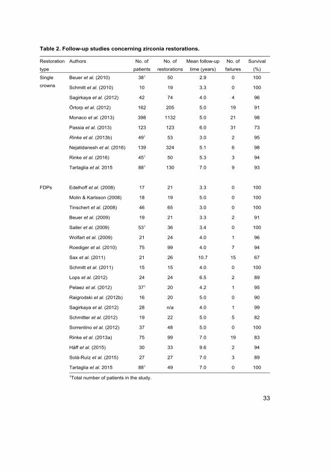
Follow-up studies concerning zirconia restorations are shown in Table 2. A
few review articles and meta-analyses, however, have summarized the clinical
success and survival of zirconia restorations. A meta-analysis by Sailer et al. (2015) estimated a survival rate of 91.2% for zirconia single crowns and 95.7%
for metal ceramic single crowns after five years. Larsson & Wennerberg (2014)
estimated five-year cumulative survival rate of 95.9% for zirconia single crowns.
A meta-analysis by Pjetursson et al. (2015) estimated a five-year survival rate of
90.4% for zirconia FDPs and 94.4% for metal ceramic FDPs.
33
Table 2. Follow-up studies concerning zirconia restorations.
Restoration
type
Authors No. of
patients
No. of
restorations
Mean follow-up
time (years)
No. of
failures
Survival
(%)
Single
crowns
Beuer et al. (2010) 381 50 2.9 0 100
Schmitt et al. (2010) 10 19 3.3 0 100
Sagirkaya et al. (2012) 42 74 4.0 4 96
Örtorp et al. (2012) 162 205 5.0 19 91
Monaco et al. (2013) 398 1132 5.0 21 98
Passia et al. (2013) 123 123 6.0 31 73
Rinke et al. (2013b) 491 53 3.0 2 95
Nejatidanesh et al. (2016) 139 324 5.1 6 98
Rinke et al. (2016) 451 50 5.3 3 94
Tartaglia et al. 2015 881 130 7.0 9 93
FDPs Edelhoff et al. (2008) 17 21 3.3 0 100
Molin & Karlsson (2008) 18 19 5.0 0 100
Tinschert et al. (2008) 46 65 3.0 0 100
Beuer et al. (2009) 19 21 3.3 2 91
Sailer et al. (2009) 531 36 3.4 0 100
Wolfart et al. (2009) 21 24 4.0 1 96
Roediger et al. (2010) 75 99 4.0 7 94
Sax et al. (2011) 21 26 10.7 15 67
Schmitt et al. (2011) 15 15 4.0 0 100
Lops et al. (2012) 24 24 6.5 2 89
Pelaez et al. (2012) 371 20 4.2 1 95
Raigrodski et al. (2012b) 16 20 5.0 0 90
Sagirkaya et al. (2012) 28 n/a 4.0 1 99
Schmitter et al. (2012) 19 22 5.0 5 82
Sorrentino et al. (2012) 37 48 5.0 0 100
Rinke et al. (2013a) 75 99 7.0 19 83
Håff et al. (2015) 30 33 9.6 2 94
Solá-Ruíz et al. (2015) 27 27 7.0 3 89
Tartaglia et al. 2015 881 49 7.0 0 100
1Total number of patients in the study.

34

35
3 Aims of the study
Zirconia has excellent mechanical and biological properties, but whether it is a
durable material for conventional fixed prosthetic restorations is unclear. These
properties could also make zirconia a useful material for abutment teeth of partial
removable dental prostheses (RDPs), but no clinical studies were available. The
hypothesis was that zirconia crowns, FDPs and abutment teeth of RDPs would
experience few early and short-term complications and would perform well in
both anterior and posterior regions in dentition, although more complications can
be expected with increasing time of function.
The specific aims were
1. to evaluate the incidence of early complications during prosthetic treatment
and short-term failures during the first year of use of zirconia single crowns
and fixed dental prostheses (FDPs)
2. to evaluate the success and survival of zirconia single crowns after two to six
years of clinical service
3. to evaluate the outcome of short- and long-span zirconia FDPs after three to
seven years of clinical service
4. to evaluate the usefulness of zirconia single crowns in abutment teeth of
partial removable dental prostheses (RDPs)

36

37
4 Material and methods
4.1 Description of the material
The prosthetic treatment and the clinical examinations were conducted at the
Dental Training Clinic, Oral Health Services, City of Oulu, and at the Institute of
Dentistry, University of Oulu, Finland. The study design was approved by the
Ethical Committee of the Northern Ostrobothnia Hospital District (100/2013).
Patient records (Effica; Tieto, Finland) were searched for patients treated with
zirconia restorations by predoctoral dental students between 2007 and 2010.
Anamnestic and clinical information related to prosthetic treatment was recorded
from patient records. Any dental treatment after the prosthetic treatment in Oral
Health Services, City of Oulu, was obtained from the patient records.
4.2 Study population
The material consisted of 173 patients treated with zirconia single crowns and/or
zirconia fixed dental prostheses (FDPs)(Fig. 2). Of these patients 94 were women
and 79 men (mean age 55 years, range 18–79 years). Altogether 268 zirconia
single crowns (mean 3 crowns per patient, range 1–12 crowns) were fabricated
for 88 patients and 120 zirconia FDPs (range 3–12 units, mean 4.5 units, 342
abutments and 190 pontics) for 102 patients. Seventeen patients had received both
crown(s) and FDP(s).

38
Fig. 2. Flow chart of the study material.
4.3 Materials of the restorations
The materials of the zirconia frameworks used were Zirkonzahn Zirconia
(Zirkonzahn, Germany), NobelProcera Zirconia (Nobel Biocare, Switzerland),
and Prettau Zirconia (Zirkonzahn, Germany). The frameworks of Zirkonzahn
Zirconia and Prettau Zirconia were fabricated by manual milling whereas the
frameworks of NobelProcera Zirconia were fabricated with CAD/CAM
manufacturing.
For Zirkonzahn Zirconia and Prettau Zirconia, the connector's cross-sectional
area requirement was 9.0 mm2 and the minimal thickness of the frameworks 0.4
mm. For NobelProcera Zirconia, the connector's cross-sectional area requirement
was 6.0mm2 (the anterior area) and 9.4 mm2 (the posterior area) and the minimal
thickness was 0.6 mm. Prettau Zirconia was monolithic whereas on Zirkonzahn
173 patients treated with zirconia single crowns or partial fixed dental prostheses
(FDPs) between 2007 and 2010 - 94 women (54%)- 79 men (46%)
120 zirconia partial fixed dental prosthesis(342 abutments, 190 pontics)
- 102 patients
268 zirconia single crowns - 88 patients
PAPER IEarly complications
and short-term failures of zirconia single
crowns and partial fixed dental prostheses.
- 173 patients- 120 FDPs- 264 crowns
PAPER IIOutcome of zirconia
single crowns made by predoctoral dental students: a clinical retrospective study after 2 to 6 years of
clinical service.- 66 patients- 190 crowns
PAPER IIIVeneered zirconia crowns as abutment teeth for partial removable dental prosthesis: A clinical 4-year retrospective study.- 17 patients- 37 crowns as abutment
teeth for a clasp-retained removable dental prosthesis
PAPER IVOutcome of zirconia partial fixed dental prostheses made by predoctoral dental students: A clinical retrospective study after 3 to 7 years of clinical service.- 76 patients- 102 FDPs
- 250 abutments - 137 pontics

39
Zirconia (GC Initial Zr; GC Europe) and Nobel Procera Zirconia (VITA VM 9;
VITA Zahnfabrik) the veneering porcelain was hand-layered.
4.4 Clinical procedures during prosthetic treatment
Treatments were performed by predoctoral dental students and supervised by
prosthodontists. All the patients had received pre-prosthetic treatments before
prosthetic treatment including cariological, periodontal, and endodontic treatment,
if needed.
Composite resin (Filtek Z250; 3M Deutschland GmbH, Germany) was used
to restore the abutment when needed and endodontically treated teeth were
reinforced with a fiber post (RelyX Fiber post; 3M Deutschland GmbH,
Germany). Preparations guidelines by Shillingburg et al. (2012) were followed
during prosthetic treatment for both single crowns and abutment teeth of FDPs:
heavy chamfer finish line preparation with total wall convergence of six degrees,
1.5 mm axial clearance, 2 mm anatomically adequate occlusal reduction and a
functional cusp bevel to ensure sufficient material thickness. In preparation of
abutment teeth for zirconia single crowns that serve as an abutment tooth for a
RDP with a metal framework, the extra space needed for occlusal rest seats and
guide planes were taken into account. The RDPs were fabricated according to
Scandinavian guidelines (Molin Thoren & Gunne 2012). If a zirconia single
crown was prepared to serve as an abutment tooth for a RDP with a metal
framework, the rest seat of the RDP was left with a zirconia surface and veneering
porcelain was placed only where needed for aesthetic reasons. The metal
frameworks of the RDPs were fabricated from a cobalt-chromium alloy and the
denture bases were made of acrylic resin.
Prior to cementation, the restorations were airborne-particle abraded with
aluminum oxide (110 µm, 200 kPa) and steam-cleaned in a laboratory and
cleaned with ethanol and air drying chair-side. Dual-polymerizing, self-adhesive,
universal resin cement (RelyX Unicem; 3M Deutschland GmbH, Germany) was
used for definitive cementation of the restorations. All the patients were re-called
for a check-up six months after the definitive cementation.
4.5 Early complications and short-term failures (Paper I)
The patient records of all the treated patients were searched for recorded
complications or failures. Any complications recorded from the beginning of

40
prosthetic treatment to the day of definitive cementation were registered from the
patient records (early complications). Any failures during the first year after the
definitive cementation were also recorded from the patient records (short-term
failures). Early complications and short-term failures were divided into biological
and technical subcategories.
4.6 Clinical follow-up
All the treated patients were to be invited to the clinical follow-ups 2–7 years
after definitive cementation. Some of the patients could not be reached. The
follow-up consisted of a questionnaire and a clinical examination.
4.6.1 Zirconia single crowns (Paper II)
The distribution of 268 zirconia single crowns in dentition is shown in Fig. 3. Of
all the patients, 54% had received three or four crowns but there were also
patients treated with up to 12 crowns.

41
Fig. 3. Distribution of zirconia single crowns (n=264) in dentition.
Of the total of 88 patients, 12 had moved away and one had died. Altogether 75
patients were invited to a clinical follow-up in 2013. Nine patients did not attend
and did not contact us. A total of 66 (75%) participants (30 women and 36 men)
attended the clinical follow-up and altogether 204 zirconia single crowns (mean
2.9 crowns, range 1–10 crowns) had been prepared for them between 2007 and
2010. In all 14 crowns had been lost during the follow-up period and 190 single
0
5
10
15
20
25
30
35
40
45
18 17 16 15 14 13 12 11 21 22 23 24 25 26 27 28
tooth number
0
5
10
15
20
25
30
35
40
45
48 47 46 45 44 43 42 41 31 32 33 34 35 36 37 38

42
crowns were examined. The mean age of the participants was 60.4 years (range
19–81 years) and the mean follow-up time was 3.9 years (1.9–6.0 years).
4.6.2 Veneered zirconia crowns as abutment teeth for partial
removable dental prostheses (Paper III)
Altogether 37 veneered zirconia single crowns were prepared for 17 patients (9
men and 8 women; mean age 62.5 years) to serve as abutment teeth for a clasp-
retained RDP with a metal framework. The mean follow-up time was 4.2 years
(2.9–5.4 years).
4.6.3 Zirconia fixed dental prostheses (FDPs) (Paper IV)
The 120 zirconia FDPs fabricated consisted of 527 total units, of which 342 were
abutments and 185 pontics. The distribution of abutments and pontics in dentition
is shown in Fig. 4.
Of the 102 patients who had received zirconia FDPs, one had moved away,
two had died and four had no contact details available. An invitation letter was
sent to 95 patients. Nineteen patients did not attend and did not contact us and 76
(75%) patients (48 women, 28 men) finally attended the clinical examination. In
all 88 FDPs had been fabricated, including 250 abutments and 137 pontics. The
mean follow-up time was 4.9 years (range 3–7 years).
The FDPs were divided into anterior FDPs from canine to canine and
posterior FDPs (premolar and molars). Altogether 40 FDPs had been fabricated
for the anterior region and 48 FDPs for the posterior region. The FDPs were also
divided into short- and long-span FDPs according to span length (Fig. 5). Short-
span FDPs consisted of three or four units, whereas long-span FDPs had five or
more units. The vast majority of FDPs (80 FDPs, 67%) were three or four units
(range: 3–12 units, mean: 4.5 units) but three 10-unit, one 11-unit and one 12-unit
FDPs were also fabricated. The number of restorations according to the length of
the restoration is shown in Fig. 5.

43
Fig. 4. Distribution of abutments and pontics in dentition. FDPs were divided into
anterior FDPs (from canine to canine) and posterior FDPs (premolars and molars)
according to the location of abutments and pontics.
0
5
10
15
20
25
30
35
40
45
18 17 16 15 14 13 12 11 21 22 23 24 25 26 27 28
pontics
abutments
0
5
10
15
20
25
30
35
40
45
48 47 46 45 44 43 42 41 31 32 33 34 35 36 37 38
pontics
abutments

44
Fig. 5. Zirconia FDPs were divided into short- and long-span FDPs according to span
length. A vast majority of the FDPs were short-span FDPs that consisted of three or
four units. Long-span FDPs had five or more units.
4.6.4 Clinical follow-up examination
Before the clinical examination, the participants filled in a questionnaire in which
they were asked on a Yes or No basis whether they were satisfied ("Are you
satisfied with...") with the aesthetics, colour-match, contour, and gloss of the
restoration. Personal symptoms such as pain (tooth, masticatory muscles,
temporomandibular joints), hypersensitivity to cold or heat and gingival bleeding
were also evaluated on a Yes or No basis ("Have you noticed..."). The participants
were also asked whether they had had complications or failures with the
restoration. The presence of sleep or awake bruxism as defined by Lobbezoo et al. (2013) was registered on a Yes or No basis.
In the clinical examination, modified World Dental Federation (FDI) clinical
criteria (Hickel et al. 2010) were used to evaluate the restorations. The colour
match of the restoration, marginal integrity, secondary caries, restorations in the
restoration margins, marginal discolouration, anatomic contour (under- and over-
contouring), surface texture, chipping or fractures of porcelain and wear of
restorations or antagonists were recorded and evaluated as good, acceptable, or
50
30
16
95 5
03 1 1
0
10
20
30
40
50
60
3 4 5 6 7 8 9 10 11 12
Number of restorations
Number of units in restoration
45
unacceptable (Hickel et al. 2010). Possible porcelain fractures were divided into
grade 1 (polished), grade 2 (repaired with composite-resin), and grade 3 (replaced)
according to Anusavice (2012). Retention of the restoration was also checked and
evaluated as firmly fixed or loss of retention.
The status of the periodontium of abutment and contralateral teeth was
evaluated by plaque accumulation (plaque index) and bleeding on probing (sulcus
bleeding index) according to Silness & Löe (1964) and Mombelli et al. (1987).
Plaque accumulation was divided into three categories: no detection of plaque,
plaque only detected by a probe or plaque seen with the naked eye. Bleeding on
probing was evaluated as no bleeding when the gingival margin was explored
with a periodontal probe, isolated bleeding when explored with a periodontal
probe or confluent bleeding when explored with a periodontal probe. The location
of the abutment margins (supragingival, marginal, subgingival) and the presence
of endodontic treatment through the restoration were recorded. No radiographic
evaluations were done as no clinical reasons existed to justify them.
If the restoration served as an abutment tooth for a partial removable dental
prosthesis (RDP), the surface of the ceramic rest seat was evaluated as good if
intact, acceptable if it showed mild or moderate wear and unacceptable if it had
fractures or severe wear. The stability and retention of the RDPs were evaluated
as good, moderate, or poor according to Molin Thoren & Gunne (2012).
Retention was evaluated by applying vertical pulling forces to the RPD. Retention
was evaluated as good if no displacement was seen, moderate if there was minor
displacement and poor if no retention was noted. Rotational forces were applied
to evaluate the stability of the RPD. Stability was evaluated as good if the RDP
firmly resisted rotational forces, moderate if vertical movement was seen after
forces were applied and poor if the RDP loosened when rotational forces were
applied.
If dental problems were noted at the follow-up, the participants were advised
to contact own dentist or book additional appointments at the clinic.
4.7 Statistical analysis
The mean and range were used to describe variables such as the age of the
patients and the number or length of restorations. Frequencies and percentages
were used to describe the distribution of variables such as gender, complications
and failures. Histograms were used to describe variable distribution.

46
Statistical analysis was performed with computer software (IBM SPSS
Statistics 22; IBM, USA). Fisher’s exact test (α=.05) was used to evaluate the
plaque accumulation index and bleeding on probing index of the abutment teeth
and the contralateral teeth. The survival and success of the restorations were
calculated using Kaplan-Meier analysis. The longevity of the restoration was
measured from the day of definitive cementation to the day of a complication or
to the day of a follow-up visit. According to Tan et al. (2004), a successful
restoration remains unchanged over the observation period, whereas a survived
restoration is still in situ at the examination visit, regardless of possible reversible
complications.
47
5 Results
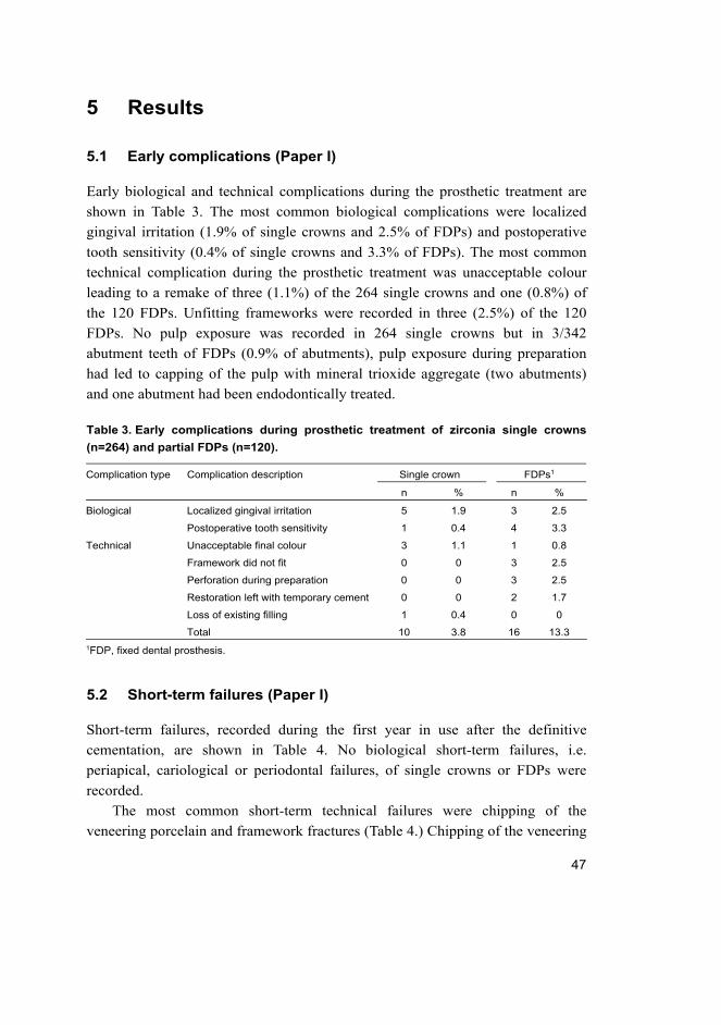
5.1 Early complications (Paper I)
Early biological and technical complications during the prosthetic treatment are
shown in Table 3. The most common biological complications were localized
gingival irritation (1.9% of single crowns and 2.5% of FDPs) and postoperative
tooth sensitivity (0.4% of single crowns and 3.3% of FDPs). The most common
technical complication during the prosthetic treatment was unacceptable colour
leading to a remake of three (1.1%) of the 264 single crowns and one (0.8%) of
the 120 FDPs. Unfitting frameworks were recorded in three (2.5%) of the 120
FDPs. No pulp exposure was recorded in 264 single crowns but in 3/342
abutment teeth of FDPs (0.9% of abutments), pulp exposure during preparation
had led to capping of the pulp with mineral trioxide aggregate (two abutments)
and one abutment had been endodontically treated.
Table 3. Early complications during prosthetic treatment of zirconia single crowns
(n=264) and partial FDPs (n=120).
Complication type Complication description Single crown FDPs1
n % n %
Biological Localized gingival irritation 5 1.9 3 2.5
Postoperative tooth sensitivity 1 0.4 4 3.3
Technical Unacceptable final colour 3 1.1 1 0.8
Framework did not fit 0 0 3 2.5
Perforation during preparation 0 0 3 2.5
Restoration left with temporary cement 0 0 2 1.7
Loss of existing filling 1 0.4 0 0
Total 10 3.8 16 13.3
1FDP, fixed dental prosthesis.
5.2 Short-term failures (Paper I)
Short-term failures, recorded during the first year in use after the definitive
cementation, are shown in Table 4. No biological short-term failures, i.e.
periapical, cariological or periodontal failures, of single crowns or FDPs were
recorded.
The most common short-term technical failures were chipping of the
veneering porcelain and framework fractures (Table 4.) Chipping of the veneering

48
porcelain in two crowns (0.8%) had occurred during the first month after
definitive cementation and was considered unrepairable (Grade 3) leading to
refabrication of the crowns. Chipping of the veneering porcelain in one FDP was
diagnosed as reparable (Grade 2) and was repaired with composite resin.
Catastrophic fractures of the framework during the first year occurred in 2 (1.7%)
out of the 120 FDPs. One crown with a loss of retention was recemented, whereas
none of the 120 FDPs had lost retention during the observation period.
Table 4. Short-term failures in year after definitive cementation of zirconia single
crowns (n=264) and partial FDPs (n=120).
Complication type Complication description Single crown FDP1
n % n %
Biological ‒ 0 0 0 0
Technical Porcelain chipping, irreparable 2 0.8 0 0
Porcelain chipping, reparable 0 0 1 0.8
Framework fracture 0 0 2 1.7
Loss of retention 1 0.4 0 0
Total 3 1.1 3 2.5
1FDP, fixed dental prosthesis.
5.3 Clinical findings at the follow-ups
5.3.1 Zirconia single crowns (Paper II)
Overall satisfaction of the patients was high: 98% were satisfied with aesthetics,
95 % with colour-match and contour and 100% with the gloss of the zirconia
single crowns. Hypersensitivity to cold in abutment teeth was described by three
participants and three participants had noticed gingival bleeding. One participant
described pain related to the zirconia crown. Self-reported bruxism was reported
by 26 participants.
Less plaque was seen on the zirconia crowns than on contralateral teeth, but
with no statistical significance (P=.376) Bleeding on probing was more frequent
in teeth with zirconia crowns (P=.012) than in contralateral teeth. The plaque
index and sulcus bleeding index are shown in Tables 5. and 6. The crown margin
placement was marginal in 53%, subgingival in 43% and supragingival in 4% of
the single crowns. Anatomic contour of the zirconia single crowns was rated good
in 156/190 (82%) and acceptable in 34/190 (18%) of the crowns. Marginal

49
integrity was rated good in all of the single crowns, whereas marginal
discolouration (acceptable) was noted in 3/190 (1.6%) of the single crowns. The
occlusal contact surface of 83/190 (44%) zirconia crowns was evaluated as
acceptable due to a slightly rough surface. Wear of opposing dentition related to
zirconia restorations was suspected with 11/190 (6%) single crowns.
Table 5. Plaque accumulation (Silness & Löe 1964, Mombelli et al. 1987) in abutment
teeth with zirconia single crowns (n=190) and in contralateral teeth.
Plaque index Abutment teeth (%) Contralateral teeth (%)
No detection of plaque 77 41
Plaque only recognized by running a probe across the
marginal surface of the crown
17 56
Plaque can be seen by the naked eye 6 3
P=.376; Fisher exact test.
Table 6. Bleeding on probing (Silness & Löe 1964, Mombelli et al. 1987) around teeth
with zirconia single crowns (n=190) and contralateral teeth.
Bleeding on probing Abutment teeth (%) Contralateral teeth (%)
No bleeding when a periodontal probe is passed
along the gingival margin
49 39
Isolated bleeding when a periodontal probe is
passed along the gingival margin
40 60
Confluent bleeding when a periodontal probe is
passed along the gingival margin
11 1
P=.012; Fisher exact test.
Biological and technical complications recorded at the clinical examination are
shown in Table 7. The complications of the zirconia single crowns were divided
according to manufacturers. No secondary caries or endodontic treatment
performed through the zirconia crowns were found in the examination. The most
frequent complications were loss of cementation (5%) and chipping of the
veneering porcelain (4%). According to Anusavice (2012), three of the porcelain
fractures were grade 3 fractures (severe) requiring replacement of the crowns and
six were polishable grade 1 fractures that did not affect function or aesthetics. The
success rate of the zirconia single crowns was 80% and the survival rate was 89%
after four-year (2–6 years) follow-up time.

50
Table 7. Complications in 204 zirconia single crowns.
Complication type Complication Zirkonzahn NobelProcera Prettau Total
n % n % n % n %
No complication No complication 138 84 17 81 18 95 173 85
Biological Secondary caries 0 0 0 0 0 0 0 0
Endodontic treatment
through the restoration
0 0 0 0 0 0 0 0
Root fracture 3 2 0 0 0 0 3 2
Periapical endodontic
infections
2 1 0 0 0 0 2 1
Total 5 3 0 0 0 0 5 3
Technical Porcelain fracture,
grade 1
5 3 1 5 0 0 6 3
Porcelain fracture,
grade 2
0 0 0 0 0 0 0 0
Porcelain fracture,
grade 3
2 1 1 5 0 0 3 1
Loss of cementation
(recemented)
5 3 2 9 1 5 8 4
Loss of cementation
(loss of crown)
3 2 0 0 0 0 3 2
Change in treatment
plan
6 4 0 0 0 0 6 3
Total 21 13 4 19 1 5 26 13
Total 164 100 21 100 19 100 204 100
5.3.2 Veneered zirconia crowns as abutment teeth for partial
removable dental prostheses (Paper III)
The clinical assessment of zirconia single crowns as abutment teeth of RPDs is
shown in Table 8. Neither secondary caries nor endodontic treatment performed
through the occlusal surface of the restoration was recorded. Fracture of the
veneering porcelain had occurred in 11% of the crowns and fracture of the
occlusal rest seat in 3%. Retention was valuated as good in all the RDPs, but
stability was rated moderate in 23% of the RDPs.

51
Table 8. Clinical assessment of 37 zirconia single crowns as abutment tooth for RDP.
Clinical assessment No (%) Moderate (%) Unacceptable (%)
Chipping of veneering porcelain 86 14 0
Over-contouring 87 13 0
Marginal discrepancy 95 5 0
Wear of ceramic surface in rest seat 100 0 0
5.3.3 Zirconia fixed dental prostheses (Paper IV)
Of the 76 participants, 96% were subjectively fully satisfied with the aesthetics of
the restoration: 100% with gloss, 99 % with colour match and 97% with the
contour of the restoration. The usual symptoms described by the participants in
the questionnaire were dull pain (9 patients, 12%), hypersensitivity to cold (8
patients, 11%), and gingival bleeding (4 patients, 11%). Altogether 28/76 (37%)
participants reported bruxism in the questionnaire.
Less plaque was seen on abutment teeth of FDPs than on contralateral teeth
(P<0.05), but bleeding on probing was more frequent in abutment teeth than in
contralateral teeth (P<0.05).Placement of abutment margins was 65% marginal,
22% subgingival and 14% supragingival. Anatomic contour of abutment teeth of
FDPs was rated good in 234/250 (94%) and acceptable in 16/250 (6%) abutments
of FDPs. Marginal integrity was good in all of the abutment teeth whereas
marginal discolouration was rated acceptable in 3/250 (1.2%) of abutment teeth.
The occlusal contact surface of 23/88 (26%) zirconia FDPs was evaluated as
acceptable due to a slightly rough surface. Wear of opposing dentition related to
the zirconia FDP was suspected in 7/88 (8%) FDPs.
Complications of the zirconia FDPs are shown in Table 9. The survival rate of
the zirconia fixed dental prostheses was 100% and the success rate was 89% after
five years. None of the 88 FDPs had lost cementation during the observation
period and no secondary caries was found, either. Endodontic periapical infection
had led to endodontic treatment through the occlusal surface of an abutment tooth
in one FDP.
Chipping of the veneering porcelain was noticed in 13 units (11 patients), of
which seven were in anterior and five in posterior regions. The distribution of
veneering porcelain fractures is shown in Table 10. and Fig. 6. Nine of the
chippings were in long-span FDPs and four in short-span FDPs. Of these
porcelain fractures, 12 were easily polished without aesthetically compromising
appearance and therefore evaluated as a grade 1 fracture according to Anusavice

52
(2012). In one FDP a fracture in the first incisor had been repaired with
composite-resin, compromising the appearance of the restoration and therefore
was evaluated as a grade 2 fracture (Anusavice 2012).
Table 9. Complications in 88 zirconia FDPs.
Complication type Complication Zirkonzahn NobelProcera Prettau Total
n % n % n % n %
No complication No complication 63 83 5 83 6 100 74 84
Biological Secondary caries 0 0 0 0 0 0 0 0
Endodontic treatment
through the restoration
0 0 0 0 0 0 0 0
Root fracture 0 0 0 0 0 0 0 0
Periapical endodontic
infections
1 1 0 0 0 0 1 1
Total 1 1 0 0 0 0 1 1
Technical Porcelain fracture,
grade 1
11 15 1 17 0 0 12 14
Porcelain fracture,
grade 2
1 1 0 0 0 0 1 1
Porcelain fracture,
grade 3
0 0 0 0 0 0 0 0
Loss of cementation
(recemented)
0 0 0 0 0 0 0 0
Loss of cementation (loss
of crown)
0 0 0 0 0 0 0 0
Change in treatment
plan
0 0 0 0 0 0 0 0
Total 12 16 1 17 0 0 13 15
Total 76 100 6 100 6 100 88 100
Table 10. Distribution of veneering porcelain fractures (n=13) in FDPs (n=88)
according to span length, jaw, and region of fracture.
Region Span length Maxilla (n=61) Mandibula (n=27) Total (n=88)
anterior region
FDPs (n=40)
3-4 unit FDPs (n=18) 3 0 3
over 5 unit FDPs (n=22) 3 2 5
posterior region
FDPs (n=48)
3-4 unit FDPs (n=40) 1 0 1
over 5 units FDPs (n=8) 3 1 4
total (n=88) 10 3 13

53
Fig. 6. Chipping of veneering porcelain (n=13) recorded in 11 patients. Arrows indicate
site of chipping, abutments are shown in black, pontics are rasterized and missing
teeth are marked “X”. Patients 1-8. had chipping in maxilla, patients 9. and 10. both in
maxilla and mandible, and patient 11. in mandible.

54

55
6 Discussion
6.1 Material and methods
The material of the study consisted of 173 patients treated with zirconia
restorations by predoctoral dental students between 2007 and 2010 at the Dental
Training Clinic, Oral Health Services, City of Oulu. Altogether 88 patients had
received zirconia single crowns and 102 patients had received zirconia FDPs.
According to literature, this is one of the largest materials of clinical studies
related to zirconia restorations (Al-Amleh et al. 2010, Raigrodski et al. 2012a,
Larsson & Wennerberg 2014, Pjetursson et al. 2015, Sailer et al. 2015). However,
the retrospective nature of the clinical study caused some difficulties in the
analysis of the results. The results of the study have to be evaluated with care,
because all the original patients were not interested in participating in the clinical
follow-up after up to seven years. The reasons can only be suspected, i.e. either
dissatisfaction with the restoration or changes in personal circumstances. In
Papers II and IV the drop-out rate was 25%. Similar drop-out rates have been
noted in other clinical studies related to zirconia restorations (Schmitter et al. 2012, Tartaglia et al. 2015), but much lower drop-out rates have also been
reported (Pjetursson et al. 2015, Sailer et al. 2015), and especially when patients
have been recalled annually (Kern et al. 2012, Chaar et al. 2015). The drop-out
rate here was relatively high, but on the other hand, prosthetic treatment with
fixed restorations is expensive and this is why it can be expected that patients who
have had problems would have contacted Oral Health Services, City of Oulu.
The relatively short observation period and the lack of a control group are
also study limitations. It would have been beneficial to be able to compare the
results with metal ceramic restorations that were fabricated during the same
period at the Dental Training Clinic. The study was started in 2012 and at that
time there were only a few 4–5-year clinical follow-ups on zirconia crowns and
FDPs with moderate size study populations. The failure rate is expected to
increase after a longer observation period (Rinke et al. 2013b, Sax et al. 2011),
although different results have also been reported (Håff et al. 2015, Tartaglia et al. 2015). Rinke et al. (2013b) reported a decrease in the survival rate of 94% at a
four-year follow-up to 83.4% at a seven-year follow-up and an increase in the
annual failure rate from 1.5% to 3.3%. On the contrary, Solá-Ruíz et al. (2015)
concluded that if a zirconia restoration survived three years without

56
complications, it more likely survived the seven-year follow-up time without
complications. Another limitation of this study is the impossibility to reliably
compare monolithic zirconia system with veneered zirconia systems or veneered
zirconia systems with each other because the number of these restorations was
low.
The pre-prosthetic and prosthetic treatments were performed by predoctoral
dental students. This reflects the usefulness of zirconia restorations, as all the
treatments were performed by unexperienced students, early complications during
prosthetic treatment were few and the survival and success rates are comparable
with other clinical studies. All the patients treated with zirconia restorations
between 2007 and 2010 were invited to the clinical examination, no exclusion
was performed and all the restorations were evaluated by the same examiners
using the same clinical criteria.
6.2 Patient satisfaction
Satisfaction with the aesthetics and function of the zirconia restorations in this
study was high. A few complaints about aesthetics regarded colour match and
bulky contour, whereas complaints regarding function were related to biting of
the cheek mucosa. These results are in accord with other clinical studies reporting
high satisfaction with veneered zirconia restorations (Molin & Karlsson 2008,
Sorrentino et al. 2012, Håff et al. 2015, Nejatidanesh et al. 2016). In literature,
opaqueness is depicted as a negative feature of zirconia, but overall satisfaction
regarding aesthetics seems to be high. It has to be taken into consideration,
however, that the mean age here was 55, and the aesthetic demands of elder
patients are usually lower.
6.3 Biological complications and failures
Early biological complications during prosthetic treatment were few and
temporary. Only a little more localized gingival irritation and postoperative tooth
sensitivity was noted in the zirconia FDPs than in single crowns. Localized
gingival irritation was generally caused by excess cement. No biological short-
term failures had occurred during the first year of use. Tooth sensitivity during
preparation was reported as temporary and needed no additional treatment, except
in two cases in which the FDP had been left with temporary cement for a longer
period of time to monitor symptoms. Roediger et al. (2010) also described tooth

57
sensitivity as a common but transient symptom and Monaco et al. (2013) reported
that 8% of vital abutment teeth had temporary symptoms of tooth sensitivity.
In this study no secondary caries was observed. In recent meta-analyses, a
significantly higher rate of secondary caries had been reported for zirconia single
crowns and FDPs (Pjetursson et al. 2015, Sailer et al. 2015). On the contrary, no
secondary caries had been reported after five years in several studies (Molin &
Karlsson 2008, Schmitter et al. 2012, Monaco et al. 2013 Nejantidenesh et al. 2015). Monaco et al. (2013) reported no secondary caries in 1132 zirconia single
crowns after five years. Tartaglia et al. (2015) reported a failure rate of 0.7% due
to secondary caries after seven years.
In this study no marginal discrepancies and only a few marginal
discolourations were found. The high rate of secondary caries in some studies is
likely related to the poor marginal fit of these restorations due to early-stage
prototype CAM systems. Sax et al. (2011) reported a high rate of marginal
discrepancy and secondary caries when a prototype CAM system (direct ceramic
machining, DCM) was used. Rinke et al. (2013b) noted that they started the study
as early as 2001 and an early-stage CAM system was used. Roediger et al. (2010)
noted a decrease in the rate of secondary caries after a software update of
CAD/CAM system. In this study, newer versions of CAD/CAM systems were
used for dental education should reflect new trends in dental practice, but the
materials and techniques should also be reliable and relatively easy to handle. A
problem in analysing review articles is that they can overestimate some problems
as they do not always take into account the developments within the materials and
fabrication techniques.
More plaque was seen on contralateral teeth than on abutment teeth, but more
bleeding on probing was seen with abutment teeth, which indicates that bacterial
adhesion is not the only factor affecting periodontal health. Other major factors
that damage the periodontium are compromised biological width and improper
reproduction of the dental contour (Becker & Kaldahl 2005, Vasconcelos et al. 2009). No significant differences regarding periodontal health between abutment
teeth and control teeth have been found in other studies (Tinschert et al. 2008,
Edelhoff et al. 2008, Beuer et al. 2009, Sailer et al. 2009a, Beuer et al. 2010,
Roediger et al. 2010, Monaco et al. 2013, Håff et al. 2015). Örtorp et al. (2009)
reported that gingival bleeding and calculus were evident with 17/25 crowns after
three years.
In this study, anatomic contour was rated as slightly over-contoured
(acceptable) in 18% of single crowns and in 6% of abutment teeth of FDPs. Molin

58
& Karlsson (2008) reported slight over-contouring in 5-10% of zirconia FDPs.
Under-contouring of buccal and lingual surfaces may promote gingival health,
whereas a bulky restoration may damage the periodontium, possibly because it
obstructs cleaning, and therefore buccal and lingual contours should be kept flat
(Becker & Kaldahl 2005). Adequate tooth preparation (dimensional requirements
of the restorative materials) and being familiar with crown morphology (both
dentist and technician) are essential for proper reproduction of the dental contour
(Becker & Kaldahl 2005). Of the restoration margins, 57% were placed
marginally or supragingivally and 43% subgingivally in single crowns, whereas
79% of margins were marginal or supragingival in FDPs. Plaque accumulation,
inflammation and gingivitis have been reported to be more frequent in teeth with
subgingival crown margins than with supragingival margins (Becker & Kaldahl
2005, Podhorsky et al. 2015). A subgingival restoration margin may compromise
biological width and cause chronic inflammation in the periodontium (Podhorsky
et al. 2015). The type of subgingival finish line may have an effect on periodontal
health (Paniz et al. 2015).
Patients treated with conventional fixed prosthodontics should undergo
thorough removal of excess cement and professional cleaning and should be
given improved home instructions. Studies have concluded that proper home care
is more important than the choice of restorative material in terms of bacterial
adhesion (Litonjua et al. 2012). All the patients in this study received
cariological, periodontal, and endodontic treatment prior to prosthetic treatment,
if needed. Professional tooth cleaning, including education and instructions for
proper home care, was performed on all the patients.
6.4 Technical complications and failures
6.4.1 Technical early complications
There were few technical complications during the prosthetic treatment. The most
common complications were unacceptable colour, poor fit of the framework, and
pulp exposure during preparation. Pulp exposure during preparation did not occur
in preparation of the single crowns, but the pulps of 3/342 (0.9%) abutment teeth
of the FDPs were exposed. This is comparable with metal ceramic FDPs as
Raustia et al. (1998) reported 2/221 (0.9%) perforations during preparation of
abutment teeth for metal ceramic FDPs.

59
In this study, a deep chamfer finish line was used, but some studies have
indicated that a knife-edge margin is also suitable preparation for zirconia and
other ceramic materials with less removal of tooth substance (Schmitt et al. 2010,
Cortellini & Canale 2012, Monaco et al. 2013). No margin preparation design has
been proven to be more durable or better fitting than other designs (Podhorsky et al. 2015). Preparation design should be chosen with regard to the specific clinical
situation. In situations where preservation of sound tooth substance is required, a
knife-edge margin might have more benefits than a chamfer or shoulder finish
line (Monaco et al. 2013). The visibility of the margin by a technician or with a
scanning device should be considered when choosing the margin preparation
(Podhorsky et al. 2015). Mini-invasive techniques are coming to fixed
prosthodontics, but adhesive bonding is needed in those cases.
6.4.2 Technical short-term failures
In this study, short-term failures were chipping of the veneering porcelain, loss of
retention and framework fractures. The two chippings in crowns had occurred
within the first few weeks after cementation, and were diagnosed as unrepairable,
grade 3 chippings according to Anusavice (2012). The crowns were remade.
Chipping in FDP was considered Grade 2 chipping and was repaired with
composite resin. None of the FDPs, but 1/264 (0.4%) of the crowns had lost
retention and was recemented.
If the material fails shortly after definitive cementation, the failure is likely
caused by material weakness or errors during the fabrication process. At this
point, patient-related factors such as bruxism or diet are only minor factors.
Identifying these possibly rectifiable initial problems is important, because studies
have indicated that an early repairable complication is a risk factor for
unrepairable failure later (De Backer et al. 2006a, De Backer et al. 2006b).
Sagirkaya et al. (2012) found in a four-year follow-up of zirconia FDPs that four
out of five failures occurred during the first year. Sola-Ruiz et al. (2015) found
that if the zirconia FDP survived the first three years without complications, the
risk for failures during a seven-year follow-up period decreased.
Framework fracture was noted in 2/120 (1.7%) FDPs and the restorations
were remade. The first framework fracture had occurred at the six-month recall
when an attempt was made to remove the FDP that had been left with temporary
cement (sensitivity problems). The other framework fracture occurred for an
unknown reason shortly after definitive cementation in an occlusal area of a molar

60
abutment tooth. A new FDP was remade and was still in situ at the follow-up
meeting.
6.4.3 Chipping
Most of the chippings of the veneering porcelain noted at the clinical
examination were polishable. It was noticed that the majority of the chippings had
occurred on occlusal surfaces, and they were possibly related to advanced wear of
the surface. Similar findings have been made in other studies (Sailer et al. 2009a,
Koenig et al. 2013). Most of the porcelain fractures were found during the follow-
up and the participant had not noticed it. Most of the chippings could be polished
without compromising esthetics and function. It has been noted in other studies
that chippings are usually incidental findings during follow-ups and rarely leads
to renewal of the restoration (Sax et al. 2011, Schmitter et al. 2012, Rinke et al. 2013b). Nejatidanesh et al. (2016) reported chipping of the veneering porcelain in
45/556 zirconia single crowns, of which 42 were minor and could be polished
without compromising aesthetics and function.
A porcelain veneer on zirconia seems to be more fragile than on metal
(Augstín-Panadero et al. 2012). In metal ceramic restorations the fracture usually
occurs at the metal-porcelain interface, whereas with zirconia restorations the
fracture usually occurs within the porcelain veneer (Schmitt et al. 2011, Augstín-
Panadero et al. 2012) and is cohesive in nature (Fischer et al. 2008, Schmitt et al. 2011, Preiss et al. 2013). There is evidence that cohesive porcelain fractures are
related to a compressive stress induced by overly fast cooling after firing and can
be prevented by using a slower cooling procedure (Denry & Kelly 2014, Paula et al. 2015) Nowadays, most porcelain veneer manufacturers have modified firing
guidelines for zirconia restorations (Paula et al. 2015). Promising clinical results
have been seen when an anatomical design of the framework and a slow cooling
protocol have been adopted (Örtorp et al. 2012, Rinke et al. 2013a, Rinke et al. 2016, Tartaglia et al. 2015).
6.4.4 Loss of retention
None of the 88 FDPs, but 8/204 (4%) of the zirconia single crowns lost retention
during the follow-up period. Recent meta-analyses by Sailer et al. (2015) and
Pjetursson et al. (2015) reported significantly more loss of retention for zirconia
single crowns and FDPs than for metal ceramic restorations. This was argued to

61
be related to inaccuracies in the fit of the framework. Another factor that may
explain the loss of retention issue could be differences in the preparation designs
of metal ceramic and zirconia abutments. In some studies, different findings have
been made, as Tartaglia et al. (2015) reported that 3/130 (2.3%) zirconia single
crowns and 0/49 (0%) zirconia FDPs had lost retention after seven years and
Monaco et al. (2013) described only 2/1127 (0.2%) losses of retention after five
years.
Strong and durable adhesive cementation increases retention and fracture
resistance and improves marginal adaptation of the restoration (Thompson et al. 2011). Cleaning and roughening the inner surface of the zirconia restoration with
moderate-pressure airborne-particle abrasion and cementation with phosphate
monomer-containing primer or luting resin seem to provide a durable bond to
zirconia (Sasse & Kern 2013, Kern 2015). These bonding procedures were not
completely followed in this study, as the adhesive cement used here did not
contain MDP monomer. To date, zirconia has mostly been cemented with
conventional cements like zinc phosphate or with glass-ionomer or resin-based
cements without MDP monomer. It seems that strong and durable adhesive
cementation is needed to increase the retention of zirconia restorations, especially
single crowns, and zirconia bonding procedures (Kern 2015) are therefore
recommended.
6.4.5 Wear of the restoration and antagonist teeth
In this study, a slightly rough occlusal surface was a common find. Clinical
follow-up studies have revealed a high incidence of occlusal surface roughness in
the veneering porcelain of zirconia restorations Molin & Karlsson 2008, Sailer et al. 2009a, Schmitt et al. 2011, Koenig et al. 2013). It was reported by Schmitt et al. (2011) that after 48 months 59% of the units of zirconia FDPs were evaluated
clinically as slightly rough or pitted and 65% after evaluation with a scanning
electron microscope (SEM). The glaze is worn away over time (Heintze et al. 2008, Janyavula et al. 2013). Some studies recommended repolishing the
zirconia restorations at recall visits (Sax et al. 2011, Sripetchdanond & Leevailoj
2014)
Veneering porcelain is reported to be more abrasive than natural enamel or
zirconia (Kim et al. 2012, Burgess et al. 2014, Lawson et al. 2014). Wear of
opposing dentition was not a major problem in this study, but may increase due to
a roughened porcelain surface. The surface roughness of ceramic restorations

62
seems to increase enamel wear of the antagonist tooth (Ghazal & Kern 2009).
Lawson et al. (2014) even recommended avoiding porcelain on the occlusal
contact area to prevent enamel wear of antagonist teeth. Promising clinical results
have been seen with monolithic and minimally veneered zirconia restorations
(Moscovitch 2015, Venezia et al. 2015).
6.4.6 Veneered zirconia crowns as abutment teeth for partial
removable dental prostheses
The lack of strength has contraindicated the use of all-ceramic materials in
removable partial denture (RPD) abutments (Kancyper et al. 2000, Carracho &
Razzoog 2006). The excellent physical properties of zirconia would allow the use
of tooth-coloured single crowns in these situations, but no clinical studies existed
to confirm this hypothesis. In this study no wear was seen on zirconia surface of
the occlusal rest seat, but chipping of the veneering porcelain was a common
problem. The occlusal rest in a zirconia single crown is more rounded than in
metal ceramic crowns because of material-related factors, but stability and
retention were rated good in most of the RDPs. The usefulness of monolithic
zirconia should be evaluated or veneering porcelain should be applied only to
non-functional areas, as described by Venezia et al. (2015).
6.5 Success and survival of zirconia single crowns and fixed
dental prosthesis
In this study, the survival rate of zirconia single crowns after 3.9 years was 89%
and the success rate was 80%, whereas the survival rate of zirconia FDPs after 4.9
years was 100% and the success rate was 89%. The results agree with clinical
follow-up studies, as in recent meta-analyses the estimated five-year survival rate
of zirconia single crowns in the anterior region was 98.5% and it was 95% in the
posterior region, and the estimated five-year survival rate of zirconia FDPs was
90.4% (Pjetursson et al. 2015, Sailer et al. 2015). In the studies with a follow-up
time of at least four years, the survival rates of zirconia single crowns have varied
from 91% to 98% (Beuer et al. 2010, Schmitt et al. 2010, Sagirkaya et al. 2012,
Örtorp et al. 2012, Monaco et al. 2013, Rinke et al. 2013b, Nejatidanesh et al. 2016, Rinke et al. 2016, Tartaglia et al. 2015) The survival rates of zirconia FDPs
has varied from 94% to 100% after four to five years (Molin & Karlsson 2008,
Wolfart et al. 2009, Roediger et al. 2010, Schmitt et al. 2011, Peleaz et al. 2012,

63
Raigrodski et al. 2012a, Sagirkaya et al. 2012, Sorrentino et al. 2012). In the
longer 6–10-year follow-ups, the survival rate has varied from 86% to 100%
(Lops et al. 2012, Rinke et al. 2013a, Håff et al. 2015Solá-Ruíz et al. 2015,
Tartaglia et al. 2015). Lower survival rates have been reported in a few studies
(Sax. et al. 2011, Schmitter et al. 2012, Passia et al. 2013).
In this study, 17/204 single crowns were evaluated as total failures. One
participant had lost a crown because of a loss of cementation, two crowns were
lost because of a root fracture, and thereafter the treatment plan had been changed
to a removable prosthesis and three more crowns had been removed. Another
participant had lost a crown because of a root fracture and thereafter three
adjacent crowns were removed because of the treatment plan of FDP. It seems
that problems accumulate in the same patients and one failure or complication
increases the risk for another.
Parafunctional habits like bruxism and endodontic treatment/non-vitality of
the teeth have been reported to increase the risk for complications and failures of
all-ceramic restorations (van Dijken & Hasselrot 2010, Beier et al. 2012). This
has also been noted with zirconia crowns, as Monaco et al. (2013) found an
almost three times higher risk for chipping of the veneering porcelain in
endodontically treated teeth with fiber reinforcement or if moderate or severe
parafunctional habits were present. They also described that 23/66 patients using
an occlusal splint reported delamination or chipping of the veneering porcelain
(Monaco et al. 2013). Koenig et al. (2013) concluded that absence of an occlusal
splint, ceramic restoration as an antagonist tooth, and presence of parafunctional
habits are statistically significant factors in chipping formation.
In this study, only one participant with porcelain chipping had reported
bruxism. It has to be taken into account that bruxism was self-reported here. Low
correlation between self-reported sleep bruxism and clinical manifestation of
bruxism has been reported in TMD patients (Paesani et al. 2013). Oral appliances
are a standard treatment for sleep bruxism (Manfredini et al. 2015) and are also
often recommended after treatment with a fixed prosthesis (Koenig et al. 2013),
even though it seems that an occlusal splint does not decrease the rate of chipping
of the veneering porcelain (Monaco et al. 2013).
In this study, the long-span FDPs (five-unit or more) seemed to have an
increased risk for chipping of the veneering porcelain. This has been noted in
other clinical studies (Sax et al. 2011, Solá-Ruíz et al. 2015) although only a few
studies have examined both short- and long-span FDPs. A meta-analysis of single
crowns indicated that a higher risk of technical complications in zirconia crowns

64
is seen in posterior regions than in anterior region (Sailer et al. 2015). Different
results have also been reported, as in a retrospective cohort study of 1132 zirconia
crowns Monaco et al. (2013) noted that chippings of veneering porcelain were
evenly distributed in the anterior and posterior regions. Rinke et al. (2015) found
that, within the molar region, a 5.5 times higher risk for chipping was seen if a
crown was placed in a terminal abutment i.e. in an abutment with no adjacent
teeth distally.
6.6 Zirconia in fixed prosthodontics
The survival and success of zirconia single crowns in this study are comparable,
but not superior to the survival of other recent all-ceramic crowns (Sailer et al. 2015). If no parafunctional habits exist, the choice of an all-ceramic material for a
single crown can be made according to the site of the restoration, taking into
account aesthetic and functional demands. From the material point of view,
zirconia single crowns cannot be recommended over any other recent ceramic
materials.
The survival and success rates of zirconia FDPs here seem to be superior to
those of other all-ceramic FDPs. There are follow-up studies available concerning
these other all-ceramic FDPs with survival rates comparable to zirconia, but they
include only short FDPs in the anterior and premolar regions of dentition (Kern et al. 2012, Chaar et al. 2015).
From the biological point of view, monolithic or minimally veneered zirconia
would be beneficial over other all-ceramic materials as less preparation of the
tooth structure is needed. The minimal preparation concept is the future of fixed
prosthodontics, potentially also with zirconia materials, because adhesive long-
term bonding of zirconia seems promising.

65
7 Summary and conclusion
The results of the study show that zirconia restorations are a suitable treatment
alternative in fixed prosthodontics. Biological complications during prosthetic
treatment and follow-up were few. Chipping of the veneering porcelain is
common, but in most cases it is a repairable complication. The surface of zirconia
restorations should be evaluated with care at recall visits, because chippings seem
to be related to advanced wear of the porcelain surface. However, developments
in fabrication processes seem to have decreased the rate of chipping, and with
new fabrication guidelines chipping may be comparable with metal ceramic
restorations. Monolithic and minimally veneered zirconia restorations are a
promising treatment alternative, but possible effects on antagonist teeth and long-
term low-temperature degradation of unveneered zirconia should be considered.
Loss of retention is a common problem with zirconia single crowns, and clinical
studies with updated fabrication guidelines and bonding procedures are needed.
Based on the findings of the study, the following conclusions can be drawn:
1. Early complications during prosthetic treatment and short-term failures
during the first year of use of zirconia single crowns and fixed dental
prostheses (FDPs) were few.
2. The success and survival of zirconia single crowns were found to be
comparable, but not superior to other recent ceramic materials.
3. Short- and long-span zirconia FDPs performed well in both anterior and
posterior regions of dentition.
4. Zirconia single crowns performed well as abutment teeth for partial RDPs
with a metal framework, but fractures in the veneering porcelain remained a
problem.

66

67
List of references
Abduo J, Lyons K & Swain M (2010) Fit of zirconia fixed partial denture: a systematic review. J Oral Rehabil 37(11): 866–876.
Aboushelib MN, Feilzer AJ & Kleverlaan CJ (2010) Bonding to zirconia using a new surface treatment. J Prosthodont 19(5): 340–346.
Aboushelib MN, Kleverlaan CJ & Feilzer AJ (2006) Microtensile bond strength of different components of core veneered all-ceramic restorations. Part II: Zirconia veneering ceramics. Dent Mater 22(9): 857–863.
Al-Amleh B, Lyons K & Swain M (2010) Clinical trials in zirconia: a systematic review. J Oral Rehabil 37(8): 641–652.
Amaral M, Belli R, Cesar PF, Valandro LF, Petschelt A & Lohbauer U (2014) The potential of novel primers and universal adhesives to bond to zirconia. J Dent 42(1): 90–98.
Anusavice KJ (2012) Standardizing failure, success, and survival decisions in clinical studies of ceramic and metal-ceramic fixed dental prostheses. Dent Mater 28(1): 102–111.
Apratim A, Eachempati P, Krishnappa Salian KK, Singh V, Chhabra S & Shah S (2015) Zirconia in dental implantology: A review. J Int Soc Prev Community Dent 5(3): 147–156.
Atsu SS, Kilicarslan MA, Kucukesmen HC & Aka PS (2006) Effect of zirconium-oxide ceramic surface treatments on the bond strength to adhesive resin. J Prosthet Dent 95(6): 430–436.
Augstin-Panadero R, Fons-Font A, Roman-Rodriguez JL, Granell-Ruiz M, del Rio-Highsmith J & Sola-Ruiz MF (2012) Zirconia versus metal: a preliminary comparative analysis of ceramic veneer behavior. Int J Prosthodont 25(3): 294–300.
Becker CM & Kaldahl WB (2005) Current theories of crown contour, margin placement, and pontic design. 1981. J Prosthet Dent 93(2): 107–115.
Beier US, Kapferer I & Dumfahrt H (2012) Clinical long-term evaluation and failure characteristics of 1,335 all-ceramic restorations. Int J Prosthodont 25(1): 70–78.
Benetti P, Kelly JR, Sanchez M & Della Bona A (2014) Influence of thermal gradients on stress state of veneered restorations. Dent Mater 30(5): 554–563.
Beuer F, Edelhoff D, Gernet W & Sorensen JA (2009) Three-year clinical prospective evaluation of zirconia-based posterior fixed dental prostheses (FDPs). Clin Oral Investig 13(4): 445–451.
Beuer F, Stimmelmayr M, Gernet W, Edelhoff D, Guh JF & Naumann M (2010) Prospective study of zirconia-based restorations: 3-year clinical results. Quintessence Int 41(8): 631–637.
Biscaro L, Bonfiglioli R, Soattin M & Vigolo P (2013) An in vivo evaluation of fit of zirconium-oxide based ceramic single crowns, generated with two CAD/CAM systems, in comparison to metal ceramic single crowns. J Prosthodont 22(1): 36–41.
Blatz MB, Sadan A & Kern M (2003) Resin-ceramic bonding: a review of the literature. J Prosthet Dent 89(3): 268–274.

68
Bottino MA, Valandro LF, Scotti R & Buso L (2005) Effect of surface treatments on the resin bond to zirconium-based ceramic. Int J Prosthodont 18(1): 60–65.
Brawek PK, Wolfart S, Endres L, Kirsten A & Reich S (2013) The clinical accuracy of single crowns exclusively fabricated by digital workflow--the comparison of two systems. Clin Oral Investig 17(9): 2119–2125.
Bremer F, Grade S, Kohorst P & Stiesch M (2011) In vivo biofilm formation on different dental ceramics. Quintessence Int 42(7): 565–574.
Burgess JO, Janyavula S, Lawson NC, Lucas TJ & Cakir D (2014) Enamel wear opposing polished and aged zirconia. Oper Dent 39(2): 189–194.
Busscher HJ, Rinastiti M, Siswomihardjo W & van der Mei HC (2010) Biofilm formation on dental restorative and implant materials. J Dent Res 89(7): 657–665.
Carracho JF & Razzoog ME (2006) Removable partial denture abutments restored with all-ceramic surveyed crowns. Quintessence Int 37(4): 283–288.
Chaar MS, Passia N & Kern M (2015) Ten-year clinical outcome of three-unit posterior FDPs made from a glass-infiltrated zirconia reinforced alumina ceramic (In-Ceram Zirconia). J Dent 43(5): 512–517.
Chevalier J (2006) What future for zirconia as a biomaterial? Biomaterials 27(4):535–43. Christel P, Meunier A, Heller M, Torre JP & Peille CN (1989) Mechanical properties and
short-term in-vivo evaluation of yttrium-oxide-partially-stabilized zirconia. J Biomed Mater Res 23(1): 45–61.
Contrepois M, Soenen A, Bartala M & Laviole O (2013) Marginal adaptation of ceramic crowns: a systematic review. J Prosthet Dent 110(6): 447–454.e10.
Cortellini D & Canale A (2012) Bonding lithium disilicate ceramic to feather-edge tooth preparations: a minimally invasive treatment concept. J Adhes Dent 14(1): 7–10.
Dalli M, Colak H & Mustafa Hamidi M (2012) Minimal intervention concept: a new paradigm for operative dentistry. J Investig Clin Dent 3(3): 167–175.
De Backer H, Van Maele G, De Moor N, Van den Berghe L & De Boever J (2006) An 18-year retrospective survival study of full crowns with or without posts. Int J Prosthodont 19(2): 136–142.
De Backer H, Van Maele G, De Moor N, Van den Berghe L & De Boever J (2006) A 20-year retrospective survival study of fixed partial dentures. Int J Prosthodont 19(2): 143–153.
Denry I & Kelly JR (2008) State of the art of zirconia for dental applications. Dent Mater 24(3): 299–307.
Denry I & Kelly JR (2014) Emerging ceramic-based materials for dentistry. J Dent Res 93(12): 1235–1242.
Edelhoff D, Florian B, Florian W & Johnen C (2008) HIP zirconia fixed partial dentures--clinical results after 3 years of clinical service. Quintessence Int 39(6): 459–471.
Filoche S, Wong L & Sissons CH (2010) Oral biofilms: emerging concepts in microbial ecology. J Dent Res 89(1): 8–18.
Fischer J, Grohmann P & Stawarczyk B (2008) Effect of zirconia surface treatments on the shear strength of zirconia/veneering ceramic composites. Dent Mater J 27(3): 448–454.

69
Fischer J, Stawarzcyk B, Trottmann A & Hammerle CH (2009) Impact of thermal misfit on shear strength of veneering ceramic/zirconia composites. Dent Mater 25(4): 419–423.
Ghazal M & Kern M (2009) The influence of antagonistic surface roughness on the wear of human enamel and nanofilled composite resin artificial teeth. J Prosthet Dent 101(5): 342–349.
Gracis S, Thompson VP, Ferencz JL, Silva NR & Bonfante EA (2015) A new classification system for all-ceramic and ceramic-like restorative materials. Int J Prosthodont 28(3): 227–235.
Griggs JA (2007) Recent advances in materials for all-ceramic restorations. Dent Clin North Am 51(3): 713–27, viii.
Guess PC, Bonfante EA, Silva NR, Coelho PG & Thompson VP (2013) Effect of core design and veneering technique on damage and reliability of Y-TZP-supported crowns. Dent Mater 29(3): 307–316.
Haff A, Lof H, Gunne J & Sjogren G (2015) A retrospective evaluation of zirconia-fixed partial dentures in general practices: an up to 13-year study. Dent Mater 31(2): 162–170.
Heintze SD, Cavalleri A, Forjanic M, Zellweger G & Rousson V (2008) Wear of ceramic and antagonist - a systematic evaluation of influencing factors in vitro. Dent Mater 2008 24(4):433–449.
Heintze SD & Rousson V (2010) Survival of zirconia- and metal-supported fixed dental prostheses: a systematic review. Int J Prosthodont 23(6): 493–502.
Hickel R, Peschke A, Tyas M, Mjor I, Bayne S, Peters M, Hiller KA, Randall R, Vanherle G & Heintze SD (2010) FDI World Dental Federation: clinical criteria for the evaluation of direct and indirect restorations-update and clinical examples. Clin Oral Investig 14(4): 349–366.
Inokoshi M, De Munck J, Minakuchi S & Van Meerbeek B (2014a) Meta-analysis of bonding effectiveness to zirconia ceramics. J Dent Res 93(4): 329–334.
Inokoshi M, Zhang F, De Munck J, Minakuchi S, Naert I, Vleugels J, Van Meerbeek B & Vanmeensel K (2014b) Influence of sintering conditions on low-temperature degradation of dental zirconia. Dent Mater 30(6): 669–678.
Janyavula S, Lawson N, Cakir D, Beck P, Ramp LC & Burgess JO (2013) The wear of polished and glazed zirconia against enamel. J Prosthet Dent 109(1): 22–29.
Kancyper S, Sierraalta M & Razzoog ME (2000) All-ceramic surveyed crowns for removable partial denture abutments. J Prosthet Dent 84(4): 400–402.
Kelly JR (2004) Dental ceramics: current thinking and trends. Dent Clin North Am 48(2): viii, 513–30.
Kelly JR & Benetti P (2011) Ceramic materials in dentistry: historical evolution and current practice. Aust Dent J 56 Suppl 1: 84–96.
Kelly JR & Denry I (2008) Stabilized zirconia as a structural ceramic: an overview. Dent Mater 24(3): 289–298.
Kelly JR, Nishimura I & Campbell SD (1996) Ceramics in dentistry: historical roots and current perspectives. J Prosthet Dent 75(1): 18–32.
70
Kern M (2015) Bonding to oxide ceramics-laboratory testing versus clinical outcome. Dent Mater 31(1): 8–14.
Kern M, Sasse M & Wolfart S (2012) Ten-year outcome of three-unit fixed dental prostheses made from monolithic lithium disilicate ceramic. J Am Dent Assoc 143(3): 234–240.
Khmaj MR, Khmaj AB, Brantley WA, Johnston WM & Dasgupta T (2014) Comparison of the metal-to-ceramic bond strengths of four noble alloys with press-on-metal and conventional porcelain layering techniques.J Prosthet Dent 112(5):1194–1200.
Kim MJ, Oh SH, Kim JH, Ju SW, Seo DG, Jun SH, Ahn JS & Ryu JJ (2012) Wear evaluation of the human enamel opposing different Y-TZP dental ceramics and other porcelains. J Dent 40(11): 979–988.
Kim H, Kim S, Lee J, Han J & Yeo I (2013) Effect of polishing and glazing on the color and spectral distribution of monolithic zirconia. J Adv Prosthodont 5(3): 296–304.
Kim HK & Kim SH (2014) Effect of the number of coloring liquid applications on the optical properties of monolithic zirconia. Dent Mater 30(9): e229–37.
Koenig V, Vanheusden AJ, Le Goff SO & Mainjot AK (2013) Clinical risk factors related to failures with zirconia-based restorations: an up to 9-year retrospective study. J Dent 41(12): 1164–1174.
Larsson C & Wennerberg A (2014) The clinical success of zirconia-based crowns: a systematic review. Int J Prosthodont 27(1): 33–43.
Lawson NC, Janyavula S, Syklawer S, McLaren EA & Burgess JO (2014) Wear of enamel opposing zirconia and lithium disilicate after adjustment, polishing and glazing. J Dent 42(12): 1586–1591.
Lee JH (2016) Digital approach to a ceramic-pressed-to-metal restoration. J Prosthet Dent 115(2):141–144.
Li RW, Chow TW & Matinlinna JP (2014) Ceramic dental biomaterials and CAD/CAM technology: state of the art. J Prosthodont Res 58(4): 208–216.
Lima EM, Koo H, Vacca Smith AM, Rosalen PL & Del Bel Cury AA (2008) Adsorption of salivary and serum proteins, and bacterial adherence on titanium and zirconia ceramic surfaces. Clin Oral Implants Res 19(8): 780–785.
Litonjua LA, Cabanilla LL & Abbott LJ (2012) Plaque formation and marginal gingivitis associated with restorative materials. Compend Contin Educ Dent 33(1): e6–10.
Lobbezoo F, Ahlberg J, Glaros AG, Kato T, Koyano K, Lavigne GJ, de Leeuw R, Manfredini D, Svensson P & Winocur E (2013) Bruxism defined and graded: an international consensus. J Oral Rehabil 40(1): 2–4.
Lops D, Mosca D, Casentini P, Ghisolfi M & Romeo E (2012) Prognosis of zirconia ceramic fixed partial dentures: a 7-year prospective study. Int J Prosthodont 25(1): 21–23.
Lughi V & Sergo V (2010) Low temperature degradation -aging- of zirconia: A critical review of the relevant aspects in dentistry. Dent Mater 26(8): 807–820.
Magne P, Carvalho AO, Bruzi G & Giannini M (2015) Fatigue resistance of ultrathin CAD/CAM complete crowns with a simplified cementation process. J Prosthet Dent 114(4): 574–579.

71
Manfredini D, Ahlberg J, Winocur E & Lobbezoo F (2015) Management of sleep bruxism in adults: a qualitative systematic literature review. J Oral Rehabil 42(11): 862–874.
Manicone PF, Rossi Iommetti P & Raffaelli L (2007) An overview of zirconia ceramics: basic properties and clinical applications. J Dent 35(11): 819–826.
Marchack BW, Sato S, Marchack CB & White SN (2011) Complete and partial contour zirconia designs for crowns and fixed dental prostheses: a clinical report. J Prosthet Dent 106(3): 145–152.
Melo RM, Souza R, Dursun E, Monteiro E, Valandro LF & Bottino MA (2015) Surface Treatments of Zirconia to Enhance Bonding Durability. Oper Dent 40(6): 636–643.
Miyazaki T & Hotta Y (2011) CAD/CAM systems available for the fabrication of crown and bridge restorations. Aust Dent J 56 Suppl 1: 97–106.
Miyazaki T, Nakamura T, Matsumura H, Ban S & Kobayashi T (2013) Current status of zirconia restoration. J Prosthodont Res 57(4): 236–261.
Molin Thoren M & Gunne J (2012) Textbook of Removable Prosthodontics - The Scandinavian Approach. Copenhagen, Munksgaard Denmark.
Molin MK & Karlsson SL (2008) Five-year clinical prospective evaluation of zirconia-based Denzir 3-unit FPDs. Int J Prosthodont 21(3): 223–227.
Mombelli A, van Oosten MA, Schurch E,Jr & Land NP (1987) The microbiota associated with successful or failing osseointegrated titanium implants. Oral Microbiol Immunol 2(4): 145–151.
Monaco C, Caldari M, Scotti R & AIOP Clinical Research Group (2013) Clinical evaluation of 1,132 zirconia-based single crowns: a retrospective cohort study from the AIOP clinical research group. Int J Prosthodont 26(5): 435–442.
Moscovitch M (2015) Consecutive case series of monolithic and minimally veneered zirconia restorations on teeth and implants: up to 68 months. Int J Periodontics Restorative Dent 35(3): 315–323.
Mundhe K, Jain V, Pruthi G & Shah N (2015) Clinical study to evaluate the wear of natural enamel antagonist to zirconia and metal ceramic crowns. J Prosthet Dent 114(3): 358–363.
Nakamura K, Harada A, Ono M, Shibasaki H, Kanno T, Niwano Y, Adolfsson E, Milleding P & Ortengren U (2016) Effect of low-temperature degradation on the mechanical and microstructural properties of tooth-colored 3Y-TZP ceramics. J Mech Behav Biomed Mater 53: 301–311.
Napankangas R & Raustia A (2008) Twenty-year follow-up of metal-ceramic single crowns: a retrospective study. Int J Prosthodont 21(4): 307–311.
Napankangas R & Raustia A (2011) An 18-year retrospective analysis of treatment outcomes with metal-ceramic fixed partial dentures. Int J Prosthodont 24(4): 314–319.
Nejatidanesh F, Moradpoor H & Savabi O (2016) Clinical outcomes of zirconia-based implant- and tooth-supported single crowns. Clin Oral Investig 20(1):169–178.
O’Brien W (2002) Dental materials and their selection - 3rd ed. Chicago, Quintessence Publishing Co, Inc: 200–209.
Oh WS, Delong R & Anusavice KJ (2002) Factors affecting enamel and ceramic wear: a literature review. J Prosthet Dent 87(4): 451–459.
72
Oh GJ, Yun KD, Lee KM, Lim HP & Park SW (2010) Sintering behavior and mechanical properties of zirconia compacts fabricated by uniaxial press forming. J Adv Prosthodont 2(3): 81–87.
Ortorp A, Kihl ML & Carlsson GE (2012) A 5-year retrospective study of survival of zirconia single crowns fitted in a private clinical setting. J Dent 40(6): 527–530.
Ozcan M & Vallittu PK (2003) Effect of surface conditioning methods on the bond strength of luting cement to ceramics. Dent Mater 19(8): 725–731.
Ozkurt Z & Kazazoglu E (2010) Clinical success of zirconia in dental applications. J Prosthodont 19(1): 64–68.
Paesani DA, Lobbezoo F, Gelos C, Guarda-Nardini L, Ahlberg J & Manfredini D (2013) Correlation between self-reported and clinically based diagnoses of bruxism in temporomandibular disorders patients. J Oral Rehabil 40(11): 803–809.
Palmqvist S & Swartz B (1993) Artificial crowns and fixed partial dentures 18 to 23 years after placement. Int J Prosthodont 6(3): 279–285.
Paniz G, Nart J, Gobbato L, Chierico A, Lops D & Michalakis K (2015) Periodontal response to two different subgingival restorative margin designs: a 12-month randomized clinical trial. Clin Oral Investig, Epub ahead of print.
Papia E, Larsson C, du Toit M & Vult von Steyern P (2014) Bonding between oxide ceramics and adhesive cement systems: a systematic review. J Biomed Mater Res B Appl Biomater 102(2): 395–413.
Passia N, Stampf S & Strub JR (2013) Five-year results of a prospective randomised controlled clinical trial of posterior computer-aided design-computer-aided manufacturing ZrSiO4 -ceramic crowns. J Oral Rehabil 40(8): 609–617.
Passos SP, Torrealba Y, Major P, Linke B, Flores-Mir C & Nychka JA (2014) In vitro wear behavior of zirconia opposing enamel: a systematic review. J Prosthodont 23(8): 593–601.
Paula VG, Lorenzoni F, Bonfante E, Silva NR, Thompson VP & Bonfante G (2015) Slow cooling protocol improves fatigue life of zirconia crowns. Dent Mater 31(2): 77–87.
Pelaez J, Cogolludo PG, Serrano B, Serrano JF & Suarez MJ (2012) A four-year prospective clinical evaluation of zirconia and metal-ceramic posterior fixed dental prostheses. Int J Prosthodont 25(5): 451–458.
Phark JH, Duarte S,Jr, Hernandez A, Blatz MB & Sadan A (2009) In vitro shear bond strength of dual-curing resin cements to two different high-strength ceramic materials with different surface texture. Acta Odontol Scand 67(6): 346–354.
Piconi C & Maccauro G (1999) Zirconia as a ceramic biomaterial. Biomaterials 20(1): 1–25.
Pieger S, Salman A & Bidra AS (2014) Clinical outcomes of lithium disilicate single crowns and partial fixed dental prostheses: a systematic review. J Prosthet Dent 112(1): 22–30.
Pjetursson BE, Sailer I, Makarov NA, Zwahlen M & Thoma DS (2015) All-ceramic or metal-ceramic tooth-supported fixed dental prostheses (FDPs)? A systematic review of the survival and complication rates. Part II: Multiple-unit FDPs. Dent Mater 31(6): 624–639.

73
Podhorsky A, Rehmann P & Wostmann B (2015) Tooth preparation for full-coverage restorations-a literature review. Clin Oral Investig 19(5): 959–968.
Preis V, Letsch C, Handel G, Behr M, Schneider-Feyrer S & Rosentritt M (2013) Influence of substructure design, veneer application technique, and firing regime on the in vitro performance of molar zirconia crowns. Dent Mater 29(7): e113–21.
Raigrodski AJ (2004) Contemporary materials and technologies for all-ceramic fixed partial dentures: a review of the literature. J Prosthet Dent 92(6): 557–562.
Raigrodski AJ, Hillstead MB, Meng GK & Chung KH (2012a) Survival and complications of zirconia-based fixed dental prostheses: a systematic review. J Prosthet Dent 107(3): 170–177.
Raigrodski AJ, Yu A, Chiche GJ, Hochstedler JL, Mancl LA & Mohamed SE (2012b) Clinical efficacy of veneered zirconium dioxide-based posterior partial fixed dental prostheses: five-year results. J Prosthet Dent 108(4): 214–222.
Ramos GF, Monteiro EB, Bottino MA, Zhang Y & Marques de Melo R (2015) Failure Probability of Three Designs of Zirconia Crowns. Int J Periodontics Restorative Dent 35(6): 843–849.
Raustia AM, Napankangas R & Salonen AM (1998) Complications and primary failures related to fixed metal ceramic bridge prostheses made by dental students. J Oral Rehabil 25(9): 677–680.
Reich S (2015) Tooth-colored CAD/CAM monolithic restorations. Int J Comput Dent 18(2): 131–146.
Ring M (1985) Dentistry: An Illustrated History. New York, Harry N. Abrams, Inc. Rinke S, Fornefett D, Gersdorff N, Lange K & Roediger M (2012) Multifactorial analysis
of the impact of different manufacturing processes on the marginal fit of zirconia copings. Dent Mater J 31(4): 601–609.
Rinke S, Gersdorff N, Lange K & Roediger M (2013a) Prospective evaluation of zirconia posterior fixed partial dentures: 7-year clinical results. Int J Prosthodont 26(2): 164–171.
Rinke S, Schafer S, Lange K, Gersdorff N & Roediger M (2013b) Practice-based clinical evaluation of metal-ceramic and zirconia molar crowns: 3-year results. J Oral Rehabil 40(3): 228–237.
Rinke S, Kramer K, Burgers R & Roediger M (2016) A practice-based clinical evaluation of the survival and success of metal-ceramic and zirconia molar crowns: 5-year results. J Oral Rehabil 43(2): 136–144.
Roediger M, Gersdorff N, Huels A & Rinke S (2010) Prospective evaluation of zirconia posterior fixed partial dentures: four-year clinical results. Int J Prosthodont 23(2): 141–148.
Rosentritt M, Steiger D, Behr M, Handel G & Kolbeck C (2009) Influence of substructure design and spacer settings on the in vitro performance of molar zirconia crowns. J Dent 37(12): 978–983.
Sagirkaya E, Arikan S, Sadik B, Kara C, Karasoy D & Cehreli M (2012) A randomized, prospective, open-ended clinical trial of zirconia fixed partial dentures on teeth and implants: interim results. Int J Prosthodont 25(3): 221–231.
74
Sailer I, Gottnerb J, Kanelb S & Hammerle CH (2009a) Randomized controlled clinical trial of zirconia-ceramic and metal-ceramic posterior fixed dental prostheses: a 3-year follow-up. Int J Prosthodont 22(6): 553–560.
Sailer I, Philipp A, Zembic A, Pjetursson BE, Hammerle CH & Zwahlen M (2009b) A systematic review of the performance of ceramic and metal implant abutments supporting fixed implant reconstructions. Clin Oral Implants Res 20 Suppl 4: 4–31.
Sailer I, Makarov NA, Thoma DS, Zwahlen M & Pjetursson BE (2015) All-ceramic or metal-ceramic tooth-supported fixed dental prostheses (FDPs)? A systematic review of the survival and complication rates. Part I: Single crowns (SCs). Dent Mater 31(6): 603–623.
Sasse M & Kern M (2013) CAD/CAM single retainer zirconia-ceramic resin-bonded fixed dental prostheses: clinical outcome after 5 years. Int J Comput Dent 16(2): 109–118.
Sax C, Hammerle CH & Sailer I (2011) 10-Year Clinical Outcomes of Fixed Dental Prostheses with Zirconia Frameworks. Int J Comput Dent 14(3): 183–202.
Sbordone L & Bortolaia C (2003) Oral microbial biofilms and plaque-related diseases: microbial communities and their role in the shift from oral health to disease. Clin Oral Investig 7(4): 181–188.
Schepke U, Meijer HJ, Kerdijk W & Cune MS (2015) Digital versus analog complete-arch impressions for single-unit premolar implant crowns: Operating time and patient preference. J Prosthet Dent 114(3): 403–406.e1.
Schmitt J, Wichmann M, Holst S & Reich S (2010) Restoring severely compromised anterior teeth with zirconia crowns and feather-edged margin preparations: a 3-year follow-up of a prospective clinical trial. Int J Prosthodont 23(2): 107–109.
Schmitt J, Wichmann M, Karl M, Gollner M, Lohbauer U & Holst S (2011) Surface characteristics of zirconia-based posterior restorations: clinical and scanning electron microscopic analysis. J Can Dent Assoc 77: b31.
Schmitter M, Mussotter K, Rammelsberg P, Gabbert O & Ohlmann B (2012) Clinical performance of long-span zirconia frameworks for fixed dental prostheses: 5-year results. J Oral Rehabil 39(7): 552–557.
Seydler B & Schmitter M (2015) Clinical performance of two different CAD/CAM-fabricated ceramic crowns: 2-Year results. J Prosthet Dent 114(2): 212–216.
Shenoy A & Shenoy N (2010) Dental ceramics: An update. J Conserv Dent 13(4): 195–203.
Shillingburg HT, Staher DA, Wilson EL,Cain JR, Mitchell DL, Blanco LJ, et al. (2012) Fundamentals of fixed prosthodontics. 4th ed. Chicago, Quintessence Publishing Co; p. 131–48, 161–2.
Silness J & Loe H (1964) Periodontal Disease in Pregnancy. Ii. Correlation between Oral Hygiene and Periodontal Condtion. Acta Odontol Scand 22: 121–135.
Sola-Ruiz MF, Agustin-Panadero R, Fons-Font A & Labaig-Rueda C (2015) A prospective evaluation of zirconia anterior partial fixed dental prostheses: Clinical results after seven years. J Prosthet Dent 113(6): 578–584.

75
Song TJ, Kwon TK, Yang JH, Han JS, Lee JB, Kim SH & Yeo IS (2013) Marginal fit of anterior 3-unit fixed partial zirconia restorations using different CAD/CAM systems. J Adv Prosthodont 5(3): 219–225.
Sorrentino R, De Simone G, Tete S, Russo S & Zarone F (2012) Five-year prospective clinical study of posterior three-unit zirconia-based fixed dental prostheses. Clin Oral Investig 16(3): 977–985.
Sripetchdanond J & Leevailoj C (2014) Wear of human enamel opposing monolithic zirconia, glass ceramic, and composite resin: an in vitro study. J Prosthet Dent 112(5): 1141–1150.
Stansbury JW & Idacavage MJ (2016) 3D printing with polymers: Challenges among expanding options and opportunities. Dent Mater 32(1): 54–64.
Stawarczyk B, Ozcan M, Schmutz F, Trottmann A, Roos M & Hammerle CH (2013) Two-body wear of monolithic, veneered and glazed zirconia and their corresponding enamel antagonists. Acta Odontol Scand 71(1): 102–112.
Sulaiman TA, Abdulmajeed AA, Donovan TE, Ritter AV, Lassila LV, Vallittu PK & Narhi TO (2015a) Degree of conversion of dual-polymerizing cements light polymerized through monolithic zirconia of different thicknesses and types. J Prosthet Dent 114(1): 103–108.
Sulaiman TA, Abdulmajeed AA, Donovan TE, Ritter AV, Vallittu PK, Narhi TO & Lassila LV (2015b) Optical properties and light irradiance of monolithic zirconia at variable thicknesses. Dent Mater 31(10): 1180–1187.
Sulaiman TA, Abdulmajeed AA, Shahramian K, Hupa L, Donovan TE, Vallittu P & Narhi TO (2015c) Impact of gastric acidic challenge on surface topography and optical properties of monolithic zirconia. Dent Mater 31(12):1445-1452.
Swain MV (2009) Unstable cracking (chipping) of veneering porcelain on all-ceramic dental crowns and fixed partial dentures. Acta Biomater 5(5): 1668–1677.
Tan JP, Sederstrom D, Polansky JR, McLaren EA & White SN (2012) The use of slow heating and slow cooling regimens to strengthen porcelain fused to zirconia. J Prosthet Dent 107(3): 163–169.
Tan K, Pjetursson BE, Lang NP & Chan ES (2004) A systematic review of the survival and complication rates of fixed partial dentures (FPDs) after an observation period of at least 5 years. Clin Oral Implants Res 15(6): 654–666.
Tanaka S, Takaba M, Ishiura Y, Kamimura E & Baba K (2015) A 3-year follow-up of ceria-stabilized zirconia/alumina nanocomposite (Ce-TZP/A) frameworks for fixed dental prostheses. J Prosthodont Res 59(1): 55–61.
Tartaglia GM, Sidoti E & Sforza C (2015) Seven-year prospective clinical study on zirconia-based single crowns and fixed dental prostheses. Clin Oral Investig 19(5): 1137–1145.
Thompson JY, Stoner BR, Piascik JR & Smith R (2011) Adhesion/cementation to zirconia and other non-silicate ceramics: where are we now? Dent Mater 27(1): 71–82.
Tinschert J, Schulze KA, Natt G, Latzke P, Heussen N & Spiekermann H (2008) Clinical behavior of zirconia-based fixed partial dentures made of DC-Zirkon: 3-year results. Int J Prosthodont 21(3): 217–222.

76
van Dijken JW & Hasselrot L (2010) A prospective 15-year evaluation of extensive dentin-enamel-bonded pressed ceramic coverages. Dent Mater 26(9): 929–939.
van Noort R (2012) The future of dental devices is digital. Dent Mater 28(1): 3–12. Vasconcelos FS, Neves AC, Silva-Concilio LR, Cunha LG & Rode Sde M (2009)
Influence of anatomic reference on the buccal contour of prosthetic crowns. Braz Oral Res 23(3): 230–235.
Venezia P, Torsello F, Cavalcanti R & D'Amato S (2015) Retrospective analysis of 26 complete-arch implant-supported monolithic zirconia prostheses with feldspathic porcelain veneering limited to the facial surface. J Prosthet Dent 114(4): 506—512.
Wang Z, Shen Y & Haapasalo M (2014) Dental materials with antibiofilm properties. Dent Mater 30(2): e1–16.
Wolfart M, Lehmann F, Wolfart S & Kern M (2007) Durability of the resin bond strength to zirconia ceramic after using different surface conditioning methods. Dent Mater 23(1): 45–50.
Wolfart S, Harder S, Eschbach S, Lehmann F & Kern M (2009) Four-year clinical results of fixed dental prostheses with zirconia substructures (Cercon): end abutments vs. cantilever design. Eur J Oral Sci 117(6): 741–749.
Yang B, Lange-Jansen HC, Scharnberg M, Wolfart S, Ludwig K, Adelung R & Kern M (2008) Influence of saliva contamination on zirconia ceramic bonding. Dent Mater 24(4): 508–513.
Yang B, Wolfart S, Scharnberg M, Ludwig K, Adelung R & Kern M (2007) Influence of contamination on zirconia ceramic bonding. J Dent Res 86(8): 749–753.
Yi YA, Ahn JS, Park YJ, Jun SH, Lee IB, Cho BH, Son HH & Seo DG (2015) The effect of sandblasting and different primers on shear bond strength between yttria-tetragonal zirconia polycrystal ceramic and a self-adhesive resin cement. Oper Dent 40(1): 63–71.
Zhang Y, Lee JJ, Srikanth R & Lawn BR (2013) Edge chipping and flexural resistance of monolithic ceramics. Dent Mater 29(12): 1201–1208.

77
Original publications
I Pihlaja J, Näpänkangas R, Raustia A (2014) Early complications and short-term failures of zirconia single crowns and partial fixed dental prostheses. J Prosthet Dent. 112(4):778–783.
II Näpänkangas R, Pihlaja J, Raustia A (2015) Outcome of zirconia single crowns made by predoctoral dental students: a clinical retrospective study after 2 to 6 years of clinical service. J Prosthet Dent. 113(4):289–294.
III Pihlaja J, Näpänkangas R, Kuoppala R, Raustia A (2015) Veneered zirconia crowns as abutment teeth for partial removable dental prostheses: A clinical 4-year retrospective study. J Prosthet Dent. J Prosthet Dent. 114(5):633–636.
IV Pihlaja J, Näpänkangas R, Raustia A (2016) Outcome of zirconia partial fixed dental prostheses made by predoctoral dental students: A clinical retrospective study after 3 to 7 years of clinical service. J Prosthet Dent, in press.
Reprinted with permission from Journal of Prosthetic Dentistry (I-V).
Reproduction of this information and its figures was approved by the Editorial
Council for The Journal of Prosthetic Dentistry, Incorporated, on December 8,
2015.
Original publications are not included in the electronic version of the dissertation.

78

A C T A U N I V E R S I T A T I S O U L U E N S I S
Book orders:Granum: Virtual book storehttp://granum.uta.fi/granum/
S E R I E S D M E D I C A
1346. Löppönen, Pekka (2016) Preceding medication, inflammation, and hematomaevacuation predict outcome of intracerebral hemorrhage : a population basedstudy
1347. Kokkonen, Tuomo (2016) Endothelial FasL in lymph nodes and in intestinallymphatic tissue
1348. Rytty, Riikka (2016) Resting-state functional MRI in behavioral variant offrontotemporal dementia
1349. Kelempisioti, Anthi (2016) Genetic risk factors for intervertebral discdegeneration
1350. Nätynki, Marjut (2016) Venous malformation causative mutations affect TIE2receptor trafficking, downstream signaling and vascular endothelial cell functions
1351. Ruotsalainen, Heidi (2016) Elintapaohjausinterventioiden vaikuttavuusylipainoisten ja lihavien nuorten fyysiseen aktiivisuuteen ja elintapamuutokseensitoutumiseen
1352. Tuisku, Anna (2016) Tobacco and health : a study of young adults in NorthernFinland
1353. Forsman, Minna (2016) Histological characteristics and gene expression profilingof Dupuytren’s disease
1354. Moilanen, Jyri (2016) Functional analysis of collagen XVII in epithelial cancers anda mouse model
1355. Selkälä, Eija (2016) Role of α-methylacyl-CoA racemase in lipid metabolism
1356. Ohukainen, Pauli (2016) Molecular profiling of calcific aortic valve disease
1357. Oikarinen, Anne (2016) Effects of risk factor targeted lifestyle counsellingintervention on quality of lifestyle counselling and on adherence to lifestylechange in stroke patients
1358. Rannikko, Irina (2016) Change in cognitive performance and its predictors ingeneral population and schizophrenia in early midlife : The Northern Finland BirthCohort 1966 Study
1359. Lantto, Iikka (2016) Acute Achilles tendon rupture : Epidemiology and treatment
1360. Räsänen, Päivi (2016) Kotona asuvien ikääntyvien itsestä huolenpito :Hoitotieteen keskitason teorian ydinrakenteen testaaminen
1361. Hannila, Ilkka (2016) T2 relaxation of articular cartilage : Normal variation,repeatability and detection of patellar cartilage lesions

UNIVERSITY OF OULU P .O. Box 8000 F I -90014 UNIVERSITY OF OULU FINLAND
A C T A U N I V E R S I T A T I S O U L U E N S I S
Professor Esa Hohtola
University Lecturer Santeri Palviainen
Postdoctoral research fellow Sanna Taskila
Professor Olli Vuolteenaho
University Lecturer Veli-Matti Ulvinen
Director Sinikka Eskelinen
Professor Jari Juga
University Lecturer Anu Soikkeli
Professor Olli Vuolteenaho
Publications Editor Kirsti Nurkkala
ISBN 978-952-62-1201-2 (Paperback)ISBN 978-952-62-1202-9 (PDF)ISSN 0355-3221 (Print)ISSN 1796-2234 (Online)
U N I V E R S I TAT I S O U L U E N S I S
MEDICA
ACTAD
D 1362
ACTA
Juha Pihlaja
OULU 2016
D 1362
Juha Pihlaja
TREATMENT OUTCOME OF ZIRCONIA SINGLE CROWNS AND FIXED DENTAL PROSTHESES
UNIVERSITY OF OULU GRADUATE SCHOOL;UNIVERSITY OF OULU,FACULTY OF MEDICINE;MEDICAL RESEARCH CENTER OULU;OULU UNIVERSITY HOSPITAL