Embed Size (px)
Citation preview
Otolaryngology for DentistsOtolaryngology for Dentists
Prof. Samy Elwany
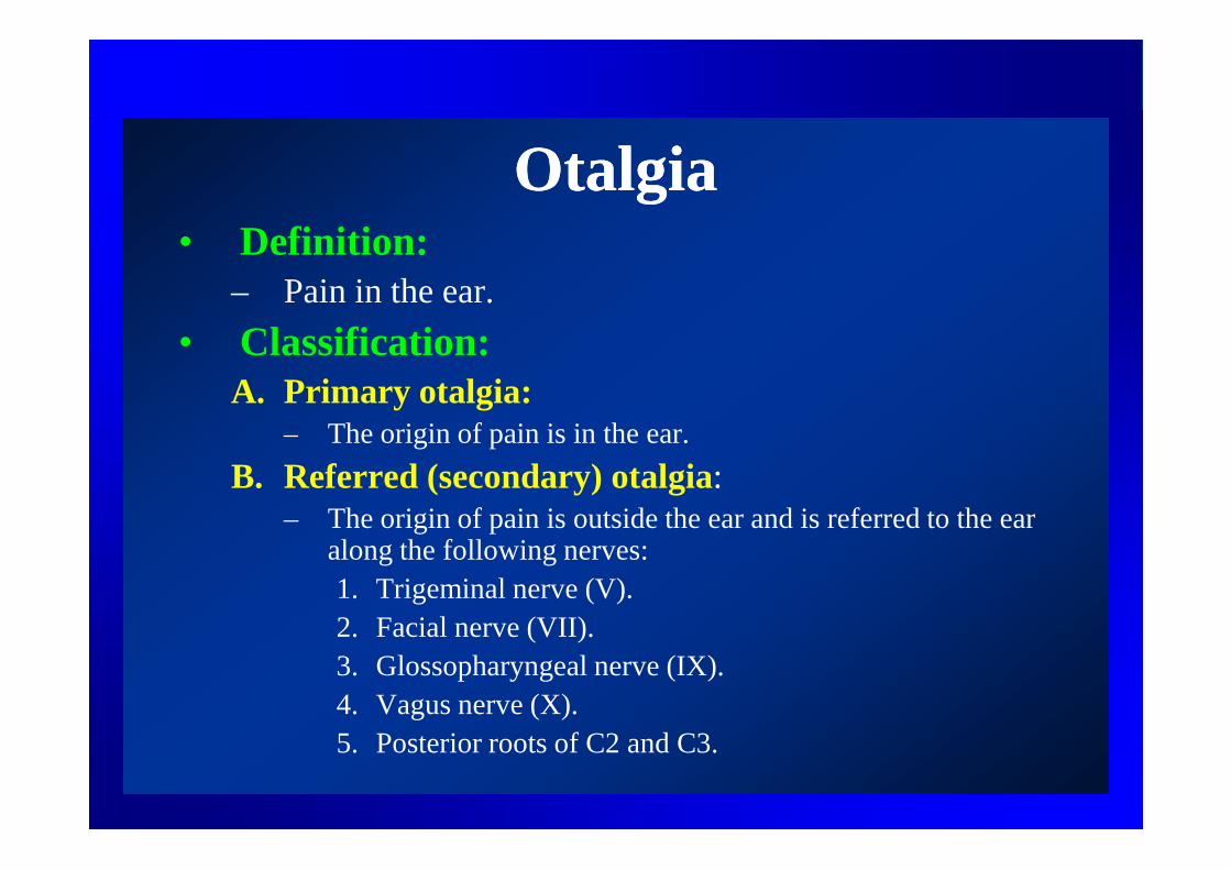
OtalgiaOtalgia• Definition:
– Pain in the ear.
• Classification:A. Primary otalgia:
– The origin of pain is in the ear.– The origin of pain is in the ear.
B. Referred (secondary) otalgia:– The origin of pain is outside the ear and is referred to the ear
along the following nerves:1. Trigeminal nerve (V).2. Facial nerve (VII).3. Glossopharyngeal nerve (IX).4. Vagus nerve (X).5. Posterior roots of C2 and C3.
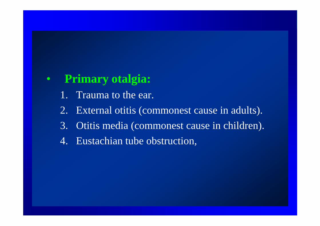
• Primary otalgia:1. Trauma to the ear.
2. External otitis (commonest cause in adults).2. External otitis (commonest cause in adults).
3. Otitis media (commonest cause in children).
4. Eustachian tube obstruction,
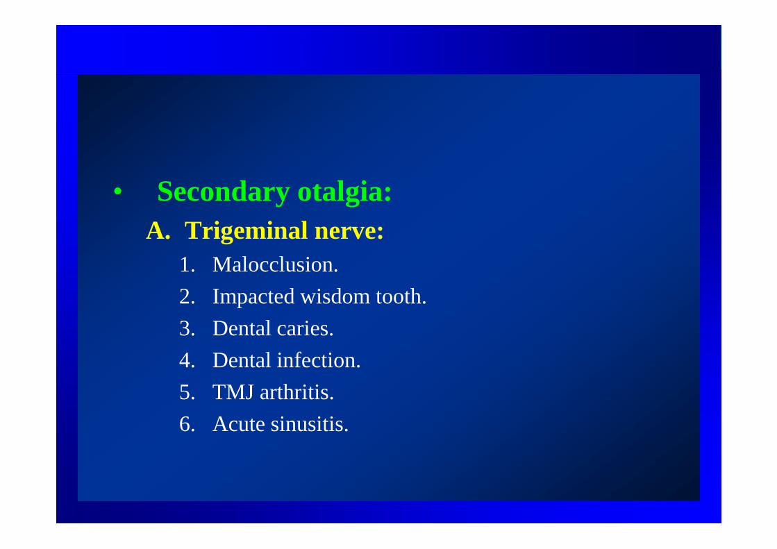
• Secondary otalgia:A. Trigeminal nerve:
1. Malocclusion.1. Malocclusion.
2. Impacted wisdom tooth.
3. Dental caries.
4. Dental infection.
5. TMJ arthritis.
6. Acute sinusitis.
B. Facial nerve:– Herpes zoster of the geniculate ganglion (Ramsay
Hunt syndrome).
C. Glossopharyngeal nerve:C. Glossopharyngeal nerve:1. Acute tonsillitis.
2. Peritonsillar abscess.
3. Glossopharyngeal neuralgia.
D. Vagus nerve (X):– Ulcers of the larynx e.g. tuberculosis.
E. C2 and C3:1. Cervical disc lesions.
2. Cervical osteoarthritis.
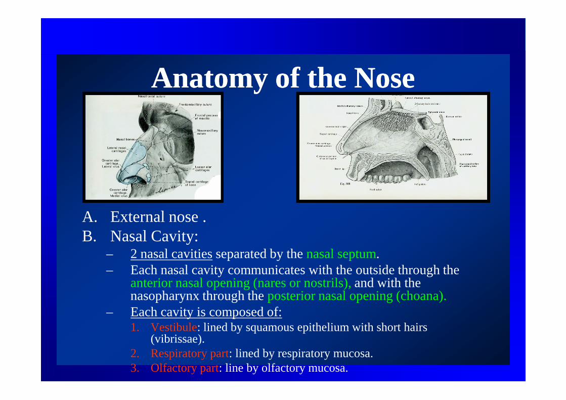
Anatomy of the NoseAnatomy of the Nose
A. External nose .B. Nasal Cavity:
– 2 nasal cavities separated by the nasal septum.– Each nasal cavity communicates with the outside through the
anterior nasal opening (nares or nostrils), and with the nasopharynx through the posterior nasal opening (choana).
– Each cavity is composed of:1. Vestibule: lined by squamous epithelium with short hairs
(vibrissae).2. Respiratory part: lined by respiratory mucosa.3. Olfactory part: line by olfactory mucosa.
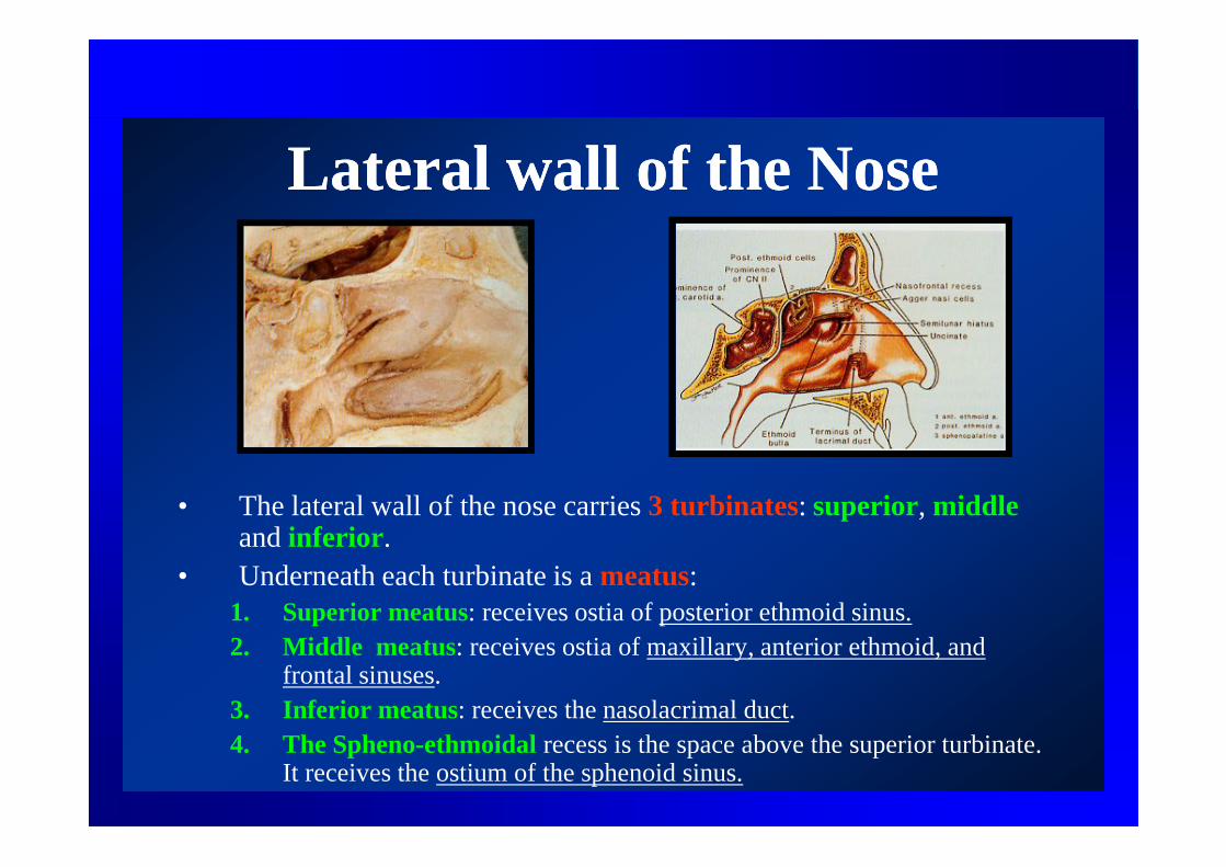
Lateral wall of the NoseLateral wall of the Nose
• The lateral wall of the nose carries 3 turbinates: superior, middleand inferior.
• Underneath each turbinate is a meatus:1. Superior meatus: receives ostia of posterior ethmoid sinus.2. Middle meatus: receives ostia of maxillary, anterior ethmoid, and
frontal sinuses.3. Inferior meatus: receives the nasolacrimal duct.4. The Spheno-ethmoidal recess is the space above the superior turbinate.
It receives the ostium of the sphenoid sinus.
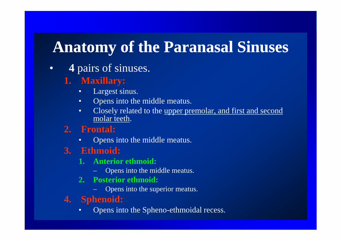
Anatomy of the Paranasal SinusesAnatomy of the Paranasal Sinuses• 4 pairs of sinuses.
1. Maxillary:• Largest sinus.• Opens into the middle meatus.• Closely related to the upper premolar, and first and second
molar teeth.molar teeth.2. Frontal:
• Opens into the middle meatus.3. Ethmoid:
1. Anterior ethmoid:– Opens into the middle meatus.
2. Posterior ethmoid:– Opens into the superior meatus.
4. Sphenoid:• Opens into the Spheno-ethmoidal recess.
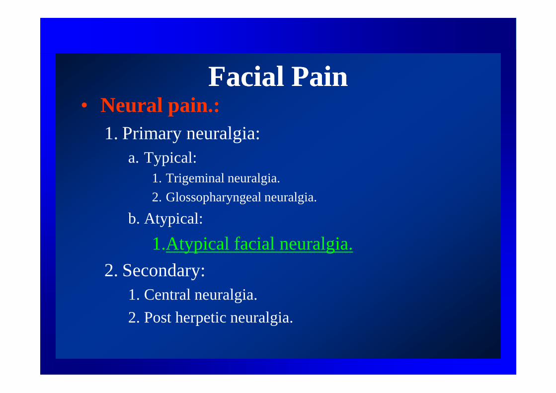
Facial PainFacial Pain• Neural pain.:
1. Primary neuralgia:a. Typical:
1. Trigeminal neuralgia.
2. Glossopharyngeal neuralgia.2. Glossopharyngeal neuralgia.
b. Atypical:
1.Atypical facial neuralgia.
2. Secondary:1. Central neuralgia.
2. Post herpetic neuralgia.
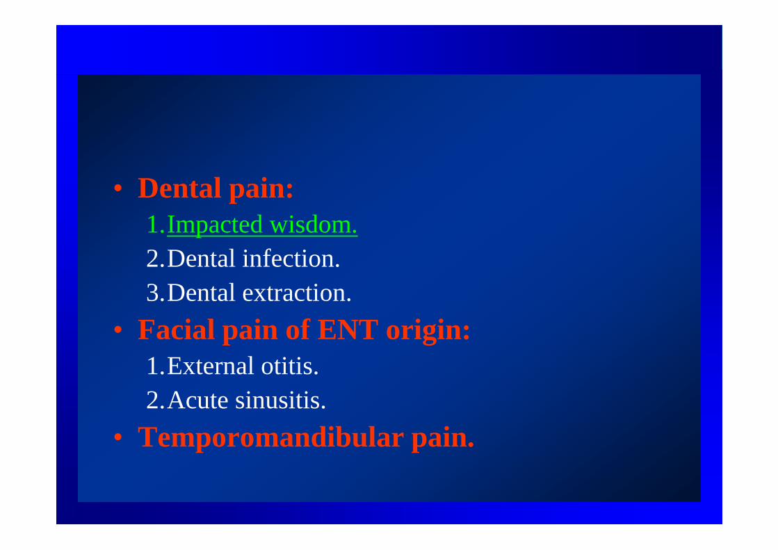
• Dental pain:1.Impacted wisdom.2.Dental infection.3.Dental extraction.
• Facial pain of ENT origin:1.External otitis.2.Acute sinusitis.
• Temporomandibular pain.
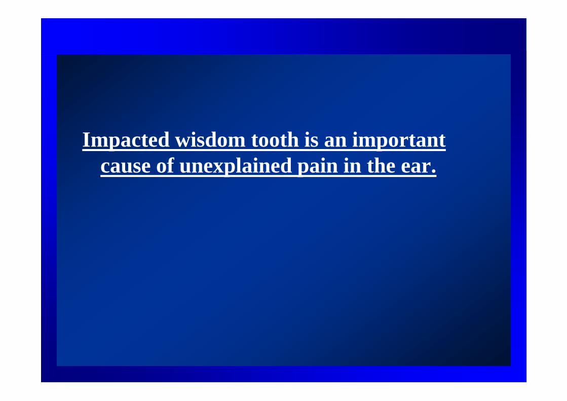
Impacted wisdom tooth is an important cause of unexplained pain in the ear.
• Facial pain of ENT origin:A. Otitis Externa:
• Inflammatory conditions of the external ear canal.• Inflammatory conditions of the external ear canal.
• The pain may be severe and throbbing. The characteristically increases with jaw movements and with pressure on the tragus.
B. Acute sinusitis:• Pain is a constant feature of acute sinusitis.
• The pain typically increases on straining, coughing, and bending down.bending down.
• Maxillary pain is over the cheek and may radiates to the upper teeth especially on bending and coughing.
• Ethmoid pain is between the eyes and over the bridge of the nose.
• Frontal pain is over the forehead and is usually associated with generalized headache. It commonly shows morning periodicity (vacuum effect).
• Sphenoid pain is usually deep seated behind the eyes, and is associated with occipital or vertical headache.
Atypical Facial NeuralgiaAtypical Facial Neuralgia
• An important cause of unnecessary dental extractions.
• Middle aged females.• Middle aged females.
• Frequently there is a psychological factor.
• Characterized by recurrent pain over the cheek and teeth.
• May be bilateral.
EpistaxisEpistaxis
• Bleeding from the nose.
• Dental causes:1. Traumatic dental extractions.1. Traumatic dental extractions.
2. Maxillofacial trauma.
3. Ora-antral fistula.
4. Dental maxillary sinusitis.
5. Tumors of dental origin.
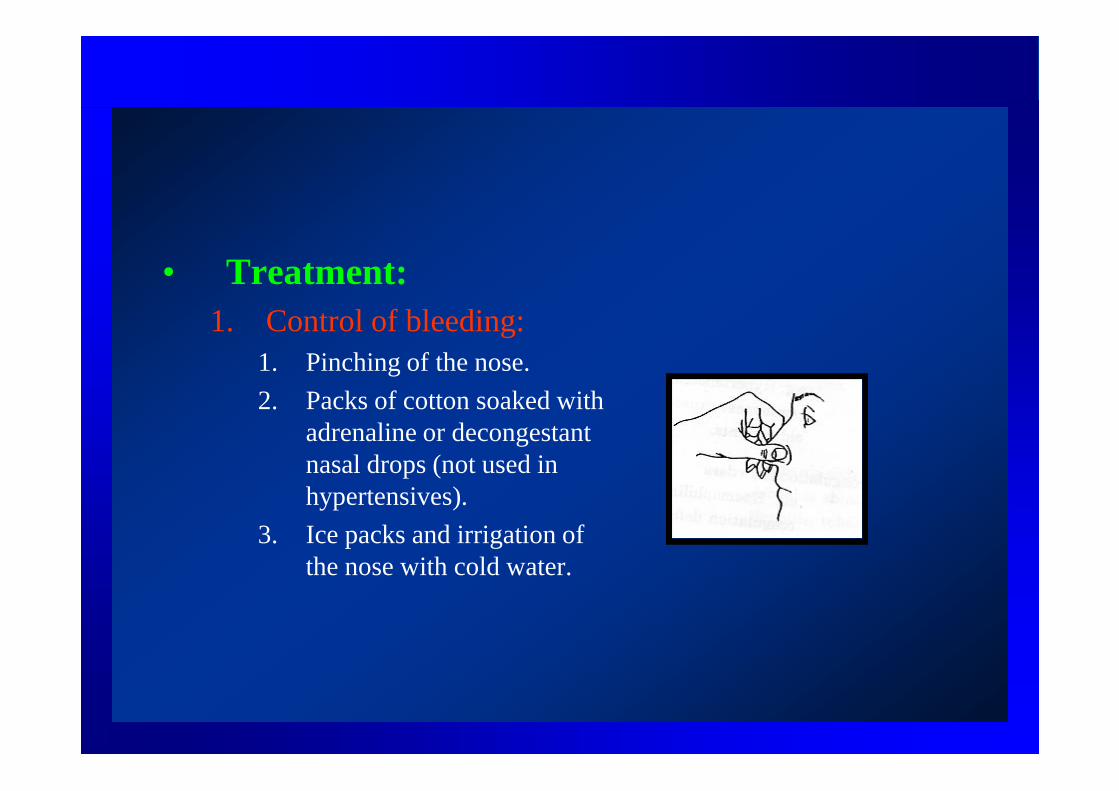
• Treatment:1. Control of bleeding:
1. Pinching of the nose.
2. Packs of cotton soaked with 2. Packs of cotton soaked with adrenaline or decongestant nasal drops (not used in hypertensives).
3. Ice packs and irrigation of the nose with cold water.
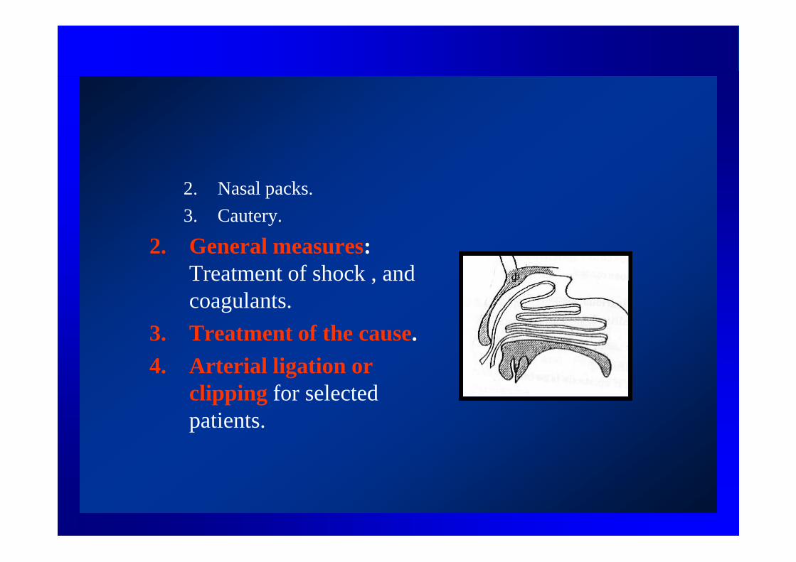
2. Nasal packs.
3. Cautery.
2. General measures:Treatment of shock , and Treatment of shock , and coagulants.
3. Treatment of the cause.
4. Arterial ligation or clipping for selected patients.
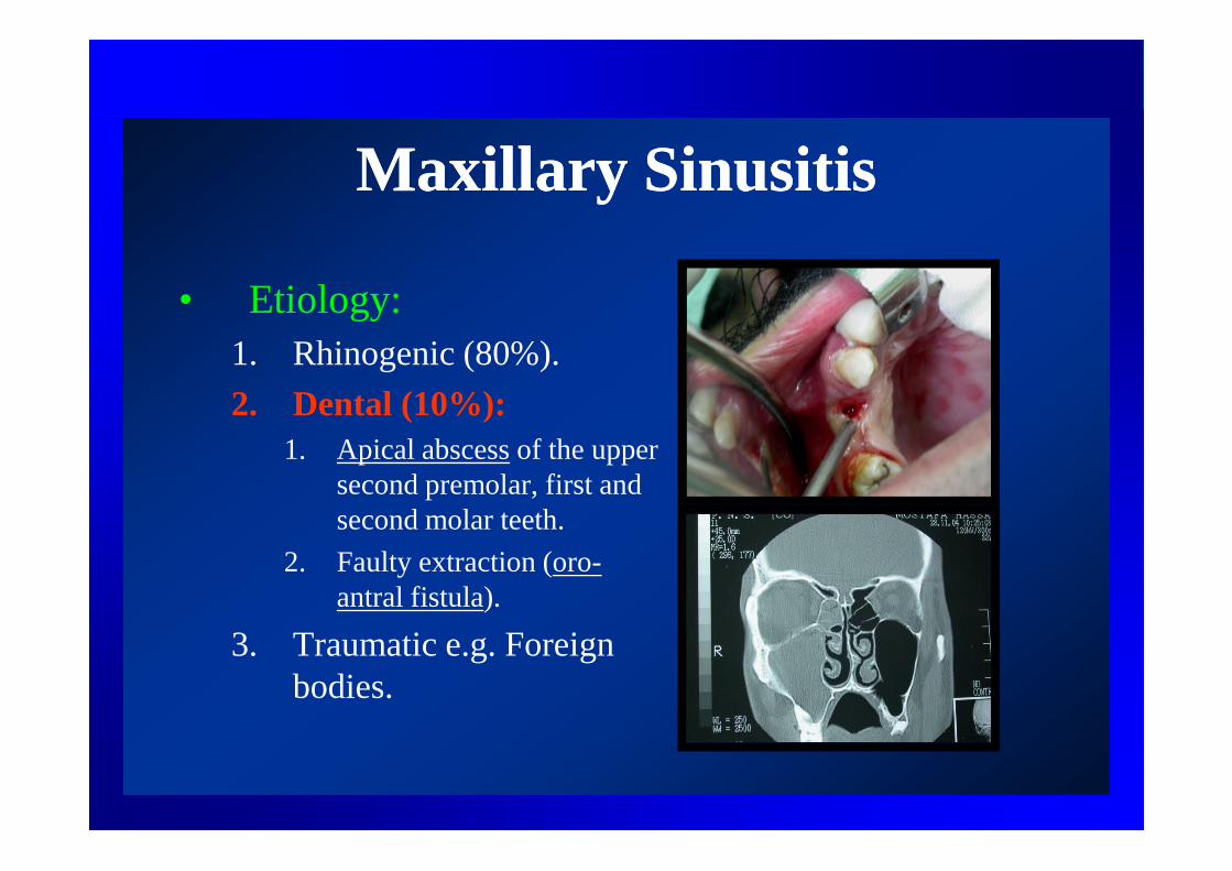
Maxillary SinusitisMaxillary Sinusitis
• Etiology:1. Rhinogenic (80%).
2. Dental (10%):1. Apical abscess of the upper 1. Apical abscess of the upper
second premolar, first and second molar teeth.
2. Faulty extraction (oro-antral fistula).
3. Traumatic e.g. Foreign bodies.
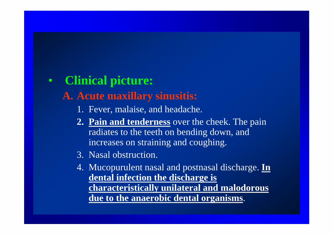
• Clinical picture:A. Acute maxillary sinusitis:
1. Fever, malaise, and headache.2. Pain and tenderness over the cheek. The pain 2. Pain and tenderness over the cheek. The pain
radiates to the teeth on bending down, and increases on straining and coughing.
3. Nasal obstruction.4. Mucopurulent nasal and postnasal discharge. In
dental infection the discharge is characteristically unilateral and malodorous due to the anaerobic dental organisms.
A. Chronic maxillary sinusitis:1. Nasal and postnasal mucopurulent discharge.
2. Nasal obstruction.
3. Sense of heaviness or recurrent pain over the 3. Sense of heaviness or recurrent pain over the cheek.
4. Headache.
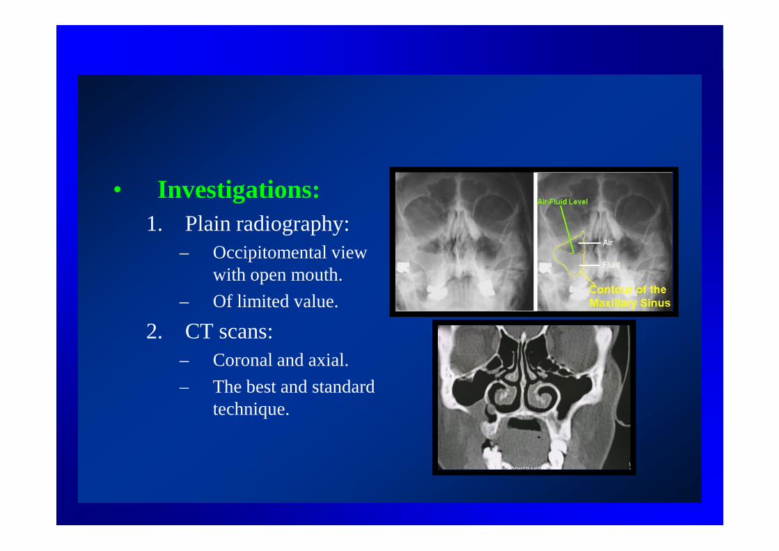
• Investigations:1. Plain radiography:
– Occipitomental view with open mouth.with open mouth.
– Of limited value.
2. CT scans:– Coronal and axial.
– The best and standard technique.
• Treatment:A. Acute sinusitis:
1. Antibiotics.2. Nasal decongestants (local and systemic).2. Nasal decongestants (local and systemic).3. Antipyretic analgesics.4. Steam inhalations
B. Chronic sinusitis:1. Medical treatment:
– Antibiotics, anti-allergics, nasal washes.
2. Surgical treatment:– Appropriate sinus procedure. Endoscopic sinus
procedures are now the standard procedures.
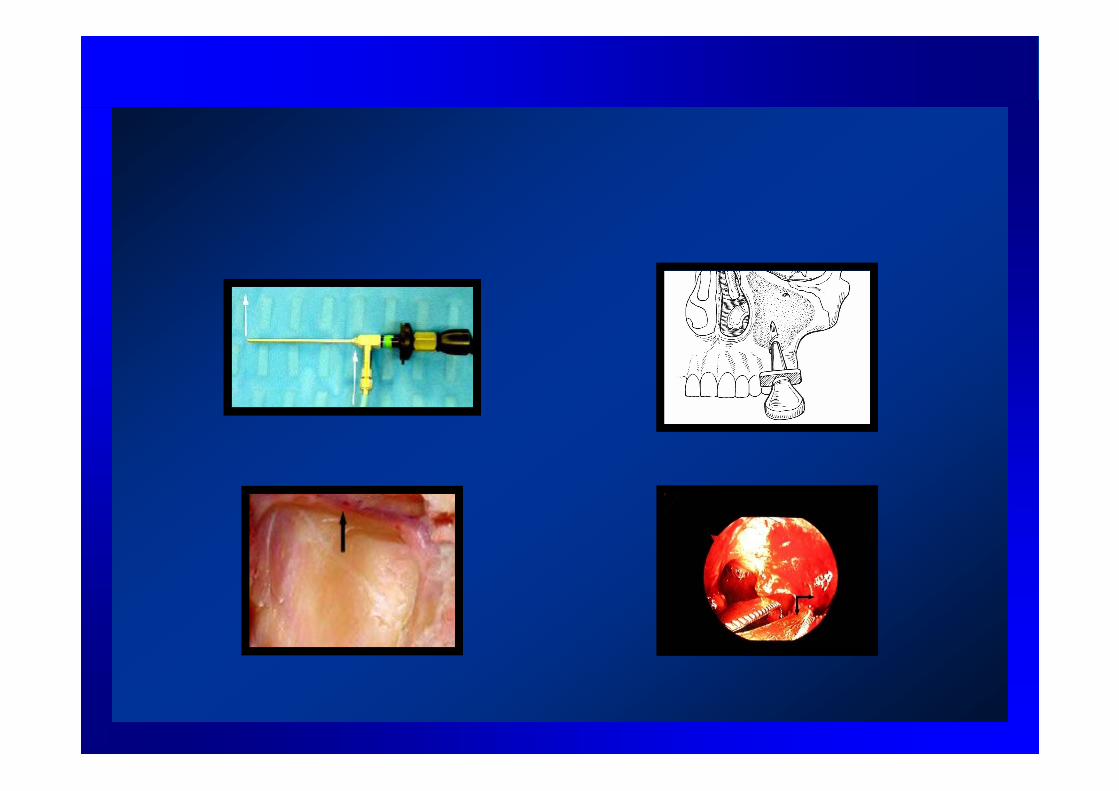
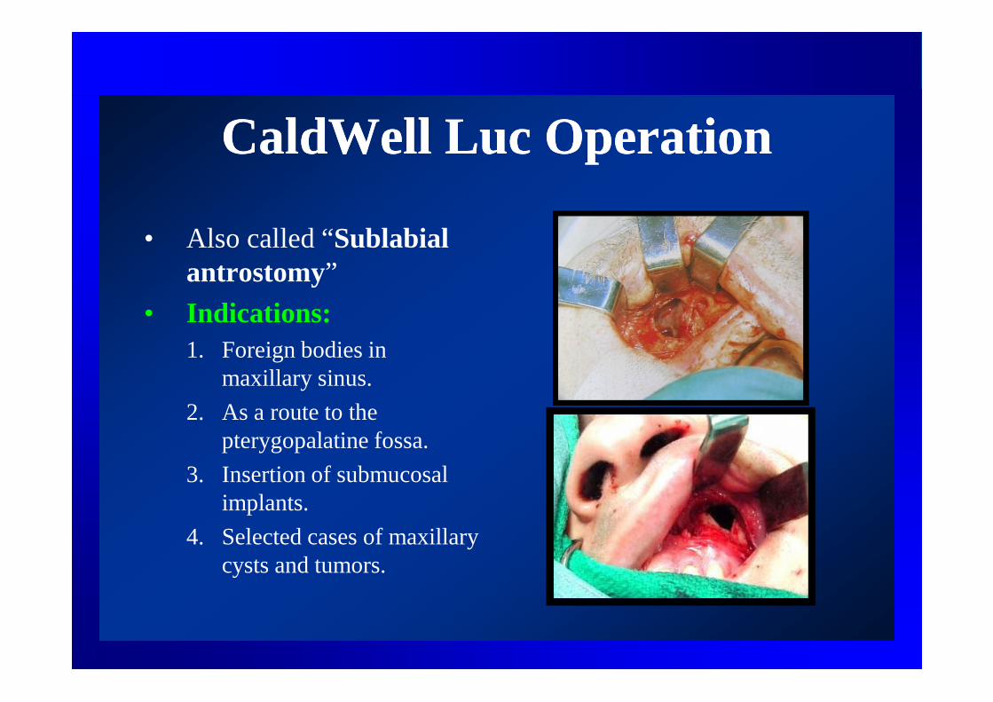
CaldWell Luc OperationCaldWell Luc Operation
• Also called “Sublabial antrostomy”
• Indications:1. Foreign bodies in 1. Foreign bodies in
maxillary sinus.
2. As a route to the pterygopalatine fossa.
3. Insertion of submucosal implants.
4. Selected cases of maxillary cysts and tumors.
• Complications:1. Sublabial oro-antral fistula.
2. Trauma to the root of the teeth or their nerve 2. Trauma to the root of the teeth or their nerve and blood supply (devitalization of teeth).
3. Infraorbital neuralgia ( trauma to the infraorbital nerve).
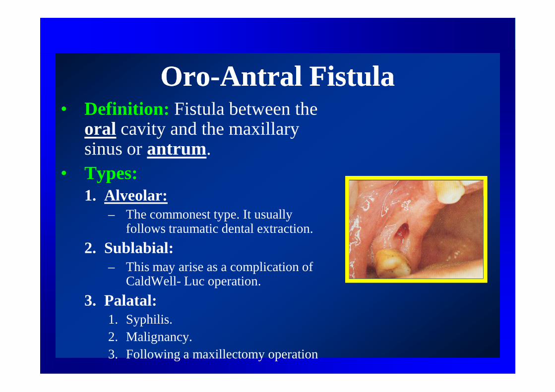
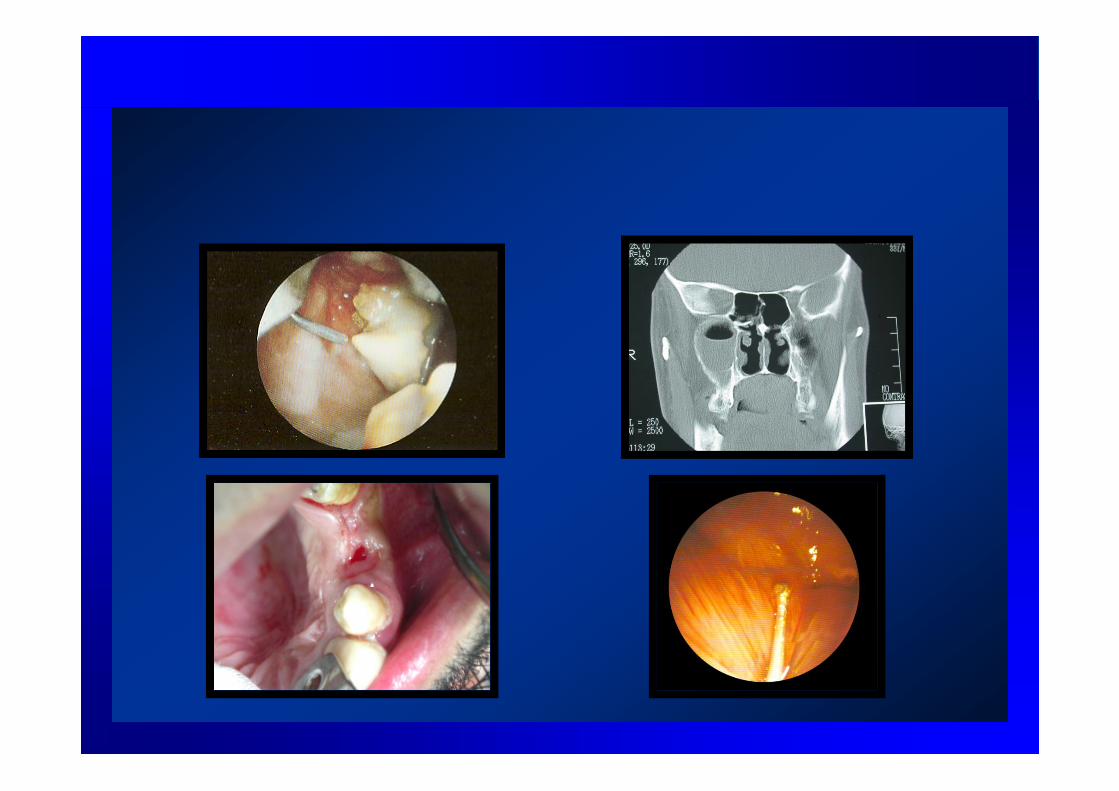
OroOro--Antral FistulaAntral Fistula• Definition: Fistula between the
oral cavity and the maxillary sinus or antrum.
• Types:1. Alveolar:1. Alveolar:
– The commonest type. It usually follows traumatic dental extraction.
2. Sublabial:– This may arise as a complication of
CaldWell- Luc operation.
3. Palatal:1. Syphilis.2. Malignancy.3. Following a maxillectomy operation
• Clinical picture of alveolar fistula:1. Mild epistaxis at the time of extraction.2. Escape of fluid or food from the nose.3. Escape of air from the fistula on blowing the nose.3. Escape of air from the fistula on blowing the nose.4. Unilateral nasal discharge with bad odor.5. Pain over the cheek.6. A probe may be passed through the fistula.
• Investigations:1. Plain X-Rays (occipitomental) +/- probe.2. CT scans
• Treatment:1. Recent fistula:
– Primary closure.
2. Old fistula:1. Small:
– Suturing after freshening of the edges of the fistula.– Suturing after freshening of the edges of the fistula.
2. Large:1. Clearing infection from the maxillary sinus by repeated
punctures or endoscopically.
2. Closure of the fistula by a buccal or palatal flap +/- bone graft:
– The palatal flap is thicker and has better blood supply, but is more traumatic.
– The buccal flap is easier, but is thin and may obliterate the buccogingival sulcus.
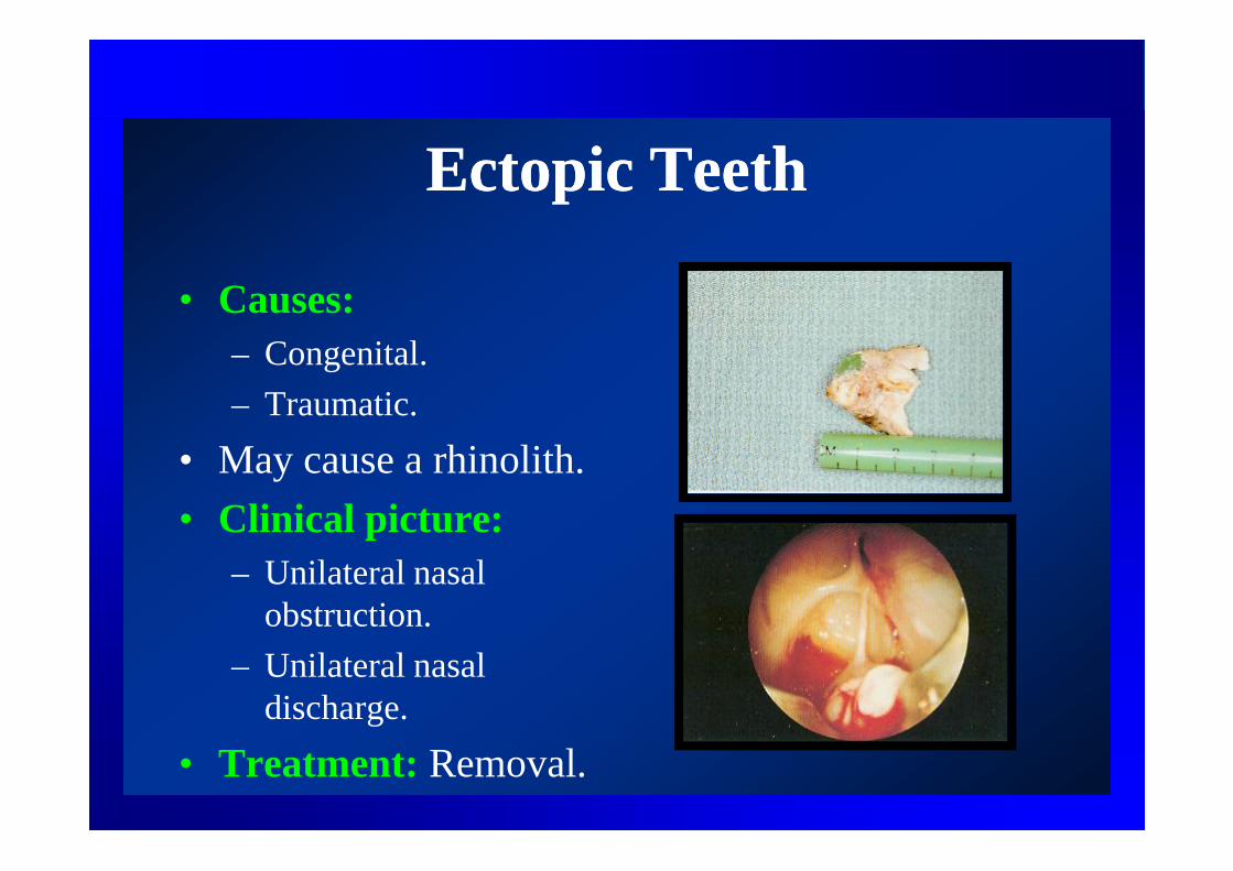
Ectopic TeethEctopic Teeth
• Causes:– Congenital.
– Traumatic.
• May cause a rhinolith.
• Clinical picture:– Unilateral nasal
obstruction.
– Unilateral nasal discharge.
• Treatment: Removal.
Cysts of the MaxillaCysts of the Maxilla1. Congenital:
1. Medial:1. Median alveolar:
– Between the upper central incisors.2. Median palatal:
– Between the palatine processes of the developing maxilla.– Between the palatine processes of the developing maxilla.3. Nasopalatine:
– Related to the incisive canal.2. Lateral:
1. Lateral alveolar.• Between the upper lateral incisor and canine.
2. Naso-alveolar:• In the lateral half of the floor of the nose.
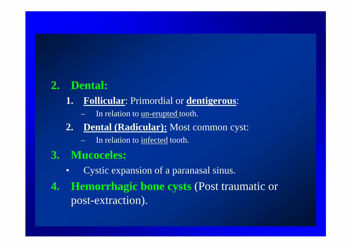
2. Dental:1. Follicular: Primordial or dentigerous:
– In relation to un-erupted tooth.
2. Dental (Radicular): Most common cyst:2. Dental (Radicular): Most common cyst:– In relation to infected tooth.
3. Mucoceles:• Cystic expansion of a paranasal sinus.
4. Hemorrhagic bone cysts (Post traumatic or post-extraction).
Benign tumorsBenign tumors
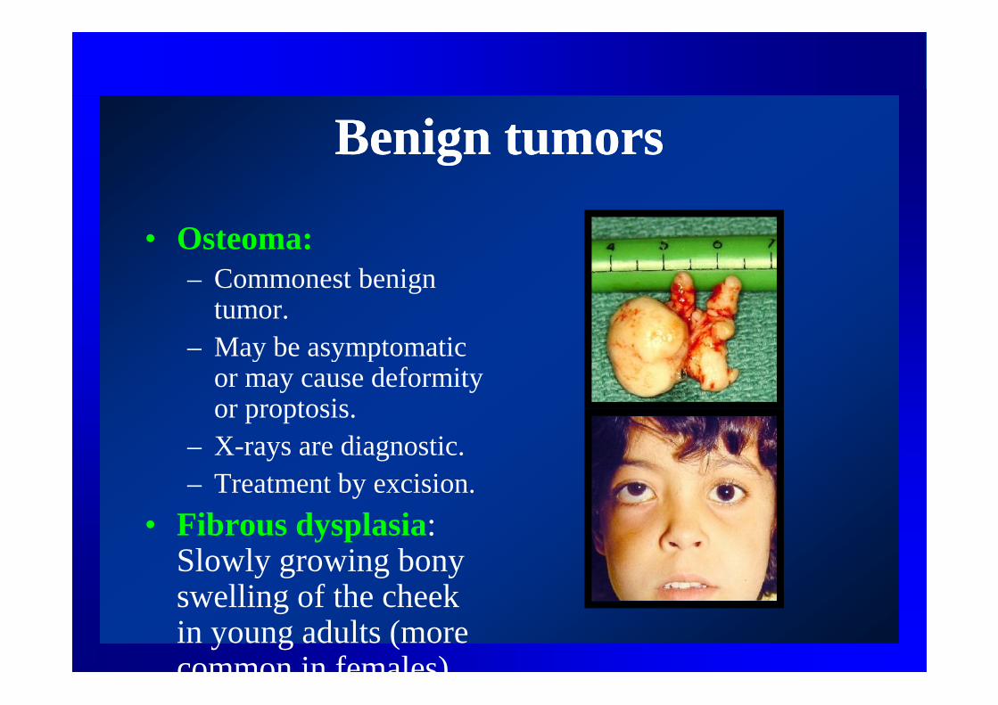
• Osteoma:– Commonest benign
tumor.– May be asymptomatic – May be asymptomatic
or may cause deformity or proptosis.
– X-rays are diagnostic.– Treatment by excision.
• Fibrous dysplasia: Slowly growing bony swelling of the cheek in young adults (more common in females).
Locally Malignant tumorsLocally Malignant tumors
• Osteoclastoma:– Young patients.– Most common in maxilla.– Reddish fleshy mass expanding the maxilla.– False capsule.– May be eggshell crackling.– X-ray shows soap-bubble appearance (do not
fill with dye).– Treatment by excision.
Locally Malignant tumorsLocally Malignant tumors
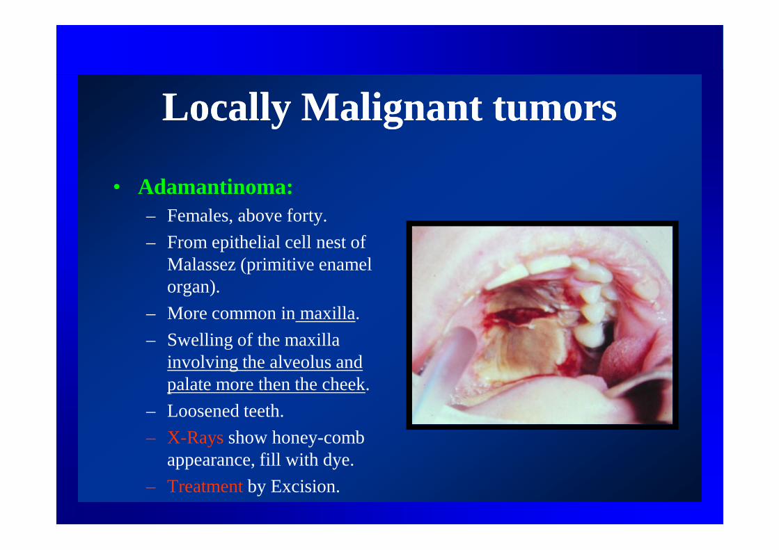
• Adamantinoma:– Females, above forty.
– From epithelial cell nest of Malassez (primitive enamel organ).
– More common in maxilla.
– Swelling of the maxilla involving the alveolus and palate more then the cheek.
– Loosened teeth.
– X-Rays show honey-comb appearance, fill with dye.
– Treatment by Excision.
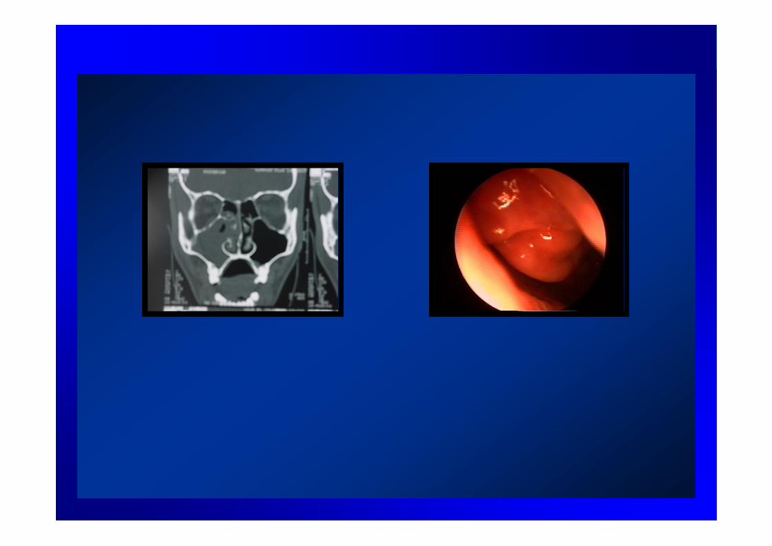
Maxillary CarcinomaMaxillary Carcinoma• Most common malignant tumor.• Males.• Squamous cell carcinoma.• Maxillary sinus is one of the sites of occult primaries in head
and neck.and neck.• Clinical picture:
– Unilateral nasal obstruction, discharge, and epistaxis.– Swelling of the face.– Proptosis.– Loosened teeth.
• CT scans and MRI are important.• Treatment: combined surgical excision, radiotherapy, and
may be chemotherapy.
PharynxPharynx
HalitosisHalitosis• Offensive breath.• Causes:
– Oral:• Caries.• Caries.• Dental infections.• Ulcers.• Poor oral hygiene.
Halitosis (Cont’)Halitosis (Cont’)
– Extra-oral:• Sinuses:
– Dental maxillary sinusitis.– Chronic sinusitis.– Chronic sinusitis.
• Tonsils: – Chronic tonsillitis.
• GIT:– Dyspepsia and maldigestion.– GERD.– Colonic problems: diarrhea, constipation.
• Tracheo-bronchial tree: Bronchiectasis.
TrismusTrismus
• Limitation of Jaw opening.• Causes:
1. Dental infections.1. Dental infections.2. Impacted wisdom.3. Suppuration around the pharynx (
peritonsillar and parapharyngeal).4. External otitis.5. Tumors.6. Tetanus.
AdenoidsAdenoids
• 3-7 years.
• Bilateral nasal obstruction causing adenoid facies:1. Open mouth and thick dry lips.2. Hitched-up upper lips.2. Hitched-up upper lips.3. Protruding incisors , faulty bite, caries.4. Receding chin.5. High arched palate.6. Flat nasolabial folds (expressionless face).7. Mucoid or mucopurulent nasal discharge.8. Inactive ala nasi.
• Investigations: X-Rays lateral skull view.• Treatment: Adenoidectomy.
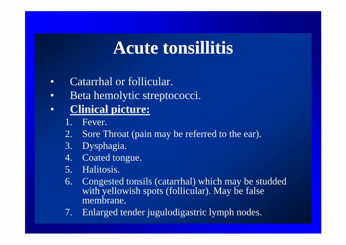
Acute tonsillitisAcute tonsillitis
• Catarrhal or follicular.• Beta hemolytic streptococci.• Clinical picture:
1. Fever.1. Fever.2. Sore Throat (pain may be referred to the ear).3. Dysphagia.4. Coated tongue.5. Halitosis.6. Congested tonsils (catarrhal) which may be studded
with yellowish spots (follicular). May be false membrane.
7. Enlarged tender jugulodigastric lymph nodes.
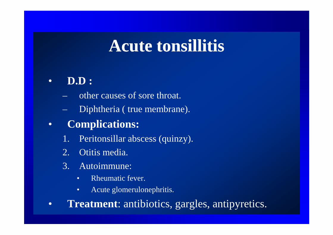
Acute tonsillitisAcute tonsillitis
• D.D : – other causes of sore throat.
– Diphtheria ( true membrane).
• Complications:1. Peritonsillar abscess (quinzy).
2. Otitis media.
3. Autoimmune:• Rheumatic fever.
• Acute glomerulonephritis.
• Treatment: antibiotics, gargles, antipyretics.
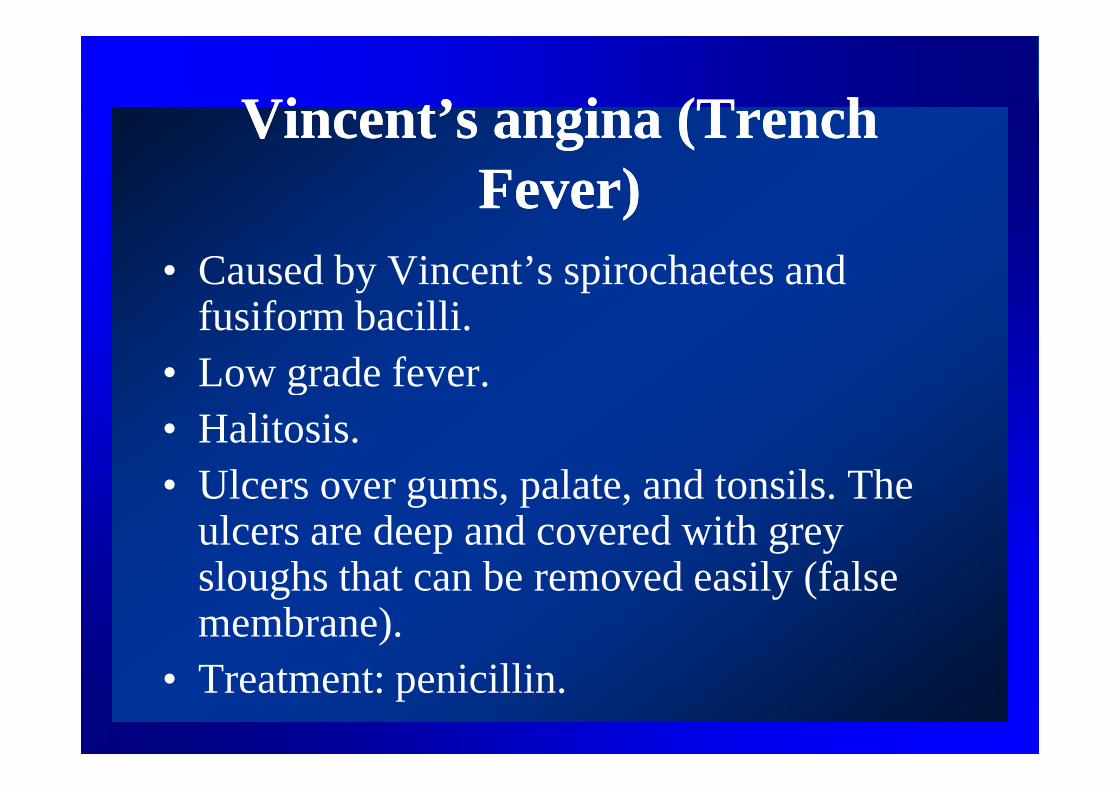
Vincent’s angina (Trench Vincent’s angina (Trench Fever)Fever)
• Caused by Vincent’s spirochaetes and fusiform bacilli.
• Low grade fever.• Low grade fever.• Halitosis.• Ulcers over gums, palate, and tonsils. The
ulcers are deep and covered with grey sloughs that can be removed easily (false membrane).
• Treatment: penicillin.
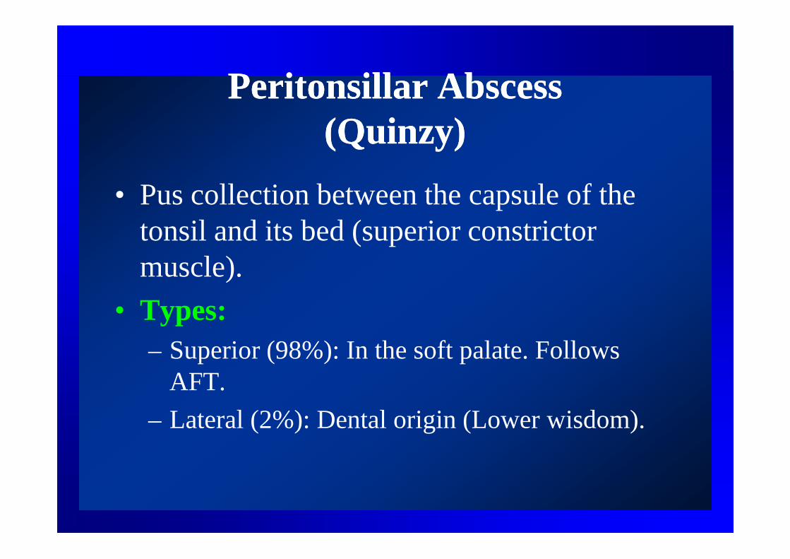
Peritonsillar AbscessPeritonsillar Abscess(Quinzy)(Quinzy)
• Pus collection between the capsule of the tonsil and its bed (superior constrictor muscle).muscle).
• Types:– Superior (98%): In the soft palate. Follows
AFT.
– Lateral (2%): Dental origin (Lower wisdom).
• Clinical picture:– Symptoms:
1. Throat pain ( more severe on one side).2. Dysphagia.3. Halitosis.4. Trismus.5. Otalgia.6. Fever.
• Signs:1. Tonsils are congested and pushed medially.2. Soft swelling above and lateral to the tonsils.3. Edematous uvula.4. Torticollis.5. Enlarged tender JD lymph nodes.6. Coated tongue.
• Complications:1. Parapharyngeal abscess.2. Laryngeal edema and stridor.3. Septicemia.
• Treatment:– Pre-suppurative stage (cellulitis): antibiotics,
gargles.– Suppurative: Drainage, antibiotics , +/-
tonsillectomy after one month.
Parapharyngeal abscessParapharyngeal abscess• Suppuration in the parapharyngeal space.
• Etiology:1. Dental infections.
2. Quinzy.2. Quinzy.
3. Trauma.
• Clinical picture:• Symptoms:
1. Throat pain.2. Neck pain.2. Neck pain.3. Dysphagia.4. Fever and malaise.
• Signs:1. The tonsil and pharyngeal wall are pushed medially.2. Tender soft neck swelling.3. Trismus.4. Torticollis.
• Complications:1. Laryngeal edema.
2. Spread of infection to other neck spaces and 2. Spread of infection to other neck spaces and mediastinum.
3. Jugular vein thrombosis.
• Treatment:1. External drainage.
2. Antibiotics.
Retropharyngeal AbscessRetropharyngeal Abscess• Acute:
– Infants and children.– Clinical picture:
1. Difficult feeding and breathing.2. Chocking.3. Croupy cough.3. Croupy cough.4. Cystic hyperemic swelling to one side of the midline.
• X-rays: Widened retropharyngeal space.• Complications:
1. Stridor.2. Rupture.
• Treatment:1. Drainage (Trendlenburg position).2. Antibiotics.
• Chronic:– Adults.
– Cervical T.B. infection (Pott’s disease).
– Clinical picture:– Clinical picture:1. Dysphagia.
2. Chocking.
3. Painful cervical spine.
4. Soft cystic swelling in the midline.
• X-Rays is diagnostic.
• Treatment: External drainage , anti TB drugs.
Ludwig’s AnginaLudwig’s Angina
• Cellulitis of the floor of the mouth and the submandibular space.• Etiology:
1. Dental infections.2. Trauma.
• Bacteriology: Anaerobic organisms, Staph.• Clinical picture:• Clinical picture:
1. Fever.2. Dysphagia.3. May be stridor.4. Firm tender swelling in the floor of mouth and submandibular region.5. Tongue is edematous and is pushed upwardsand backwards.
• Complications: suffocation.• Treatment:
1. Drainage.2. Antibiotics.3. May be tracheostomy.
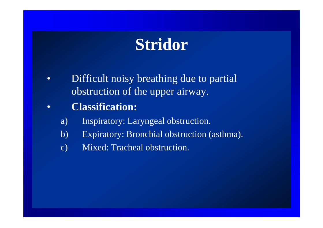
StridorStridor
• Difficult noisy breathing due to partial obstruction of the upper airway.
• Classification:a) Inspiratory: Laryngeal obstruction.
b) Expiratory: Bronchial obstruction (asthma).
c) Mixed: Tracheal obstruction.
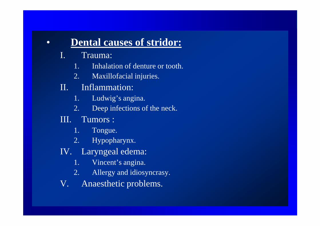
• Dental causes of stridor:I. Trauma:
1. Inhalation of denture or tooth.2. Maxillofacial injuries.
II. Inflammation:1. Ludwig’s angina.2. Deep infections of the neck.
III. Tumors :1. Tongue.2. Hypopharynx.
IV. Laryngeal edema:1. Vincent’s angina.2. Allergy and idiosyncrasy.
V. Anaesthetic problems.
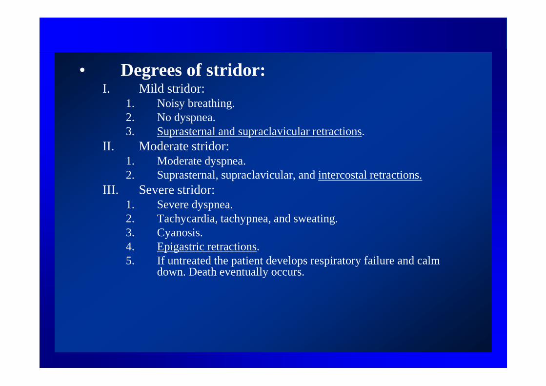
• Degrees of stridor:I. Mild stridor:
1. Noisy breathing.2. No dyspnea.3. Suprasternal and supraclavicular retractions.
II. Moderate stridor:1. Moderate dyspnea.2. Suprasternal, supraclavicular, and intercostal retractions.
III. Severe stridor:III. Severe stridor:1. Severe dyspnea.2. Tachycardia, tachypnea, and sweating.3. Cyanosis.4. Epigastric retractions.5. If untreated the patient develops respiratory failure and calm
down. Death eventually occurs.
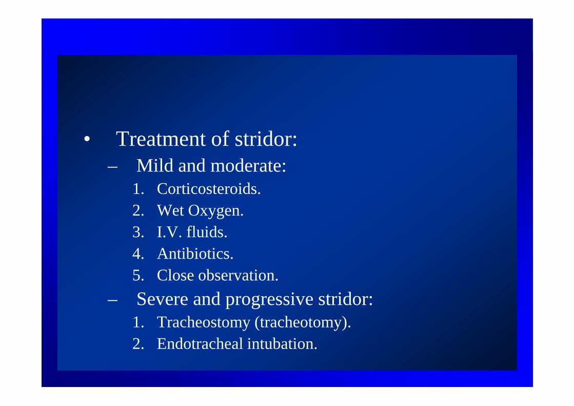
• Treatment of stridor:– Mild and moderate:
1. Corticosteroids.2. Wet Oxygen.2. Wet Oxygen.3. I.V. fluids.4. Antibiotics.5. Close observation.
– Severe and progressive stridor:1. Tracheostomy (tracheotomy).2. Endotracheal intubation.
Foreign BodiesForeign Bodies• May lodge in the subglottic region (may be fatal) or right
main bronchus (most common).• Clinical picture:
1. Stridor.2. Cough.3. Blood stained expectoration3. Blood stained expectoration4. Localized wheezes.
• Investigations:1. X-rays.2. Endoscopy.
• Treatment:1. Bronchoscopy and removal.2. May be tracheostomy.3. May be thoracotomy.
Tracheostomy (Tracheotomy)Tracheostomy (Tracheotomy)
• Laryngotomy: opening in the cricopthyroid membrane.
• Tracheostomy: Opening in the trachea.• Tracheostomy: Opening in the trachea.
• Indications:1. Severe stridor.
2. Progressive stridor.
3. Non-obstructive indications as respiratory failure and secretory obstruction.
• Levels:I. High : 1st and 2nd tracheal rings.II. Middle: 3rd and 4th tracheal rings .III. Low: 5th and 6th tracheal rings.III. Low: 5th and 6th tracheal rings.
• Complications:A. During operation:
1. Hemorrhage.2. Apnea.3. Cardiac arrest.4. TE fistula.5. Pneumothorax.
B. Early postoperative:1. Surgical emphysema.2. Obstructed tube.3. Dislodged tube.3. Dislodged tube.4. Wound infection.5. Chest infection.6. Atelectasis.
C. Late postoperative:1. Tracheal stenosis.
2. Rupture of innominate vessels.
3. Failed decannulation.3. Failed decannulation.
4. TE fistula.
5. Contracted neck scar.