Embed Size (px)
Citation preview
Otitis & PharyngitisPhysical Assessment for the Certified School
Nurse
Jackie Zolotsky, RN, MSN, CRNP, CSNParkland School District
August 12, 2015
Goals1. Be able to identify the anatomy of the
ear and pharynx and their purpose.2. Perform a basic Otoscopic exam.3. Recognize the signs and symptoms of
Otitis Externa and Otitis Media.4. Recognize the signs and symptoms of
Pharyngitis.5. Apply this knowledge to clinical
practice.
Overview of Topics
• Anatomy of the Outer, Middle and Inner Ear• How we hear• How to perform an Otoscopic Exam• What to expect during a “normal” Otoscopic Exam• Otitis Externa – Definition, Causes, Signs and
Symptoms, Physical Examination, Otoscopic Exam, and Examples
• Otitis Media – Definition, Causes, Signs and Symptoms, Physical Examination, Otoscopic Exam and Examples
• Differences between Children and Adults when it comes to Ear Infections
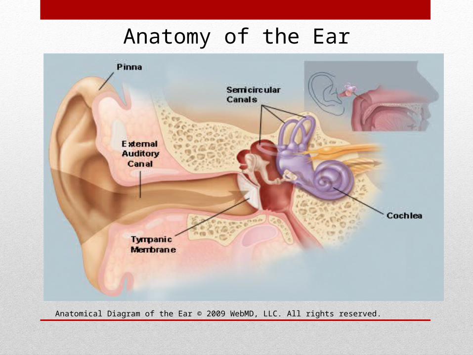
Anatomical Diagram of the Ear © 2009 WebMD, LLC. All rights reserved.
Anatomy of the Ear
• The ear has 3 main parts - Outer, Middle and Inner portions
OUTER EAR • The Pinna – made of cartilage and covered with skin.
• It includes everything we see on the outside—the curved flap of the ear leading down to the earlobe—but it also includes the ear canal, which begins at the opening to the ear and extends to the tympanic membrane.
• The tympanic membrane separates the outer ear from the middle ear.
Anatomy of the Ear

The Outer Ear
http://www.nidcd.nih.gov/health/hearing/pages/earinfections.aspx
Anatomy of the Middle Ear
• It is located between the eardrum and the inner ear.
• Within the middle ear are three tiny bones called the malleus, incus, and stapes.
• These bones transmit sound vibrations from the eardrum to the inner ear.
• The bones of the middle ear are surrounded by air.

The Middle Ear
http://www.nidcd.nih.gov/health/hearing/pages/earinfections.aspx
Anatomy of the Inner Ear
• Contains the fluid-filled semicircular canals (labyrinth) which attach to the cochlea and nerves in the inner ear. They send information on balance and head position to the brain.
• The cochlea, a part of the labyrinth, is a snail-shaped organ that converts sound vibrations from the middle ear into electrical signals.
• The auditory nerve carries these signals from the cochlea to the brain.
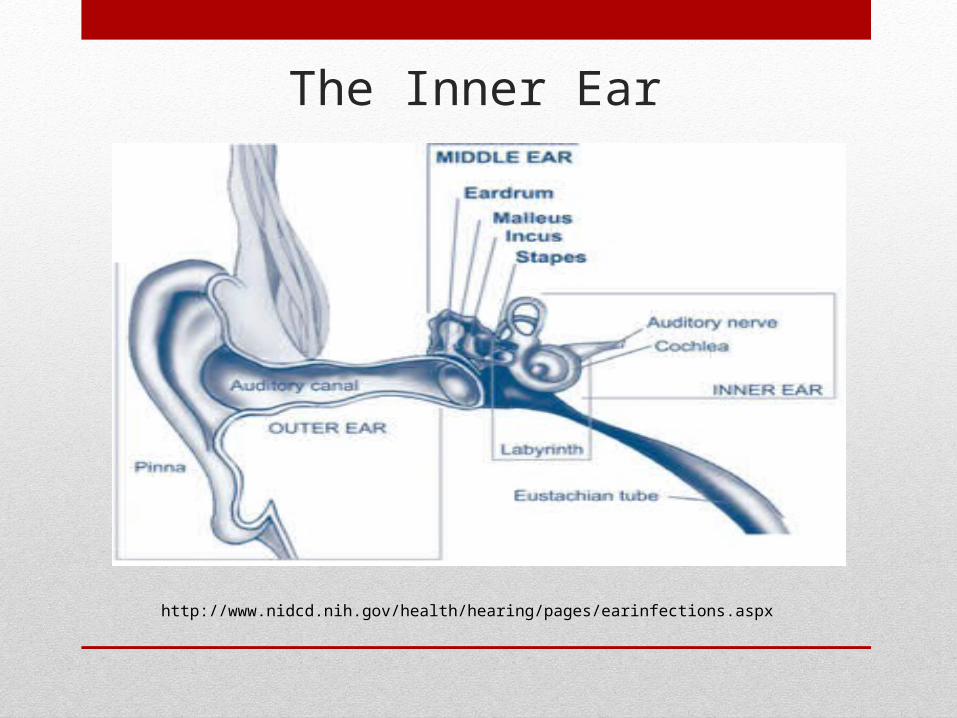
The Inner Ear
http://www.nidcd.nih.gov/health/hearing/pages/earinfections.aspx
Other Ear Anatomy
• The eustachian tube is a small passageway that connects the upper part of the throat to the middle ear.
• The eustachian tube’s job is to supply fresh air to the middle ear. It also drains fluid from the middle ear into the pharynx behind the nose. It also keeps air pressure at a steady level between the nose and the ear.
• Adenoids are small pads of tissue located behind the back of the nose, above the throat, and near the eustachian tubes. Adenoids are mostly made up of immune system cells. They fight off infection by trapping bacteria that enter through the mouth.
How do we Hear?
• Sound funnels through pinna into the external auditory canal.
• Sound causes the tympanic membrane and the tiny attached bones in the middle portion of the ear to vibrate. The vibrations are conducted to the cochlea.
• The cochlea transforms sound into nerve impulses that travel to the brain.
• When an infection is introduced to various parts of the ear, it affects a person’s ability to hear correctly.
How to perform an Otoscopic Exam
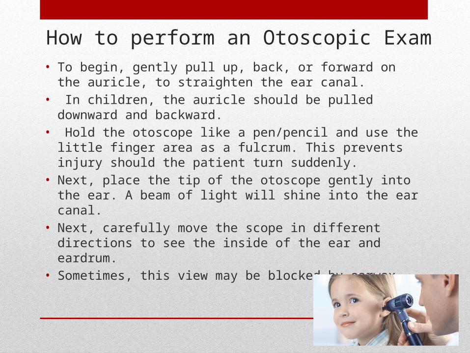
• To begin, gently pull up, back, or forward on the auricle, to straighten the ear canal.
• In children, the auricle should be pulled downward and backward.
• Hold the otoscope like a pen/pencil and use the little finger area as a fulcrum. This prevents injury should the patient turn suddenly.
• Next, place the tip of the otoscope gently into the ear. A beam of light will shine into the ear canal.
• Next, carefully move the scope in different directions to see the inside of the ear and eardrum.
• Sometimes, this view may be blocked by earwax.
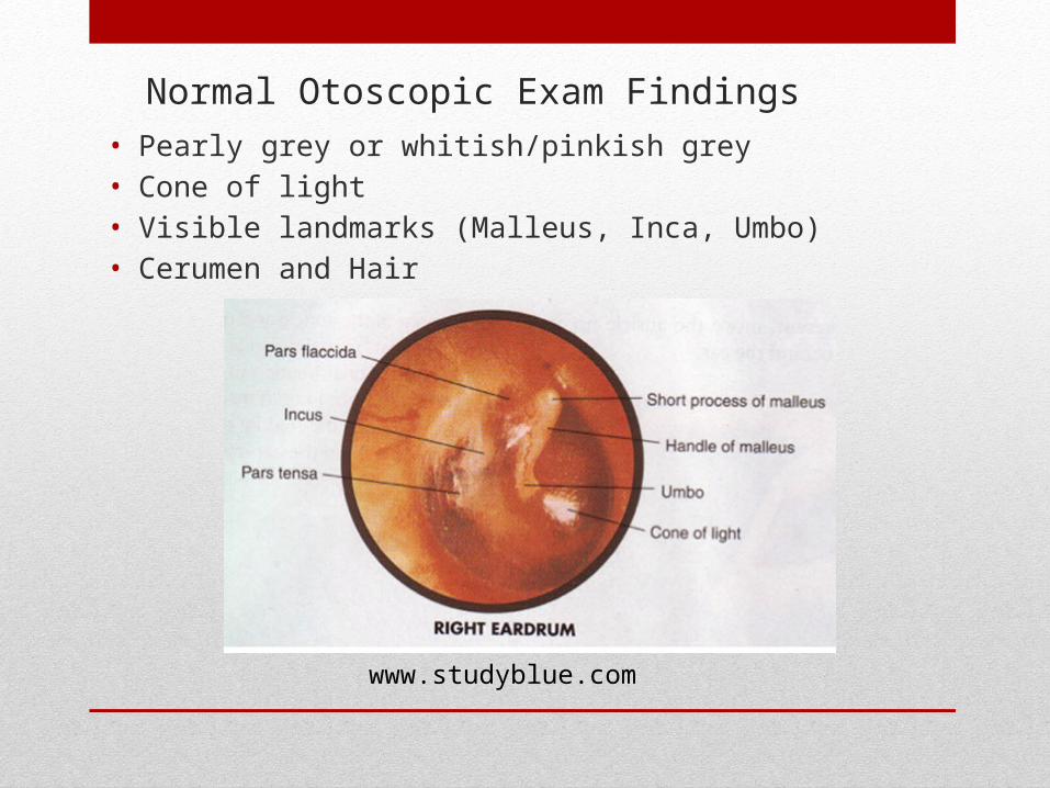
Normal Otoscopic Exam Findings• Pearly grey or whitish/pinkish grey• Cone of light• Visible landmarks (Malleus, Inca, Umbo)• Cerumen and Hair
www.studyblue.com
Otitis Externa
• Otitis externa is an inflammation or infection of the external auditory canal, the auricle, or both.
• This condition can be found in all age groups.
• Commonly seen in swimmers.
Otitis Externa - Causes
• History of exposure to or activities in water (e.g., swimming, surfing, kayaking)
• History of ear trauma (e.g., forceful ear cleaning, use of cotton swabs, or water in the ear canal)
• Viral, Bacterial and Fungal causes
Otitis Externa – Signs and Symptoms
• Ear pain (OTALGIA) - Ranges from mild to severe, progressing over 1-2 days
• Hearing loss• Ear fullness or pressure• Erythema, edema, and narrowing of the ear canal• Tinnitus• Fever (occasionally)• Itching • Severe deep pain - Immunocompromised patients may
have necrotizing (malignant) OE• Discharge - Initially, clear; quickly becomes purulent
and foul-smelling• Cellulitis of the face or neck or lymphadenopathy of
the neck (occasionally)• Bilateral symptoms (rare)
Otitis Externa – Physical Assessment
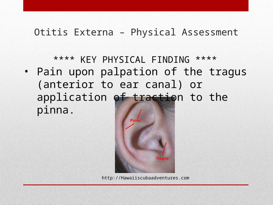
**** KEY PHYSICAL FINDING **** • Pain upon palpation of the tragus
(anterior to ear canal) or application of traction to the pinna.
http://Hawaiiscubaadventures.com
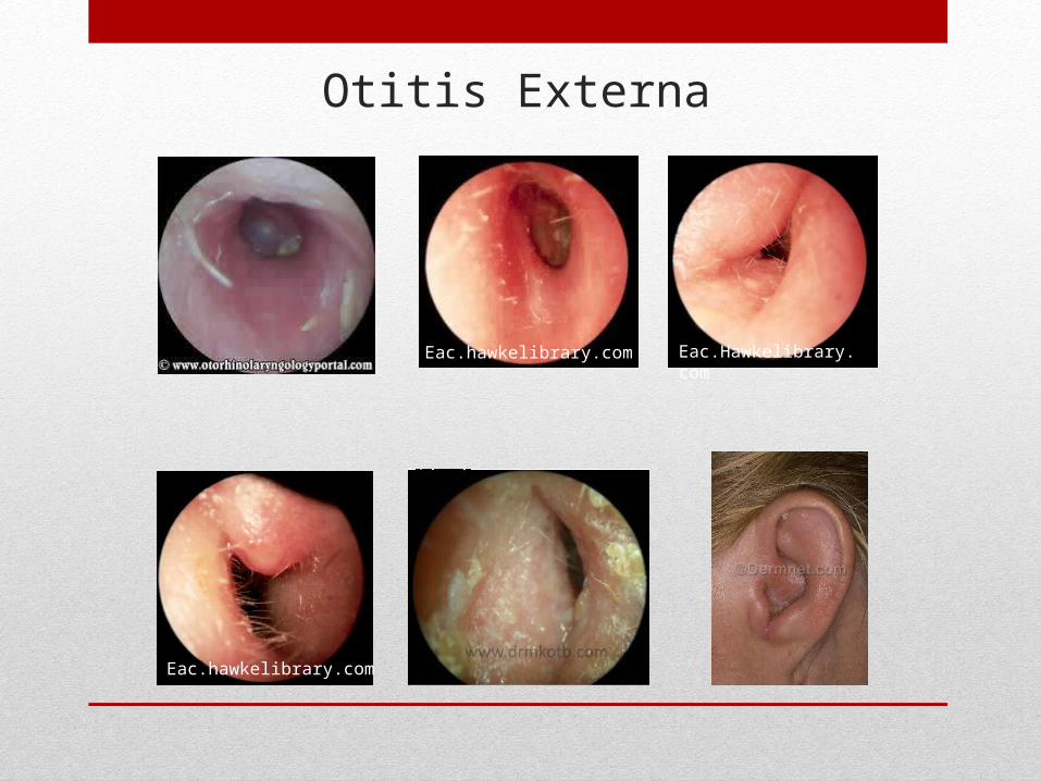
Otitis Externa
Eac.hawkelibrary.com Eac.Hawkelibrary.com
Eac.hawkelibrary.com
Acute Otitis Media• Acute Otitis Media is an inflammation of
the middle ear.
• Children get ear infections more often than adults.
• Five out of six children will have at least one ear infection by their third birthday.
• Ear infections are the most common reason parents bring their child to a doctor.
Causes of Acute Otitis Media
• Usually caused by bacteria and often begins after a child has a sore throat, cold, or other upper respiratory infection.
• If the upper respiratory infection is bacterial, these same bacteria may spread to the middle ear.
• If the upper respiratory infection is caused by a virus, such as a cold, bacteria may be drawn to the microbe-friendly environment and move into the middle ear as a secondary infection.
• Because of the infection, fluid builds up behind the tympanic membrane – causing swelling and pain.
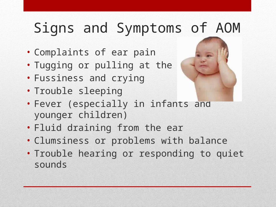
Signs and Symptoms of AOM
• Complaints of ear pain• Tugging or pulling at the ear(s)• Fussiness and crying• Trouble sleeping• Fever (especially in infants and younger
children)• Fluid draining from the ear• Clumsiness or problems with balance• Trouble hearing or responding to quiet sounds
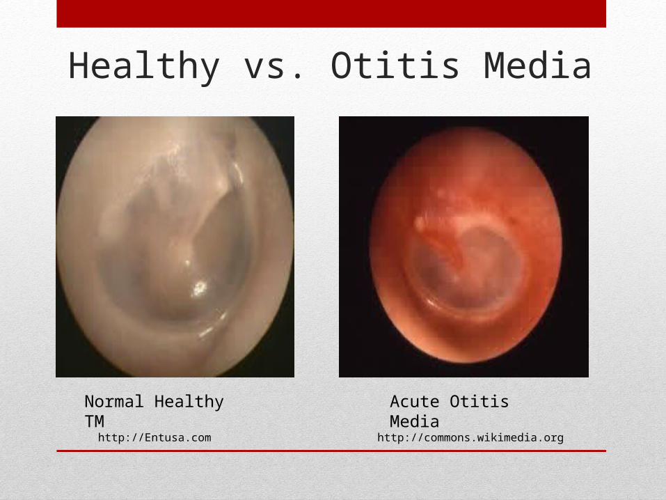
Healthy vs. Otitis Media
Normal Healthy TM Acute Otitis Media
http://commons.wikimedia.orghttp://Entusa.com
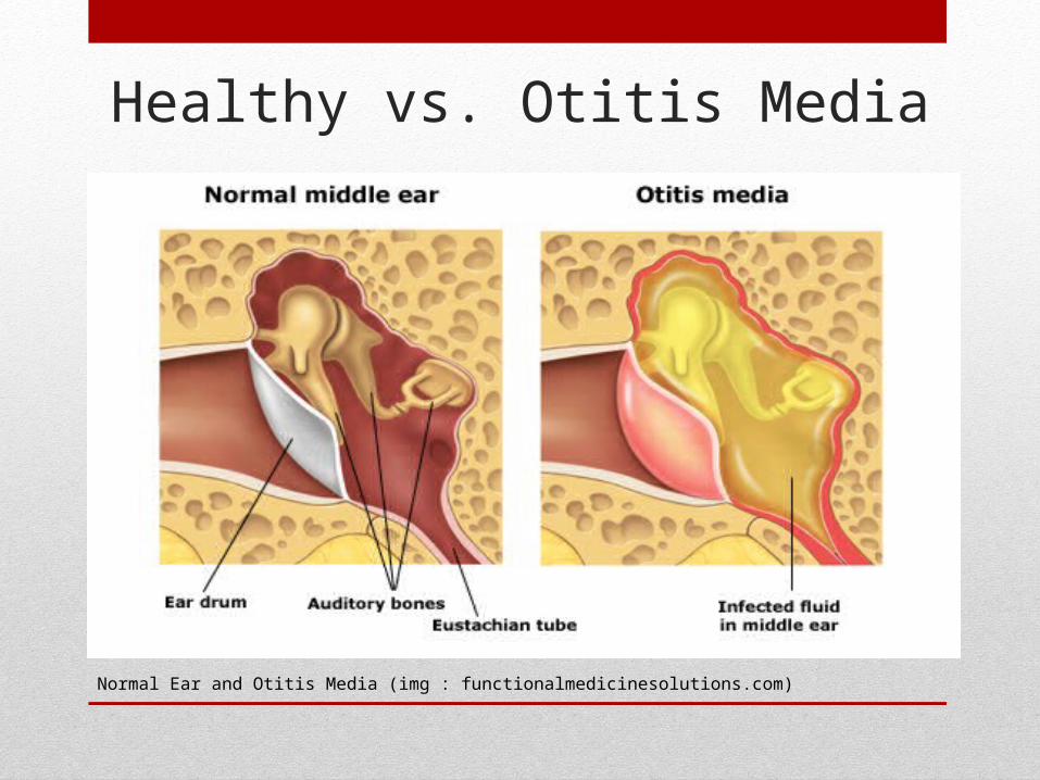
Healthy vs. Otitis Media
Normal Ear and Otitis Media (img : functionalmedicinesolutions.com)
Ear Infections: Children vs. Adults
• Eustachian tubes are smaller and more level in children. This makes it difficult for fluid to drain out of the ear. If the eustachian tubes are swollen or blocked with mucus due to a cold or other respiratory illness, fluid may not be able to drain.
• A child’s immune system isn’t as effective because it’s still developing. This makes it harder for children to fight infections.
• As part of the immune system, the adenoids respond to bacteria passing through the nose and mouth. Sometimes bacteria get trapped in the adenoids, causing a chronic infection that can then pass on to the eustachian tubes and the middle ear.
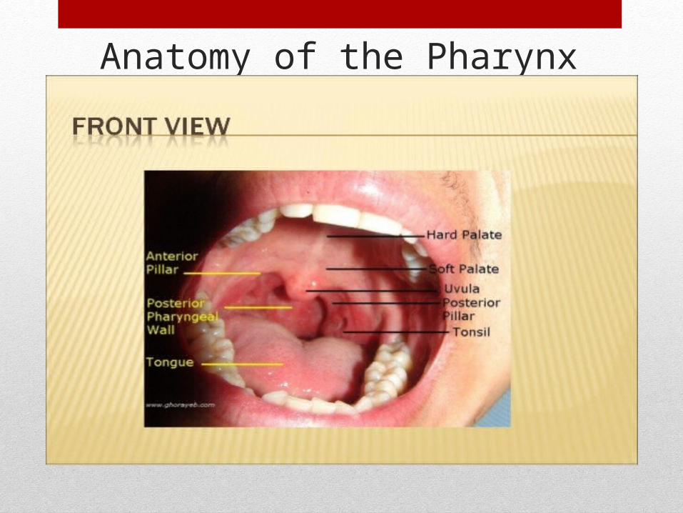
Anatomy of the Pharynx
Physical Assessment of the Pharynx
• Smell breath – unusually malodorous?
• Any noticeable lesions or ulcers (aptheous)
• Is tongue midline?
• Does the student have tonsils?
• Notice shape of uvula
• Is there cobblestoning?
• Is there exudate? White patches?
Pharyngitis: A Definition
Pharyngitis is inflammation of the pharynx—the back of the throat. This can cause a sore throat, as well as scratchiness in the
throat and difficulty swallowing.
(wwww.healthline.com)
Pharyngitis Signs and SymptomsThe symptoms that accompany a sore throat can vary,
depending on what's causing it.
• Sore throat with a cold (likely viral):• Sneezing, Cough, Runny nose, A low fever (less than 102 °F), Mild headache
• Sore throat – sudden onset – no cold symptoms (possibly bacterial)• Fever greater than 101, Sudden onset, white patches on tonsils/pharynx,
Lethargy/fatigue
• Sore throat with flu:• Fatigue, Body aches, Chills, Fever higher than 102 °F
• Sore throat with mononucleosis:• Enlarged lymph nodes in neck and armpits, Swollen tonsils, Headache, Loss
of appetite, Swollen spleen, Liver inflammation
Source: Pharyngitis | University of Maryland Medical Center http://umm.edu/health/medical/altmed/condition/pharyngitis#ixzz3Ps1gquRk University of Maryland Medical Center
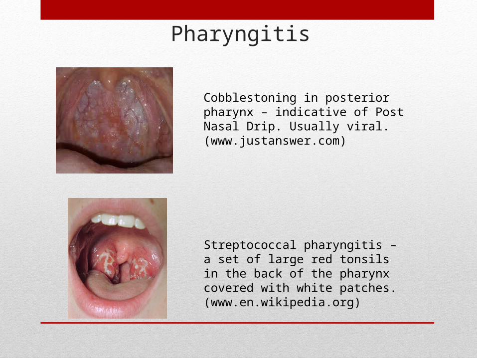
Pharyngitis
Cobblestoning in posterior pharynx – indicative of Post Nasal Drip. Usually viral. (www.justanswer.com)
Streptococcal pharyngitis – a set of large red tonsils in the back of the pharynx covered with white patches. (www.en.wikipedia.org)
Summary: How does all of this knowledge affect the CSN’s Clinical Practice?
• Otalgia and Pharyngitis are two of the most common complaints at the doctor’s office as well as in our Health rooms.
• Understanding basic ENT anatomy will enhance the CSN’s ability to perform a more accurate physical assessment – thus determining the severity of the child’s ailment. It can prevent many unnecessary phone calls and early dismissals home.
• Recognizing and understanding the normal physical findings on otoscopic and oral examinations can help the CSN inform the parents/guardians of any abnormal findings; thus expediting physician appointments.
Summary – Continued
• REMEMBER: It is not the job of the CSN to diagnose, but rather to note abnormalities when present – and then refer as needed.
• Finally, being confident in our overall ENT knowledge and physical assessment skills will give us opportunity to educate our students and families – hopefully keeping our students healthy and in school.

THANK YOU!!!!!
References
• Anatomical Diagram of the Ear © 2009 WebMD, LLC.• http://www.commonswikimedia.org • http://www.dermnet.com• http://www.drmkotb.com• http://www.eachawklibrary.com• http://www.entusa.com• http://www.en.wikipedia.org• http://www.functionalmedicines.com• http://www.ghorayeb.com• http://www.hawaiiscubaadventures.com• http://www.healthline.com• http://www.justanswer.com• http://www.otorhinolaryngoportal.com• http://www.nidcd.nih.gov/health/hearing/pages/earinfections.aspx• http://www.studyblue.com• http
://umm.edu/health/medical/altmed/condition/pharyngitis#ixzz3Ps1gquRk