Embed Size (px)
Citation preview
Osteosarcoma Biomarkers Discovery Using“Omics” Approaches
Giulia Bernardini, Maurizio Orlandini, Federico Galvagni,and Annalisa Santucci
ContentsKey Facts of Osteosarcoma . . . . . . . . . . . . . . . . . . . . . . . . . . . . . . . . . . . . . . . . . . . . . . . . . . . . . . . . . . . . . . . . . . . . . . 2Definitions of Words and Terms . . . . . . . . . . . . . . . . . . . . . . . . . . . . . . . . . . . . . . . . . . . . . . . . . . . . . . . . . . . . . . . . . . 3Introduction . . . . . . . . . . . . . . . . . . . . . . . . . . . . . . . . . . . . . . . . . . . . . . . . . . . . . . . . . . . . . . . . . . . . . . . . . . . . . . . . . . . . . . . 4MicroRNAs . . . . . . . . . . . . . . . . . . . . . . . . . . . . . . . . . . . . . . . . . . . . . . . . . . . . . . . . . . . . . . . . . . . . . . . . . . . . . . . . . . . . . . . 5Transcriptomics . . . . . . . . . . . . . . . . . . . . . . . . . . . . . . . . . . . . . . . . . . . . . . . . . . . . . . . . . . . . . . . . . . . . . . . . . . . . . . . . . . . 8Proteomics . . . . . . . . . . . . . . . . . . . . . . . . . . . . . . . . . . . . . . . . . . . . . . . . . . . . . . . . . . . . . . . . . . . . . . . . . . . . . . . . . . . . . . . . 12Potential Applications to Prognosis, Other Diseases, or Conditions . . . . . . . . . . . . . . . . . . . . . . . . . . . . 18Summary Points . . . . . . . . . . . . . . . . . . . . . . . . . . . . . . . . . . . . . . . . . . . . . . . . . . . . . . . . . . . . . . . . . . . . . . . . . . . . . . . . . . 19References . . . . . . . . . . . . . . . . . . . . . . . . . . . . . . . . . . . . . . . . . . . . . . . . . . . . . . . . . . . . . . . . . . . . . . . . . . . . . . . . . . . . . . . . 19
AbstractOsteosarcoma is the most common malignant primary cancer of bone tissueaffecting mostly children and young adults. Nowadays, reliable circulating orcellular/tissue biomarkers do not exist for early diagnosis, drug resistance, andrelapses of osteosarcoma. Post-genomics represents an invaluable tool to disclosecancer complexity at a molecular as well as to discover novel diagnostic andprognostic biomarkers.
Although “omics” research on osteosarcoma has only been undertakenrecently in respect to that on many other tumor types, these studies have broughtto light several potential molecular biomarkers that represent the basis to developnovel and better strategies for early detection, outcome prediction, detection ofdisease recurrence, and therapeutic approach.
G. Bernardini • M. Orlandini • F. Galvagni • A. Santucci (*)Dipartimento di Biotecnologie, Chimica e Farmacia, Università di Siena, Siena, Italye-mail: [email protected]; [email protected]; [email protected];[email protected]
# Springer Science+Business Media Dordrecht 2015V.R. Preedy (ed.), Biomarkers in Bone Disease, Biomarkers in Disease: Methods,Discoveries and Applications, DOI 10.1007/978-94-007-7745-3_17-1
1
In this chapter, the discovery of such molecular markers through the emergingomics technologies, including miRNA-omics, transcriptomics, and proteomics,will be extensively reviewed.
KeywordsOsteosarcoma • Post-genomics • Omics approaches • miRNA • Transcriptomics •Proteomics • Biomarker
List of Abbreviations1DE Monodimensional polyacrylamide gel electrophoresis2D-DIGE Two-dimensional difference in gel electrophoresis2DE Two-dimensional polyacrylamide gel electrophoresisCSC Cancer stem cellELISA Enzyme-linked immunosorbent assayESI Electrospray ionizationFACS Fluorescence-activated cell sortingFT-ICR Fourier transform ion cyclotron resonanceIHC ImmunohistochemistryiTRAQ Isobaric tags for relative and absolute quantitationLC Liquid chromatographyLTQ Linear ion trapmiRNA Micro RNAMS Mass spectrometryMS/MS Tandem mass spectrometryOB OsteoblastOC OsteochondromaOS OsteosarcomaPCA Principal component analysisPMF Peptide mass spectrometryq RT-PCR Quantitative real time PCRQToF Quadrupole time of flightSELDI-ToF/MS Surface-enhanced laser desorption/ionization time of flight
mass spectrometryWB Western blotting
Key Facts of Osteosarcoma
• Osteosarcoma is the most common type of bone cancer, which develops ingrowing bones and occurs more frequently in children and adolescents.
• The most common early signs of osteosarcoma are pain and swelling. Thepresence of a bone tumor has to be confirmed by a complete medical examinationincluding blood test, since bone tumors can be associated with increased levels ofcertain enzymes in the blood; X-rays and other scans of the bone(s); and then a
2 G. Bernardini et al.
biopsy (removal of a sample of tissue) that will be examined by a pathologist todetermine whether it is cancerous and if so what type of cancer it is.
• Osteosarcomas can be localized or metastasize to other parts of the body (mainlylungs). Microscopic spreads can occur even at the early phases of cancer pro-gression, when the primary tumor has a very small size.
• Modern treatments of osteosarcoma require surgery (to remove all visible tumortissue) and chemotherapy, given before (to shrink tumor size and to preventmetastasis) and after surgery (to kill cancer cells not completely removed bysurgery). Many factors including site and location of the main tumor and otherindividual factors affect the type of surgery. When it is possible, limb-sparingprocedures by an artificial device (endoprosthesis) or bones from other places inthe body (bone graft) are preferred to amputation.
• Several factors affect the prognosis of osteosarcoma patients, including cancerspreading, size and location of tumor, type of osteosarcoma, surgery outcome,responsiveness to chemotherapy, and patient’s general health.
Definitions of Words and Terms
Biological marker (Biomarker) A characteristic that is objectively mea-sured and evaluated as an indicator ofnormal biologic processes, pathogenicprocesses, or pharmacologic responsesto a therapeutic intervention. (BiomarkerDefinitions Working Group – 1998).
miRNA miRNAs are short noncoding RNA mol-ecules that regulate gene expression atthe posttranscriptional level by bindingto the 30 untranslated regions of targetmessenger RNAs. To date, nearly 2000human miRNAs have been identifiedand any single miRNA can regulatedozens or hundreds of target genes.
Proteome Large-scale inventory of the proteinsexpressed in cells, tissues, or organisms.Proteome reflects a specific develop-mental stage or physiological condition.
Proteomics Comprehensive study of a specific pro-teome with the aim to catalog all proteinspecies, to determine their structure andfunction, and to quantify the changingexpression levels of each protein speciesduring development and under differentconditions. Proteomics approaches are
Osteosarcoma Biomarkers Discovery Using “Omics” Approaches 3
conducted by means of high-throughputtechnologies.
Transcriptome Large-scale inventory of the RNA tran-scripts (mRNAs, noncoding RNAs, andsmall RNAs) produced by the genome incells, tissues, or organisms.Transcriptome reflects a specific devel-opmental stage or physiologicalcondition.
Transcriptomics The study of a specific transcriptomewith the aim to catalog all species oftranscript, to determine the transcrip-tional structure of genes, and to quantifythe changing expression levels of eachtranscript during development and underdifferent conditions. Transcriptomicsapproaches are conducted by means ofhigh-throughput technologies.
Introduction
Osteosarcoma (OS) is a rare neoplasm of bone that affects mainly young patients.Since OS have a high tendency to metastasize, they are classified among the mostfrequent sources of cancer-related death in childhood tumors (Botter et al. 2014).Despite the survival rate of OS patients has been improved as a result of refinedsurgical techniques and multiagent chemotherapy, the survival of patients thatdevelop metastases still remains low (Anninga et al. 2011). Therefore, a moredetailed understanding of the molecular mechanisms and specific identifying ofbiomarkers involved in tumor initiation, progression, and metastasis formation isof immediate importance to develop new and improved treatment strategies for OS.
Currently the diagnosis of OS occurs around 4 months from the onset of symp-toms. Diagnosis of OS is based, after a first complete medical history of the patient,on imaging analysis, including radiographs, magnetic resonance imaging, bonescintigraphy, and biopsy which provide a definite diagnosis and grading/staging ofthe tumor. So far, reliable OS circulating markers do not exist. In fact, alkalinephosphatase (ALP) exhibits high plasmatic level only in 40 % of cases, while lactatedehydrogenase (LDH) is elevated in around 30 % of cases. These laboratory valuesalso possess a moderate prognostic relevance: normal ALP and LDH levels inchemonaive patients have been associated with 5-year disease-free survival and alonger time to disease recurrence (Geller and Gorlick 2010). However, the mostimportant prognostic factors in OS are represented by the presence of metastatic
4 G. Bernardini et al.
disease at the time of diagnosis and the histological response to preoperativechemotherapy.
Nowadays, the greatest challenge in OS management is the lack of reliablemarkers able to detect the tumor at an early stage, when there is a better chancefor its treatment, or to predict the prognosis or the response to chemotherapy.
In the last few years, the significant progress in “omics” technologies(epigenomics, transcriptomics, and proteomics), allowing the simultaneous detec-tion of thousands of molecular species in a large amount of biological samples,provided researchers with the opportunity to discover a variety of biomarkers withdiagnostic and prognostic purposes. In this regard, the development of bioinformat-ics analytical tools suitable to mine the massive flood of data provided by high-throughput experiments is mandatory to integrate different “omics” approaches aswell as to achieve robust and reliable finding with clinical relevance as well as to getnovel clues for understanding cancer biology and pathophysiology (Bernardiniet al. 2012, 2014).
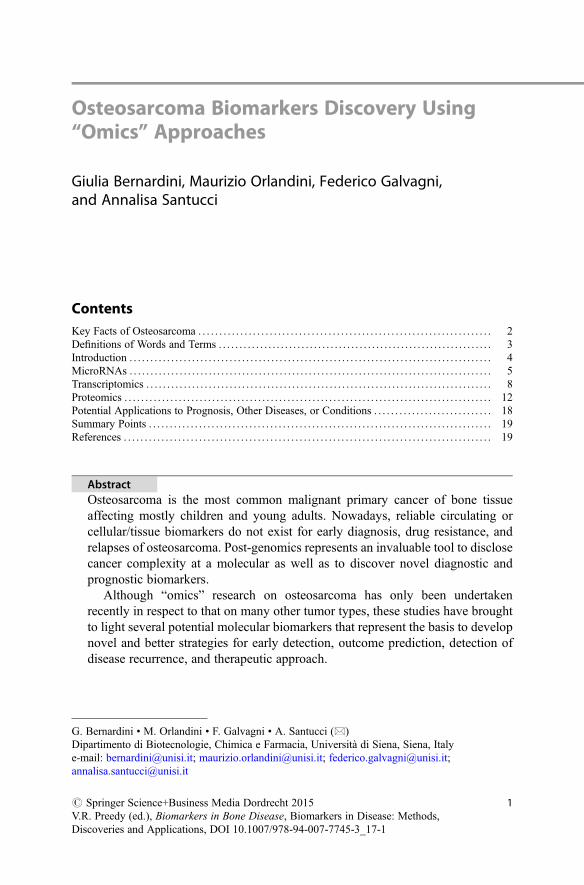
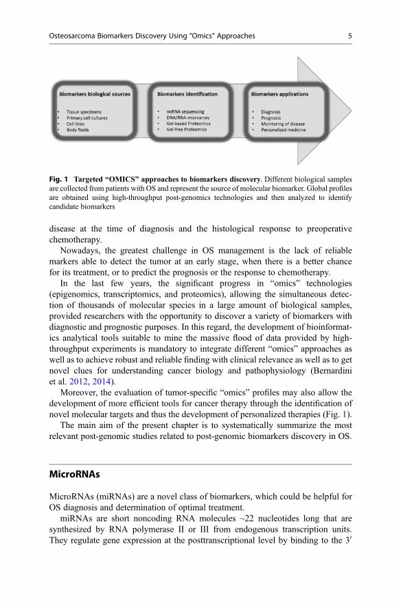
Moreover, the evaluation of tumor-specific “omics” profiles may also allow thedevelopment of more efficient tools for cancer therapy through the identification ofnovel molecular targets and thus the development of personalized therapies (Fig. 1).
The main aim of the present chapter is to systematically summarize the mostrelevant post-genomic studies related to post-genomic biomarkers discovery in OS.
MicroRNAs
MicroRNAs (miRNAs) are a novel class of biomarkers, which could be helpful forOS diagnosis and determination of optimal treatment.
miRNAs are short noncoding RNA molecules ~22 nucleotides long that aresynthesized by RNA polymerase II or III from endogenous transcription units.They regulate gene expression at the posttranscriptional level by binding to the 30
Fig. 1 Targeted “OMICS” approaches to biomarkers discovery. Different biological samplesare collected from patients with OS and represent the source of molecular biomarker. Global profilesare obtained using high-throughput post-genomics technologies and then analyzed to identifycandidate biomarkers
Osteosarcoma Biomarkers Discovery Using “Omics” Approaches 5
untranslated regions (30 UTRs) of target messenger RNAs (Ambros 2004). To date,nearly 2000 human miRNAs have been identified (miRBase, Homo sapiensmiRNAs database, Manchester University), and any single miRNA can regulatedozens or hundreds of target genes (Rana 2007).
In the context of cancer cells, miRNAs can act as oncogenes (oncomiR) or tumorsuppressor genes (anti-oncomiR) based on their inhibition of tumor-suppressive andoncogenic mRNAs, respectively, and expression deregulation of one or moremiRNAs was demonstrated to be involved in development and progression of cancer(Calin et al. 2002; Sotiropoulou et al. 2009). The expression profiling of miRNAs isalready used into cancer clinics as diagnostic and prognostic biomarkers to assesstumor initiation, progression, and response to treatment (Reddy 2015).
miRNAs Expression in OS
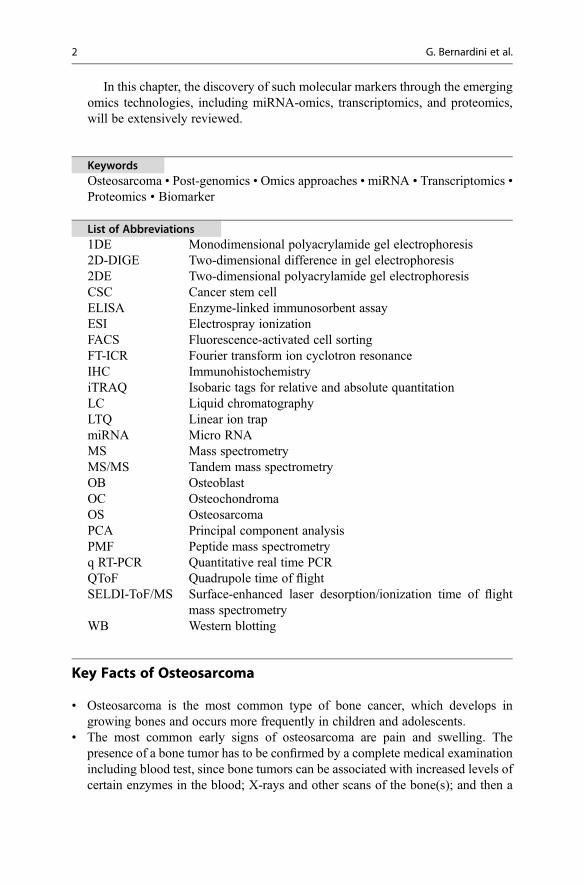
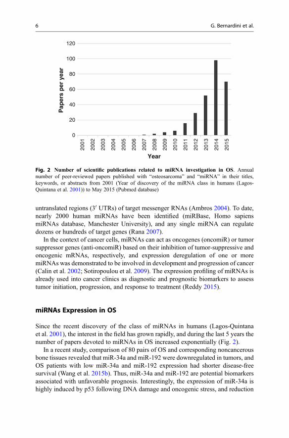
Since the recent discovery of the class of miRNAs in humans (Lagos-Quintanaet al. 2001), the interest in the field has grown rapidly, and during the last 5 years thenumber of papers devoted to miRNAs in OS increased exponentially (Fig. 2).
In a recent study, comparison of 80 pairs of OS and corresponding noncancerousbone tissues revealed that miR-34a and miR-192 were downregulated in tumors, andOS patients with low miR-34a and miR-192 expression had shorter disease-freesurvival (Wang et al. 2015b). Thus, miR-34a and miR-192 are potential biomarkersassociated with unfavorable prognosis. Interestingly, the expression of miR-34a ishighly induced by p53 following DNA damage and oncogenic stress, and reduction
120
100
80
60
40
20
0
Year
Pap
ers
per
yea
r
2001
2002
2003
2004
2005
2006
2007
2008
2009
2010
2011
2012
2013
2014
2015
Fig. 2 Number of scientific publications related to miRNA investigation in OS. Annualnumber of peer-reviewed papers published with “osteosarcoma” and “miRNA” in their titles,keywords, or abstracts from 2001 (Year of discovery of the miRNA class in humans (Lagos-Quintana et al. 2001)) to May 2015 (Pubmed database)
6 G. Bernardini et al.
of miR-34 function attenuates p53-mediated cell death (He et al. 2007). Moreover, inOS models, miR-34a inhibits proliferation, angiogenesis, and metastasis of tumorcells by targeting Notch-1, mTOR, c-Met, MDM4, and Eag1 (Li et al. 2013; Tianet al. 2014b; Wu et al. 2013; Yan et al. 2012).
In a sample of 52 patients, miRNA-22 was identified as a novel potentialbiomarker of unfavorable prognosis in OS (Wang et al. 2015a). In fact, miR-22 isdownregulated in OS in comparison with noncancerous bone tissues, and its lowexpression level correlates with recurrence, metastasis, chemotherapy response, andpoorer overall survival and DFS. miR-22 seems to act as tumor suppressor gene bytargeting the 30UTR of high-mobility group box 1 (HMGB1) and inhibiting itstranslation. In OS cells, high levels of HMGB1 (due to miR-22 downregulation)promote autophagy and consequent drug resistance (Guo et al. 2014).
As example of upregulated miRNAs in OS, miR-27a was found to be prognosticof metastatic disease in a sample of 18 patients (Jones et al. 2012). miR-27a isdescribed to promote metastasis in OS, at least in part, through targeting the tumorsuppressor CBFA2T3, which is downregulated in a majority of patients (Salahet al. 2015).
Up to now, several other miRNAs have been found to be implicated in OS (Zhanget al. 2015), and a tool was needed to manage the information regarding theexpression patterns. To this aim, Korsching and coworkers constructed the Osteo-sarcoma Database, which provides a structured, annotated, and easy accessibleoverview of the protein-coding and miRNAs genes whose expression correlateswith disease progression and that might be used as biomarkers (http://osteosarcoma-db.uni-muenster.de). At the time of the last update (October 2013), the Osteosar-coma Database contains 911 protein-coding genes and 81 microRNAs associatedwith OS according to 1,331 PubMed abstracts. The Osteosarcoma Database offers“the possibility to rank and sort the literature according to various parameters,including therapeutic and prognostic value of specific genes and microRNA andthe type of sample used” (Poos et al. 2014).
miRNAs Detection Methods
Quantitative real-time PCR (qRT-PCR) technique is the most popular reference testto quantify miRNA expression, because of its speed, simplicity, low cost of exercise,and high sensitivity and specificity. The disadvantage is that this technique is timeconsuming and laborious if large number of miRNA has to be analyzed. Microarraysor microRNA sequencing (miRNA-seq), instead, are used when high throughput isdesired, even if they need more complex steps of standardization and validation.Microarray platforms allow the analysis of thousands of miRNAs in a singleexperiment, and it is widely used in order to detect and quantify miRNAs.miRNA-seq uses next-generation sequencing technology to massively sequencemiRNAs; it is relatively recent but is replacing microarrays. This miRNA-seqtechnology has the advantage of quantifying and identifying known miRNAs, aswell as novel miRNAs.
Osteosarcoma Biomarkers Discovery Using “Omics” Approaches 7
A major difficulty in miRNA quantification from patient tissues is the availabilityof frozen samples. Recently, Spentzos and colleagues overcome this problem andpublished a large OS profiling study (Kelly et al. 2013). They used the IlluminacDNA-mediated annealing, selection, extension, and ligation (DASL) assay toanalyze the expression of 1,146 miRNAs from the partially degraded RNAsextracted from 91 formalin-fixed, paraffin-embedded (FFPE) OS diagnostic biopsyspecimens and identified a cluster of miRNAs with predictive value for OS recur-rence and survival. This cluster is located at the 14q32 locus, already linked to thistype of cancer. Through this technology, they also identify nonoverlapping miRNAprofiles predictive of chemoresponse.
Circulating miRNAs
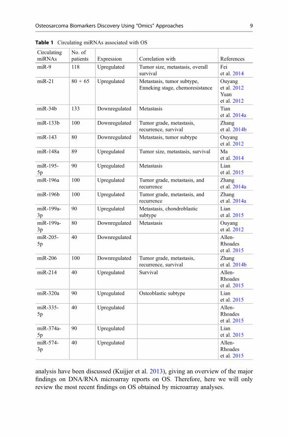
miRNAs are not only regulators of gene expression in the same cell in which they aresynthesized, but they can be secreted and transferred horizontally between cells,assuming also a role in intercellular communication and long-distance signaling,regulating target RNAs in recipient cells (Chen et al. 2012). Circulating miRNAshave been found in serum, plasma, and other body fluids and represent attractivebiomarkers in noninvasive serological tests for the diagnosis or prognosis of cancer.This type of analysis presents the advantage of easier samples achievement and,consequently, it allows analyzing larger cohort of patients. Recent findings oncirculating miRNA associated with OS are summarized in Table 1.
In some interesting example, such as miR-9 and miR-214, the differential expres-sion of miRNAs and their prognostic value in OS were described for both plasmaand tumoral tissue (Allen-Rhoades et al. 2015; Fei et al. 2014; Wang et al. 2014; Xuet al. 2014).
In conclusion, it is clear that the expression profiles of circulating miRNA areuseful as biomarkers for OS diagnosis, prognosis, and chemoresponse. Butdespite the large progress in the field, a lot of work is still needed to identify,characterize, and validate the most predictive biomarkers, and several researchfindings have to be clarified, such as the opposite expression profile found indifferent studies for miR-199a-3p in plasma of patients (Lian et al. 2015; Ouyanget al. 2012).
Transcriptomics
Over the last few years, the analysis of differentially expressed genes by microarraycombined with bioinformatics analysis has been used to identify key genes andcellular signaling pathways involved in OS progression and metastasis. However,OS genome-wide studies resulted extremely hard due to the rarity of the disease, thehigh genomically unstable OS cells, and the heterogeneity of tumor clinical samples(Kuijjer et al. 2013). In a recent review, the challenges of high-grade OS data
8 G. Bernardini et al.
analysis have been discussed (Kuijjer et al. 2013), giving an overview of the majorfindings on DNA/RNA microarray reports on OS. Therefore, here we will onlyreview the most recent findings on OS obtained by microarray analyses.
Table 1 Circulating miRNAs associated with OS
CirculatingmiRNAs
No. ofpatients Expression Correlation with References
miR-9 118 Upregulated Tumor size, metastasis, overallsurvival
Feiet al. 2014
miR-21 80 + 65 Upregulated Metastasis, tumor subtype,Enneking stage, chemoresistance
Ouyanget al. 2012Yuanet al. 2012
miR-34b 133 Downregulated Metastasis Tianet al. 2014a
miR-133b 100 Downregulated Tumor grade, metastasis,recurrence, survival
Zhanget al. 2014b
miR-143 80 Downregulated Metastasis, tumor subtype Ouyanget al. 2012
miR-148a 89 Upregulated Tumor size, metastasis, survival Maet al. 2014
miR-195-5p
90 Upregulated Metastasis Lianet al. 2015
miR-196a 100 Upregulated Tumor grade, metastasis, andrecurrence
Zhanget al. 2014a
miR-196b 100 Upregulated Tumor grade, metastasis, andrecurrence
Zhanget al. 2014a
miR-199a-3p
90 Upregulated Metastasis, chondroblasticsubtype
Lianet al. 2015
miR-199a-3p
80 Downregulated Metastasis Ouyanget al. 2012
miR-205-5p
40 Downregulated Allen-Rhoadeset al. 2015
miR-206 100 Downregulated Tumor grade, metastasis,recurrence, survival
Zhanget al. 2014b
miR-214 40 Upregulated Survival Allen-Rhoadeset al. 2015
miR-320a 90 Upregulated Osteoblastic subtype Lianet al. 2015
miR-335-5p
40 Upregulated Allen-Rhoadeset al. 2015
miR-374a-5p
90 Upregulated Lianet al. 2015
miR-574-3p
40 Upregulated Allen-Rhoadeset al. 2015
Osteosarcoma Biomarkers Discovery Using “Omics” Approaches 9
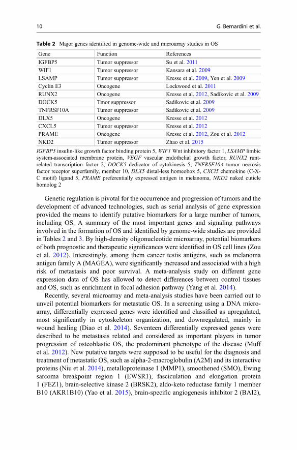
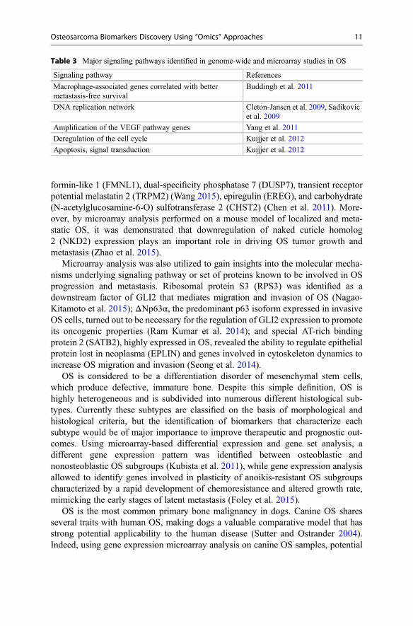
Genetic regulation is pivotal for the occurrence and progression of tumors and thedevelopment of advanced technologies, such as serial analysis of gene expressionprovided the means to identify putative biomarkers for a large number of tumors,including OS. A summary of the most important genes and signaling pathwaysinvolved in the formation of OS and identified by genome-wide studies are providedin Tables 2 and 3. By high-density oligonucleotide microarray, potential biomarkersof both prognostic and therapeutic significances were identified in OS cell lines (Zouet al. 2012). Interestingly, among them cancer testis antigens, such as melanomaantigen family A (MAGEA), were significantly increased and associated with a highrisk of metastasis and poor survival. A meta-analysis study on different geneexpression data of OS has allowed to detect differences between control tissuesand OS, such as enrichment in focal adhesion pathway (Yang et al. 2014).
Recently, several microarray and meta-analysis studies have been carried out tounveil potential biomarkers for metastatic OS. In a screening using a DNA micro-array, differentially expressed genes were identified and classified as upregulated,most significantly in cytoskeleton organization, and downregulated, mainly inwound healing (Diao et al. 2014). Seventeen differentially expressed genes weredescribed to be metastasis related and considered as important players in tumorprogression of osteoblastic OS, the predominant phenotype of the disease (Muffet al. 2012). New putative targets were supposed to be useful for the diagnosis andtreatment of metastatic OS, such as alpha-2-macroglobulin (A2M) and its interactiveproteins (Niu et al. 2014), metalloproteinase 1 (MMP1), smoothened (SMO), Ewingsarcoma breakpoint region 1 (EWSR1), fasciculation and elongation protein1 (FEZ1), brain-selective kinase 2 (BRSK2), aldo-keto reductase family 1 memberB10 (AKR1B10) (Yao et al. 2015), brain-specific angiogenesis inhibitor 2 (BAI2),
Table 2 Major genes identified in genome-wide and microarray studies in OS
Gene Function References
IGFBP5 Tumor suppressor Su et al. 2011
WIF1 Tumor suppressor Kansara et al. 2009
LSAMP Tumor suppressor Kresse et al. 2009, Yen et al. 2009
Cyclin E3 Oncogene Lockwood et al. 2011
RUNX2 Oncogene Kresse et al. 2012, Sadikovic et al. 2009
DOCK5 Tmor suppressor Sadikovic et al. 2009
TNFRSF10A Tumor suppressor Sadikovic et al. 2009
DLX5 Oncogene Kresse et al. 2012
CXCL5 Tumor suppressor Kresse et al. 2012
PRAME Oncogene Kresse et al. 2012, Zou et al. 2012
NKD2 Tumor suppressor Zhao et al. 2015
IGFBP5 insulin-like growth factor binding protein 5,WIF1Wnt inhibitory factor 1, LSAMP limbicsystem-associated membrane protein, VEGF vascular endothelial growth factor, RUNX2 runt-related transcription factor 2, DOCK5 dedicator of cytokinesis 5, TNFRSF10A tumor necrosisfactor receptor superfamily, member 10, DLX5 distal-less homeobox 5, CXCl5 chemokine (C-X-C motif) ligand 5, PRAME preferentially expressed antigen in melanoma, NKD2 naked cuticlehomolog 2
10 G. Bernardini et al.
formin-like 1 (FMNL1), dual-specificity phosphatase 7 (DUSP7), transient receptorpotential melastatin 2 (TRPM2) (Wang 2015), epiregulin (EREG), and carbohydrate(N-acetylglucosamine-6-O) sulfotransferase 2 (CHST2) (Chen et al. 2011). More-over, by microarray analysis performed on a mouse model of localized and meta-static OS, it was demonstrated that downregulation of naked cuticle homolog2 (NKD2) expression plays an important role in driving OS tumor growth andmetastasis (Zhao et al. 2015).
Microarray analysis was also utilized to gain insights into the molecular mecha-nisms underlying signaling pathway or set of proteins known to be involved in OSprogression and metastasis. Ribosomal protein S3 (RPS3) was identified as adownstream factor of GLI2 that mediates migration and invasion of OS (Nagao-Kitamoto et al. 2015); ΔNp63α, the predominant p63 isoform expressed in invasiveOS cells, turned out to be necessary for the regulation of GLI2 expression to promoteits oncogenic properties (Ram Kumar et al. 2014); and special AT-rich bindingprotein 2 (SATB2), highly expressed in OS, revealed the ability to regulate epithelialprotein lost in neoplasma (EPLIN) and genes involved in cytoskeleton dynamics toincrease OS migration and invasion (Seong et al. 2014).
OS is considered to be a differentiation disorder of mesenchymal stem cells,which produce defective, immature bone. Despite this simple definition, OS ishighly heterogeneous and is subdivided into numerous different histological sub-types. Currently these subtypes are classified on the basis of morphological andhistological criteria, but the identification of biomarkers that characterize eachsubtype would be of major importance to improve therapeutic and prognostic out-comes. Using microarray-based differential expression and gene set analysis, adifferent gene expression pattern was identified between osteoblastic andnonosteoblastic OS subgroups (Kubista et al. 2011), while gene expression analysisallowed to identify genes involved in plasticity of anoikis-resistant OS subgroupscharacterized by a rapid development of chemoresistance and altered growth rate,mimicking the early stages of latent metastasis (Foley et al. 2015).
OS is the most common primary bone malignancy in dogs. Canine OS sharesseveral traits with human OS, making dogs a valuable comparative model that hasstrong potential applicability to the human disease (Sutter and Ostrander 2004).Indeed, using gene expression microarray analysis on canine OS samples, potential
Table 3 Major signaling pathways identified in genome-wide and microarray studies in OS
Signaling pathway References
Macrophage-associated genes correlated with bettermetastasis-free survival
Buddingh et al. 2011
DNA replication network Cleton-Jansen et al. 2009, Sadikovicet al. 2009
Amplification of the VEGF pathway genes Yang et al. 2011
Deregulation of the cell cycle Kuijjer et al. 2012
Apoptosis, signal transduction Kuijjer et al. 2012
Osteosarcoma Biomarkers Discovery Using “Omics” Approaches 11
new biomarkers and novel pathways that may be targeted for therapeutic interven-tion were identified (O’Donoghue et al. 2010).
Gene expression profiling by microarray combined with other techniques resultedsuccessful in several studies. Indeed, expression microarray analysis combined withthe investigation of focal copy number aberrations has allowed identifying CKLF-like Marvel transmembrane domain containing 8 (CMTM8) as a new candidatetumor suppressor and G protein-coupled receptor 177 (GPR177) as a new putativeoncogene in OS (Both et al. 2014). Furthermore, combining proteomic analysis withpreviously obtained cDNA microarray results allowed detecting aldolase A fructose-bisphosphate (ALDOA) and sulfotransferase family cytosolic 1A phenol-preferring3 (SULT1A3) as predictors of clinical outcomes for OS patients (Chen et al. 2014),while microarray-based comparative genomic hybridization (aCGH) allowed to gaina comprehensive understanding of the key driving pathways for OS, elucidating thecontradictory role of Wnt signaling (Du et al. 2014), and identifying a functionalcrosstalk between vascular endothelial growth factor (VEGF) and runt-related tran-scription factor 2 (RUNX2) essential for the pathogenesis and angiogenesis of thedisease (Yang et al. 2013).
Finally, although in some studies microarray failed to predict biomarkers for OSpatients’ outcome (Sabile et al. 2013), for researchers it certainly provides an usefultool to characterize the altered expression of genes involved in the development andbehavior of OS subtypes and to identify the gene signature of an individual OSpatient revealing distinct signaling events, which might account for the biologicalfeatures specific for each tumor type.
Proteomics
Proteomic approaches to cancer research offer several advantages in respect to otherhigh-throughput technologies such as genomics or transcriptomics. In addition toglobal protein profiling and protein identification, proteomics provides powerfultools to investigate the complexity of these highly dynamic macromolecules. Infact, disease-associated phenotypic alterations are consequences not only ofderegulated (increasing/decreasing) expression of proteins but also of functionalregulations by various processes such as proteins degradations, posttranslationalmodifications (e.g., phosphorylation, glycosylation, methylation), involvement incomplex structures, and differential compartmentalization (e.g., nuclearlocalization).
Moreover, proteomic approaches can be applied to a variety of biospecimensranging from biological models, such as cell lines, primary cell cultures, or animalmodels of disease, to clinical samples, including serum/plasma, urine, spinal fluid,synovial fluid, and tissue.
Comprehensive analysis of proteomic data from cancer patients’ samples hasnotably improved our understanding of tumor pathogenesis and treatment,uncovering the different processes involved in cancer development and progression,along with the identification of novel target for cancer therapy.
12 G. Bernardini et al.
The discovery of biomarkers with clinical relevance using proteomics is affectedby several critical challenges, in particular the biological variability among patients’samples and the huge dynamic range of biomarkers concentration in biologicalfluids. In addition to these, another major obstacle to be taken into account is thethousands of cancer-associated proteins detected by high-throughput proteomicapproaches that have to be properly validated.
Nevertheless, in the last decade proteomic approaches lead to the discovery ofclinically relevant biomarkers for several types of cancers such as breast, esophageal,lung, liver, and colorectal cancer. All these biomarkers possess high values ofspecificity and sensitivity and represent unvaluable tool to be used for screening,early detection, and prediction of response to therapy in oncology (Sallam 2014).
Proteomics technologies include gel-based methods (1DE, 2DE, and 2D-DIGE),gel-free methods based on mass spectrometry (SELDI and MALDI ToF/MS,LC-MS/MS), or based on array (antibody array, reverse phase protein microarray(RPMA)) and bioinformatics.
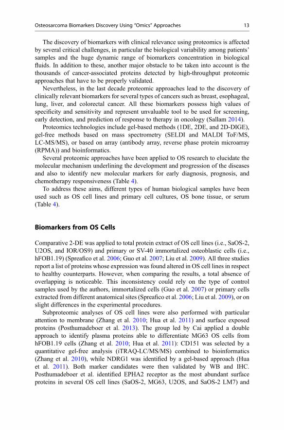
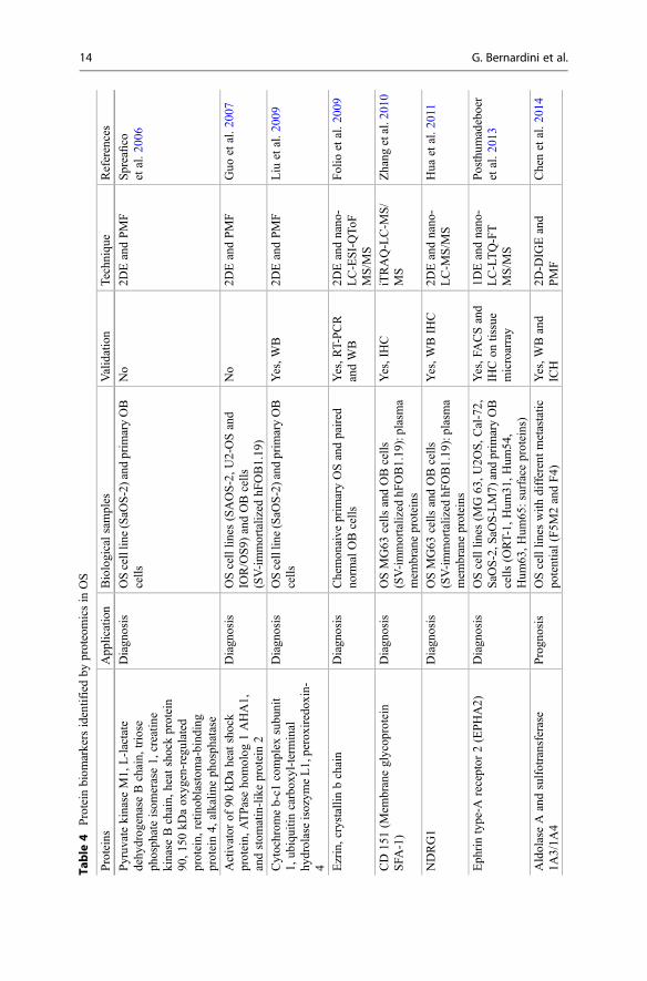
Several proteomic approaches have been applied to OS research to elucidate themolecular mechanism underlining the development and progression of the diseasesand also to identify new molecular markers for early diagnosis, prognosis, andchemotherapy responsiveness (Table 4).
To address these aims, different types of human biological samples have beenused such as OS cell lines and primary cell cultures, OS bone tissue, or serum(Table 4).
Biomarkers from OS Cells
Comparative 2-DE was applied to total protein extract of OS cell lines (i.e., SaOS-2,U2OS, and IOR/OS9) and primary or SV-40 immortalized osteoblastic cells (i.e.,hFOB1.19) (Spreafico et al. 2006; Guo et al. 2007; Liu et al. 2009). All three studiesreport a list of proteins whose expression was found altered in OS cell lines in respectto healthy counterparts. However, when comparing the results, a total absence ofoverlapping is noticeable. This inconsistency could rely on the type of controlsamples used by the authors, immortalized cells (Guo et al. 2007) or primary cellsextracted from different anatomical sites (Spreafico et al. 2006; Liu et al. 2009), or onslight differences in the experimental procedures.
Subproteomic analyses of OS cell lines were also performed with particularattention to membrane (Zhang et al. 2010; Hua et al. 2011) and surface exposedproteins (Posthumadeboer et al. 2013). The group led by Cai applied a doubleapproach to identify plasma proteins able to differentiate MG63 OS cells fromhFOB1.19 cells (Zhang et al. 2010; Hua et al. 2011): CD151 was selected by aquantitative gel-free analysis (iTRAQ-LC/MS/MS) combined to bioinformatics(Zhang et al. 2010), while NDRG1 was identified by a gel-based approach (Huaet al. 2011). Both marker candidates were then validated by WB and IHC.Posthumadeboer et al. identified EPHA2 receptor as the most abundant surfaceproteins in several OS cell lines (SaOS-2, MG63, U2OS, and SaOS-2 LM7) and
Osteosarcoma Biomarkers Discovery Using “Omics” Approaches 13
Table
4Protein
biom
arkersidentifiedby
proteomicsin
OS
Proteins
App
lication
Biologicalsamples
Validation
Techn
ique
References
Pyruv
atekinase
M1,
L-lactate
dehy
drog
enaseBchain,
triose
phosph
ateisom
erase1,
creatin
ekinase
Bchain,
heatshockprotein
90,1
50kD
aox
ygen-regulated
protein,
retin
oblastom
a-bind
ing
protein4,
alkalin
eph
osph
atase
Diagn
osis
OScelllin
e(SaO
S-2)and
prim
aryOB
cells
No
2DEandPMF
Spreafico
etal.2
006
Activator
of90
kDaheatshock
protein,
ATPaseho
molog
1AHA1,
andstom
atin-likeprotein2
Diagn
osis
OScelllin
es(SAOS-2,U
2-OSand
IOR/OS9)
andOBcells
(SV-immortalized
hFOB1.19
)
No
2DEandPMF
Guo
etal.2
007
Cytochrom
eb-c1
complex
subu
nit
1,ub
iquitin
carbox
yl-terminal
hydrolaseisozym
eL1,peroxiredo
xin-
4
Diagn
osis
OScelllin
e(SaO
S-2)and
prim
aryOB
cells
Yes,W
B2D
EandPMF
Liu
etal.2
009
Ezrin,crystallin
bchain
Diagn
osis
Chemon
aive
prim
aryOSandpaired
norm
alOBcells
Yes,R
T-PCR
andWB
2DEandnano
-LC-ESI-QToF
MS/M
S
Folio
etal.2
009
CD
151(M
embraneglycop
rotein
SFA
-1)
Diagn
osis
OSMG63
cells
andOBcells
(SV-immortalized
hFOB1.19
):plasma
mem
braneproteins
Yes,IHC
iTRAQ-LC-M
S/
MS
Zhang
etal.201
0
NDRG1
Diagn
osis
OSMG63
cells
andOBcells
(SV-immortalized
hFOB1.19
):plasma
mem
braneproteins
Yes,W
BIH
C2D
Eandnano
-LC-M
S/M
SHua
etal.2
011
Eph
rintype-A
receptor
2(EPHA2)
Diagn
osis
OScelllin
es(M
G63
,U2O
S,C
al-72,
SaO
S-2,S
aOS-LM7)
andprim
aryOB
cells
(ORT-1,
Hum
31,H
um54
,Hum
63,H
um65
:surfaceproteins)
Yes,F
ACSand
IHCon
tissue
microarray
1DEandnano
-LC-LTQ-FT
MS/M
S
Posthum
adeboer
etal.2
013
AldolaseAandsulfotransferase
1A3/1A
4Progn
osis
OScelllin
eswith
differentmetastatic
potential(F5M
2andF4)
Yes,W
Band
ICH
2D-D
IGEand
PMF
Chenetal.2
014
14 G. Bernardini et al.
Translatio
nally
controlledtumor
protein,
malatedehy
drog
enase,
CBX3,
dihy
drop
yrim
idinase-related
protein2,
fructose-bisph
osph
ate
aldo
lase
C
Diagn
osis
OScancer
stem
cells
CHA59
CBX3:
Yes,
RT-PCR
2DEandLC-M
S/
MS
Saini
etal.2
012
Activationof
MAPKspathway
Diagn
osis
OSCSC(3-A
BOS)andits
parental
OScelllin
e(M
G-63)
No
Antibod
yarray
andkn
owledg
e-basedanalysis
Gem
eietal.2
013
Protein
sign
atureof
10proteinspots
Therapy
respon
seChemon
aive
OStissues
classified
asgo
od/poo
rrespon
ders
No
2D-D
IGEand
PCA
Kaw
aietal.200
8
Vim
entin
,tub
ulin-a1c,lam
inB2,
coatom
erproteincomplex,epsilo
nsubu
nit,zinc
fing
erprotein
133,
ferritinlig
htpo
lypeptide,
myo
sin,
light
chain6,
ezrin,
transferrin,
a1-antitryp
sin,
chaperon
in-con
tainingTCP1
Diagn
osis
OSandbenign
bone
tumor
tissues
Yes,W
Band
IHC
2DEandPMF
Lietal.2
010
Perox
iredox
in-2
Therapy
respon
seChemon
aive
OStissues
classified
asgo
od/poo
rrespon
ders
Yes,W
B2D
-DIG
Eand
LC-LTQiontrap
MS/M
S
Kikuta
etal.2
010,
Kub
ota
etal.2
013
Heatshockprotein90
andclusterin
Progn
osis
OStissues
from
olderadultsand
desm
oidtumor
tissues
Yes,T
MA
LC-M
S/M
SRao
etal.2
013
Serum
amyloidA
Diagn
osis
Plasm
aform
OSandOC
Yes,W
BSELDI-ToF
/MS
Lietal.2
006
Serum
amyloidA
Diagn
osis
Serum
from
OSpatientsandhealthy
subjects
Yes,W
Band
ELISA
2D-D
IGEand
PMF
Jinetal.2
007
(con
tinued)
Osteosarcoma Biomarkers Discovery Using “Omics” Approaches 15
Table
4(con
tinue
d)
Proteins
App
lication
Biologicalsamples
Validation
Techn
ique
References
Cytochrom
e-c1
Diagn
osis
Serum
from
OSpatientsandhealthy
subjects
Yes,W
BSELDI-ToF
/MS
Lietal.2
009
Serum
amyloidAandtransthy
retin
Therapy
respon
sePlasm
afrom
OSpatientsbefore
and
afterpreoperativ
echem
otherapy
and
classified
asgo
od/poo
rrespon
ders
Yes,W
BSELDI-ToF
/MS
Lietal.2
011
Gelsolin
(decrease)
Diagn
osis
Serum
from
OSpatientsandhealthy
subjects
Yes,W
Band
ELISA
2D-D
IGEand
PMF
Jinetal.2
012
TwoproteinpeaksatM/Z
of39
54Da
and64
38Da(not
identified)
Plasm
afrom
OS,O
C,and
healthy
volunteers
No
SELDI-ToF
/MS
Guetal.2
014
16 G. Bernardini et al.
significantly overexpressed in OS cells and tissues in respect to normal samples(Posthumadeboer et al. 2013).
OS-specific proteins were also investigated by Folio in primary cells isolatedfrom five paired samples of OS tumor and normal bone tissue (Folio et al. 2009).2DE global protein profiling showed the upregulation of 56 protein spots intransformed cells. The overexpression of two of these, namely, ezrin and alpha-crystallin B chain, were confirmed by immune histochemistry or real-time PCR.
Recently, researchers in the field of experimental and clinical oncology havefocused their attention on cancer stem cells (CSC). Several lines of evidencesindicate that CSCs posses an elevated genotypic and phenotypic plasticity respon-sible for the heterogenicity of tumors and are involved not only in carcinogenesis butalso in the metastatic process, invasion, therapeutic poor responsiveness, and recur-rence of cancer. Although our comprehension of OS CSCs has notably improved,their role in OS pathophysiology is still largely unknown (Bernardini et al. 2014). Tostrengthen our knowledge of OS CSCs, global protein profiling can be extremelyuseful in uncovering their complexity as well as in selecting novel putative bio-markers. Several phenotypic changes were detected in two OS CSCs when com-pared to their parental cell lines (Table 1; Saini et al. 2012; Gemei et al. 2013).However, since none of these potential markers have been validated, their use asdiagnostic or prognostic factors is still to be demonstrated.
Biomarkers from OS Tissues
Proteomic studies to identify specific OS protein markers were also conducted ontissue samples obtained from patients’ biopsies (Kawai et al. 2008; Kikutaet al. 2010; Li et al. 2010; Kubota et al. 2013; Rao et al. 2013).
Li et al. compared the protein expression profile of malignant (osteoblastic,chondroblastic, and fibroblastic OS) and several benign (chondroblastoma,osteoblastoma, and giant cell) bone tumors using 2DE combined to PMF(Li et al. 2010). The overexpression in OS of two (TUBA1C and ZNF133) out of12 upregulated protein spots was validated by WB and IHC and thus selected aspotential OS biomarkers. Although authors did not extend the validation phase tonormal bone tissue, these two proteins represent a starting point for the developmentof important molecular tools for OS diagnostic.
Analogously, a gel-based proteomic approach was carried out by Kondo toidentify prognostic markers of OS responsiveness to chemotherapy (Kawaiet al. 2008; Kikuta et al. 2010; Kubota et al. 2013). Authors detected theoverexpression of peroxiredoxin 2 in OS tissue samples from chemonaive patientswho were afterwards classified as poor responder to different chemotherapy pro-tocols: combination of IFO, DOX, and CDDP (Kikuta et al. 2010) or combination ofMTX, DOX, and CDDP (Kubota et al. 2013). The overexpression of peroxiredoxin2 was further validated in a larger cohort of OS patients by WB and ROC analysis(AUC = 0.90, sensitivity = 83.3 %, specificity = 85.7 %, p = 0.015) that dem-onstrated the reliability of such a prognostic marker (Kubota et al. 2013).
Osteosarcoma Biomarkers Discovery Using “Omics” Approaches 17
Heat shock protein 90 and clusterin were found by a gel-free proteomic approachto be able to differentiate OS tissues from benign desmoid tissues (Rao et al. 2013).In particular, OS tissues were isolated from older adult patients with differentbackground (Paget’s disease, OS associated to dedifferentiated liposarcoma,extraosseus OS, dedifferentiated periosteal OS) with the aim to define the proteinprofile related to a highly metastatic cancer.
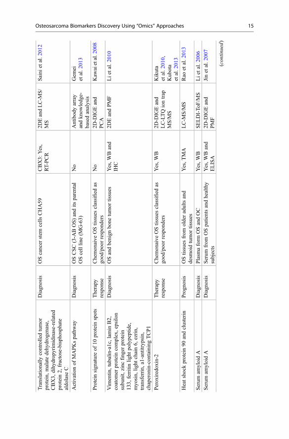
Circulating OS Biomarkers
Finally, the occurrence of specific OS protein biomarkers was also explored inplasma samples.
Serum amyloid protein A (SAA) was found to be present in higher amount in OSpatients than in osteochondroma patients (Li et al. 2006) or in healthy controls (Jinet al. 2007). Moreover, several authors demonstrated that SAA levels in OS patientsmight be used as marker to monitor relapses or response to chemotherapy (Jinet al. 2007; Li et al. 2011). Other OS plasmatic biomarkers include high level ofcytochrome C as an early diagnostic indicator, while high level of transthyretinsuggests a poor response to therapy (Li et al. 2009, 2011).
Potential Applications to Prognosis, Other Diseases, orConditions
Osteosarcoma is a heterogeneous tumor. This is due to the lack of characteristicchromosomal translocations or alterations, the occurrence in different anatomicalsites, and the presence of different histologic subtypes. This heterogeneity, inaddition to biospecimens variability (tissues, cells, body fluids, etc.), and to thelow incidence of the pathology, is reflected in post-genomics studies leading tononoverlapping or discordant results and to a very challenging validation phase ofpotential biomarkers. Therefore it is likely to be difficult to identify genes, miRNAs,or proteins that could have reliable diagnostic and/or prognostic value in osteosar-coma. To overcome the problem related to the scarcity of clinical cases, the scientificand medical community should promote networks of biobanks by means of nationaland international reference centers. These networks should be committed to harmo-nize procedures and set common standards for biospecimens and clinical datacollection and storage and to facilitate access to biological samples. A similarapproach should be used with high-throughput approaches and comprehensive andintegrated post-genomic investigations of patients should be required in order toovercome the intrinsic limitation of each related technology.
Our chance to understand the relationships between the individual molecularasset and the pathogenesis of disease, as well as the diversity of clinical outcomesor responses to therapies, will only be guaranteed by the use of high-qualitybiological samples with accurately phenotyped clinical data. This will likely leadto personalized medicines for OS patients.
18 G. Bernardini et al.
Summary Points
• Osteosarcoma is the most common primary bone cancer in adolescents and youngadults.
• The presence of metastatic disease at the time of diagnosis and responsiveness tochemotherapy are the principal prognostic factors for OS.
• There is an urgency for reliable biomarkers for early detection of OS, predictionof chemoresponsiveness, monitoring of treatment or relapses.
• “Omics” approaches identified biomarkers for several types of cancers as diag-nostic/prognostic indicators.
• Novel diagnostic and prognostic biomarkers can also represent novel target formore effective and personalized therapies.
• Rarity of OS hinders the large and proper validation of biomarkers selected bypost-genomics approaches.
References
Allen-Rhoades W, Kurenbekova L, Satterfield L, Parikh N, Fuja D, Shuck RL, Rainusso N,Trucco M, Barkauskas DA, Jo E, Ahern C, Hilsenbeck S, Donehower LA, Yustein JT. Cross-species identification of a plasma microRNA signature for detection, therapeutic monitoring,and prognosis in osteosarcoma. Can Med. 2015. doi:10.1002/cam4.438.
Ambros V. The functions of animal microRNAs. Nature. 2004;431:350–5.Anninga JK, Gelderblom H, Fiocco M, Kroep JR, Taminiau AHM, Hogendoorn PCW, Egeler
RM. Chemotherapeutic adjuvant treatment for osteosarcoma: where do we stand? Eur J Cancer.2011;47:2431–45.
Bernardini G, Braconi D, Spreafico A, Santucci A. Post-genomics of bone metabolic dysfunctionsand neoplasias. Proteomics. 2012;12(4–5):708–21.
Bernardini G, Laschi M, Geminiani M, Santucci A. Proteomics of osteosarcoma. Expert RevProteomics. 2014;11:331–43.
Both J, Krijgsman O, Bras J, Schaap GR, Baas F, Ylstra B, Hulsebos TJM. Focal chromosomalcopy number aberrations identify CMTM8 and GPR177 as new candidate driver genes inosteosarcoma. PLoS One. 2014;9:e115835.
Botter SM, Neri D, Fuchs B. Recent advances in osteosarcoma. Curr Opin Pharmacol.2014;16:15–23.
Buddingh EP, Kuijjer ML, Duim RAJ, B€urger H, Agelopoulos K, Myklebost O, Serra M,Mertens F, Hogendoorn PCW, Lankester AC, Cleton-Jansen A-M. Tumor-Infiltrating macro-phages are associated with metastasis suppression in high-grade osteosarcoma: a rationale fortreatment with aacrophage activating agents. Clin Cancer Res. 2011;17:2110–9.
Calin GA, Dumitru CD, Shimizu M, Bichi R, Zupo S, Noch E, Aldler H, Rattan S, Keating M,Rai K, Rassenti L, Kipps T, Negrini M, Bullrich F, Croce CM. Frequent deletions and down-regulation of micro- RNA genes miR15 and miR16 at 13q14 in chronic lymphocytic leukemia.Proc Natl Acad Sci. 2002;99:15524–9.
Chen X, Yang T-T, Qiu X-C, Ji Z-G, Li C-X, Long H, Zhou Y, Ma B-A, Ma Q, Zhang X, Fan Q-Y.Gene expression profiles of human osteosarcoma cell sublines with different pulmonary meta-static potentials. Cancer Biol Ther. 2011;11:287–92.
Chen X, Liang H, Zhang J, Zen K, Zhang C. Secreted microRNAs: a new form of intercellularcommunication. Trends Cell Biol. 2012;22:125–32.
Osteosarcoma Biomarkers Discovery Using “Omics” Approaches 19
Chen X, Yang T-T, Zhou Y, WangW, Qiu X-C, Gao J, Li C-X, Long H,Ma B-A, Ma Q, X-z Z, YangL-J, Fan Q-Y. Proteomic profiling of osteosarcoma cells identifies ALDOA and SULT1A3 asnegative survival markers of human osteosarcoma. Mol Carcinog. 2014;53:138–44.
Cleton-Jansen AM, Anninga JK, Briaire-de Bruijn IH, Romeo S, Oosting J, Egeler RM,Gelderblom H, Taminiau AHM, Hogendoorn PCW. Profiling of high-grade central osteosar-coma and its putative progenitor cells identifies tumourigenic pathways. Br J Cancer.2009;101:1909–18.
Diao C, Guo H, Ouyang Y, Zhang H, Liu L, Bu J, Wang Z, Xiao T. Screening for metastaticosteosarcoma biomarkers with a DNA microarray. Asian Pac J Cancer Prev. 2014;14:1817–22.
Du X, Yang J, Yang D, Tian W, Zhu Z. The genetic basis for inactivation of Wnt pathway in humanosteosarcoma. BMC Cancer. 2014;14:450.
Fei D, Li Y, Zhao D, Zhao K, Dai L, Gao Z. Serum miR-9 as a prognostic biomarker in patients withosteosarcoma. J Int Med Res. 2014;42:932–7.
Foley J, Scholten D, Monks N, Cherba D, Monsma D, Davidson P, Dylewski D, Dykema K,Winn M, Steensma M. Anoikis-resistant subpopulations of human osteosarcoma display sig-nificant chemoresistance and are sensitive to targeted epigenetic therapies predicted by expres-sion profiling. J Transl Med. 2015;13:110.
Folio C, Mora MI, Zalacain M, Corrales FJ, Segura V, Sierrasesumaga L, Toledo G, San-Julian M,Patino-Garcia A. Proteomic analysis of chemonaive pediatric osteosarcomas and correspondingnormal bone reveals multiple altered molecular targets. J Proteome Res. 2009;8:3882–8.
Geller DS, Gorlick R. Osteosarcoma: a review of diagnosis, management, and treatment strategies.Clin Adv Hematol Oncol. 2010;8(10):705–18.
Gemei M, Corbo C, D’Alessio F, Di Noto R, Vento R, Del Vecchio L. Surface proteomic analysis ofdifferentiated versus stem-like osteosarcoma human cells. Proteomics. 2013;13:3293–7.
Gu J, Li J, Huang M, Zhang Z, Li D, Song G, Ding X, Li W. Identification of osteosarcoma-relatedspecific proteins in serum samples using surface-enhanced laser desorption/ionization-time-of-flight mass spectrometry. J Immunol Res. 2014;2014:649075.
Guo QC, Shen JN, Jin S, Wang J, Huang G, Zhang LJ, Huang G, Yin JQ, Zou CY, LiMT. Comparative proteomic analysis of human osteosarcoma and SV40-immortalized normalosteoblastic cell lines. Acta Pharmacol Sin. 2007;28:850–8.
Guo S, Bai R, Liu W, Zhao A, Zhao Z, Wang Y, Wang Y, Zhao W, Wang W. miR-22 inhibitsosteosarcoma cell proliferation and migration by targeting HMGB1 and inhibiting HMGB1-mediated autophagy. Tumor Biol. 2014;35:7025–34.
He X, He L, Hannon GJ. The guardian’s little helper: microRNAs in the p53 tumor suppressornetwork. Cancer Res. 2007;67:11099–101.
Hua Y, Jia X, Sun M, Zheng L, Yin L, Zhang L, Cai Z. Plasma membrane proteomic analysis ofhuman osteosarcoma and osteoblastic cells: revealing NDRG1 as a marker for osteosarcoma.Tumour Biol. 2011;32:1013–21.
Jin S, Shen JN, Guo QC, Zhou JG, Wang J, Huang G, Zou CY, Yin JQ, Liu SJ, Liu W, Li MT, WangLN. 2-D DIGE and MALDI-TOF-MS analysis of the serum proteome in human osteosarcoma.Proteomics Clin Appl. 2007;1:272–85.
Jin S, Shen JN, Peng JQ, Wang J, Huang G, Li MT. Increased expression of serum gelsolin inpatients with osteosarcoma. Chin Med J (Engl). 2012;125:262–9.
Jones KB, Salah Z, Del Mare S, Galasso M, Gaudio E, Nuovo GJ, Lovat F, LeBlanc K, Palatini J,Randall RL, Volinia S, Stein GS, Croce CM, Lian JB, Aqeilan RI. miRNA signatures associatewith pathogenesis and progression of osteosarcoma. Cancer Res. 2012;72:1865–77.
Kansara M, Tsang M, Kodjabachian L, Sims NA, Trivett MK, Ehrich M, Dobrovic A, Slavin J,Choong PFM, Simmons PJ, Dawid IB, Thomas DM. Wnt inhibitory factor 1 is epigeneticallysilenced in human osteosarcoma, and targeted disruption accelerates osteosarcomagenesis inmice. J Clin Invest. 2009;119:837–51.
Kawai A, Kondo T, Suehara Y, Kikuta K, Hirohashi S. Global protein-expression analysis of boneand soft tissue sarcomas. Clin Orthop Relat Res. 2008;466:2099–106.
20 G. Bernardini et al.
Kelly A, Haibe-Kains B, Janeway K, Hill K, Howe E, Goldsmith J, Kurek K, Perez-Atayde A,Francoeur N, Fan J, April C, Schneider H, Gebhardt M, Culhane A, Quackenbush J, SpentzosD. MicroRNA paraffin-based studies in osteosarcoma reveal reproducible independent prog-nostic profiles at 14q32. Genome Med. 2013;5:2.
Kikuta K, Tochigi N, Saito S, Shimoda T, Morioka H, Toyama Y, Hosono A, Suehara Y, Beppu Y,Kawai A, Hirohashi S, Kondo T. Peroxiredoxin 2 as a chemotherapy responsiveness biomarkercandidate in osteosarcoma revealed by proteomics. Proteomics Clin Appl. 2010;4:560–7.
Kresse SH, Ohnstad HO, Paulsen EB, Bjerkehagen B, Szuhai K, Serra M, Schaefer K-L,Myklebost O, Meza-Zepeda LA. LSAMP, a novel candidate tumor suppressor gene in humanosteosarcomas, identified by array comparative genomic hybridization. Genes ChromosomesCancer. 2009;48:679–93.
Kresse SH, Rydbeck H, Skårn M, Namløs HM, Barragan-Polania AH, Cleton-Jansen A-M,Serra M, Liestøl K, Hogendoorn PCW, Hovig E, Myklebost O, Meza-Zepeda LA. Integrativeanalysis reveals relationships of genetic and epigenetic alterations in osteosarcoma. PLoS One.2012;7:e48262.
Kubista B, Klinglmueller F, Bilban M, Pfeiffer M, Lass R, Giurea A, Funovics P, Toma C,Dominkus M, Kotz R, Thalhammer T, Trieb K, Zettl T, Singer C. Microarray analysis identifiesdistinct gene expression profiles associated with histological subtype in human osteosarcoma.Int Orthop. 2011;35:401–11.
Kubota D, Mukaihara K, Yoshida A, Tsuda H, Kawai A, Kondo T. Proteomics study of open biopsysamples identifies peroxiredoxin 2 as a predictive biomarker of response to induction chemo-therapy in osteosarcoma. J Proteomics. 2013;91:393–404.
Kuijjer ML, Rydbeck H, Kresse SH, Buddingh EP, Lid AB, Roelofs H, B€urger H, Myklebost O,Hogendoorn PCW,Meza-Zepeda LA, Cleton-Jansen A-M. Identification of osteosarcoma drivergenes by integrative analysis of copy number and gene expression data. Genes ChromosomesCancer. 2012;51:696–706.
Kuijjer ML, Hogendoorn PCW, Cleton-Jansen A-M. Genome-wide analyses on high-grade osteo-sarcoma: making sense of a genomically most unstable tumor. Int J Cancer. 2013;133:2512–21.
Lagos-Quintana M, Rauhut R, Lendeckel W, Tuschl T. Identification of novel genes coding forsmall expressed RNAs. Science. 2001;294:853–8.
Li Y, DanTA SJ, Perlaky L, Hicks J, Murray J, Meyer W, Chintagumpala M, Lau CC, ManTK. Identification of a plasma proteomic signature to distinguish pediatric osteosarcoma frombenign osteochondroma. Proteomics. 2006;6:3426–35.
Li G, Zhang W, Zeng H, Chen L, Wang W, Liu J, Zhang Z, Cai Z. An integrative multi-platformanalysis for discovering biomarkers of osteosarcoma. BMC Cancer. 2009;9:150.
Li Y, Liang Q,Wen YQ, Chen LL,Wang LT, Liu YL, Luo CQ, Liang HZ, Li MT, Li Z. Comparativeproteomics analysis of human osteosarcomas and benign tumor of bone. Cancer GenetCytogenet. 2010;198:97–106.
Li Y, Dang TA, Shen J, Hicks J, Chintagumpala M, Lau CC, Man TK. Plasma proteome predictschemotherapy response in osteosarcoma patients. Oncol Rep. 2011;25:303–14.
Li Y, Zhang J, Zhang L, Si M, Yin H, Li J. Diallyl trisulfide inhibits proliferation, invasion andangiogenesis of osteosarcoma cells by switching on suppressor microRNAs and inactivating ofNotch-1 signaling. Carcinogenesis. 2013;34:1601–10.
Lian F, Cui Y, Zhou C, Gao K, Wu L. Identification of a plasma four-microRNA panel as potentialnoninvasive biomarker for osteosarcoma. PLoS One. 2015;10:e0121499.
Liu X, Zeng B, Ma J, Wan C. Comparative proteomic analysis of osteosarcoma cell and humanprimary cultured osteoblastic cell. Cancer Invest. 2009;27:345–52.
Lockwood WW, Stack D, Morris T, Grehan D, O’Keane C, Stewart GL, Cumiskey J, Lam WL,Squire JA, Thomas DM, O’Sullivan MJ. Cyclin E1 is amplified and overexpressed in osteosar-coma. J Mol Diagn. 2011;13:289–96.
Ma W, Zhang X, Chai J, Chen P, Ren P, Gong M. Circulating miR-148a is a significant diagnosticand prognostic biomarker for patients with osteosarcoma. Tumor Biol. 2014;35:12467–72.
Osteosarcoma Biomarkers Discovery Using “Omics” Approaches 21
Muff R, Ram Kumar RM, Botter SM, Born W, Fuchs B. Genes regulated in metastatic osteosar-coma: evaluation by microarray analysis in four human and two mouse cell line systems.Sarcoma. 2012;2012:937506.
Nagao-Kitamoto H, Setoguchi T, Kitamoto S, Nakamura S, Tsuru A, Nagata M, Nagano S,Ishidou Y, Yokouchi M, Kitajima S, Yoshioka T, Maeda S, Yonezawa S, KomiyaS. Ribosomal protein S3 regulates GLI2-mediated osteosarcoma invasion. Cancer Lett.2015;356:855–61.
Niu F, Zhao S, Xu C, Chen L, Ye L, Bi G, Tian G, Gong P, Nie T. Identification and functionalanalysis of differentially expressed genes related to metastatic osteosarcoma. Asian Pac J CancerPrev. 2014;15:10797–801.
O’Donoghue L, Ptitsyn A, Kamstock D, Siebert J, Thomas R, Duval D. Expression profiling incanine osteosarcoma: identification of biomarkers and pathways associated with outcome. BMCCancer. 2010;10:506.
Ouyang L, Liu P, Yang S, Ye S, Xu W, Liu X. A three-plasma miRNA signature serves as novelbiomarkers for osteosarcoma. Med Oncol. 2012;30(1):340.
Poos K, Smida J, Nathrath M, Maugg D, Baumhoer D, Neumann A, Korsching E. Structuringosteosarcoma knowledge: an osteosarcoma-gene association database based on literature min-ing and manual annotation. Database. 2014; 2014:1–9. pii: bau042.
Posthumadeboer J, Piersma SR, Pham TV, van Egmond PW, Knol JC, Cleton-Jansen AM, van GeerMA, van Beusechem VW, Kaspers GJ, van Royen BJ, Jimenez CR, Helder MN. Surfaceproteomic analysis of osteosarcoma identifies EPHA2 as receptor for targeted drug delivery.Br J Cancer. 2013;109:2142–54.
Ram Kumar R, Betz M, Robl B, Born W, Fuchs B. DeltaNp63alpha enhances the oncogenicphenotype of osteosarcoma cells by inducing the expression of GLI2. BMC Cancer.2014;14:559.
Rana TM. Illuminating the silence: understanding the structure and function of small RNAs. NatRev Mol Cell Biol. 2007;8:23–36.
Rao UN, Hood BL, Jones-Laughner JM, Sun M, Conrads TP. Distinct profiles of oxidative stress-related and matrix proteins in adult bone and soft tissue osteosarcoma and desmoid tumors: aproteomics study. Hum Pathol. 2013;44:725–33.
Reddy K. MicroRNA (miRNA) in cancer. Cancer Cell Int. 2015;15:38.Sabile AA, Arlt MJE, Muff R, Husmann K, Hess D, Bertz J, Langsam B, Aemisegger C, Ziegler U,
Born W, Fuchs B. Caprin-1, a novel Cyr61-interacting protein, promotes osteosarcoma tumorgrowth and lung metastasis in mice. Biochim Biophys Acta. 2013;1832:1173–82.
Sadikovic B, Yoshimoto M, Chilton-MacNeill S, Thorner P, Squire JA, Zielenska M. Identificationof interactive networks of gene expression associated with osteosarcoma oncogenesis byintegrated molecular profiling. Hum Mol Genet. 2009;18:1962–75.
Saini V, Hose CD, Monks A, Nagashima K, Han B, Newton DL, Millione A, Shah J, HollingsheadMG, Hite KM, Burkett MW, Delosh RM, Silvers TE, Scudiero DA, ShoemakerRH. Identification of CBX3 and ABCA5 as putative biomarkers for tumor stem cells inosteosarcoma. PLoS One. 2012;7:e41401.
Salah Z, Arafeh R, Maximov V, Galasso M, Khawaled S, Abou-Sharieha S, Volinia S, Jones KB,Croce CM, Aqeilan RI. miR-27a and miR-27a* contribute to metastatic properties of osteosar-coma cells. Oncotarget. 2015;6(7):4920–35.
Sallam RM. Proteomics in cancer biomarkers discovery: challenges and applications. Dis Markers.2014;2015:321370.
Seong BKA, Lau J, Adderley T, Kee L, Chaukos D, Pienkowska M, Malkin D, Thorner P, IrwinMS. SATB2 enhances migration and invasion in osteosarcoma by regulating genes involved incytoskeletal organization. Oncogene. 2014;34(27):3582–92.
Sotiropoulou G, Pampalakis G, Lianidou E, Mourelatos Z. Emerging roles of microRNAs asmolecular switches in the integrated circuit of the cancer cell. RNA. 2009;15:1443–61.
22 G. Bernardini et al.
Spreafico A, Frediani B, Capperucci C, Chellini F, Paffetti A, D’Ambrosio C, Bernardini G, Mini R,Collodel G, Scaloni A, Marcolongo R, Santucci A. A proteomic study on human osteoblasticcells proliferation and differentiation. Proteomics. 2006;6:3520–32.
Su Y,Wagner ER, Luo Q, Huang J, Chen L, He BC, Zuo GW, Shi Q, Zhang BQ, Zhu G, Bi Y, Luo J,Luo X, Kim SH, Shen J, Rastegar F, Huang E, Gao Y, Gao JL, Yang K, Wietholt C, Li M, Qin J,Haydon RC, He TC, Luu HH. Insulin-like growth factor binding protein 5 suppresses tumorgrowth and metastasis of human osteosarcoma. Oncogene. 2011;30:3907–17.
Sutter NB, Ostrander EA. Dog star rising: the canine genetic system. Nat Rev Genet.2004;5:900–10.
Tian Q, Jia J, Ling S, Liu Y, Yang S, Shao Z. A causal role for circulating miR-34b in osteosarcoma.Eur J Surg Oncol. 2014a;40:67–72.
Tian Y, Zhang YZ, Chen W. MicroRNA-199a-3p and microRNA-34a regulate apoptosis in humanosteosarcoma cells. Biosci Rep. 2014b;34:281–6.
Wang Q. Identification of biomarkers for metastatic osteosarcoma based on DNA microarray data.Neoplasma. 2015;62:365–71.
Wang Z, Cai H, Lin L, TangM, Cai H. Upregulated expression of microRNA-214 is linked to tumorprogression and adverse prognosis in pediatric osteosarcoma. Pediatr Blood Cancer.2014;61:206–10.
Wang G, Shen N, Cheng L, Lin J, Li K. Downregulation of miR-22 acts as an unfavorableprognostic biomarker in osteosarcoma. Tumor Biol. 2015a. doi:10.1007/s13277-015-3379-1.
Wang Y, Jia L, Yuan W, Wu Z, Wang H, Xu T, Sun J, Cheng K, Shi J. Low miR-34a and miR-192are associated with unfavorable prognosis in patients suffering from osteosarcoma. Am J TranslRes. 2015b;7:111–9.
Wu X, Zhong D, Gao Q, Zhai W, Ding Z, Wu J. MicroRNA-34a inhibits human osteosarcomaproliferation by downregulating ether à go-go 1 expression. Int J Med Sci. 2013;10:676–82.
Xu SH, Yang Y, Han SM, Wu ZH. MicroRNA-9 expression is a prognostic biomarker in patientswith osteosarcoma. World J Surg Oncol. 2014;12:195.
Yan K, Gao J, Yang T, Ma Q, Qiu X, Fan Q, Ma B. MicroRNA-34a inhibits the proliferation andmetastasis of osteosarcoma cells both in vitro and in vivo. PLoS One. 2012;7:e33778.
Yang J, Yang D, Sun Y, Sun B, Wang G, Trent JC, Araujo DM, Chen K, Zhang W. Geneticamplification of the vascular endothelial growth factor (VEGF) pathway genes, includingVEGFA, in human osteosarcoma. Cancer. 2011;117:4925–38.
Yang J, Zhao L, Tian W, Liao Z, Zheng H, Wang G, Chen K. Correlation of WWOX, RUNX2 andVEGFA protein expression in human osteosarcoma. BMC Med Genomics. 2013;6:56.
Yang Z, Chen Y, Fu Y, Yang Y, Zhang Y, Chen Y, Li D. Meta-analysis of differentially expressedgenes in osteosarcoma based on gene expression data. BMC Med Genet. 2014;15:80.
Yao P, Wang Z, Ding Y, Ma J, Hong T, Pan S, Zhang J. Regulatory network of differentiallyexpressed genes in metastatic osteosarcoma. Mol Med Rep. 2015;11:2104–10.
Yen C, Chen W, Chen T, Chen WY, Chen PC, Chiou H, Hung G, Wu HH, Wei C, Shiau C, Wu Y,Chao T, Tzeng C, Chen P, Lin C, Chen Y, Fletcher JA. Identification of chromosomal aberra-tions associated with disease progression and a novel 3q13.31 deletion involving LSAMP genein osteosarcoma. Int J Oncol. 2009;35:775–88.
Yuan J, Chen L, Chen X, Sun W, Zhou X. Identification of serum microRNA-21 as abiomarker for chemosensitivity and prognosis in human osteosarcoma. J Int Med Res.2012;40:2090–7.
Zhang Z, Zhang L, Hua Y, Jia X, Li J, Hu S, Peng X, Yang P, Sun M, Ma F, Cai Z. Comparativeproteomic analysis of plasma membrane proteins between human osteosarcoma and normalosteoblastic cell lines. BMC Cancer. 2010;10:206.
Zhang C, Yao C, Li H, Wang G, He X. Combined elevation of microRNA-196a and microRNA-196b in sera predicts unfavorable prognosis in patients with osteosarcomas. Int J Mol Sci.2014a;15:6544–55.
Osteosarcoma Biomarkers Discovery Using “Omics” Approaches 23

Zhang C, Yao C, Li H, Wang G, He X. Serum levels of microRNA-133b and microRNA-206expression predict prognosis in patients with osteosarcoma. Int J Clin Exp Pathol.2014b;7:4194–203.
Zhang J, Yan Y, Wang C, Zhang S, Yu X, Wang W. MicroRNAs in osteosarcoma. Clin Chim Acta.2015;444:9–17.
Zhao S, Kurenbekova L, Gao Y, Roos A, Creighton CJ, Rao P, Hicks J, Man TK, Lau C, BrownAMC, Jones SN, Lazar AJ, Ingram D, Lev D, Donehower LA, Yustein JT. NKD2, a negativeregulator of Wnt signaling, suppresses tumor growth and metastasis in osteosarcoma. Onco-gene. 2015;34(39):5069–79.
Zou C, Shen J, Tang Q, Yang Z, Yin J, Li Z, Xie X, Huang G, Lev D, Wang J. Cancer-testis antigensexpressed in osteosarcoma identified by gene microarray correlate with a poor patient prognosis.Cancer. 2012;118:1845–55.
24 G. Bernardini et al.


![Insights on biomarkers from Chinese hamster ovary ‘omics ... · future science rou 3 Insights on biomarkers from Chinese hamster ovary ‘omics’ studies Perspective Rps6) [5,8–10]](https://img.pdfslide.us/doc/110x75/5c73fcbb09d3f22e5a8b968b/insights-on-biomarkers-from-chinese-hamster-ovary-omics-future-science.jpg)