Embed Size (px)
Citation preview

Osteopontin Is Induced by TGF-b2 and RegulatesMetabolic Cell Activity in Cultured Human Optic NerveHead AstrocytesCarolin Neumann1., Fabian Garreis1*., Friedrich Paulsen1, Christian M. Hammer1, Marco T. Birke2",
Michael Scholz1"
1Department of Anatomy II, Friedrich-Alexander-University Erlangen-Nurnberg, Erlangen, Germany, 2Department of Ophthalmology, Tufts University, Boston,
Massachusetts, United States of America
Abstract
The aqueous humor (AH) component transforming growth factor (TGF)-b2 is strongly correlated to primary open-angleglaucoma (POAG), and was shown to up-regulate glaucoma-associated extracellular matrix (ECM) components, members ofthe ECM degradation system and heat shock proteins (HSP) in primary ocular cells. Here we present osteopontin (OPN) as anew TGF-b2 responsive factor in cultured human optic nerve head (ONH) astrocytes. Activation was initially demonstratedby Oligo GEArray microarray and confirmed by semiquantitative (sq) RT-PCR, realtime RT-PCR and western blot. Expressionsof most prevalent OPN receptors CD44 and integrin receptor subunits aV, a4, a 5, a6, a9, b1, b3 and b5 by ONH astrocyteswere shown by sqRT-PCR and immunofluorescence labeling. TGF-b2 treatment did not affect their expression levels. OPNdid not regulate gene expression of described TGF-b2 targets shown by sqRT-PCR. In MTS-assays, OPN had a time- anddose-dependent stimulating effect on the metabolic activity of ONH astrocytes, whereas TGF-b2 significantly reducedmetabolism. OPN signaling via CD44 mediated a repressive outcome on metabolic activity, whereas signaling via integrinreceptors resulted in a pro-metabolic effect. In summary, our findings characterize OPN as a TGF-b2 responsive factor that isnot involved in TGF-b2 mediated ECM and HSP modulation, but affects the metabolic activity of astrocytes. A potentialinvolvement in a protective response to TGF-b2 triggered damage is indicated, but requires further investigation.
Citation: Neumann C, Garreis F, Paulsen F, Hammer CM, Birke MT, et al. (2014) Osteopontin Is Induced by TGF-b2 and Regulates Metabolic Cell Activity inCultured Human Optic Nerve Head Astrocytes. PLoS ONE 9(4): e92762. doi:10.1371/journal.pone.0092762
Editor: Raghavan Raju, University of Alabama, Birmingham, United States of America
Received July 12, 2013; Accepted February 25, 2014; Published April 9, 2014
Copyright: � 2014 Neumann et al. This is an open-access article distributed under the terms of the Creative Commons Attribution License, which permitsunrestricted use, distribution, and reproduction in any medium, provided the original author and source are credited.
Funding: FG was supported in part by the Muck Foundation, University Erlangen Nuremberg. MS was supported by the Johannes and Frieda MarohnFoundation, University Erlangen Nuremberg. The authors acknowledge support by Deutsche Forschungsgemeinschaft (DFG) and Friedrich-Alexander-UniversityErlangen-Nurnberg (FAU) within the funding program Open Access Publishing. The funders had no role in study design, data collection and analysis, decision topublish, or preparation of the manuscript.
Competing Interests: The authors have declared that no competing interests exist.
* E-mail: [email protected]
. These authors contributed equally to this work.
" These authors also contributed equally to this work.
Introduction
Glaucoma is a generic term for a heterogeneous group of ocular
neuropathies generally defined as progressive degeneration of
retinal ganglion cells (RGCs) and loss of optic nerve axons.
Without therapeutic intervention, this will lead to a confined visual
field and finally to complete blindness. In the year 2020 it is
estimated that more than 80 million people will suffer from a
glaucomatous disease worldwide [1]. The molecular pathophys-
iology of glaucoma is poorly understood, reflecting its complex
multifactorial etiology [2]. In regard to their etiology, glaucomas
can be sub grouped into primary and secondary glaucomas.
Whereas secondary glaucomas are caused by a distinct initial
event, like trauma, steroid therapy, intraocular tumors or
inflammatory processes such as uveitis, primary glaucomas
develop in an idiopathic manner. The most common and best
studied primary glaucoma worldwide is primary open-angle
glaucoma (POAG). Today, an estimated 4.5 million people are
blind due to a POAG, which represents more than twelve percent
of all global causes for blindness [1]. Evident causes and the basic
pathomechanisms of POAG are still not satisfyingly elucidated.
Advanced age and elevated intraocular pressure (IOP) are the
most important risk factors for developing a POAG. Clinical
studies, various data from experimental animal models and
morphology studies point out that the optic nerve head (ONH)
and the lamina cribrosa are the initial sites of neurodegenerative
processes [3]. The typical loss of axons is frequently accompanied
by an accumulation of ECM in an unstructured distribution
throughout the ONH known as ONH tissue remodeling [4–6]. It
is generally accepted that the main source of the ECM within the
ONH are astrocytes. We already demonstrated that cultured
human ONH astrocytes respond with a strong increase of ECM
protein secretion and produce high levels of the inhibitor of ECM
degradation, PAI-1, when exposed to TGF-b2 [7–11], the most
frequently increased aqueous humor (AH) factor in POAG
patients [12,13].
In a previous study we introduced osteopontin (OPN) as a novel
AH factor that increases with age in DBA/2J mice, a widely used
PLOS ONE | www.plosone.org 1 April 2014 | Volume 9 | Issue 4 | e92762
animal model for glaucomatous neurodegeneration in the eye [14–
19]. Moreover, OPN showed a significant correlation with the
progressive degree of optic nerve degeneration and RGC loss in
these mice [20]. OPN is a secreted ECM protein with a broad
variety of biological activities. It is encoded by the gene secreted
phosphoprotein (spp1) and expressed by a wide spectrum of different
cells during embryogenesis, wound healing, inflammation and
tumorigenesis [21]. Under physiological conditions OPN expres-
sion is low, but slightly raised during inflammation [22,23]. OPN
protein levels are significantly increased in neurodegenerative
diseases such as Alzheimer’s, Parkinson’s, multiple sclerosis and
stroke [24–30]. The precise function of OPN in these conditions is
not yet confirmed, but data suggest either a role as an active
mediator of the degeneration process or as part of the
neuroprotective response.
The present study was carried out to investigate the regulatory
effects of TGF-b2 on OPN expression in cultured human ONH
astrocytes. We also examined the expression of the most prevalent
OPN receptors in cultivated ONH astrocytes and their respon-
siveness to TGF-b2. Furthermore, we analyzed potential regula-
tory effects of OPN regarding expression of (i) ECM proteins, (ii)
proteins of the ECM degradation system, (iii) POAG-associated
stress proteins, and (iv) the metabolic activity of cultured ONH
astrocytes.
Materials and Methods
Ethics StatementMethods included proper consent and approval, complied with
the declaration of Helsinki, and were approved by the ethic
committee of the Ludwig Maximilian University, Munich,
Germany as described before [31].
Human Optic Nerve Head (ONH) Astrocyte ExplantCulturesHuman donor eyes from 11 donors with no history of eye
diseases (19–62 years old, 4–8 h post mortem) were obtained from
the eye bank and the Department of Ophthalmology of the
Ludwig Maximilian University, Munich, Germany. Preparation of
ONH astrocytes and their characterization was done as described
before [8]. Monolayer ONH astrocytes were cultured in DMEM/
F12 (1:1; PAA Laboratories) supplemented with 10% FCS
(Invitrogen) in a humidified 5% CO2 incubator at 37uC. For cellculture experiments ONH astrocytes of passages 3–5 were used.
Cell CultureAstrocytes were collected from subcultures and 26103 cells/well
were seeded to 96 well plates (MTS assay) or 16104 cells/well in
6 well plates in DMEM/F-12 with 10% FCS. At confluence,
before treatment cells were starved for 24 h in serum-free
DMEM/F-12. Then medium was changed to serum-free
DMEM/F-12 containing one or more of the fowling substances:
1 ng/ml active TGF-b2 (R&D Systems, 302-B2-010/CF), 250,
1000 or 2000 ng/ml human recombinant OPN (R&D Systems,
1433-OP-050/CF), 100 nM RGD peptide (Sigma, A8052), or an
anti-CD44 blocking antibody (1:100, Abcam, ab41478). In each
experiment, control cultures were incubated with the solvent in
serum-free medium alone.
RNA Preparation and Complementary DNA (cDNA)SynthesisTotal RNA from cultured ONH astrocytes was extracted using
TRIZOL reagent (Invitrogen). Crude RNA was purified with
isopropanol and repeated ethanol precipitation, and contaminated
DNA was destroyed by digestion with RNase-free DNase I
(Boehringer). Structural integrity, yield and purity of RNA were
determined photometrically and confirmed by electrophoresis.
First-strand complementary DNA (cDNA) was amplified from
2.5 mg total RNA using a Superscript II reverse transcriptase kit
(Invitrogen) according to the manufacturer’s protocol.
Oligo GEArray AnalysisFor the Oligo GEArray human extracellular matrix and
adhesion molecules microarray (Sabiosciences OHS-013) astrocyte
total RNA from one donor cell line (donor age: 54 years) was
isolated and purified using an ArrayGrade total RNA isolation kit
(SuperArray). The Oligo GEArray microarray was performed
according to the manufacturer’s protocol (http://saweb2.
sabiosciences.com/gene_array_product/HTML/OHS-013.html).
Different gene expression from untreated and 1 ng/ml TGF-b2treated ONH astrocytes were detected by chemiluminescence
signals with a Lumi imager (Boehringer). Quantification was
performed with the Lumi-Analyst software (Boehringer).
Semiquantitative (sq) RT-PCRFor gene-specific RT-PCRs, each reaction was prepared with
5 ml cDNA, 2.5 ml 106 PCR buffer (Mg2+-free), 0.75 ml 50 mM
MgCl2, 0.5 ml 10 mM dNTPs, 0.5 ml 10 mM primer mix, 0.1 mlTaq polymerase (5 U/ml; all solution from Invitrogen) in a total
volume of 25 ml. PCR cycles were 30 s denaturation at 96uC, 30 s
annealing and 45 s extension at 72uC, followed by a final
extension for 5 min at 72uC. Primer sequences, annealing
temperatures, cycle numbers and product sizes are given in table
S1. The RT-PCR conditions and cycle numbers were chosen so
that none of the gene-specific amplicons reached a plateau at the
end of the PCR protocol, i.e. they were in the exponential phase of
amplification. Functionality of primers was tested on cDNAs
obtained from different tissues prior to the experiments to exclude
false-negative results. Ten microliters of the PCR were loaded on a
1.5% agarose gel and after electrophoresis, PCR products were
visualized by ethidium bromide staining. Fluorescence signals were
detected with a Lumi imager (Boehringer) and quantification was
performed with the Lumi-Analyst software (Boehringer). Band
intensities were expressed as relative absorbance units. The ratio
between the gene-specific PCR amplification product and
reference gene glyceraldehyde 3-phosphate dehydrogenase (GAPDH)
was calculated to normalize for initial variations in sample
concentration and as a control for reaction efficiency. Mean and
standard deviation (SD) of all experiments were calculated after
normalization to GAPDH.
Realtime (rt) RT-PCRInduction of OPN gene expression was analyzed by real time
RT-PCR using a LightCyler480 (Roche). Each reaction contained
5 ml cDNA, 4 ml LightCycler480 56 probe mastermix, 0.2 mlOPN forward primer (59-gagggcttggttgtcagc-39), 0.2 ml OPN
reverse primer (59-caattctcatggtagtgagttttcc-39), 0.2 ml Universal
ProbeLibrary (UPL) probe #18 (10 mM), and 12.5 ml nuclease-free water. OPN primers and the corresponding probe was
performed using the ProbeFinder software (Version 2.04, Roche).
Each plate was run at 95uC for 2 min, then 50 cycles of 95uC for
15 s, 60uC for 30 s, and 72uC for 30 s. A standard curve was
generated by six-fold serial dilutions of cDNA from non-stimulated
cells to examine PCR efficiency. To standardize mRNA concen-
tration transcript levels of small ribosomal subunit (18S rRNA)
were determined in parallel for each sample, and relative
transcript levels were corrected by normalization based on the
Effects of TGF-b2 Induced OPN in ONH Astrocytes
PLOS ONE | www.plosone.org 2 April 2014 | Volume 9 | Issue 4 | e92762
18S rRNA transcript levels. All real-time RT-PCRs were
performed in triplicate, and the changes in gene expression were
calculated using the delta delta Ct method [32].
Protein Extraction and Western Blot AnalysisONH astrocytes were directly lysed in 250 ml RIPA lysis buffer
(150 mM NaCl, 1% NP-40, 0.5% DOC, 0.1% SDS, 50 mM Tris
pH 8) and protein purification was carried out as previously
described [8]. 25 ml aliquots were separated by SDS-polyacryl-
amide gel electrophoresis (PAGE) and transferred onto a
nitrocellulose membrane (Protran BA83, 0.2 mm; Schleicher &
Schull) at 70 V for 0.75 h in 16 transfer buffer (10 mM CAPS
pH 11, 20% methanol, 0.1% SDS) by the tank blot method.
Membranes were blocked in TBST/5% BSA (tris-buffered saline,
0.1% tween-20, 5% bovine serum albumin, pH 7.2) for 1 hour.
After washing in TBST, the anti-osteopontin antibody (1:500,
Abcam, ab8448) was added in TBST/1% BSA for 1 hour at room
temperature. After washing twice for 5 min with TBST, a
horseradish peroxidase (HRP) conjugated secondary antibody
(1:10,000, Caltag) diluted in TBST/1% BSA was added for
30 min at room temperature. Blots were washed three times in
TBST for 5 min and once in detection buffer. For detection CDP-
star (Roche) was added to the membranes and chemiluminescence
signals were visualized by exposure to light-sensitive films
(Hyperfilm ECL; Amersham Biosciences/GE Healthcare) for 1–
10 min. Quantification was done with the Lumi-Analyst software
(Boehringer).
Immunofluorescence (IF)Cultured ONH astrocytes were grown on 4 well microscope
chamber slides (Nunc). At semi-confluence cells were washed three
times with PBS, fixed in methanol for 4 min and air-dried. Tissues
from the human optic nerve head were embedded with OCT and
5 mm cryosections were done with a cryostat. Sections were
thawed at room temperature and dried for 5 min. Labeling of
OPN receptors in cultured human ONH astrocytes was done with
primary antibodies against CD44 (St. Cruz, 1:200), IntaV (St.
Cruz, 1:200), Intb3 (St. Cruz, 1:200) and Intb5 (St. Cruz, 1:200)
and detection with an Alexa Fluor 488-conjugated secondary
antibody (Mobitec, 1:500–1:2.000) as previously described [8].
Controls were incubated with non-immune IgGs and secondary
antibodies alone to determine unspecific binding. Nuclei were
counterstained with 49,6-diamidino-2-phenylindole (DAPI) for
3 min and slides mounted with fluorescent mounting medium
(Dako). Slides were analyzed under a Leitz Aristoplan fluorescence
microscope.
Metabolic Cell Activity Assay (MTS Assay)The metabolic activity was assessed by the CellTiter 96
AQueous MTS Assay System (Promega) as described before
[20]. In brief, ONH astrocytes were cultured in serum-free
medium supplemented with 1 ng/ml active TGF-b2 or human
recombinant OPN (250, 1000 and 2000 ng/ml), respectively. To
test the effect of blocking OPN receptors, either 100 mM of an
RGD-pathway blocking peptide (aa sequence: GRGDS, Sigma,
A8052) or a CD44-blocking antibody (1:100, Abcam, ab41478
[33]) was added. Metabolic activity was measured photometrically
at the indicated time points in a plate reader (MWG) at 490 nm.
Control cells were cultured in serum-free medium and analyzed at
the same time points.
Statistical AnalysisAll data are represented as the mean average (m.a.) 6 standard
deviation (SD). OligoArrays were performed on 1 human ONH
astrocyte line derived from 1 donor (aged 54 years). For all other
experiments (RT-PCR, rtPCR, WB, IF and MTS assay) 11
different human ONH astrocyte lines derived from 11 different
donors (aged 19 to 62 years) were used. Statistical significance was
evaluated by a student’s t-test using the InStat statistical software.
P values of less than 0.05 were considered as statistically
significant.
Results
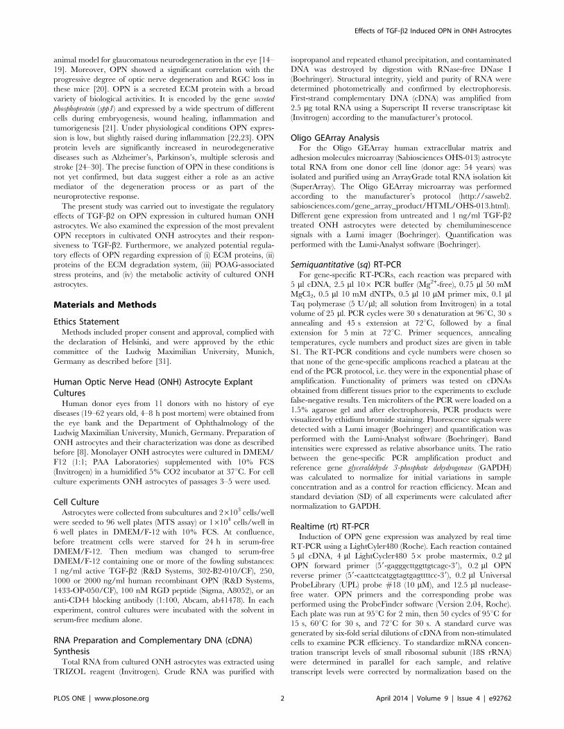
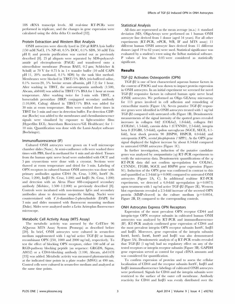
TGF-b2 Activates Osteopontin (OPN)TGF-b2 is one of best characterized aqueous humor factors in
the context of PAOG and was shown to impact protein expression
in ONH astrocytes. In an initial experiment we screened for novel
TGF-b2 responsive factors in cultured human optic nerve head
(ONH) astrocytes. We performed an Oligo GEArray microarray
for 113 genes involved in cell adhesion and remodeling of
extracellular matrix (Figure 1A). Seven putative TGF-b2 respon-
sive genes were identified in ONH astrocytes treated with 1 ng/ml
TGF-b2 compared with untreated cells (Figure 1B). Densitometric
measurements of the signal intensity of the spotted genes revealed
increases in collagen 6a2 (COL6a2, 1.8-fold), collagen 8a1(COL8a1, 1.5-fold), catenin delta 1 (CTNND1, 2.5-fold), integrin
beta 8 (ITGB8, 5.9-fold), epsilon sarcoglycan (SGCE, SECE, 4.1-
fold), heat shock protein 90 (HSP90, HSPCB, 4.0-fold) and
osteopontin (OPN, secreted phosphoprotein 1 SPP1). As the OPN gene
signal displayed the highest increase by about 8.3-fold compared
to untreated ONH astrocytes (Figure 1C).
In further investigation, induction of the putative candidate
genes was analyzed by semiquantitative (sq) RT-PCR experiments to
verify the microarray data. Densitometric quantifications of the sq
RT-PCR data did not confirm up-regulation for COL8a1,CTNND1, ITGB8, SGCE and HSP90 gene expression (Figure
S1). Induction of the OPN gene was confirmed in contrast to this
and quantified as 2.3-fold (p = 0.008) compared to untreated ONH
astrocytes (Figure 2A, C). In additional realtime RT-PCR
experiments, we detected a 6.0-fold up-regulation (p= 0.0073)
upon treatment with 1 ng/ml active TGF-b2 (Figure 2E). Western
blot experiments revealed a 2.5-fold increase of the secreted OPN
protein (MMP-cleaved) in the culture medium (p= 0.0054,
Figure 2B, D) compared to the corresponding controls.
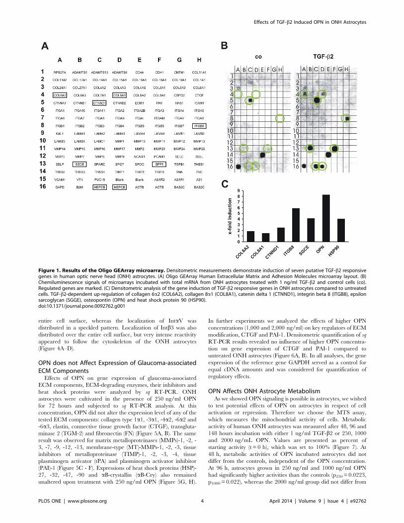
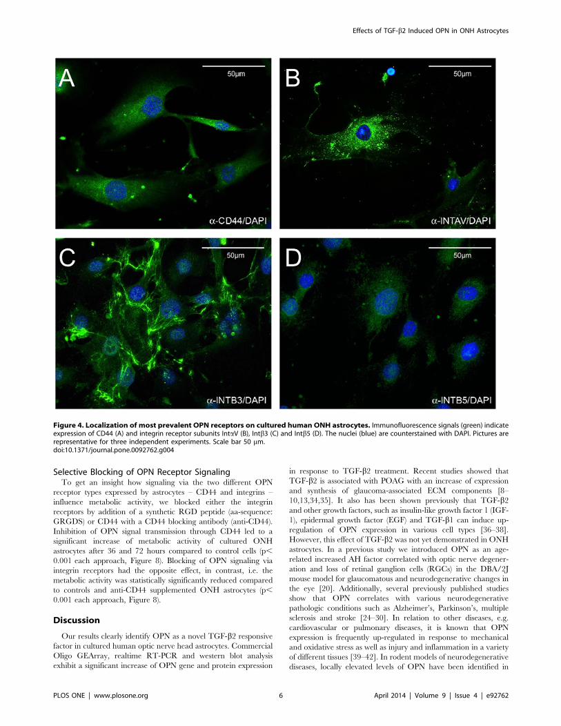
ONH Astrocytes Express OPN ReceptorsExpression of the most prevalent OPN receptor CD44 and
integrin-type OPN receptor subunits in cultivated human ONH
astrocytes was analyzed by RT-PCR and immunofluorescence
(IF). RT-PCR analysis confirmed gene expression of CD44 and
the most prevalent integrin OPN receptor subunits IntaV, Intb3and Intb5. Moreover, gene expression of the integrin subunits
Inta4, Inta5, Inta6, Inta9 and Intb1 was also demonstrated
(Figure 3A). Densitometric analysis of sq RT-PCR results revealed
that TGF-b2 (1 ng/ml) had no regulatory effect on any of the
tested receptors or integrin receptor subunits (Figure 3B). GAPDH
gene expression served as control for equal cDNA amounts and
was considered for quantification.
To confirm expression of proteins and to assess the cellular
localization of CD44 and the receptor subunits IntaV, Intb3 and
Intb5 immunofluorescence labelings of cultivated ONH astrocytes
were performed. Signals for CD44 and the integrin subunits were
restricted to the surface of the outer cell membrane. Antibody
reactivity for CD44 and Intb5 was evenly distributed over the
Effects of TGF-b2 Induced OPN in ONH Astrocytes
PLOS ONE | www.plosone.org 3 April 2014 | Volume 9 | Issue 4 | e92762
entire cell surface, whereas the localization of IntaV was
distributed in a speckled pattern. Localization of Intb3 was also
distributed over the entire cell surface, but very intense reactivity
appeared to follow the cytoskeleton of the ONH astrocytes
(Figure 4A–D).
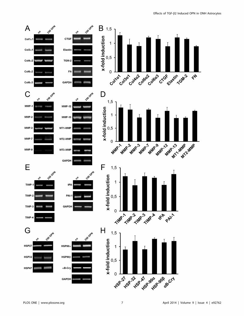
OPN does not Affect Expression of Glaucoma-associatedECM ComponentsEffects of OPN on gene expression of glaucoma-associated
ECM components, ECM-degrading enzymes, their inhibitors and
heat shock proteins were analyzed by sq RT-PCR. ONH
astrocytes were cultivated in the presence of 250 ng/ml OPN
for 72 hours and subjected to sq RT-PCR analysis. At this
concentration, OPN did not alter the expression level of any of the
tested ECM components: collagen type 1a1, -3a1, -4a2, -6a2 and
-6a3, elastin, connective tissue growth factor (CTGF), transgluta-
minase 2 (TGM-2) and fibronectin (FN) (Figure 5A, B). The same
result was observed for matrix metalloproteinases (MMPs)-1, -2, -
3, -7, -9, -12, -13, membrane-type (MT)-MMPs-1, -2, -3, tissue
inhibitors of metalloproteinase (TIMP)-1, -2, -3, -4, tissue
plasminogen activator (tPA) and plasminogen activator inhibitor
(PAI)-1 (Figure 5C - F). Expressions of heat shock proteins (HSP)-
27, -32, -47, -90 and aB-crystallin (aB-Cry) also remained
unaltered upon treatment with 250 ng/ml OPN (Figure 5G, H).
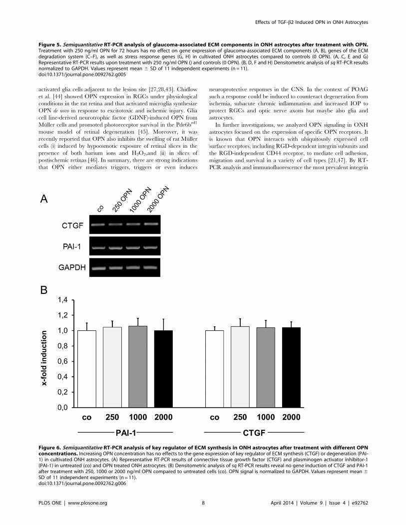
In further experiments we analyzed the effects of higher OPN
concentrations (1,000 and 2,000 ng/ml) on key regulators of ECM
modification, CTGF and PAI-1. Densitometric quantification of sq
RT-PCR results revealed no influence of higher OPN concentra-
tion on gene expression of CTGF and PAI-1 compared to
untreated ONH astrocytes (Figure 6A, B). In all analyses, the gene
expression of the reference gene GAPDH served as a control for
equal cDNA amounts and was considered for quantification of
regulatory effects.
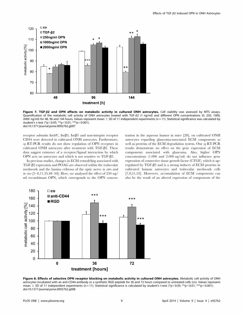
OPN Affects ONH Astrocyte MetabolismAs we showed OPN signaling is possible in astrocytes, we wished
to test potential effects of OPN on astrocytes in respect of cell
activation or repression. Therefore we choose the MTS assay,
which measures the mitochondrial activity of cells. Metabolic
activity of human ONH astrocytes was measured after 48, 96 and
148 hours incubation with either 1 ng/ml TGF-b2 or 250, 1000
and 2000 ng/mL OPN. Values are presented as percent of
starting activity (t = 0 h), which was set to 100% (Figure 7). At
48 h, metabolic activities of OPN incubated astrocytes did not
differ from the controls, independent of the OPN concentration.
At 96 h, astrocytes grown in 250 ng/ml and 1000 ng/ml OPN
had significantly higher activities than the controls (p250 = 0.0223,
p1000 = 0.022), whereas the 2000 ng/ml group did not differ from
Figure 1. Results of the Oligo GEArray microarray. Densitometric measurements demonstrate induction of seven putative TGF-b2 responsivegenes in human optic nerve head (ONH) astrocytes. (A) Oligo GEArray Human Extracellular Matrix and Adhesion Molecules microarray layout. (B)Chemiluminescence signals of microarrays incubated with total mRNA from ONH astrocytes treated with 1 ng/ml TGF-b2 and control cells (co).Regulated genes are marked. (C) Densitometric analysis of the gene induction of TGF-b2 responsive genes in ONH astrocytes compared to untreatedcells. TGF-b2-dependent up-regulation of collagen 6a2 (COL6A2), collagen 8a1 (COL8A1), catenin delta 1 (CTNND1), integrin beta 8 (ITGB8), epsilonsarcoglycan (SGGE), osteopontin (OPN) and heat shock protein 90 (HSP90).doi:10.1371/journal.pone.0092762.g001
Effects of TGF-b2 Induced OPN in ONH Astrocytes
PLOS ONE | www.plosone.org 4 April 2014 | Volume 9 | Issue 4 | e92762
the controls. Quantification at 144 h showed a similar result, i.e.
activities of the 250 ng/ml and 1000 ng/ml astrocytes were
significantly increased (p250 = 0.0056, p1000 = 0.0105) and activity
of the 2000 ng/ml group was in the range of the controls.
Astrocytes incubated with TGF-b2, in contrast, showed signifi-
cantly lower activities at all-time points compared to control
astrocytes (p48 = 0.0312, p96 = 0.0007, p144 = 0.0026) and all
astrocytes grown in presence of OPN (p,0.05 for each approach).
Figure 2. Quantification of OPN induction in TGF-b2 treated ONH astrocytes. Analysis of OPN expression in ONH astrocytes after treatmentwith 1 ng/ml TGF-b2 for 72 hours. (A) Semiquantitative (sq) RT-PCR analysis indicates an up-regulation of OPN transcript in TGF-b2 treated ONHastrocytes compared to control cells (co). (B) Western blot analysis demonstrates an increase in secreted OPN protein (MMP-cleaved) in TGF-b2treated ONH astrocytes compared to controls. (C) Densitometric quantification of sq RT-PCR reveals significant induction of OPN mRNA (2.3-fold,p = 0.008). OPN signals are normalized to GAPDH. (D) Quantification of western blot results reveals significantly increased OPN secretion intosupernatant (2.5-fold, p = 0.0054). OPN western blots are normalized within the b-actin signal on the same nitrocellulose membrane. (E) Statisticalanalysis of real time RT-PCR shows significant up-regulation of OPN transcript (6-fold, p = 0.0073). Values represent mean 6 SD of 11 independentexperiments (n = 11).doi:10.1371/journal.pone.0092762.g002
Figure 3. Semiquantitative RT-PCR analyses of most prevalent OPN receptors in cultivated ONH astrocytes and effect of TGF-b2. (A)Representative RT-PCR results of OPN receptor gene expression in untreated (co) and TGF-b2 (1 ng/ml, 72 h) treated ONH astrocytes. (B)Densitometric analysis of sq RT-PCR results does not demonstrate any regulation of OPN receptors in TGF-b2 treated cells compared to controls. OPNsignal is normalized to GAPDH. Values represent mean 6 SD of 11 independent experiments (n = 11).doi:10.1371/journal.pone.0092762.g003
Effects of TGF-b2 Induced OPN in ONH Astrocytes
PLOS ONE | www.plosone.org 5 April 2014 | Volume 9 | Issue 4 | e92762
Selective Blocking of OPN Receptor SignalingTo get an insight how signaling via the two different OPN
receptor types expressed by astrocytes – CD44 and integrins –
influence metabolic activity, we blocked either the integrin
receptors by addition of a synthetic RGD peptide (aa-sequence:
GRGDS) or CD44 with a CD44 blocking antibody (anti-CD44).
Inhibition of OPN signal transmission through CD44 led to a
significant increase of metabolic activity of cultured ONH
astrocytes after 36 and 72 hours compared to control cells (p,
0.001 each approach, Figure 8). Blocking of OPN signaling via
integrin receptors had the opposite effect, in contrast, i.e. the
metabolic activity was statistically significantly reduced compared
to controls and anti-CD44 supplemented ONH astrocytes (p,
0.001 each approach, Figure 8).
Discussion
Our results clearly identify OPN as a novel TGF-b2 responsive
factor in cultured human optic nerve head astrocytes. Commercial
Oligo GEArray, realtime RT-PCR and western blot analysis
exhibit a significant increase of OPN gene and protein expression
in response to TGF-b2 treatment. Recent studies showed that
TGF-b2 is associated with POAG with an increase of expression
and synthesis of glaucoma-associated ECM components [8–
10,13,34,35]. It also has been shown previously that TGF-b2and other growth factors, such as insulin-like growth factor 1 (IGF-
1), epidermal growth factor (EGF) and TGF-b1 can induce up-
regulation of OPN expression in various cell types [36–38].
However, this effect of TGF-b2 was not yet demonstrated in ONH
astrocytes. In a previous study we introduced OPN as an age-
related increased AH factor correlated with optic nerve degener-
ation and loss of retinal ganglion cells (RGCs) in the DBA/2J
mouse model for glaucomatous and neurodegenerative changes in
the eye [20]. Additionally, several previously published studies
show that OPN correlates with various neurodegenerative
pathologic conditions such as Alzheimer’s, Parkinson’s, multiple
sclerosis and stroke [24–30]. In relation to other diseases, e.g.
cardiovascular or pulmonary diseases, it is known that OPN
expression is frequently up-regulated in response to mechanical
and oxidative stress as well as injury and inflammation in a variety
of different tissues [39–42]. In rodent models of neurodegenerative
diseases, locally elevated levels of OPN have been identified in
Figure 4. Localization of most prevalent OPN receptors on cultured human ONH astrocytes. Immunofluorescence signals (green) indicateexpression of CD44 (A) and integrin receptor subunits IntaV (B), Intb3 (C) and Intb5 (D). The nuclei (blue) are counterstained with DAPI. Pictures arerepresentative for three independent experiments. Scale bar 50 mm.doi:10.1371/journal.pone.0092762.g004
Effects of TGF-b2 Induced OPN in ONH Astrocytes
PLOS ONE | www.plosone.org 6 April 2014 | Volume 9 | Issue 4 | e92762
Effects of TGF-b2 Induced OPN in ONH Astrocytes
PLOS ONE | www.plosone.org 7 April 2014 | Volume 9 | Issue 4 | e92762
activated glia cells adjacent to the lesion site [27,28,43]. Chidlow
et al. [44] showed OPN expression in RGCs under physiological
conditions in the rat retina and that activated microglia synthesize
OPN de novo in response to excitotoxic and ischemic injury. Glia
cell line-derived neurotrophic factor (GDNF)-induced OPN from
Muller cells and promoted photoreceptor survival in the Pde6brd1
mouse model of retinal degeneration [45]. Moreover, it was
recently reported that OPN also inhibits the swelling of rat Muller
cells (i) induced by hypoosmotic exposure of retinal slices in the
presence of both barium ions and H2O2,and (ii) in slices of
postischemic retinas [46]. In summary, there are strong indications
that OPN either mediates triggers, triggers or even induces
neuroprotective responses in the CNS. In the context of POAG
such a response could be induced to counteract degeneration from
ischemia, subacute chronic inflammation and increased IOP to
protect RGCs and optic nerve axons but maybe also glia and
astrocytes.
In further investigations, we analyzed OPN signaling in ONH
astrocytes focused on the expression of specific OPN receptors. It
is known that OPN interacts with ubiquitously expressed cell
surface receptors, including RGD-dependent integrin subunits and
the RGD-independent CD44 receptor, to mediate cell adhesion,
migration and survival in a variety of cell types [21,47]. By RT-
PCR analysis and immunofluorescence the most prevalent integrin
Figure 5. Semiquantitative RT-PCR analysis of glaucoma-associated ECM components in ONH astrocytes after treatment with OPN.Treatment with 250 ng/ml OPN for 72 hours has no effect on gene expression of glaucoma-associated ECM components (A, B), genes of the ECMdegradation system (C–F), as well as stress response genes (G, H) in cultivated ONH astrocytes compared to controls (0 OPN). (A, C, E and G)Representative RT-PCR results upon treatment with 250 ng/ml OPN () and controls (0 OPN). (B, D, F and H) Densitometric analysis of sq RT-PCR resultsnormalized to GAPDH. Values represent mean 6 SD of 11 independent experiments (n = 11).doi:10.1371/journal.pone.0092762.g005
Figure 6. Semiquantitative RT-PCR analysis of key regulator of ECM synthesis in ONH astrocytes after treatment with different OPNconcentrations. Increasing OPN concentration has no effects to the gene expression of key regulator of ECM synthesis (CTGF) or degeneration (PAI-1) in cultivated ONH astrocytes. (A) Representative RT-PCR results of connective tissue growth factor (CTGF) and plasminogen activator inhibitor-1(PAI-1) in untreated (co) and OPN treated ONH astrocytes. (B) Densitometric analysis of sq RT-PCR results reveal no gene induction of CTGF and PAI-1after treatment with 250, 1000 or 2000 ng/ml OPN compared to untreated cells (co). OPN signal is normalized to GAPDH. Values represent mean 6SD of 11 independent experiments (n = 11).doi:10.1371/journal.pone.0092762.g006
Effects of TGF-b2 Induced OPN in ONH Astrocytes
PLOS ONE | www.plosone.org 8 April 2014 | Volume 9 | Issue 4 | e92762
receptor subunits IntaV, Intb3, Intb5 and non-integrin receptor
CD44 were detected in cultivated ONH astrocytes. Furthermore,
sq RT-PCR results do not show regulation of OPN receptors in
cultivated ONH astrocytes after treatment with TGF-b2. Thesedata suggest existence of a receptor/ligand interaction by which
OPN acts on astrocytes and which is not sensitive to TGF-b2.In previous studies, changes in ECM remodeling associated with
TGF-b2 expression and POAG are observed within the trabecular
meshwork and the lamina cribrosa of the optic nerve in vitro and
in vivo [5–8,11,35,48–50]. Here, we analyzed the effect of 250 ng/
ml recombinant OPN, which corresponds to the OPN concen-
tration in the aqueous humor in mice [20], on cultivated ONH
astrocytes regarding glaucoma-associated ECM components as
well as proteins of the ECM degradation system. Our sq RT-PCR
results demonstrate no effect on the gene expression of ECM
components associated with glaucoma. Also, higher OPN
concentrations (1.000 and 2.000 ng/ml) do not influence gene
expression of connective tissue growth factor (CTGF), which is up-
regulated by TGF-b2 and is a strong inducer of ECM proteins in
cultivated human astrocytes and trabecular meshwork cells
[7,8,51,52]. Moreover, accumulation of ECM components can
also be the result of an altered expression of components of the
Figure 7. TGF-b2 and OPN effects on metabolic activity in cultured ONH astrocytes. Cell viability was assessed by MTS assays.Quantification of the metabolic cell activity of ONH astrocytes treated with TGF-b2 (1 ng/ml) and different OPN concentrations (0, 250, 1000,2000 ng/ml) for 48, 96 and 144 hours. Values represent mean 6 SD of 11 independent experiments (n = 11). Statistical significance was calculated bystudent’s t-test (*p,0.05; **p,0.01; ***p,0.001).doi:10.1371/journal.pone.0092762.g007
Figure 8. Effects of selective OPN receptor blocking on metabolic activity in cultured ONH astrocytes. Metabolic cell activity of ONHastrocytes incubated with an anti-CD44 antibody or a synthetic RGD peptide for 36 and 72 hours compared to untreated cells (co). Values representmean 6 SD of 11 independent experiments (n = 11). Statistical significance is calculated by student’s t-test (*p,0.05; **p,0.01; ***p,0.001).doi:10.1371/journal.pone.0092762.g008
Effects of TGF-b2 Induced OPN in ONH Astrocytes
PLOS ONE | www.plosone.org 9 April 2014 | Volume 9 | Issue 4 | e92762
ECM degradation system. Apart from a basal expression in
cultivated ONH astrocytes, no up-regulation of matrix metallo-
proteinases (MMPs), tissue inhibitors of metalloproteinases
(TIMPs) and genes involved in the activation of pro-MMP, e.g.
tissue plasminogen activator (tPA) or plasminogen activator
inhibitor (PAI)-1, is detectable at the mRNA level after OPN
treatment. Gene expression of PAI-1, an inhibitor of the
plasminogen activation system induced by TGF-b2 [53], is not
influenced by higher OPN concentrations. Furthermore, our sq
RT-PCR analysis with heat shock proteins (HSP), frequently
described for glaucomatous diseases, revealed no activation of
stress response in ONH astrocytes by OPN. Yu et al. were able to
show in a previous study that H2O2-induced oxidative stress and
TGF-b2 reactivate ONH astrocytes by increasing Hsp32 and -47
expression in vitro [54]. In summary, we conclude from these data
that OPN is not involved in the TGF-b2 induced activation of
POAG-associated ECM and stress response genes. However, other
studies show that OPN is required for the activation, migration,
proliferation, and differentiation of fibroblasts and is up-regulated
in several fibrotic diseases [55–57]. In a dystrophic mouse model
(mdx mice), OPN promotes fibrosis in muscle by modulating
immune cell subsets and intramuscular TGF-beta [58]. Abu El-
Asrar et al. [59] demonstrated that OPN and other regulators of
angiogenesis and fibrogenesis contribute to the pathogenesis of
proliferative vitreoretinal disorders in humans. Another study
showed that OPN is expressed in injured lens epithelial cells in
association with fibrotic scar formation in mice and humans [60].
In early stages of POAG, an increase of astrocytes correspond-
ing to an astrogliosis is often detectable. In late-stage glaucoma,
however, a decreased count of astrocytes in the optic nerve is
observed [4,6,49,50,61]. These findings suggest that proliferation
and/or cell survival are deregulated in glaucomatous eyes. For that
reason we analyzed the effect of OPN and TGF-b2 on the cell
viability of cultivated human ONH astrocytes by means of
metabolic activity assays. Our results show a time- and dose-
dependent pro-metabolic effect of OPN on cultured human ONH
astrocytes. This positive effect on cell viability might also indicate a
neuroprotective impact of OPN. Tambuyzer et al., [62] showed
that OPN containing medium of a pig renal epithelial cell line
(LLC-PK1) doubled the rate of proliferation of porcine microglia
in vitro. The additional application of an anti-OPN polyclonal
antibody completely reversed this effect [62]. Interestingly, a
higher OPN concentration (2.000 ng/ml) does not further
increase the metabolic activity of ONH astrocytes. These data
are in line with a previous study, which indicated a limiting OPN
concentration and anti-proliferative effect at higher OPN doses in
murine neuronal precursor cells (RGC5) and ex vivo cultivated
DBA/2J eyes in a long-time cultivation assay [20]. In contrast,
ONH astrocytes treated with TGF-b2 displayed significantly
reduced metabolic activity at all observed time points. This result
shows that ONH astrocytes are TGF-b2 sensitive and indicate an
anti-proliferation effect on these cells. Thus, it is tempting to
speculate that OPN might be involved in counteracting the anti-
proliferation TGF-b2 effect on ONH astrocytes.
Even our OPN receptor blocking experiments in cultivated
astrocytes provide pro- and anti-metabolic effects by blocking
OPN specific receptors. Blocking of RGD-independent CD44 led
to increased metabolic activity, thus perhaps indicating an
activation of the anti-proliferative pathway via the CD44 receptor.
This effect may be ONH astrocyte-specific. In IL-3-dependent
bone marrow cells, OPN promotes proliferation and survival via
CD44 [63]. Blocking of RGD-dependent integrin receptors
otherwise significantly reduced metabolic activity in astrocytes,
which could be a link to a pro-proliferation signal transduced by
integrin receptors. Meller et al. [28] demonstrated that OPN is a
potent neuroprotective factor against ischemic injury depending
on integrin-binding RGD motif and the activation of Akt and
p42/p44 MAPK pathways. Several other studies have indicated
that OPN mediates a pro-survival and anti-apoptotic signal to
different cell types by inhibiting apoptosis induced by different
pathological events and lack of growth factors [21]. Our initial
data, however, require supplementation by further studies focusing
on the correlation of OPN and astrocyte counts in different clinical
stages of POAG eyes. The above-mentioned astrogliosis and
degeneration of astrocytes is very likely only an accompanying
phenomenon of glaucoma, but the disease does correlate with the
degeneration of optic nerve axons and retinal ganglion cells.
In conclusion, we show in this study that OPN is a novel TGF-
b2 responsive factor in cultured human ONH astrocytes and
might be part of a rescue mechanism to counteract neurodegen-
erative effects of glaucoma-associated TGF-b2. It is conceivablethat OPN is secreted by ONH astrocytes to protect the neurons of
the optic nerve from mechanical and oxidative stress. Neverthe-
less, further studies are required to confirm a neuroprotective
function of OPN in relation to retinal ganglion cells (RGCs) and
optic nerve axons in glaucoma models.
Supporting Information
Figure S1 RT-PCR confirmation of the Oligo GEArraymicroarray data. (A) Semiquantitative (sq) RT-PCR analysis of
COL8a1, CTNND1, ITGB8, SGCE, HSP90 and OPN gene
expression in untreated (co) and TGF-b2 treated astrocytes. (B)
Densitometric analysis of the sq RT-PCR results. Signals are
normalized to GAPDH. Values represent mean 6 SD of 11
independent experiments (n = 11). Statistical significance is
calculated by student’s t-test (**p#0.01).
(TIFF)
Table S1 Primers and settings used for semiquantita-tive (sq) RT-PCR analysis. At = annealing temperature.
(DOCX)
Acknowledgments
The authors thank Julia Muller-Mausolf, Hong Nguyen and Anke Fischer
for excellent technical assistance. We thank Sylvia Dyczek for helpful
advice with the translation of the manuscript.
Author Contributions
Conceived and designed the experiments: CN FG MTB MS. Performed
the experiments: CN FG MTB MS. Analyzed the data: CN FG FP CMH
MTB MS. Contributed reagents/materials/analysis tools: CN FG FP
CMH MTB MS. Wrote the paper: CN FG FP CMH MTB MS.
References
1. Quigley HA, Broman AT (2006) The number of people with glaucoma
worldwide in 2010 and 2020. Br J Ophthalmol 90: 262–267.
2. Diekmann H, Fischer D (2013) Glaucoma and optic nerve repair. Cell Tissue
Res. 353(2): 327–37.
3. Nickells RW, Howell GR, Soto I, John SW (2012) Under pressure: cellular and
molecular responses during glaucoma, a common neurodegeneration with
axonopathy. Annu Rev Neurosci 35: 153–179.
4. Tektas OY, Lutjen-Drecoll E, Scholz M (2010) Qualitative and quantitative
morphologic changes in the vasculature and extracellular matrix of the
Effects of TGF-b2 Induced OPN in ONH Astrocytes
PLOS ONE | www.plosone.org 10 April 2014 | Volume 9 | Issue 4 | e92762
prelaminar optic nerve head in eyes with POAG. Invest Ophthalmol Vis Sci 51:5083–5091.
5. Lutjen-Drecoll E (2005) Morphological changes in glaucomatous eyes and the
role of TGFbeta2 for the pathogenesis of the disease. Exp Eye Res 81: 1–4.
6. Gottanka J, Kuhlmann A, Scholz M, Johnson DH, Lutjen-Drecoll E (2005)Pathophysiologic changes in the optic nerves of eyes with primary open angle
and pseudoexfoliation glaucoma. Invest Ophthalmol Vis Sci 46: 4170–4181.
7. Fuchshofer R, Birke M, Welge-Lussen U, Kook D, Lutjen-Drecoll E (2005)Transforming growth factor-beta 2 modulated extracellular matrix component
expression in cultured human optic nerve head astrocytes. Invest OphthalmolVis Sci 46: 568–578.
8. Neumann C, Yu A, Welge-Lussen U, Lutjen-Drecoll E, Birke M (2008) The
effect of TGF-beta2 on elastin, type VI collagen, and components of theproteolytic degradation system in human optic nerve astrocytes. Invest
Ophthalmol Vis Sci 49: 1464–1472.
9. Wordinger RJ, Fleenor DL, Hellberg PE, Pang IH, Tovar TO, et al. (2007)Effects of TGF-beta2, BMP-4, and gremlin in the trabecular meshwork:
implications for glaucoma. Invest Ophthalmol Vis Sci 48: 1191–1200.
10. Fuchshofer R, Tamm ER (2012) The role of TGF-beta in the pathogenesis ofprimary open-angle glaucoma. Cell Tissue Res 347: 279–290.
11. Fuchshofer R, Welge-Lussen U, Lutjen-Drecoll E (2003) The effect of TGF-beta2 on human trabecular meshwork extracellular proteolytic system. Exp Eye
Res 77: 757–765.
12. Picht G, Welge-Luessen U, Grehn F, Lutjen-Drecoll E (2001) Transforminggrowth factor beta 2 levels in the aqueous humor in different types of glaucoma
and the relation to filtering bleb development. Graefes Arch Clin Exp
Ophthalmol 239: 199–207.
13. Tripathi RC, Li J, Chan WF, Tripathi BJ (1994) Aqueous humor in
glaucomatous eyes contains an increased level of TGF-beta 2. Exp Eye Res
59: 723–727.
14. Anderson MG, Libby RT, Mao M, Cosma IM, Wilson LA, et al. (2006) Genetic
context determines susceptibility to intraocular pressure elevation in a mousepigmentary glaucoma. BMC Biol 4: 20.
15. Heiduschka P, Julien S, Schuettauf F, Schnichels S (2010) Loss of retinal
function in aged DBA/2J mice - New insights into retinal neurodegeneration.Exp Eye Res 91: 779–783.
16. Howell GR, Libby RT, John SW (2008) Mouse genetic models: an ideal system
for understanding glaucomatous neurodegeneration and neuroprotection. ProgBrain Res 173: 303–321.
17. Inman DM, Sappington RM, Horner PJ, Calkins DJ (2006) Quantitative
correlation of optic nerve pathology with ocular pressure and corneal thicknessin the DBA/2 mouse model of glaucoma. Invest Ophthalmol Vis Sci 47: 986–
996.
18. Jakobs TC, Libby RT, Ben Y, John SW, Masland RH (2005) Retinal ganglioncell degeneration is topological but not cell type specific in DBA/2J mice. J Cell
Biol 171: 313–325.
19. Scholz M, Buder T, Seeber S, Adamek E, Becker CM, et al. (2008) Dependencyof intraocular pressure elevation and glaucomatous changes in DBA/2J and
DBA/2J-Rj mice. Invest Ophthalmol Vis Sci 49: 613–621.
20. Birke MT, Neumann C, Birke K, Kremers J, Scholz M (2010) Changes ofosteopontin in the aqueous humor of the DBA2/J glaucoma model correlated
with optic nerve and RGC degenerations. Investigative ophthalmology & visualscience 51: 5759–5767.
21. Wang KX, Denhardt DT (2008) Osteopontin: Role in immune regulation and
stress responses. Cytokine Growth Factor Rev.
22. Hashimoto M, Sun D, Rittling SR, Denhardt DT, Young W (2007)
Osteopontin-deficient mice exhibit less inflammation, greater tissue damage,
and impaired locomotor recovery from spinal cord injury compared with wild-type controls. J Neurosci 27: 3603–3611.
23. Scatena M, Liaw L, Giachelli CM (2007) Osteopontin: a multifunctional
molecule regulating chronic inflammation and vascular disease. Arteriosclerosis,thrombosis, and vascular biology 27: 2302–2309.
24. Comi C, Carecchio M, Chiocchetti A, Nicola S, Galimberti D, et al. (2009)Osteopontin is Increased in the Cerebrospinal Fluid of Patients with Alzheimer’s
Disease and Its Levels Correlate with Cognitive Decline. J Alzheimers Dis.
25. Iczkiewicz J, Jackson MJ, Smith LA, Rose S, Jenner P (2006) Osteopontinexpression in substantia nigra in MPTP-treated primates and in Parkinson’s
disease. Brain Res 1118: 239–250.
26. Jin JK, Na YJ, Moon C, Kim H, Ahn M, et al. (2006) Increased expression ofosteopontin in the brain with scrapie infection. Brain Res 1072: 227–233.
27. Maetzler W, Berg D, Schalamberidze N, Melms A, Schott K, et al. (2007)
Osteopontin is elevated in Parkinson’s disease and its absence leads to reducedneurodegeneration in the MPTP model. Neurobiol Dis 25: 473–482.
28. Meller R, Stevens SL, Minami M, Cameron JA, King S, et al. (2005)
Neuroprotection by osteopontin in stroke. J Cereb Blood Flow Metab 25: 217–225.
29. Schroeter M, Zickler P, Denhardt DT, Hartung HP, Jander S (2006) Increased
thalamic neurodegeneration following ischaemic cortical stroke in osteopontin-deficient mice. Brain : a journal of neurology 129: 1426–1437.
30. McFarland HF, Martin R (2007) Multiple sclerosis: a complicated picture of
autoimmunity. 8: 913–919.
31. Yu AL, Fuchshofer R, Birke M, Priglinger SG, Eibl KH, et al. (2007) Hypoxia/
reoxygenation and TGF-beta increase alphaB-crystallin expression in human
optic nerve head astrocytes. Experimental eye research 84: 694–706.
32. Pfaffl MW (2001) A new mathematical model for relative quantification in real-
time RT-PCR. Nucleic Acids Res 29: e45.
33. Weber GF, Ashkar S, Glimcher MJ, Cantor H (1996) Receptor-ligand
interaction between CD44 and osteopontin (Eta-1). Science 271: 509–512.
34. Yu AL, Birke K, Moriniere J, Welge-Lussen U (2010) TGF-{beta}2 induces
senescence-associated changes in human trabecular meshwork cells. Invest
Ophthalmol Vis Sci 51: 5718–5723.
35. Gottanka J, Chan D, Eichhorn M, Lutjen-Drecoll E, Ethier CR (2004) Effects of
TGF-beta2 in perfused human eyes. Invest Ophthalmol Vis Sci 45: 153–158.
36. Padanilam BJ, Martin DR, Hammerman MR (1996) Insulin-like growth factor
I-enhanced renal expression of osteopontin after acute ischemic injury in rats.
Endocrinology 137: 2133–2140.
37. Zhang GX, Zhao ZQ, Wang HD, Hao B (2004) Enhancement of osteopontin
expression in HepG2 cells by epidermal growth factor via phosphatidylinositol 3-
kinase signaling pathway. World J Gastroenterol 10: 205–208.
38. Noda M, Yoon K, Prince CW, Butler WT, Rodan GA (1988) Transcriptional
regulation of osteopontin production in rat osteosarcoma cells by type beta
transforming growth factor. J Biol Chem 263: 13916–13921.
39. Gross TS, King KA, Rabaia NA, Pathare P, Srinivasan S (2005) Upregulation of
osteopontin by osteocytes deprived of mechanical loading or oxygen. J Bone
Miner Res 20: 250–256.
40. Okamoto H (2007) Osteopontin and cardiovascular system. Mol Cell Biochem
300: 1–7.
41. Heguy A, O’Connor TP, Luettich K, Worgall S, Cieciuch A, et al. (2006) Gene
expression profiling of human alveolar macrophages of phenotypically normal
smokers and nonsmokers reveals a previously unrecognized subset of genes
modulated by cigarette smoking. J Mol Med (Berl) 84: 318–328.
42. Denhardt DT, Noda M (1998) Osteopontin expression and function: role in
bone remodeling. J Cell Biochem Suppl 30–31: 92–102.
43. Lee MY, Shin SL, Choi YS, Kim EJ, Cha JH, et al. (1999) Transient
upregulation of osteopontin mRNA in hippocampus and striatum following
global forebrain ischemia in rats. Neurosci Lett 271: 81–84.
44. Chidlow G, Wood JP, Manavis J, Osborne NN, Casson RJ (2008) Expression of
osteopontin in the rat retina: effects of excitotoxic and ischemic injuries. Invest
Ophthalmol Vis Sci 49: 762–771.
45. Del Rio P, Irmler M, Arango-Gonzalez B, Favor J, Bobe C, et al. (2011) GDNF-
induced osteopontin from Muller glial cells promotes photoreceptor survival in
the Pde6brd1 mouse model of retinal degeneration. Glia 59: 821–832.
46. Wahl V, Vogler S, Grosche A, Pannicke T, Ueffing M, et al. (2013) Osteopontin
inhibits osmotic swelling of retinal glial (Muller) cells by inducing release of
VEGF. Neuroscience.
47. Kazanecki CC, Uzwiak DJ, Denhardt DT (2007) Control of osteopontin
signaling and function by post-translational phosphorylation and protein folding.
J Cell Biochem 102: 912–924.
48. Hernandez MR (2000) The optic nerve head in glaucoma: role of astrocytes in
tissue remodeling. Prog Retin Eye Res 19: 297–321.
49. Hernandez MR, Miao H, Lukas T (2008) Astrocytes in glaucomatous optic
neuropathy. Prog Brain Res 173: 353–373.
50. Pena JD, Agapova O, Gabelt BT, Levin LA, Lucarelli MJ, et al. (2001) Increased
elastin expression in astrocytes of the lamina cribrosa in response to elevated
intraocular pressure. Invest Ophthalmol Vis Sci 42: 2303–2314.
51. Iyer P, Maddala R, Pattabiraman PP, Rao PV (2012) Connective tissue growth
factor-mediated upregulation of neuromedin U expression in trabecular
meshwork cells and its role in homeostasis of aqueous humor outflow. Invest
Ophthalmol Vis Sci 53: 4952–4962.
52. Junglas B, Yu AH, Welge-Lussen U, Tamm ER, Fuchshofer R (2009)
Connective tissue growth factor induces extracellular matrix deposition in
human trabecular meshwork cells. Exp Eye Res 88: 1065–1075.
53. Eitzman DT, Ginsburg D (1997) Of mice and men. The function of
plasminogen activator inhibitors (PAIs) in vivo. Adv Exp Med Biol 425: 131–
141.
54. Yu AL, Moriniere J, Birke M, Neumann C, Fuchshofer R, et al. (2009)
Reactivation of optic nerve head astrocytes by TGF-beta2 and H2O2 is
accompanied by increased Hsp32 and Hsp47 expression. Invest Ophthalmol Vis
Sci 50: 1707–1717.
55. Kohan M, Breuer R, Berkman N (2009) Osteopontin induces airway remodeling
and lung fibroblast activation in a murine model of asthma. Am J Respir Cell
Mol Biol 41: 290–296.
56. Pardo A, Gibson K, Cisneros J, Richards TJ, Yang Y, et al. (2005) Up-regulation
and profibrotic role of osteopontin in human idiopathic pulmonary fibrosis.
PLoS Med 2: e251.
57. Sabo-Attwood T, Ramos-Nino ME, Eugenia-Ariza M, Macpherson MB, Butnor
KJ, et al. (2011) Osteopontin modulates inflammation, mucin production, and
gene expression signatures after inhalation of asbestos in a murine model of
fibrosis. Am J Pathol 178: 1975–1985.
58. Vetrone SA, Montecino-Rodriguez E, Kudryashova E, Kramerova I, Hoffman
EP, et al. (2009) Osteopontin promotes fibrosis in dystrophic mouse muscle by
modulating immune cell subsets and intramuscular TGF-beta. J Clin Invest 119:
1583–1594.
59. Abu El-Asrar AM, Imtiaz Nawaz M, Kangave D, Siddiquei MM, Geboes K
(2012) Osteopontin and other regulators of angiogenesis and fibrogenesis in the
vitreous from patients with proliferative vitreoretinal disorders. Mediators of
inflammation 2012: 493043.
Effects of TGF-b2 Induced OPN in ONH Astrocytes
PLOS ONE | www.plosone.org 11 April 2014 | Volume 9 | Issue 4 | e92762

60. Saika S, Miyamoto T, Ishida I, Ohnishi Y, Ooshima A (2003) Osteopontin: a
component of matrix in capsular opacification and subcapsular cataract. Invest
Ophthalmol Vis Sci 44: 1622–1628.
61. Hernandez MR, Andrzejewska WM, Neufeld AH (1990) Changes in the
extracellular matrix of the human optic nerve head in primary open-angle
glaucoma. Am J Ophthalmol 109: 180–188.
62. Tambuyzer BR, Casteleyn C, Vergauwen H, Van Cruchten S, Van Ginneken C
(2012) Osteopontin alters the functional profile of porcine microglia in vitro. CellBiol Int 36: 1233–1238.
63. Lin YH, Huang CJ, Chao JR, Chen ST, Lee SF, et al. (2000) Coupling of
osteopontin and its cell surface receptor CD44 to the cell survival responseelicited by interleukin-3 or granulocyte-macrophage colony-stimulating factor.
Mol Cell Biol 20: 2734–2742.
Effects of TGF-b2 Induced OPN in ONH Astrocytes
PLOS ONE | www.plosone.org 12 April 2014 | Volume 9 | Issue 4 | e92762