Embed Size (px)
Citation preview
For personal use. Only reproduce with permission from The Lancet publishing Group.
SEMINAR
Osteogenesis imperfecta is a genetic disorder of increasedbone fragility and low bone mass. Severity varies widely,ranging from intrauterine fractures and perinatal lethality tovery mild forms without fractures.1 Typical extraskeletalmanifestations can be associated variably with the disorder.These include blue sclera, dentinogenesis imperfecta,hyperlaxity of ligaments and skin, hearing impairment, andpresence of wormian bones on skull radiographs. Mostpatients with a clinical diagnosis of osteogenesis imperfectahave a mutation in one of the two genes that encode the� chains of collagen type 1 (COL1A1 and COL1A2).
Diagnosis and classification Diagnosis The clinical diagnosis of osteogenesis imperfecta is basedmainly on the signs and symptoms outlined above.Traditionally, much emphasis has been laid on the presenceor absence of blue sclera and dentinogenesis imperfecta asdiagnostic signs of osteogenesis imperfecta. This practicestill holds true, but some limitations should be recognised.Dark or bluish sclerae are very typical in healthy infants, andtherefore this finding is not of much diagnostic use in thisage-group. Dentinogenesis imperfecta is more frequentlyclinically evident in primary than in permanent teeth ofpatients with osteogenesis imperfecta.2 Radiological orhistological examinations frequently show abnormalities,even in individuals whose teeth look normal oninspection.3–5
Clinically evident hearing loss is rare in the first twodecades of life, even though subtle audiometric abnor-malities can be recorded in a large proportion of childrenand adolescents with osteogenesis imperfecta.6–8 About halfof patients older than age 50 years report hearing loss, andan even higher proportion of adults have clearly pathologicalaudiometric findings.8,9
Diagnosis of osteogenesis imperfecta is straightforward inindividuals with a positive family history or in whom severaltypical features are present, but can be difficult in theabsence of affected family members and when bone fragility
Lancet 2004; 363: 1377–85
Genetics Unit, Shriners Hospital for Children and McGill University,1529 Cedar Avenue, Montréal, Québec, Canada H3G 1A6 (F Rauch MD, Prof F H Glorieux MD)
Correspondence to: Prof Francis H Glorieux (e-mail: [email protected])
is not associated with obvious extraskeletal abnormalities.The uncertainty in such cases is compounded by the factthat there are no agreed minimum criteria that establish aclinical diagnosis of the disorder. In this situation, analysisof the collagen type 1 genes can provide helpful infor-mation, which can be done by investigating the amount andstructure of type 1 procollagen molecules that are derivedfrom the patient’s cultured skin fibroblasts.10 Alternatively,genomic DNA can be extracted from white blood cells andthe coding region of the COL1A1 and COL1A2 genes canthen be screened for mutations.11 Both of these approachesare thought to detect almost 90% of all collagen type 1mutations.12 A positive collagen type 1 study thus confirmsthe diagnosis of osteogenesis imperfecta. However, anegative result leaves open the possibility that either acollagen type 1 mutation is present but was not detected orthe patient has a form of the disorder that is not associatedwith collagen type 1 mutations (see below). Therefore, anegative collagen type 1 study does not rule outosteogenesis imperfecta.
Classification Even though the range of clinical severity in osteogenesisimperfecta is a continuum, categorisation of patients intoseparate types can be useful to assess prognosis and to helpassess the effects of therapeutic interventions. The mostwidely used classification of osteogenesis imperfecta is bySillence and colleagues13 and distinguishes four clinicaltypes. We have further delineated three additional groups ofpatients who had a clinical diagnosis of the disorder but whopresented clearly distinct features (table 1).14–16 The most
Osteogenesis imperfecta
Frank Rauch, Francis H Glorieux
Osteogenesis imperfecta is a genetic disorder of increased bone fragility, low bone mass, and other connective-tissuemanifestations. The most frequently used classification outlines four clinical types, which we have expanded to sevendistinct types. In most patients the disorder is caused by mutations in one of the two genes encoding collagen type 1,but in some individuals no such mutations are detectable. The most important therapeutic advance is the introductionof bisphosphonate treatment for moderate to severe forms of osteogenesis imperfecta. However, at present, the besttreatment regimen and the long-term outcomes of bisphosphonate therapy are unknown. Although this treatment doesnot constitute a cure, it is an adjunct to physiotherapy, rehabilitation, and orthopaedic care. Gene-based therapypresently remains in the early stages of preclinical research.
Seminar
THE LANCET • Vol 363 • April 24, 2004 • www.thelancet.com 1377
Search strategy and selection criteriaWe searched PubMed with the keywords “osteogenesisimperfecta”. On June 30, 2003, the database contained680 such articles that were published in January, 1995, orlater. We assessed all these database entries. This Seminardiscusses topics where, from the authors’ perspective,clinically relevant progress has taken place in recent years. Themost important publications dealing with these topics wereincluded in this Seminar. Frequently cited older published workwas also taken into account. Articles in English, French, andGerman were used. As a result of space constraints, importantcontributions had to be left out if they could not besummarised under the main headings selected by the authors.
For personal use. Only reproduce with permission from The Lancet publishing Group.
relevant clinical characteristic of all types of osteogenesisimperfecta is bone fragility, the severity of which increases inthe order type I < types IV, V, VI, VII < type III < type II.
Osteogenesis imperfecta type I includes patients withmild disease and absence of major bone deformities(table 1). However, vertebral fractures are typical and canlead to mild scoliosis. Type II is lethal in the perinatalperiod, usually because of respiratory failure resulting frommultiple rib fractures. Osteogenesis imperfecta type III isthe most severe form in children surviving the neonatalperiod. These patients are of very short stature and havelimb and spine deformities secondary to multiple fractures,which can lead to respiratory difficulties—identified as aleading cause of death in this patient group.17,18 Patients withmild to moderate bone deformities and variable shortstature are classified as osteogenesis imperfecta type IV.
This last group includes all individuals who are not clearlypart of the first three types. From this heterogeneous groupwe have identified three separate clinical entities on thebasis of distinct clinical and bone histological features.These disorders have been named osteogenesis imperfectatype V, VI, and VII.14–16
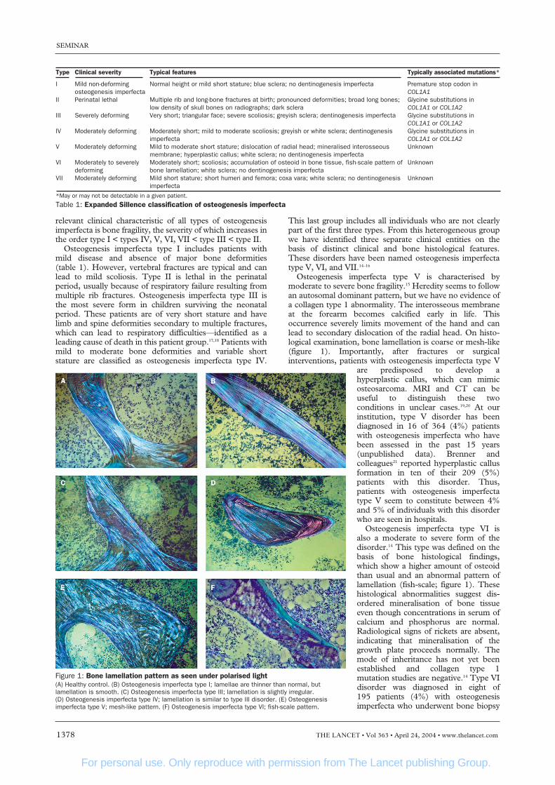
Osteogenesis imperfecta type V is characterised bymoderate to severe bone fragility.15 Heredity seems to followan autosomal dominant pattern, but we have no evidence ofa collagen type 1 abnormality. The interosseous membraneat the forearm becomes calcified early in life. Thisoccurrence severely limits movement of the hand and canlead to secondary dislocation of the radial head. On histo-logical examination, bone lamellation is coarse or mesh-like(figure 1). Importantly, after fractures or surgicalinterventions, patients with osteogenesis imperfecta type V
are predisposed to develop ahyperplastic callus, which can mimicosteosarcoma. MRI and CT can beuseful to distinguish these twoconditions in unclear cases.19,20 At ourinstitution, type V disorder has beendiagnosed in 16 of 364 (4%) patientswith osteogenesis imperfecta who havebeen assessed in the past 15 years(unpublished data). Brenner andcolleagues21 reported hyperplastic callusformation in ten of their 209 (5%)patients with this disorder. Thus,patients with osteogenesis imperfectatype V seem to constitute between 4%and 5% of individuals with this disorderwho are seen in hospitals.
Osteogenesis imperfecta type VI isalso a moderate to severe form of thedisorder.14 This type was defined on thebasis of bone histological findings,which show a higher amount of osteoidthan usual and an abnormal pattern oflamellation (fish-scale; figure 1). Thesehistological abnormalities suggest dis-ordered mineralisation of bone tissueeven though concentrations in serum ofcalcium and phosphorus are normal.Radiological signs of rickets are absent,indicating that mineralisation of thegrowth plate proceeds normally. Themode of inheritance has not yet beenestablished and collagen type 1mutation studies are negative.14 Type VIdisorder was diagnosed in eight of195 patients (4%) with osteogenesisimperfecta who underwent bone biopsy
SEMINAR
1378 THE LANCET • Vol 363 • April 24, 2004 • www.thelancet.com
Type Clinical severity Typical features Typically associated mutations*
I Mild non-deforming Normal height or mild short stature; blue sclera; no dentinogenesis imperfecta Premature stop codon in osteogenesis imperfecta COL1A1
II Perinatal lethal Multiple rib and long-bone fractures at birth; pronounced deformities; broad long bones; Glycine substitutions in low density of skull bones on radiographs; dark sclera COL1A1 or COL1A2
III Severely deforming Very short; triangular face; severe scoliosis; greyish sclera; dentinogenesis imperfecta Glycine substitutions in COL1A1 or COL1A2
IV Moderately deforming Moderately short; mild to moderate scoliosis; greyish or white sclera; dentinogenesis Glycine substitutions in imperfecta COL1A1 or COL1A2
V Moderately deforming Mild to moderate short stature; dislocation of radial head; mineralised interosseous Unknownmembrane; hyperplastic callus; white sclera; no dentinogenesis imperfecta
VI Moderately to severely Moderately short; scoliosis; accumulation of osteoid in bone tissue, fish-scale pattern of Unknowndeforming bone lamellation; white sclera; no dentinogenesis imperfecta
VII Moderately deforming Mild short stature; short humeri and femora; coxa vara; white sclera; no dentinogenesis Unknownimperfecta
*May or may not be detectable in a given patient.
Table 1: Expanded Sillence classification of osteogenesis imperfecta
Figure 1: Bone lamellation pattern as seen under polarised light(A) Healthy control. (B) Osteogenesis imperfecta type I; lamellae are thinner than normal, butlamellation is smooth. (C) Osteogenesis imperfecta type III; lamellation is slightly irregular. (D) Osteogenesis imperfecta type IV; lamellation is similar to type III disorder. (E) Osteogenesisimperfecta type V; mesh-like pattern. (F) Osteogenesis imperfecta type VI; fish-scale pattern.
For personal use. Only reproduce with permission from The Lancet publishing Group.
at our institution during the past 15 years (unpublisheddata). Typical histological features of type VI disorder havebeen described by Sarathchandra and colleagues22 in threeof 36 patients (8%) with osteogenesis imperfecta.
Type VII osteogenesis imperfecta is a recessive disorder,which so far has been reported only in a community ofNative Americans in northern Quebec.16 Apart from bonefragility, rhizomelia is a prominent clinical feature, and coxavara can be present even in infancy. The disease has beenlocalised to chromosome 3p22–24.1, which is outside theloci for collagen type 1 genes.23
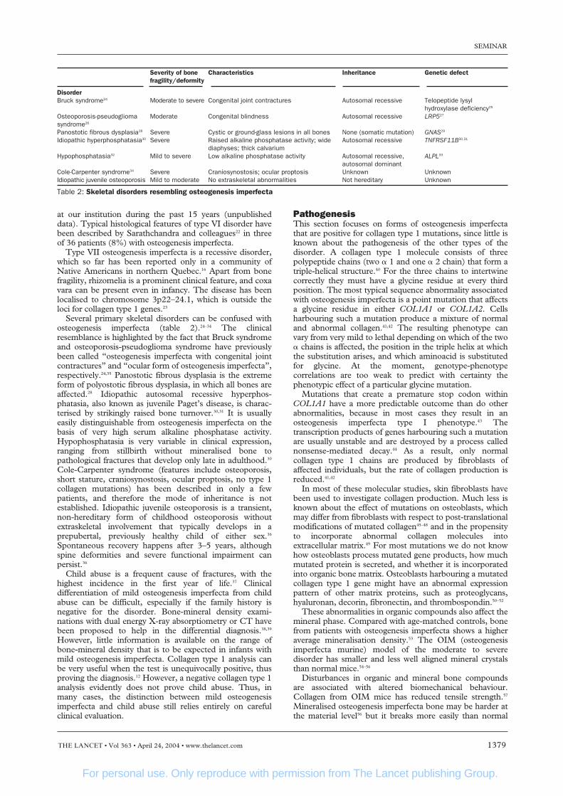
Several primary skeletal disorders can be confused withosteogenesis imperfecta (table 2).24–34 The clinicalresemblance is highlighted by the fact that Bruck syndromeand osteoporosis-pseudoglioma syndrome have previouslybeen called “osteogenesis imperfecta with congenital jointcontractures” and “ocular form of osteogenesis imperfecta”,respectively.24,35 Panostotic fibrous dysplasia is the extremeform of polyostotic fibrous dysplasia, in which all bones areaffected.28 Idiopathic autosomal recessive hyperphos-phatasia, also known as juvenile Paget’s disease, is charac-terised by strikingly raised bone turnover.30,31 It is usuallyeasily distinguishable from osteogenesis imperfecta on thebasis of very high serum alkaline phosphatase activity.Hypophosphatasia is very variable in clinical expression,ranging from stillbirth without mineralised bone topathological fractures that develop only late in adulthood.30
Cole-Carpenter syndrome (features include osteoporosis,short stature, craniosynostosis, ocular proptosis, no type 1collagen mutations) has been described in only a fewpatients, and therefore the mode of inheritance is notestablished. Idiopathic juvenile osteoporosis is a transient,non-hereditary form of childhood osteoporosis withoutextraskeletal involvement that typically develops in aprepubertal, previously healthy child of either sex.36
Spontaneous recovery happens after 3–5 years, althoughspine deformities and severe functional impairment canpersist.36
Child abuse is a frequent cause of fractures, with thehighest incidence in the first year of life.37 Clinicaldifferentiation of mild osteogenesis imperfecta from childabuse can be difficult, especially if the family history isnegative for the disorder. Bone-mineral density exami-nations with dual energy X-ray absorptiometry or CT havebeen proposed to help in the differential diagnosis.38,39
However, little information is available on the range ofbone-mineral density that is to be expected in infants withmild osteogenesis imperfecta. Collagen type 1 analysis canbe very useful when the test is unequivocally positive, thusproving the diagnosis.12 However, a negative collagen type 1analysis evidently does not prove child abuse. Thus, inmany cases, the distinction between mild osteogenesisimperfecta and child abuse still relies entirely on carefulclinical evaluation.
Pathogenesis This section focuses on forms of osteogenesis imperfectathat are positive for collagen type 1 mutations, since little isknown about the pathogenesis of the other types of thedisorder. A collagen type 1 molecule consists of threepolypeptide chains (two � 1 and one � 2 chain) that form atriple-helical structure.40 For the three chains to intertwinecorrectly they must have a glycine residue at every thirdposition. The most typical sequence abnormality associatedwith osteogenesis imperfecta is a point mutation that affectsa glycine residue in either COL1A1 or COL1A2. Cellsharbouring such a mutation produce a mixture of normaland abnormal collagen.41,42 The resulting phenotype canvary from very mild to lethal depending on which of the two� chains is affected, the position in the triple helix at whichthe substitution arises, and which aminoacid is substitutedfor glycine. At the moment, genotype-phenotypecorrelations are too weak to predict with certainty thephenotypic effect of a particular glycine mutation.
Mutations that create a premature stop codon withinCOL1A1 have a more predictable outcome than do otherabnormalities, because in most cases they result in anosteogenesis imperfecta type I phenotype.43 Thetranscription products of genes harbouring such a mutationare usually unstable and are destroyed by a process callednonsense-mediated decay.44 As a result, only normalcollagen type 1 chains are produced by fibroblasts ofaffected individuals, but the rate of collagen production isreduced.41,42
In most of these molecular studies, skin fibroblasts havebeen used to investigate collagen production. Much less isknown about the effect of mutations on osteoblasts, whichmay differ from fibroblasts with respect to post-translationalmodifications of mutated collagen45–48 and in the propensityto incorporate abnormal collagen molecules intoextracellular matrix.49 For most mutations we do not knowhow osteoblasts process mutated gene products, how muchmutated protein is secreted, and whether it is incorporatedinto organic bone matrix. Osteoblasts harbouring a mutatedcollagen type 1 gene might have an abnormal expressionpattern of other matrix proteins, such as proteoglycans,hyaluronan, decorin, fibronectin, and thrombospondin.50–52
These abnormalities in organic compounds also affect themineral phase. Compared with age-matched controls, bonefrom patients with osteogenesis imperfecta shows a higheraverage mineralisation density.53 The OIM (osteogenesisimperfecta murine) model of the moderate to severedisorder has smaller and less well aligned mineral crystalsthan normal mice.54–56
Disturbances in organic and mineral bone compoundsare associated with altered biomechanical behaviour.Collagen from OIM mice has reduced tensile strength.57
Mineralised osteogenesis imperfecta bone may be harder atthe material level56 but it breaks more easily than normal
SEMINAR
THE LANCET • Vol 363 • April 24, 2004 • www.thelancet.com 1379
Severity of bone Characteristics Inheritance Genetic defectfragility/deformity
DisorderBruck syndrome24 Moderate to severe Congenital joint contractures Autosomal recessive Telopeptide lysyl
hydroxylase deficiency25
Osteoporosis-pseudoglioma Moderate Congenital blindness Autosomal recessive LRP527
syndrome26
Panostotic fibrous dysplasia28 Severe Cystic or ground-glass lesions in all bones None (somatic mutation) GNAS29
Idiopathic hyperphosphatasia30 Severe Raised alkaline phosphatase activity; wide Autosomal recessive TNFRSF11B30,31
diaphyses; thick calvariumHypophosphatasia32 Mild to severe Low alkaline phosphatase activity Autosomal recessive, ALPL33
autosomal dominantCole-Carpenter syndrome34 Severe Craniosynostosis; ocular proptosis Unknown UnknownIdiopathic juvenile osteoporosis Mild to moderate No extraskeletal abnormalities Not hereditary Unknown
Table 2: Skeletal disorders resembling osteogenesis imperfecta
For personal use. Only reproduce with permission from The Lancet publishing Group.
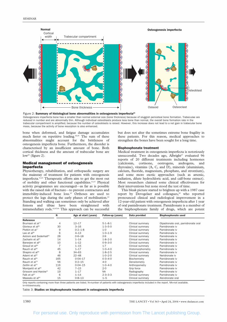
bone when deformed, and fatigue damage accumulatesmuch faster on repetitive loading.58,59 The sum of theseabnormalities might account for the brittleness ofosteogenesis imperfecta bone. Furthermore, the disorder ischaracterised by an insufficient amount of bone. Bothcortical thickness and the amount of trabecular bone arelow60 (figure 2).
Medical management of osteogenesisimperfecta Physiotherapy, rehabilitation, and orthopaedic surgery arethe mainstay of treatment for patients with osteogenesisimperfecta.61,62 Therapeutic efforts aim to get the most outof mobility and other functional capabilities.61,63 Physicalactivity programmes are encouraged—as far as is possiblewith the raised risk of fracture—to prevent contractures andimmobility-induced bone loss.62 Orthoses are used toprotect the legs during the early phases of mobilisation.64
Standing and walking can sometimes only be achieved afterfemora and tibiae have been straightened withintramedullary rods.62,65,66 This approach can be successful
but does not alter the sometimes extreme bone fragility inthese patients. For this reason, medical approaches tostrengthen the bones have been sought for a long time.
Bisphosphonate treatment Medical treatment in osteogenesis imperfecta is notoriouslyunsuccessful. Two decades ago, Albright67 evaluated 96reports of 20 different treatments including hormones(calcitonin, cortisone, oestrogens, androgens, andthyroxine), vitamins (A, C, and D), minerals (aluminium,calcium, fluoride, magnesium, phosphate, and strontium),and some more exotic approaches (such as arsenic,radiation, dilute hydrochloric acid, and calf-bone extract).Most researchers claimed some clinical effectiveness fortheir interventions but none stood the test of time.
This bleak picture started to brighten up with a 1987 casereport by Devogelaer and colleagues,68 who reportedpronounced clinical and radiological improvement in a 12-year-old patient with osteogenesis imperfecta after 1 yearof oral pamidronate treatment. Pamidronate is a member ofthe bisphosphonate family of drugs, which are potent
SEMINAR
1380 THE LANCET • Vol 363 • April 24, 2004 • www.thelancet.com
Osteogenesis imperfecta
Osteoid
Osteoblast
OsteoclastBone thickness
Corticalwidth Trabecular compartment
Normal
Figure 2: Summary of histological bone abnormalities in osteogenesis imperfecta60
Osteogenesis imperfecta bone has a smaller than normal external size (bone thickness) because of sluggish periosteal bone formation. Trabeculae arereduced in number and are abnormally thin. Although individual osteoblasts produce less bone than normal, the overall bone formation rate in thetrabecular compartment is amplified, because the number of osteoblasts is raised. However, this increase does not lead to a net gain in trabecular bonemass, because the activity of bone resorption is also enhanced.
N Age at start (years) Follow-up (years) Data provided Bisphosphonate used
ReferenceBrumsen et al74 4 12–17 5·1–8·1 Clinical summary Olpadronate oral, pamidronate oralGlorieux et al82 30 3–16 1·3–5·0 Clinical summary Pamidronate ivPlotkin et al77 9 0·2–1·8 1·0 Clinical summary Pamidronate ivLee et al86 6 4–13 1·0–1·9 Clinical summary Pamidronate ivAstrom and Soderhall83 28 0·6–18 2-9 Clinical summary Pamidronate ivZacharin et al84 14 1–14 1·8–2·0 Clinical summary Pamidronate ivBanerjee et al87 10 1–12 0·9–3·0 Clinical summary Pamidronate ivGiraud et al88 7 1–15 1-7 Clinical summary Pamidronate ivRauch et al80 45 1–17 1·0–4·0 Histomorphometry Pamidronate ivShapiro et al89 8 34–63 1·8–2·5 Clinical summary Pamidronate ivAdami et al90 46 22–48 1·0–2·0 Clinical summary Neridronate ivRauch et al81 165 0·04–17 0·3–4·0 Biochemistry Pamidronate ivRauch et al78 56 0·2–15 4·0 Densitometry Pamidronate ivZeitlin et al79 116 0·04–15 1·0–4·0 Anthropometry Pamidronate ivMontpetit et al85 42 7–15 2·0 Grip force Pamidronate ivGrissom and Harcke91 19 1–17 NA Radiography Pamidronate ivFalk et al92 6 1–14 2·3–3·3 Clinical summary Pamidronate ivMaasalu et al93 15 0·8–13 1–5 Clinical summary Alendronate oral
Only reports containing more than three patients are listed. N=number of patients with osteogenesis imperfecta included in the report. NA=not available.iv=intravenously.
Table 3: Studies on bisphosphonate treatment in osteogenesis imperfecta
For personal use. Only reproduce with permission from The Lancet publishing Group.
antiresorptive agents.69 It interferes with the mevalonatepathway of cholesterol biosynthesis in osteoclasts,70
inhibiting the function of these cells but not usually leadingto apoptosis, as was believed previously.71
The encouraging observations by Devogelaer andcolleagues,68 and findings of subsequent pilot studies,72–76
prompted investigators to treat large groups of patients withbisphosphonates. Available evidence on this treatmentapproach in children mostly stems from observational trialsin moderately to severely affected patients with osteogenesisimperfecta (table 3).74,77–93 None of the studies was placebo-controlled, but some included historical controls.77–81 Mostpatients in these studies were treated with cyclic intravenouspamidronate (table 3). The most widely used treatmentschedule is shown in table 4. At present, very little is knownabout the effect of oral bisphosphonate treatment, eventhough this regimen is under investigation in controlledtrials. A pronounced decline in chronic bone pain can beseen within a few weeks after the start of intravenouspamidronate treatment.72–84 This result is associated with anenhanced sense of well-being83 and increased musclestrength in the grip force test.85
During pamidronate treatment, vertebral bone-mineralmass increases faster than in untreated patients.82–84,86–88
Higher lumbar-spine bone mass is attributable to not onlyincreased bone-mineral density but also larger vertebralsize.78 Several investigators have reported the impressionthat crushed vertebral bodies regain a more normal shapeduring pamidronate treatment.82–84
With respect to long bones, enhanced cortical thicknesshas been noted at the second metacarpal.82 On the basis ofCT analyses in two patients with osteogenesis imperfecta,researchers suggested that pamidronate might increase themass of long-bone diaphyses but not geometric variables ofbone stability.94 However, our preliminary observations withperipheral quantitative CT in more than 20 patients with
this disorder suggest that this drug also has a beneficialeffect on indicators of long-bone stability (unpublisheddata).
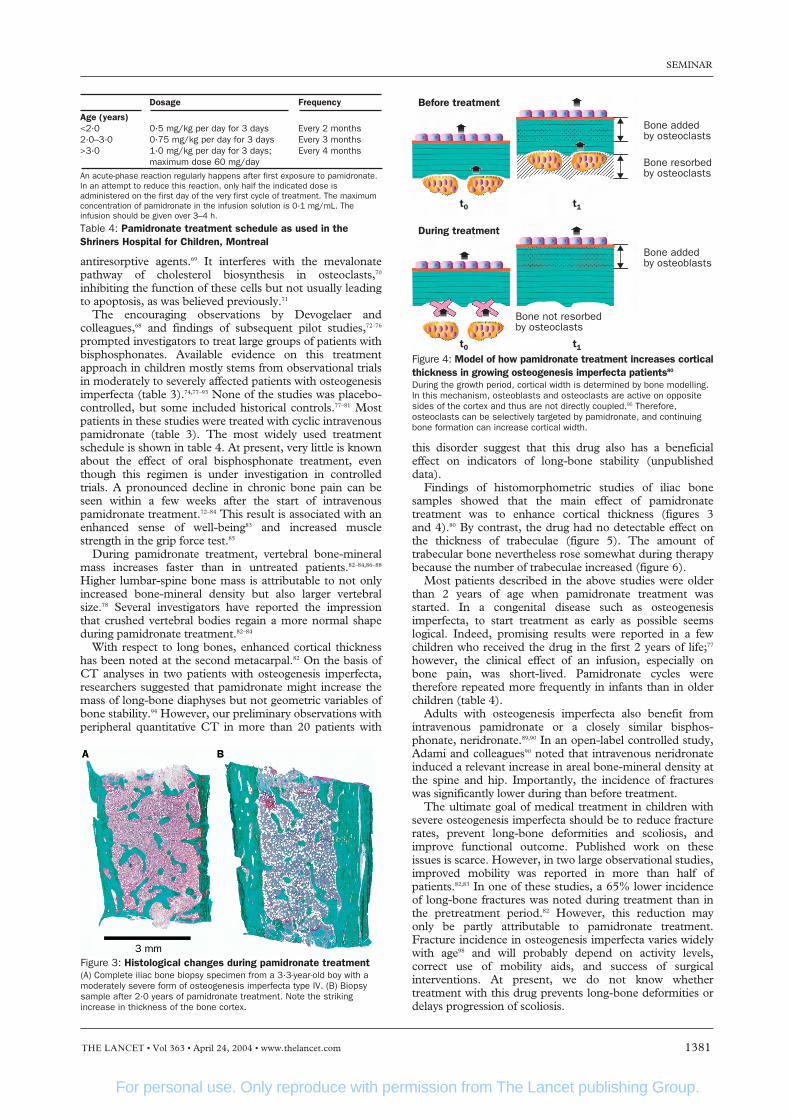
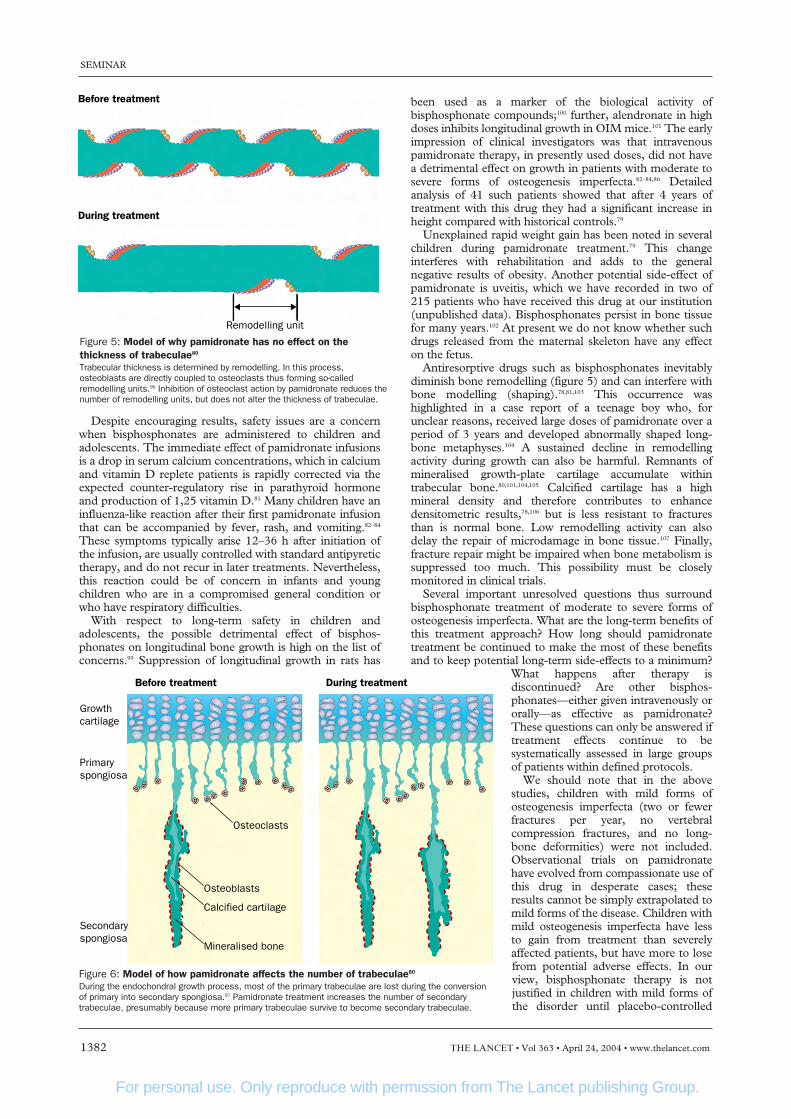
Findings of histomorphometric studies of iliac bonesamples showed that the main effect of pamidronatetreatment was to enhance cortical thickness (figures 3and 4).80 By contrast, the drug had no detectable effect onthe thickness of trabeculae (figure 5). The amount oftrabecular bone nevertheless rose somewhat during therapybecause the number of trabeculae increased (figure 6).
Most patients described in the above studies were olderthan 2 years of age when pamidronate treatment wasstarted. In a congenital disease such as osteogenesisimperfecta, to start treatment as early as possible seemslogical. Indeed, promising results were reported in a fewchildren who received the drug in the first 2 years of life;77
however, the clinical effect of an infusion, especially onbone pain, was short-lived. Pamidronate cycles weretherefore repeated more frequently in infants than in olderchildren (table 4).
Adults with osteogenesis imperfecta also benefit fromintravenous pamidronate or a closely similar bisphos-phonate, neridronate.89,90 In an open-label controlled study,Adami and colleagues90 noted that intravenous neridronateinduced a relevant increase in areal bone-mineral density atthe spine and hip. Importantly, the incidence of fractureswas significantly lower during than before treatment.
The ultimate goal of medical treatment in children withsevere osteogenesis imperfecta should be to reduce fracturerates, prevent long-bone deformities and scoliosis, andimprove functional outcome. Published work on theseissues is scarce. However, in two large observational studies,improved mobility was reported in more than half ofpatients.82,83 In one of these studies, a 65% lower incidenceof long-bone fractures was noted during treatment than inthe pretreatment period.82 However, this reduction mayonly be partly attributable to pamidronate treatment.Fracture incidence in osteogenesis imperfecta varies widelywith age98 and will probably depend on activity levels,correct use of mobility aids, and success of surgicalinterventions. At present, we do not know whethertreatment with this drug prevents long-bone deformities ordelays progression of scoliosis.
SEMINAR
THE LANCET • Vol 363 • April 24, 2004 • www.thelancet.com 1381
Dosage Frequency
Age (years)<2·0 0·5 mg/kg per day for 3 days Every 2 months2·0–3·0 0·75 mg/kg per day for 3 days Every 3 months>3·0 1·0 mg/kg per day for 3 days; Every 4 months
maximum dose 60 mg/day
An acute-phase reaction regularly happens after first exposure to pamidronate.In an attempt to reduce this reaction, only half the indicated dose isadministered on the first day of the very first cycle of treatment. The maximumconcentration of pamidronate in the infusion solution is 0·1 mg/mL. Theinfusion should be given over 3–4 h.
Table 4: Pamidronate treatment schedule as used in theShriners Hospital for Children, Montreal
Figure 3: Histological changes during pamidronate treatment(A) Complete iliac bone biopsy specimen from a 3·3-year-old boy with amoderately severe form of osteogenesis imperfecta type IV. (B) Biopsysample after 2·0 years of pamidronate treatment. Note the strikingincrease in thickness of the bone cortex.
Before treatment
Bone addedby osteoclasts
Bone not resorbedby osteoclasts
During treatment
t0 t1
t0 t1
Bone resorbedby osteoclasts
Bone addedby osteoblasts
Figure 4: Model of how pamidronate treatment increases corticalthickness in growing osteogenesis imperfecta patients80
During the growth period, cortical width is determined by bone modelling.In this mechanism, osteoblasts and osteoclasts are active on oppositesides of the cortex and thus are not directly coupled.95 Therefore,osteoclasts can be selectively targeted by pamidronate, and continuingbone formation can increase cortical width.
For personal use. Only reproduce with permission from The Lancet publishing Group.
Despite encouraging results, safety issues are a concernwhen bisphosphonates are administered to children andadolescents. The immediate effect of pamidronate infusionsis a drop in serum calcium concentrations, which in calciumand vitamin D replete patients is rapidly corrected via theexpected counter-regulatory rise in parathyroid hormoneand production of 1,25 vitamin D.81 Many children have aninfluenza-like reaction after their first pamidronate infusionthat can be accompanied by fever, rash, and vomiting.82–84
These symptoms typically arise 12–36 h after initiation ofthe infusion, are usually controlled with standard antipyretictherapy, and do not recur in later treatments. Nevertheless,this reaction could be of concern in infants and youngchildren who are in a compromised general condition orwho have respiratory difficulties.
With respect to long-term safety in children andadolescents, the possible detrimental effect of bisphos-phonates on longitudinal bone growth is high on the list ofconcerns.99 Suppression of longitudinal growth in rats has
been used as a marker of the biological activity ofbisphosphonate compounds;100 further, alendronate in highdoses inhibits longitudinal growth in OIM mice.101 The earlyimpression of clinical investigators was that intravenouspamidronate therapy, in presently used doses, did not havea detrimental effect on growth in patients with moderate tosevere forms of osteogenesis imperfecta.82–84,86 Detailedanalysis of 41 such patients showed that after 4 years oftreatment with this drug they had a significant increase inheight compared with historical controls.79
Unexplained rapid weight gain has been noted in severalchildren during pamidronate treatment.79 This changeinterferes with rehabilitation and adds to the generalnegative results of obesity. Another potential side-effect ofpamidronate is uveitis, which we have recorded in two of215 patients who have received this drug at our institution(unpublished data). Bisphosphonates persist in bone tissuefor many years.102 At present we do not know whether suchdrugs released from the maternal skeleton have any effecton the fetus.
Antiresorptive drugs such as bisphosphonates inevitablydiminish bone remodelling (figure 5) and can interfere withbone modelling (shaping).78,81,103 This occurrence washighlighted in a case report of a teenage boy who, forunclear reasons, received large doses of pamidronate over aperiod of 3 years and developed abnormally shaped long-bone metaphyses.104 A sustained decline in remodellingactivity during growth can also be harmful. Remnants ofmineralised growth-plate cartilage accumulate withintrabecular bone.80,101,104,105 Calcified cartilage has a highmineral density and therefore contributes to enhancedensitometric results,78,106 but is less resistant to fracturesthan is normal bone. Low remodelling activity can alsodelay the repair of microdamage in bone tissue.107 Finally,fracture repair might be impaired when bone metabolism issuppressed too much. This possibility must be closelymonitored in clinical trials.
Several important unresolved questions thus surroundbisphosphonate treatment of moderate to severe forms ofosteogenesis imperfecta. What are the long-term benefits ofthis treatment approach? How long should pamidronatetreatment be continued to make the most of these benefitsand to keep potential long-term side-effects to a minimum?
What happens after therapy isdiscontinued? Are other bisphos-phonates—either given intravenously ororally—as effective as pamidronate?These questions can only be answered iftreatment effects continue to besystematically assessed in large groupsof patients within defined protocols.
We should note that in the abovestudies, children with mild forms ofosteogenesis imperfecta (two or fewerfractures per year, no vertebralcompression fractures, and no long-bone deformities) were not included.Observational trials on pamidronatehave evolved from compassionate use ofthis drug in desperate cases; theseresults cannot be simply extrapolated tomild forms of the disease. Children withmild osteogenesis imperfecta have lessto gain from treatment than severelyaffected patients, but have more to losefrom potential adverse effects. In ourview, bisphosphonate therapy is notjustified in children with mild forms ofthe disorder until placebo-controlled
SEMINAR
1382 THE LANCET • Vol 363 • April 24, 2004 • www.thelancet.com
Before treatment
Growthcartilage
Primaryspongiosa
Osteoclasts
Secondaryspongiosa
During treatment
Osteoblasts
Calcified cartilage
Mineralised bone
Figure 6: Model of how pamidronate affects the number of trabeculae80
During the endochondral growth process, most of the primary trabeculae are lost during the conversionof primary into secondary spongiosa.97 Pamidronate treatment increases the number of secondarytrabeculae, presumably because more primary trabeculae survive to become secondary trabeculae.
Before treatment
During treatment
Remodelling unit
Figure 5: Model of why pamidronate has no effect on thethickness of trabeculae80
Trabecular thickness is determined by remodelling. In this process,osteoblasts are directly coupled to osteoclasts thus forming so-calledremodelling units.96 Inhibition of osteoclast action by pamidronate reduces thenumber of remodelling units, but does not alter the thickness of trabeculae.
For personal use. Only reproduce with permission from The Lancet publishing Group.
trials have established the efficacy and safety of thisapproach in this group of patients.
Other medical treatments Growth hormone was proposed as a possible treatment forosteogenesis imperfecta almost three decades ago.108
Findings of small studies suggest that growth-hormonetreatment might accelerate short-term height velocity insome patients.109–111 In calcium kinetic studies, after 1 year ofgrowth-hormone therapy bone turnover increased butcalcium retention was unchanged compared withpretreatment.110 Enhanced bone turnover during growth-hormone treatment was also reported in histomorphometricstudies of iliac bone samples.111 Since bone turnover isalready abnormally high in untreated children withosteogenesis imperfecta,60 further stimulation does not seemto be a desirable goal. Growth hormone might be moreuseful in combination with bisphosphonates, but thisregimen remains to be tested.
Parathyroid hormone is a potent bone anabolic agent thatreduces fracture incidence in postmenopausal osteo-porosis.112 These results suggest parathyroid hormone as anattractive candidate for treating children with osteogenesisimperfecta. However, a substantial proportion of young ratsreceiving parathyroid hormone subsequently developedosteosarcoma,113 and a similar effect could happen in humanbeings.114 Thus, parathyroid hormone should not be used inchildren until these issues have been resolved.
Potential treatments Bone-marrow stromal cells can differentiate into various celllineages, including osteoblasts.115 This observation led to thestraightforward hypothesis that transplanting bone-marrowstromal cells from healthy people might improve the clinicalcourse of osteogenesis imperfecta. For this approach to besuccessful, intravenously infused bone-marrow stromal cellsmust find their way into the skeleton and differentiate intoosteoblasts that start producing normal bone. Also, we musthope that the bone produced by transplanted cells is notsubjected to the same rapid removal as that formed by thepatient’s original osteoblasts.
These hopes formed the rationale for undertaking bone-marrow transplantation in a small group of children withsevere osteogenesis imperfecta:116,117 the reported resultselicited mixed reactions. Bone-marrow transplantationexperts were excited, because some of the patients’osteoblasts seemed to be of donor origin.118 Bone-diseasespecialists remained sceptical, because no convincingevidence was presented to show that the patients hadactually benefited from the procedure.41,115,119–121
Whatever the clinical effects might have been, theresearchers conceded that they were short-lived.117
Therefore, they re-treated the same patients with a modifiedapproach—this time, isolated marrow stromal cells wereinfused.122 Similar to the first study, a few transplanted cellswere detectable in several tissues, including bone. Clinicalbenefit was claimed, based mainly on increased growthvelocity in the 6 months after the procedure. However, thedifficulty of measuring short-term growth velocity inchildren with bone deformities is obvious.
In our view, enthusiasm about an innovative techniqueshould not be a substitute for scientific stringency andobjective evaluation of results. Techniques based onmarrow stem cells could offer therapeutic potential forpatients with osteogenesis imperfecta in the future.Therefore, we should not discredit this approach bypremature clinical application; basic science and technicalissues should be worked out in animals before furtherstudies are undertaken in human beings.
Presently, medical treatment options at best achievesymptomatic improvement of osteogenesis imperfecta. Theonly hope for actually curing the disease is by elimination ofthe mutated gene or gene product. Unfortunately, majorobstacles to gene-based therapy of osteogenesis imperfectaexist. Most severe cases result from the presence ofabnormal collagen molecules. Thus, we cannot simplyreplace a missing protein as is the case in many recessiveenzyme disorders. Rather, we need to first inactivate themutant allele and then substitute for its product.123 Researchis still grappling with the first of these two tasks. Variousinvestigators are studying so-called hammerheadribozymes,124–126 small RNA molecules that can cut mRNAin the absence of protein cofactors.127 Various viruses havebeen tested that allow for transfection of mesenchymalprogenitor cells.126,128 When ribozymes were transfected intosuch cells, COL1A1 mRNA amounts could be suppressedby about 50%.126 Such results are encouraging, butnevertheless they represent only a first step on a long anddifficult path.
Conflict of interest statementF H Glorieux has received support for research into use of bisphosphonatesin osteogenesis imperfecta from Merck Research Laboratories and honorariafor lectures in the area of paediatric bone diseases at academic meetingsorganised by various pharmaceutical companies. None of these sources hascontributed to the content of this Seminar. F Rauch has no conflict ofinterest with respect to this Seminar.
AcknowledgmentsThis work was supported by the Shriners of North America. We thankMark Lepik for artwork. The sponsor had no role in writing this Seminar.
References1 Shapiro JR, Stover ML, Burn VE, et al. An osteopenic nonfracture
syndrome with features of mild osteogenesis imperfecta associated withthe substitution of a cysteine for glycine at triple helix position 43 in thepro alpha 1(I) chain of type I collagen. J Clin Invest 1992; 89: 567–73.
2 Petersen K, Wetzel WE. Recent findings in classification of osteogenesisimperfecta by means of existing dental symptoms. ASDC J Dent Child1998; 65: 305–09.
3 Lygidakis NA, Smith R, Oulis CJ. Scanning electron microscopy ofteeth in osteogenesis imperfecta type I. Oral Surg Oral Med Oral PatholOral Radiol Endod 1996; 81: 567–72.
4 Lund AM, Jensen BL, Nielsen LA, Skovby F. Dental manifestations ofosteogenesis imperfecta and abnormalities of collagen I metabolism. J Craniofac Genet Dev Biol 1998; 18: 30–37.
5 Malmgren B, Norgren S. Dental aberrations in children and adolescentswith osteogenesis imperfecta. Acta Odontol Scand 2002; 60: 65–71.
6 Pedersen U. Hearing loss in patients with osteogenesis imperfecta: aclinical and audiological study of 201 patients. Scand Audiol 1984; 13:67–74.
7 Kuurila K, Grenman R, Johansson R, Kaitila I. Hearing loss in childrenwith osteogenesis imperfecta. Eur J Pediatr 2000; 159: 515–19.
8 Paterson CR, Monk EA, McAllion SJ. How common is hearingimpairment in osteogenesis imperfecta? J Laryngol Otol 2001; 115:280–02.
9 Kuurila K, Kaitila I, Johansson R, Grenman R. Hearing loss in Finnishadults with osteogenesis imperfecta: a nationwide survey. Ann Otol Rhinol Laryngol 2002; 111: 939–46.
10 Wenstrup RJ, Willing MC, Starman BJ, Byers PH. Distinct biochemicalphenotypes predict clinical severity in nonlethal variants of osteogenesisimperfecta. Am J Hum Genet 1990; 46: 975–82.
11 Korkko J, Ala-Kokko L, De Paepe A, Nuytinck L, Earley J, Prockop DJ.Analysis of the COL1A1 and COL1A2 genes by PCR amplification andscanning by conformation-sensitive gel electrophoresis identifies onlyCOL1A1 mutations in 15 patients with osteogenesis imperfecta type I:identification of common sequences of null-allele mutations. Am J Hum Genet 1998; 62: 98–110.
12 Marlowe A, Pepin MG, Byers PH. Testing for osteogenesis imperfectain cases of suspected non-accidental injury. J Med Genet 2002; 39:382–86.
13 Sillence DO, Senn A, Danks DM. Genetic heterogeneity in osteogenesisimperfecta. J Med Genet 1979; 16: 101–16.
14 Glorieux FH, Ward LM, Rauch F, Lalic L, Roughley PJ, Travers R.Osteogenesis imperfecta type VI: a form of brittle bone disease with amineralization defect. J Bone Miner Res 2002; 17: 30–38.
15 Glorieux FH, Rauch F, Plotkin H, et al. Type V osteogenesisimperfecta: a new form of brittle bone disease. J Bone Miner Res 2000;15: 1650–58.
SEMINAR
THE LANCET • Vol 363 • April 24, 2004 • www.thelancet.com 1383
For personal use. Only reproduce with permission from The Lancet publishing Group.
16 Ward LM, Rauch F, Travers R, et al. Osteogenesis imperfecta type VII:an autosomal recessive form of brittle bone disease. Bone 2002; 31:12–18.
17 McAllion SJ, Paterson CR. Causes of death in osteogenesis imperfecta. J Clin Pathol 1996; 49: 627–30.
18 Paterson CR, Ogston SA, Henry RM. Life expectancy in osteogenesisimperfecta. BMJ 1996; 312: 351.
19 Rieker O, Kreitner KF, Karbowski A. Hyperplastic callus formation inosteogenesis imperfecta: CT and MRI findings. Eur Radiol 1998; 8:1137–39.
20 Dobrocky I, Seidl G, Grill F. MRI and CT features of hyperplastic callusin osteogenesis imperfecta tarda. Eur Radiol 1999; 9: 665–68.
21 Brenner RE, Schiller B, Pontz BF, et al. Osteogenesis imperfecta inKindheit und Adoleszenz. Monatsschr Kinderheilkd 1993; 141: 940–45.
22 Sarathchandra P, Pope FM, Kayser MV, Ali SY. A light and electronmicroscopic study of osteogenesis imperfecta bone samples, withreference to collagen chemistry and clinical phenotype. J Pathol 2000;192: 385–95.
23 Labuda M, Morissette J, Ward LM, et al. Osteogenesis imperfecta typeVII maps to the short arm of chromosome 3. Bone 2002; 31: 19–25.
24 McPherson E, Clemens M. Bruck syndrome (osteogenesis imperfectawith congenital joint contractures): review and report on the first NorthAmerican case. Am J Med Genet 1997; 70: 28–31.
25 Bank RA, Robins SP, Wijmenga C, et al. Defective collagen crosslinkingin bone, but not in ligament or in Bruck syndrome: indications for abone-specific telopeptide lysyl hydroxylase on chromosome 17. Proc Natl Acad Sci USA 1999; 96: 1054–58.
26 Frontali M, Stomeo C, Dallapiccola B. Osteoporosis-pseudogliomasyndrome: report of three affected sibs and an overview. Am J Med Genet1985; 22: 35–47.
27 Gong Y, Slee RB, Fukai N, et al. LDL receptor-related protein 5 (LRP5)affects bone accrual and eye development. Cell 2001; 107: 513–23.
28 Cole DE, Fraser FC, Glorieux FH, et al. Panostotic fibrous dysplasia: acongenital disorder of bone with unusual facial appearance, bone fragility,hyperphosphatasemia, and hypophosphatemia. Am J Med Genet 1983;14: 725–35.
29 Schwindinger WF, Francomano CA, Levine MA. Identification of amutation in the gene encoding the alpha subunit of the stimulatory Gprotein of adenylyl cyclase in McCune-Albright syndrome. Proc Natl Acad Sci USA 1992; 89: 5152–56.
30 Whyte MP, Obrecht SE, Finnegan PM, et al. Osteoprotegerin deficiencyand juvenile Paget’s disease. N Engl J Med 2002; 347: 175–84.
31 Cundy T, Hegde M, Naot D, et al. A mutation in the gene TNFRSF11Bencoding osteoprotegerin causes an idiopathic hyperphosphatasiaphenotype. Hum Mol Genet 2002; 11: 2119–27.
32 Whyte MP. Hypophosphatasia and the role of alkaline phosphatase inskeletal mineralization. Endocr Rev 1994; 15: 439–61.
33 Mornet E. Hypophosphatasia: the mutations in the tissue-nonspecificalkaline phosphatase gene. Hum Mutat 2000; 15: 309–15.
34 Cole DE, Carpenter TO. Bone fragility, craniosynostosis, ocularproptosis, hydrocephalus, and distinctive facial features: a newlyrecognized type of osteogenesis imperfecta. J Pediatr 1987; 110: 76–80.
35 Beighton P, Winship I, Behari D. The ocular form of osteogenesisimperfecta: a new autosomal recessive syndrome. Clin Genet 1985; 28:69–75.
36 Smith R. Idiopathic juvenile osteoporosis: experience of twenty-onepatients. Br J Rheumatol 1995; 34: 68–77.
37 Nimkin K, Kleinman PK. Imaging of child abuse. Radiol Clin North Am2001; 39: 843–64.
38 Moore MS, Minch CM, Kruse RW, Harcke HT, Jacobson L, Taylor A.The role of dual energy x-ray absorptiometry in aiding the diagnosis ofpediatric osteogenesis imperfecta. Am J Orthop 1998; 27: 797–801.
39 Miller ME, Hangartner TN. Bone density measurements by computedtomography in osteogenesis imperfecta type I. Osteoporos Int 1999; 9:427–32.
40 Kadler KE, Holmes DF, Trotter JA, Chapman JA. Collagen fibrilformation. Biochem J 1996; 316: 1–11.
41 Byers PH. Osteogenesis imperfecta: perspectives and opportunities. Curr Opin Pediatr 2000; 12: 603–09.
42 Rowe DW, Shapiro JR. Osteogenesis imperfecta. In: Avioli LV,Krane SM, eds. Metabolic bone disease and clinically related disorders.3rd edn. San Diego Academic Press, 1998: 651–95.
43 Willing MC, Deschenes SP, Slayton RL, Roberts EJ. Premature chaintermination is a unifying mechanism for COL1A1 null alleles inosteogenesis imperfecta type I cell strains. Am J Hum Genet 1996; 59:799–809.
44 Byers PH. Killing the messenger: new insights into nonsense-mediatedmRNA decay. J Clin Invest 2002; 109: 3–6.
45 Sarafova AP, Choi H, Forlino A, et al. Three novel type I collagenmutations in osteogenesis imperfecta type IV probands are associatedwith discrepancies between electrophoretic migration of osteoblast andfibroblast collagen. Hum Mutat 1998; 11: 395–403.
46 Cabral WA, Chernoff EJ, Marini JC. G76E substitution in type I collagenis the first nonlethal glutamic acid substitution in the alpha1(I) chain andalters folding of the N-terminal end of the helix. Mol Genet Metab 2001;72: 326–35.
47 Cabral WA, Fertala A, Green LK, Korkko J, Forlino A, Marini JC.Procollagen with skipping of alpha 1(I) exon 41 has lower binding affinityfor alpha 1(I) C-telopeptide, impaired in vitro fibrillogenesis, and alteredfibril morphology. J Biol Chem 2002; 277: 4215–22.
48 Galicka A, Wolczynski S, Gindzienski A. Comparative studies ofosteoblast and fibroblast type I collagen in a patient with osteogenesisimperfecta type IV. J Pathol 2002; 196: 235–37.
49 Mundlos S, Chan D, Weng YM, Sillence DO, Cole WG, Bateman JF.Multiexon deletions in the type I collagen COL1A2 gene in osteogenesisimperfecta type IB: molecules containing the shortened alpha2(I) chainsshow differential incorporation into the bone and skin extracellularmatrix. J Biol Chem 1996; 271: 21068–74.
50 Fedarko NS, Robey PG, Vetter UK. Extracellular matrix stoichiometry inosteoblasts from patients with osteogenesis imperfecta. J Bone Miner Res1995; 10: 1122–29.
51 Fedarko NS, Sponseller PD, Shapiro JR. Long-term extracellular matrixmetabolism by cultured human osteogenesis imperfecta osteoblasts. J Bone Miner Res 1996; 11: 800–05.
52 Grzesik WJ, Frazier CR, Shapiro JR, Sponsellor PD, Robey PG, Fedarko NS. Age-related changes in human bone proteoglycan structure:impact of osteogenesis imperfecta. J Biol Chem 2002; 277: 43638–47.
53 Boyde A, Travers R, Glorieux FH, Jones SJ. The mineralization densityof iliac crest bone from children with osteogenesis imperfecta. Calcif Tissue Int 1999; 64: 185–90.
54 Fratzl P, Paris O, Klaushofer K, Landis WJ. Bone mineralization in anosteogenesis imperfecta mouse model studied by small-angle x-rayscattering. J Clin Invest 1996; 97: 396–402.
55 Camacho NP, Hou L, Toledano TR, et al. The material basis forreduced mechanical properties in oim mice bones. J Bone Miner Res1999; 14: 264–72.
56 Grabner B, Landis WJ, Roschger P, et al. Age- and genotype-dependenceof bone material properties in the osteogenesis imperfecta murine model(oim). Bone 2001; 29: 453–57.
57 Misof K, Landis WJ, Klaushofer K, Fratzl P. Collagen from theosteogenesis imperfecta mouse model (oim) shows reduced resistanceagainst tensile stress. J Clin Invest 1997; 100: 40–45.
58 Jepsen KJ, Goldstein SA, Kuhn JL, Schaffler MB, Bonadio J. Type-Icollagen mutation compromises the post-yield behavior of Mov13 longbone. J Orthop Res 1996; 14: 493–99.
59 Jepsen KJ, Schaffler MB, Kuhn JL, Goulet RW, Bonadio J,Goldstein SA. Type I collagen mutation alters the strength and fatiguebehavior of Mov13 cortical tissue. J Biomech 1997; 30: 1141–47.
60 Rauch F, Travers R, Parfitt AM, Glorieux FH. Static and dynamic bonehistomorphometry in children with osteogenesis imperfecta. Bone 2000;26: 581–89.
61 Engelbert RH, Pruijs HE, Beemer FA, Helders PJ. Osteogenesisimperfecta in childhood: treatment strategies. Arch Phys Med Rehabil1998; 79: 1590–94.
62 Zeitlin L, Fassier F, Glorieux FH. Modern approach to children withosteogenesis imperfecta. J Pediatr Orthop B 2003; 12: 77–87.
63 Engelbert RH, Beemer FA, van der Graaf Y, Helders PJ. Osteogenesisimperfecta in childhood: impairment and disability: a follow-up study.Arch Phys Med Rehabil 1999; 80: 896–903.
64 Gerber LH, Binder H, Berry R, et al. Effects of withdrawal of bracing in matched pairs of children with osteogenesis imperfecta. Arch Phys Med Rehabil 1998; 79: 46–51.
65 Wilkinson JM, Scott BW, Clarke AM, Bell MJ. Surgical stabilisation ofthe lower limb in osteogenesis imperfecta using the Sheffield TelescopicIntramedullary Rod System. J Bone Joint Surg Br 1998; 80: 999–1004.
66 Karbowski A, Schwitalle M, Brenner R, Lehmann H, Pontz B,Worsdorfer O. Experience with Bailey-Dubow rodding in children withosteogenesis imperfecta. Eur J Pediatr Surg 2000; 10: 119–24.
67 Albright JA. Systemic treatment of osteogenesis imperfecta. Clin Orthop1981; 159: 88–96.
68 Devogelaer JP, Malghem J, Maldague B, Nagant de Deuxchaisnes C.Radiological manifestations of bisphosphonate treatment with APD in achild suffering from osteogenesis imperfecta. Skeletal Radiol 1987; 16:360–63.
69 Fleisch H. Bisphosphonates: mechanisms of action. Endocr Rev 1998; 19: 80–100.
70 Fisher JE, Rogers MJ, Halasy JM, et al. Alendronate mechanism ofaction: geranylgeraniol, an intermediate in the mevalonate pathway,prevents inhibition of osteoclast formation, bone resorption, and kinaseactivation in vitro. Proc Natl Acad Sci USA 1999; 96: 133–38.
71 Halasy-Nagy JM, Rodan GA, Reszka AA. Inhibition of bone resorptionby alendronate and risedronate does not require osteoclast apoptosis.Bone 2001; 29: 553–59.
72 Huaux JP, Lokietek W. Is APD a promising drug in the treatment ofsevere osteogenesis imperfecta? J Pediatr Orthop 1988; 8: 71–72.
SEMINAR
1384 THE LANCET • Vol 363 • April 24, 2004 • www.thelancet.com
For personal use. Only reproduce with permission from The Lancet publishing Group.
73 Landsmeer-Beker EA, Massa GG, Maaswinkel-Mooy PD,van de Kamp JJ, Papapoulos SE. Treatment of osteogenesis imperfectawith the bisphosphonate olpadronate(dimethylaminohydroxypropylidene bisphosphonate). Eur J Pediatr1997; 156: 792–94.
74 Brumsen C, Hamdy NA, Papapoulos SE. Long-term effects of bisphosphonates on the growing skeleton: studies of young patientswith severe osteoporosis. Medicine (Baltimore) 1997; 76: 266–83.
75 Bembi B, Parma A, Bottega M, et al. Intravenous pamidronatetreatment in osteogenesis imperfecta. J Pediatr 1997; 131: 622–25.
76 Astrom E, Soderhall S. Beneficial effect of bisphosphonate during fiveyears of treatment of severe osteogenesis imperfecta. Acta Paediatr 1998;87: 64–68.
77 Plotkin H, Rauch F, Bishop NJ, et al. Pamidronate treatment of severeosteogenesis imperfecta in children under 3 years of age.J Clin Endocrinol Metab 2000; 85: 1846–50.
78 Rauch F, Plotkin H, Zeitlin L, Glorieux FH. Bone mass, size anddensity in children and adolescents with osteogenesis imperfecta: effectof intravenous pamidronate therapy. J Bone Miner Res 2003; 18:610–614.
79 Zeitlin L, Rauch F, Plotkin H, Glorieux FH. Height and weightdevelopment during long-term therapy with cyclical intravenouspamidronate in children and adolescents with osteogenesis imperfectatypes I, III and IV. Pediatrics 2003; 111: 1030–36.
80 Rauch F, Travers R, Plotkin H, Glorieux FH. The effects ofintravenous pamidronate on the bone tissue of children and adolescentswith osteogenesis imperfecta. J Clin Invest 2002; 110: 1293–99.
81 Rauch F, Plotkin H, Travers R, Zeitlin L, Glorieux FH. Osteogenesisimperfecta types I, III and IV: effect of pamidronate therapy on boneand mineral metabolism. J Clin Endocrinol Metab 2003; 88: 986–992.
82 Glorieux FH, Bishop NJ, Plotkin H, Chabot G, Lanoue G, Travers R.Cyclic administration of pamidronate in children with severeosteogenesis imperfecta. N Engl J Med 1998; 339: 947–52.
83 Astrom E, Soderhall S. Beneficial effect of long term intravenousbisphosphonate treatment of osteogenesis imperfecta. Arch Dis Child2002; 86: 356–64.
84 Zacharin M, Bateman J. Pamidronate treatment of osteogenesisimperfecta: lack of correlation between clinical severity, age at onset oftreatment, predicted collagen mutation and treatment response.J Pediatr Endocrinol Metab 2002; 15: 163–74.
85 Montpetit K, Plotkin H, Rauch F, et al. Rapid increase in grip forceafter start of pamidronate therapy in children and adolescents withsevere osteogenesis imperfecta. Pediatrics 2003; 111: e601–03.
86 Lee YS, Low SL, Lim LA, Loke KY. Cyclic pamidronate infusionimproves bone mineralisation and reduces fracture incidence inosteogenesis imperfecta. Eur J Pediatr 2001; 160: 641–44.
87 Banerjee I, Shortland GJ, Evans WD, Gregory JW. Osteogenesisimperfecta and intravenous pamidronate. Arch Dis Child 2002; 87:562–63.
88 Giraud F, Meunier PJ. Effect of cyclical intravenous pamidronatetherapy in children with osteogenesis imperfecta: open-label study inseven patients. Joint Bone Spine 2002; 69: 486–90.
89 Shapiro JR, McCarthy EF, Rossiter K, et al. The effect of intravenouspamidronate on bone mineral density, bone histomorphometry, andparameters of bone turnover in adults with type IA osteogenesisimperfecta. Calcif Tissue Int 2003; 103–112.
90 Adami S, Gatti D, Colapietro F, et al. Intravenous neridronate inadults with osteogenesis imperfecta. J Bone Miner Res 2003; 18:126–30.
91 Grissom LE, Harcke HT. Radiographic features of bisphosphonatetherapy in pediatric patients. Pediatr Radiol 2003; 33: 226–29.
92 Falk MJ, Heeger S, Lynch KA, et al. Intravenous bisphosphonatetherapy in children with osteogenesis imperfecta. Pediatrics 2003; 111:573–78.
93 Maasalu K, Haviko T, Martson A. Treatment of children withosteogenesis imperfecta in Estonia. Acta Paediatr 2003; 92: 452–55.
94 Roldan EJ, Pasqualini T, Plantalech L. Bisphosphonates in childrenwith osteogenesis imperfecta may improve bone mineralization but notbone strength: report of two patients. J Pediatr Endocrinol Metab 1999;12: 555–59.
95 Frost HM. Skeletal structural adaptations to mechanical usage(SATMU): 1, redefining Wolff’s law: the bone modeling problem. Anat Rec 1990; 226: 403–13.
96 Frost HM. Skeletal structural adaptations to mechanical usage(SATMU): 2, redefining Wolff’s law: the remodeling problem. Anat Rec 1990; 226: 414–22.
97 Fazzalari NL, Moore AJ, Byers S, Byard RW. Quantitative analysis oftrabecular morphogenesis in the human costochondral junction duringthe postnatal period in normal subjects. Anat Rec 1997; 248: 1–12.
98 Paterson CR, McAllion S, Stellman JL. Osteogenesis imperfecta afterthe menopause. N Engl J Med 1984; 310: 1694–96.
99 Srivastava T, Alon US. Bisphosphonates: from grandparents tograndchildren. Clin Pediatr (Phila) 1999; 38: 687–702.
100 Schenk R, Eggli P, Fleisch H, Rosini S. Quantitative morphometricevaluation of the inhibitory activity of new aminobisphosphonates onbone resorption in the rat. Calcif Tissue Int 1986; 38: 342–49.
101 Evans KD, Lau ST, Oberbauer AM, Martin RB. Alendronate affectslong bone length and growth plate morphology in the oim mouse modelfor osteogenesis imperfecta. Bone 2003; 32: 268–74.
102 Khan SA, Kanis JA, Vasikaran S, et al. Elimination and biochemicalresponses to intravenous alendronate in postmenopausal osteoporosis. J Bone Miner Res 1997; 12: 1700–07.
103 Parfitt AM, Mundy GR, Roodman GD, Hughes DE, Boyce BF. Anew model for the regulation of bone resorption, with particularreference to the effects of bisphosphonates. J Bone Miner Res 1996;11: 150–09.
104 Whyte MP, Wenkert D, Clements KL, McAlister WH, Mumm S.Bisphosphonate-induced osteopetrosis. N Engl J Med 2003; 349:457–63.
105 Bikle DD, Morey-Holton ER, Doty SB, Currier PA, Tanner SJ,Halloran BP. Alendronate increases skeletal mass of growing rats duringunloading by inhibiting resorption of calcified cartilage. J Bone Miner Res 1994; 9: 1777–87.
106 Roschger P, Grabner BM, Rinnerthaler S, et al. Structural developmentof the mineralized tissue in the human L4 vertebral body. J Struct Biol2001; 136: 126–36.
107 Burr DB. Targeted and nontargeted remodeling. Bone 2002; 30: 2–4.108 Kruse HP, Kuhlencordt F. On an attempt to treat primary and
secondary osteoporosis with human growth hormone. Horm Metab Res1975; 7: 488–91.
109 Antoniazzi F, Bertoldo F, Mottes M, et al. Growth hormone treatmentin osteogenesis imperfecta with quantitative defect of type I collagensynthesis. J Pediatr 1996; 129: 432–39.
110 Vieira NE, Marini JC, Hopkins E, Abrams SA, Yergey AL. Effect ofgrowth hormone treatment on calcium kinetics in patients withosteogenesis imperfecta type III and IV. Bone 1999; 25: 501–05.
111 Marini JC, Hopkins E, Glorieux FH, et al. Positive linear growth andbone responses to growth hormone treatment in children with types IIIand IV osteogenesis imperfecta: high predictive value of thecarboxyterminal propeptide of type I procollagen. J Bone Miner Res2003; 18: 237–43.
112 Neer RM, Arnaud CD, Zanchetta JR, et al. Effect of parathyroidhormone (1–34) on fractures and bone mineral density inpostmenopausal women with osteoporosis. N Engl J Med 2001; 344:1434–41.
113 Vahle JL, Sato M, Long GG, et al. Skeletal changes in rats given dailysubcutaneous injections of recombinant human parathyroid hormone(1–34) for 2 years and relevance to human safety. Toxicol Pathol 2002;30: 312–21.
114 Kuijpers G, Schneider B, Stadel B, Colman E. Recombinant humanparathyroid hormone. Preclinical data on rat osteosarcoma were notdismissed. BMJ 2002; 324: 1218.
115 Bianco P, Gehron Robey P. Marrow stromal stem cells. J Clin Invest2000; 105: 1663–68.
116 Horwitz EM, Prockop DJ, Fitzpatrick LA, et al. Transplantability and therapeutic effects of bone marrow-derived mesenchymal cells in children with osteogenesis imperfecta. Nat Med 1999; 5:309–13.
117 Horwitz EM, Prockop DJ, Gordon PL, et al. Clinical responses to bonemarrow transplantation in children with severe osteogenesis imperfecta.Blood 2001; 97: 1227–31.
118 Gerson SL. Mesenchymal stem cells: no longer second class marrowcitizens. Nat Med 1999; 5: 262–64.
119 Marini JC. Osteogenesis imperfecta calls for caution. Nat Med 1999; 5: 466–67.
120 Bishop NJ. Osteogenesis imperfecta calls for caution. Nat Med 1999; 5: 466–67.
121 Smith R. Severe osteogenesis imperfecta: new therapeutic options? BMJ 2001; 322: 63–64.
122 Horwitz EM, Gordon PL, Koo WK, et al. Isolated allogeneic bonemarrow-derived mesenchymal cells engraft and stimulate growth inchildren with osteogenesis imperfecta: implications for cell therapy ofbone. Proc Natl Acad Sci USA 2002; 99: 8932–37.
123 Forlino A, Marini JC. Osteogenesis imperfecta: prospects for moleculartherapeutics. Mol Genet Metab 2000; 71: 225–32.
124 Dawson PA, Marini JC. Hammerhead ribozymes selectively suppressmutant type I collagen mRNA in osteogenesis imperfecta fibroblasts.Nucleic Acids Res 2000; 28: 4013–20.
125 Toudjarska I, Kilpatrick MW, Niu J, Wenstrup RJ, Tsipouras P.Delivery of a hammerhead ribozyme specifically downregulates mutanttype I collagen mRNA in a murine model of osteogenesis imperfecta.Antisense Nucleic Acid Drug Dev 2001; 11: 341–46.
126 Millington-Ward S, Allers C, Tuohy G, et al. Validation inmesenchymal progenitor cells of a mutation-independent ex vivoapproach to gene therapy for osteogenesis imperfecta. Hum Mol Genet2002; 11: 2201–06.
127 Doudna JA, Cech TR. The chemical repertoire of natural ribozymes.Nature 2002; 418: 222–28.
128 Liu P, Kalajzic I, Stover ML, Rowe DW, Lichtler AC. Human bonemarrow stromal cells are efficiently transduced by vesicular stomatitisvirus-pseudotyped retrovectors without affecting subsequentosteoblastic differentiation. Bone 2001; 29: 331–35.
SEMINAR
THE LANCET • Vol 363 • April 24, 2004 • www.thelancet.com 1385