Embed Size (px)
Citation preview
IntroductionWhen bone is formed, osteoblasts at the bone surfacesynthesize and secrete an organic extracellular matrix(ECM) that subsequently becomes mineralized.Osteoblasts embedded in the ECM they secrete becomeosteocytes that form a syncytial network connectedthrough canaliculi that permit the passage of extracel-lular fluid (1). Newly formed osteocytes retain severalstructural features of osteoblasts with an abundant andwell-organized granular endoplasmic reticulum and alarge Golgi region, characteristics of active protein-syn-thesizing cells. Osteocytes located at increasing dis-tances from active bone-forming surfaces, however,have a scanty granular endoplasmic reticulum and asmaller Golgi apparatus. Despite their appearance as“inactive” cells, there is increasing evidence indicatingthat mature osteocytes are the cells that have a role intransduction of signals of mechanical loading, therebyacting as the mechano-sensors in bone (2, 3). Thenature of these signals, which possibly include canalic-ular flow-induced production of prostaglandins (3, 4),is currently a subject of considerable interest. Althoughosteocytes are long-lived cells with a life span estimat-ed at more than 20 years, some osteocytes die, as shownby the presence of empty lacunae in “inactive” bone (5,6). Increased osteocyte death has been observed in spec-imens of iliac bone from humans with estrogen defi-ciency associated with the use of a gonadotropin-releas-
ing hormone analogue (7) and in the bone diseaseinduced by glucocorticoid excess (8).
Bone remodeling is thought to occur in units and isgenerally initiated by osteoclastic bone resorption.Although bone formation, especially during embryon-ic development and during postnatal periods ofgrowth, is independent of bone resorption, duringbone remodeling later in life there is evidence thatresponses of osteoblastic bone formation are modulat-ed (coupled) by signals sent from osteoclasts and couldinvolve ligands such as TGF-β released from the ECMof bone and activated in the process of osteoclasticresorption. Locally generated ligands besides TGF-β,such as the bone morphogenetic proteins and the IGFs,also have stimulatory effects on osteoblast generationand differentiation (9, 10). Most recently, neural con-trols of bone formation, exerted through the action ofleptin in the central nervous system, have also beendemonstrated (11). In addition, the circulating hor-mone, parathyroid hormone (PTH), and the locallyproduced PTH-related peptide (PTHrP) act directlythrough the same receptor on mesenchymal cells suchas osteoblasts and marrow stromal fibroblasts to mod-ulate bone formation (12). A major action of PTH,however, is to induce bone resorption, an action exert-ed indirectly on osteoclasts and their precursorsthrough direct effects on the mesenchymal cells. Instimulating bone formation, as well as resorption,
The Journal of Clinical Investigation | October 2000 | Volume 106 | Number 8 941
Osteocyte and osteoblast apoptosis and excessive bone deposition accompany failure of collagenase cleavage of collagen
Weiguang Zhao, Michael H. Byrne, Yingmin Wang, and Stephen M. Krane
Department of Medicine, Harvard Medical School, Medical Services (Arthritis Unit), Massachusetts General Hospital, Boston, Massachusetts, USA
Address correspondence to: Stephen M. Krane, Arthritis Unit/Bulf 165, Massachusetts General Hospital, 55 Fruit Street,Boston, Massachusetts 02114, USA. Phone: (617) 726-2870; Fax: (617) 726-2872; E-mail: [email protected].
Received for publication April 25, 2000, and accepted in revised form September 7, 2000.
Mice carrying a targeted mutation (r) in Col1a1, encoding a collagenase-resistant form of type I col-lagen, have altered skeletal remodeling. In hematoxylin and eosin–stained paraffin sections, wedetect empty lacunae in osteocytes in calvariae from Col1a1r/r mice at age 2 weeks, increasing throughage 10–12 months. Empty lacunae appear to result from osteocyte apoptosis, since staining of osteo-cytes/periosteal osteoblasts with terminal deoxynucleotidyl transferase-mediated dUTP nick-endlabeling is increased in Col1a1r/r relative to wild-type bones. Osteocyte perilacunar matrices stainedwith Ab that recognizes collagenase collagen α1(I) chain cleavage ends in wild-type but not Col1a1r/r
calvariae. Increased calvarial periosteal and tibial/femoral endosteal bone deposition was found inCol1a1r/r mice from ages 3–12 months. Calcein labeling of calvarial surfaces was increased in Col1a1r/r
relative to wild-type mice. Daily injections of synthetic parathyroid hormone for 30 days increasedcalcein-surface labeling in wild-type but caused no further increase in the already high calcein stain-ing of Col1a1r/r bones. Thus, failure of collagenase cleavage of type I collagen in Col1a1r/r mice is asso-ciated with osteocyte/osteoblast death but increases bone deposition in a manner that mimics theparathyroid hormone–induced bone surface activation seen in wild-type mice.
J. Clin. Invest. 106:941–949 (2000).
PTH, acting on mesenchymal cells and then throughdirect cell-cell contacts mediated by cell-bound ligandssuch as osteoclast differentiation factor (RANK ligand)and/or production of soluble ligands, modulates theactivity of existing osteoclasts and the differentiationof osteoclasts from precursor cells (12–15). There is evi-dence that osteoblastic cells exposed intermittently torelatively low concentrations of PTH function anabol-ically to increase bone formation, whereas osteoblasticcells exposed continuously to relatively high concen-trations of PTH function catabolically to increase boneresorption (16, 17). The same osteoblastic cells exertthese seemingly opposing responses to PTH, probablythrough different intracellular pathways of signaltransduction (18, 19). Thus, PTH in low dosagesincreases bone formation and bone mass in normal orovariectomized rats (10, 16, 17, 20–22), as well as inhumans with osteoporosis (23–26).
We have been examining skeletal modeling andremodeling using mice in which a mutation was tar-geted to Col1a1 in the region that encodes the single sitein the helical domain of the type I collagen α1(I) chainswhere collagenase cleavage occurs (27–29). The mice,termed Col1a1tml Jae, which express the targeted muta-tion (r) on both alleles (r/r) of Col1a1, produce type Icollagen molecules in which neither the α1(I) nor theα 2(I) chains are cleaved by collagenases. The Col1a1r/r
mice, in contrast to wild-type mice, fail to mount anosteoclastic bone resorptive response to PTH injectedsubcutaneously over the calvariae (30).
We describe here further observations of the abnor-mal skeletal phenotype and the effects of PTH inCol1a1r/r mice. As early as 2 weeks of age, empty osteo-cyte lacunae were evident in the calvariae fromCol1a1r/r mice; the number of empty lacunaeincreased with increasing age. Since the loss of osteo-cytes from their lacunae could be due to apoptosis,we used terminal deoxynucleotidyl transferase-medi-ated dUTP nick-end labeling (TUNEL) staining toestimate DNA strand breaks in bone cells. Manyosteocytes as well as periosteal cells in Col1a1r/r cal-variae were TUNEL positive, whereas few TUNEL-positive cells were seen in wild-type calvariae. Emptyosteocyte lacunae were also found in the shafts oflong bones from the Col1a1r/r mice. We present evi-dence that collagenase cleavage takes place in perios-teocytic ECM in wild-type but not in Col1a1r/r cal-variae. In other cell systems, collagenase genetranscription is induced when cells bind to collagenand cleavage of the collagen with subsequent ligationthrough integrins (e.g., αvβ3) produces antiapoptot-ic signals (31, 32). We postulate that normally osteo-cytes (and osteoblasts) could use similar signals tomaintain their viability, and, if such signals are notinduced, they undergo apoptosis and their lacunaeempty. We also show here that young Col1a1r/r micebegan to develop thickening of the calvariae throughdeposition of new bone, predominantly at the innerperiosteal surface; increased deposition of endosteal
trabecular bone was found in long bones in olderCol1a1r/r mice. The increased bone deposition inuntreated Col1a1r/r mice was accounted for bymarked activation of bone-forming surfaces, judgingfrom the pattern of calcein labeling. This pattern inuntreated Col1a1r/r mice resembled that in wild-typemice treated with PTH, although we have not so fardetected significant differences in circulating levelsof PTH in the Col1a1r/r compared with the wild-typemice. Thus, the failure of collagenase to cleave type Icollagen in the Col1a1r/r mice was associated withincreased osteoblast and osteocyte apoptosis, yetincreased bone deposition.
MethodsMice. In the experiments described, we used the proge-ny of homozygous (r/r) breeding pairs of the Col1a1tml Jae mice (29, 30). Homozygous offspring hadbeen identified by genotyping the progeny of het-erozygous (Col1a1r/+) breeding pairs using a PCR-basedmethod (30). In these experiments, the mice referredto as wild-type controls were the wild-type progeny ofCol1a1r/+ breeding pairs. The Col1a1r/+ animals werederived from the J1/129 strain (the mutation was tar-geted in ES cells of this strain) and the C57BL/6 strain.
Tissue processing and analysis. After sacrifice by CO2
narcosis, calvariae and hind limbs were removedintact, soft tissues were gently dissected, and the boneswere fixed in 10% phosphate-buffered formalin for 24hours for further processing and analysis. After fixa-tion, calvariae were decalcified in 14% EDTA for 7–8days and then dehydrated in graded alcohol. Calvariaewere then bisected perpendicular to the sagittal suturethrough the central portion of the parietal bones, par-allel to the lambdoidal and coronal sutures, andembedded in paraffin to obtain sections of a standardarea. Four to six 5-µm-thick representative, noncon-secutive step sections were cut. The sections were rou-tinely stained with hematoxylin and eosin (H&E). Tofacilitate histomorphometric measurements, a stan-dard length of 5 mm of each section from the edge ofthe sagittal suture to the muscle insertion at the later-al border of each bone was used. The hind limbs, afterdissection of soft tissues and fixation as above, weredecalcified in 14% EDTA for 2 weeks before embed-ding in paraffin and sectioning. Longitudinal sectionsthrough the epiphyses of the tibias and femurs werealso obtained for analysis.
Estimation of the number of osteocytes and empty osteocytelacunae. For measurement of numbers of osteocytesand empty osteocyte lacunae in the calvariae, a stan-dard length of 2 mm of each section from the edge ofthe sagittal suture to the muscle insertion at the later-al border of each bone was obtained for staining withH&E. Measurements were made on digitized imagesusing the NIH Image program (NIH, Bethesda, Mary-land, USA) for the Macintosh computer. The area insquare millimeter per millimeter of standard length,the total number of empty osteocyte lacunae per
942 The Journal of Clinical Investigation | October 2000 | Volume 106 | Number 8
square millimeter, and the percentage of empty lacu-nae per total lacunae were calculated.
Identification of apoptotic cells. The TUNEL method wasused (33) to detect apoptotic cells in 5-mm-thick sec-tions of paraffin-embedded tissue using the kitobtained from Boehringer Mannheim BiochemicalsInc. (Indianapolis, Indiana, USA). Fluorescencemicroscopy with an inciting wavelength of 485 nm andan analyzing wavelength of 510 nm was used to identi-fy the green-fluorescing cells. Numbers of TUNEL-stained cells in calvarial sections of standard length werecounted using digitized images as described above.
Assessment of collagenase cleavage in tissue sections. Amouse mAb, 9A4, was generated (34) using the syn-thetic peptide Gly Pro Pro Gly Pro Gln Gly linked tokeyhole limpet hemocyanin as the immunogen andwas generously provided by Ivan Otterness (CentralResearch Division, Pfizer, Groton, Connecticut, USA).This sequence of amino acid residues 768–775 in theα1(II) chains precedes the collagenase-cleavage sitebetween Gly775 and Leu776 in α1(II) chains (34, 35).Whereas mAb 9A4 also recognizes the heptapeptidesequence Gly Thr Pro Gly Pro Gln Gly in the COOH-terminal end of the larger, three-quarter fragment(termed Aα1[I] or TCA α1[I]) of the α1(I) chain of type Icollagen, it does not recognize this sequence inuncleaved, denatured type I or type II collagen. To usethe Ab for assessment of collagenase cleavage of type Icollagen in mouse tissue sections, yet maintain a lowbackground of staining, the Ab was biotinylated, andbinding of Ab to epitope was assayed using an avidin-linked peroxidase system (Vector Laboratories,Burlingame, California, USA). Controls comprisedsamples with the avidin-peroxidase but without Ab.
Calcein labeling. To further define the abnormal bonegrowth in the Col1a1r/r mice, a modified histomor-phometric analysis was performed using double label-ing with calcein as the marker. Calcein (25 mg/kg;Sigma Chemical Co., St. Louis, Missouri, USA) wasinjected intraperitoneally twice at intervals of 11 days,and the mice were sacrificed 4 days after the last injec-tion. Calcein labeling in the calvariae was assessedusing undecalcified, frozen, 5-mm-thick sections oftissue fixed previously in formalin. Calcein labeling oflong bones was assessed using formalin-fixed, unde-calcified samples embedded in methyl methacrylate,and 5-mm-thick sections were prepared. A Nikonmicroscope was used and the pattern of fluorescenceanalyzed with an inciting wavelength of 485 nm andan analyzing wavelength of 510 nm.
Effects of PTH in vivo. To measure anabolic responses ofbone to low-dose systemic PTH in vivo, we followed theprotocols developed by Hock and coworkers (16, 17).Synthetic human PTH(1-34) was obtained from A. Kha-tri (Endocrine Unit, Massachusetts General Hospital,Boston, Massachusetts, USA) and was dissolved in vehi-cle (1 mM HCl, 0.1% BSA). To measure the effects ofPTH on activation of bone surfaces, 3-month-old micewere injected once daily, intraperitoneally, for 30 days
with vehicle or synthetic hPTH 1-34, 40 µg/kg, and withcalcein, 25 mg/kg, intraperitoneally, the latter at 15 and26 days. The mice were then sacrificed at 30 days.
Quantification of bone area and active bone surfaces. Forthe calvarial sections, the total areas of bone and mar-row cavities (i.e., bone and marrow between the innerand outer periosteal surfaces within the 5-mm lengthfrom the sagittal suture) were measured on digitizedimages using the NIH Image program for the Macin-tosh computer. To reduce measurement errors, at leasttwo of the most central sections among all sectionsfrom each sample were examined and quantified. Eacharea of bone per standard length was digitized twiceusing the NIH Image program, and two readings wereaveraged. All measured areas of bone within each sec-tion were then summed. The amount of inner andouter bone surfaces labeled with calcein and the dis-tances between the labels were similarly quantified. Sta-tistical significance was determined using ANOVA.
Blood measurements. Blood ionized calcium concen-tration [Ca2+] was determined with the 634 ISCCa++/pH Analyzer from Ciba Corning Diagnostics(Medfield, Massachusetts, USA). Serum PTH levelswere measured using the Rat IRMA PTH kit (Immuno-topics Inc., San Clemente, California, USA). To obtainsufficient serum required for this assay, blood wasobtained after sacrifice and serum separated. Mea-surements were made on 200–400 µl of serum. Gross-ly hemolyzed samples were not analyzed.
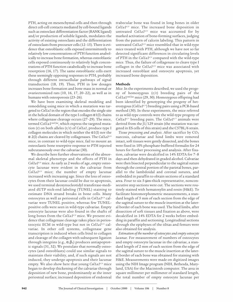
ResultsOsteocytes and empty osteocyte lacunae in Col1a1r/r mice. Wefirst noted the presence of significant increases in thenumber of empty osteocyte lacunae in 4-week-oldCol1a1r/r mice compared with 4-week-old wild-type mice,as shown in representative sections of calvariae stainedwith H&E in Figure 1a. These changes persisted in cal-variae from 10 to 12-month-old Col1a1r/r mice (Figure1b). The calvariae in the older Col1a1r/r mice were alsothicker than those of the wild-type mice (see below). Asshown in Figure 1b, although the area of the calvariaewas comparable in 4-week-old wild-type and Col1a1r/r
mice, the number of empty lacunae was increasedapproximately tenfold in the Col1a1r/r mice comparedwith the wild-type mice. In 4-week-old wild-type mice (n = 8), there were 12 ± 2 (SEM) empty lacunae per mm2
(1.8 ± 0.32% of total), whereas in 4-week-old Col1a1r/r
mice (n = 8), there were 120 ± 19 empty lacunae per mm2
(15 ± 2.5% of total). In 10- to 12-month-old wild-typemice (n = 5), there were 15 ± 5.6 empty lacunae per mm2
(3 ± 1.2% of total), whereas in 10- to 12-month-oldCol1a1r/r mice (n = 7), there were 150 ± 18 empty lacunaeper mm2 (22 ± 2.4% of total). Increased numbers ofempty osteocyte lacunae were seen in Col1a1r/r mice asyoung as 2 weeks of age, but in calvariae from neonatalwild-type and Col1a1r/r mice all osteocyte lacunae werefilled (data not shown). Empty osteocyte lacunae werealso found in tibias and femurs, the long bones exam-ined in the Col1a1r/r mice (data not shown).
The Journal of Clinical Investigation | October 2000 | Volume 106 | Number 8 943
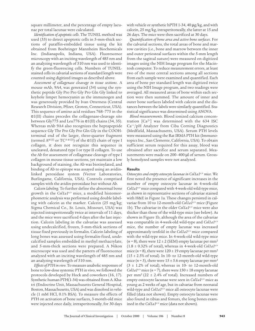
Apoptosis in Col1a1r/r mouse calvariae. In view of theremarkable increase in the number of empty osteocytelacunae in the calvariae from the Col1a1r/r mice, we test-ed for the occurrence of apoptosis in cells that filled theremaining osteocyte lacunae, using TUNEL staining. Itcan be seen in the example from 4-week-old mice shownin Figure 2a that the number of persisting TUNEL-pos-itive osteocytes in the calvariae from both 4-week-oldand 10-month-old Col1a1r/r mice was increased, com-pared with calvariae from wild-type mice of comparableage. TUNEL staining was also observed in cells in theperiosteum of the Col1a1r/r mice (Figure 2a), whichincluded osteoblasts, preosteoblasts, or stromal fibrob-lasts, but they were not further identified. In the cal-variae from the wild-type mice, however, TUNEL stain-ing was observed only in rare, scattered bone cells of alltypes. Increased TUNEL staining was also seen in thecalvariae from 2-week-old Col1a1r/r mice but not inosteoblasts or osteocytes in bones from neonatalCol1a1r/r or wild-type mice (data not shown). As shown
in Figure 2b, in calvariae from 4 week-old wild-typemice, 7.3 ± 1.0 (SEM) apoptotic osteocytes per mm2 werecounted, whereas in calvariae from Col1a1r/r mice, 73.8± 7.4 (SEM) (n = 5) apoptotic osteocytes per mm2 werecounted. In each of the two 8-month-old wild-type micesampled, 9 and 7 apoptotic osteocytes per mm2 werecounted, whereas in four Col1a1r/r mice a mean of 83.0 ±2.7 (SEM) apoptotic osteocytes per mm2 were counted.
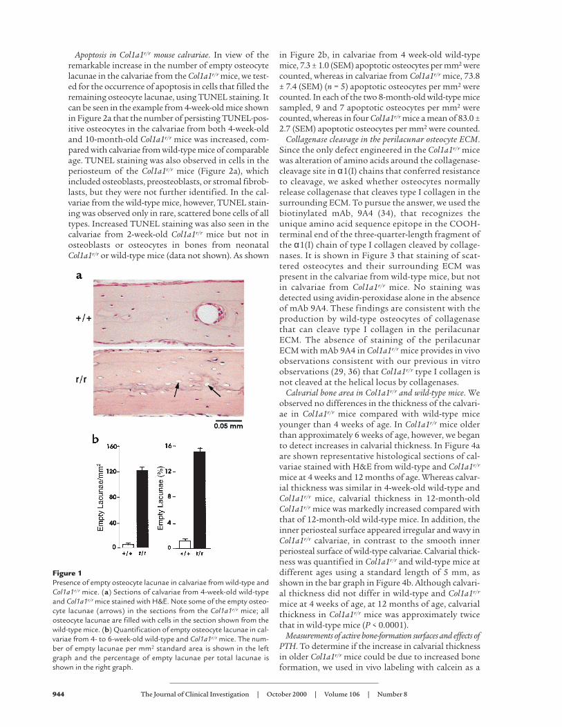
Collagenase cleavage in the perilacunar osteocyte ECM.Since the only defect engineered in the Col1a1r/r micewas alteration of amino acids around the collagenase-cleavage site in α1(I) chains that conferred resistanceto cleavage, we asked whether osteocytes normallyrelease collagenase that cleaves type I collagen in thesurrounding ECM. To pursue the answer, we used thebiotinylated mAb, 9A4 (34), that recognizes theunique amino acid sequence epitope in the COOH-terminal end of the three-quarter-length fragment ofthe α1(I) chain of type I collagen cleaved by collage-nases. It is shown in Figure 3 that staining of scat-tered osteocytes and their surrounding ECM waspresent in the calvariae from wild-type mice, but notin calvariae from Col1a1r/r mice. No staining wasdetected using avidin-peroxidase alone in the absenceof mAb 9A4. These findings are consistent with theproduction by wild-type osteocytes of collagenasethat can cleave type I collagen in the perilacunarECM. The absence of staining of the perilacunarECM with mAb 9A4 in Col1a1r/r mice provides in vivoobservations consistent with our previous in vitroobservations (29, 36) that Col1a1r/r type I collagen isnot cleaved at the helical locus by collagenases.
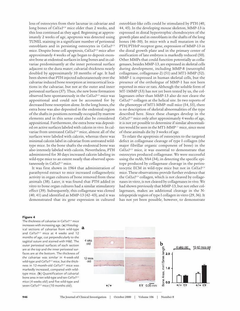
Calvarial bone area in Col1a1r/r and wild-type mice. Weobserved no differences in the thickness of the calvari-ae in Col1a1r/r mice compared with wild-type miceyounger than 4 weeks of age. In Col1a1r/r mice olderthan approximately 6 weeks of age, however, we beganto detect increases in calvarial thickness. In Figure 4aare shown representative histological sections of cal-variae stained with H&E from wild-type and Col1a1r/r
mice at 4 weeks and 12 months of age. Whereas calvar-ial thickness was similar in 4-week-old wild-type andCol1a1r/r mice, calvarial thickness in 12-month-oldCol1a1r/r mice was markedly increased compared withthat of 12-month-old wild-type mice. In addition, theinner periosteal surface appeared irregular and wavy inCol1a1r/r calvariae, in contrast to the smooth innerperiosteal surface of wild-type calvariae. Calvarial thick-ness was quantified in Col1a1r/r and wild-type mice atdifferent ages using a standard length of 5 mm, asshown in the bar graph in Figure 4b. Although calvari-al thickness did not differ in wild-type and Col1a1r/r
mice at 4 weeks of age, at 12 months of age, calvarialthickness in Col1a1r/r mice was approximately twicethat in wild-type mice (P < 0.0001).
Measurements of active bone-formation surfaces and effects ofPTH. To determine if the increase in calvarial thicknessin older Col1a1r/r mice could be due to increased boneformation, we used in vivo labeling with calcein as a
944 The Journal of Clinical Investigation | October 2000 | Volume 106 | Number 8
Figure 1Presence of empty osteocyte lacunae in calvariae from wild-type andCol1a1r/r mice. (a) Sections of calvariae from 4-week-old wild-typeand Col1a1r/r mice stained with H&E. Note some of the empty osteo-cyte lacunae (arrows) in the sections from the Col1a1r/r mice; allosteocyte lacunae are filled with cells in the section shown from thewild-type mice. (b) Quantification of empty osteocyte lacunae in cal-variae from 4- to 6-week-old wild-type and Col1a1r/r mice. The num-ber of empty lacunae per mm2 standard area is shown in the leftgraph and the percentage of empty lacunae per total lacunae isshown in the right graph.
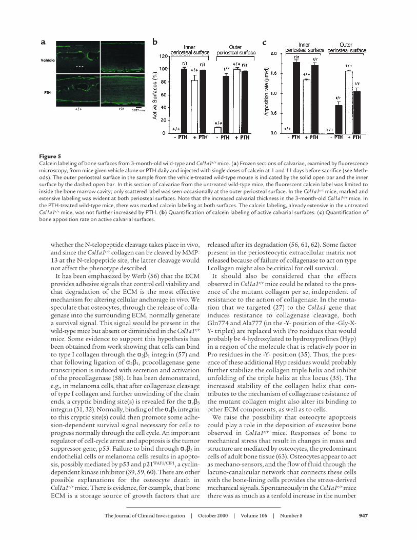
marker in animals 3 months of age or older. As shown inthe examples in Figure 5a, in calvariae from untreated(vehicle alone) wild-type mice, most of the fluorescentlabels were observed inside the bone marrow cavity.There was rarely labeling of the inner periosteal surfacesand only occasionally labeling at the outer periosteal sur-faces. In striking contrast, however, marked and exten-sive double labeling was evident at both periosteal sur-faces in untreated Col1a1r/r mice (Figure 5a). Since PTHin low doses has been demonstrated to have anaboliceffects on bone, and we have shown that catabolic effectsof high doses of PTH on bone resorption are markedlyblunted in the Col1a1r/r mice (30), we designed experi-ments to compare the anabolic effects of low-dose PTHin the wild-type and Col1a1r/r mice (see protocol in Meth-ods). PTH treatment increased calcein labeling, particu-larly evident at both periosteal surfaces. These differ-ences were quantified as shown in Figure 5b. There wasmarked activity at both periosteal surfaces in Col1a1r/r
mice with or without PTH treatment. In contrast, therewas little labeling of either surface in wild-type micetreated with vehicle, but in wild-type mice treated withPTH, labeling was markedly increased at both periostealsurfaces. There was a parallel increase in the mineralapposition rate measured as the distance between thetwo calcein labels divided by the time interval betweeninjections in wild-type mice treated with PTH (Figure5c). The spontaneous mineral apposition rate tended tobe greater at the inner compared with the outerperiosteal surface in Col1a1r/r mice.
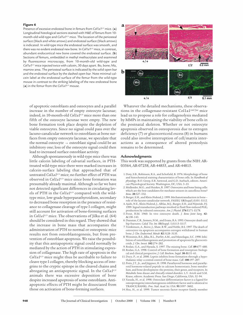
Excessive endosteal bone in femurs from Col1a1r/r mice.Changes were also consistently observed in the longbones from Col1a1r/r mice. Examples shown in Figure6a are longitudinal, histological sections of portions ofthe midfemoral shaft from 10-month-old mice. Theendosteal surface was smooth in wild-type mice, withlittle deposition of new endosteal bone. The endostealsurface in Col1a1r/r mice, however, contained abundant,new, endocortical trabecular bone surrounding activemarrow spaces. Total bone (cortical plus trabecular) inthis region of the diaphysis was greater in all samplesexamined from Col1a1r/r (n = 6) compared with wild-type mice of similar age (n = 6). The pattern of calceinlabeling in the long bones from untreated wild-typeand Col1a1r/r mice also differed markedly as shown in
Figure 6b. In Col1a1r/r mice, calcein labeling wasobserved at both periosteal and endosteal surfaces withmost of the fluorescence in the new bone at theendosteal surface. Only limited and scattered labelingwas found on the endosteal surfaces of the femurs andtibias from wild-type mice.
Blood ionized-calcium and PTH levels. Since we showedin these experiments that PTH activates bone surfaces,and we had shown previously that the resorptiveeffects of PTH are blunted in the Col1a1r/r mice, weasked whether spontaneous secondary hyperparathy-roidism was present in the Col1a1r/r mice. Levels of ion-ized calcium were not different in the Col1a1r/r com-pared with the wild-type mice (data not shown).Although the mean levels of PTH were higher inCol1a1r/r compared with wild-type mice (4.7 ± 0.6[SEM] pg/ml, n = 15, versus 3.7 ± 0.2 [SEM] pg/ml, n = 17), the differences were not significant.
DiscussionAlthough the homozygous (Col1a1r/r) mice had normaldevelopment of the appendicular skeleton duringembryogenesis and during the first week after birth,after approximately 2 weeks of age, aspects of the skele-tal phenotype became apparent. There was detectable
The Journal of Clinical Investigation | October 2000 | Volume 106 | Number 8 945
Figure 2Fluorescence photomicrographs of sec-tions of calvariae from 4-week-old wild-type and Col1a1r/r mice stained by theTUNEL method. (a) Presence of TUNEL-positive cells in calvariae from wild-typeand Col1a1r/r mice. Open bars indicateouter (top) and inner (bottom) surfacesof the periosteum. (b) Quantification ofTUNEL-positive cells in calvariae fromwild-type and Col1a1r/r mice.
Figure 3Photomicrographs of calvarial sections from 4-week-old wild-typeand Col1a1r/r mice stained with 9A4 mAb to the collagenase cleavageepitope. Two separate representative areas of sections from differentmice are shown. No counterstain was used.
loss of osteocytes from their lacunae in calvariae andlong bones of Col1a1r/r mice older than 2 weeks, andthis loss continued as they aged. Beginning at approx-imately 2 weeks of age, apoptosis was detected usingTUNEL staining in a significant number of periostealosteoblasts and in persisting osteocytes in Col1a1r/r
mice. Despite bone cell apoptosis, Col1a1r/r mice afterapproximately 4 weeks of age began to deposit exces-sive bone at endosteal surfaces in long bones and in cal-variae predominantly at the inner periosteal surfaceadjacent to the dura mater; calvarial thickness nearlydoubled by approximately 10 months of age. It hadbeen shown that PTH injected subcutaneously over thecalvariae induced bone resorption at intracortical loca-tions in the calvariae, but not at the outer and innerperiosteal surfaces (37). Thus, the new bone formationobserved here spontaneously in the Col1a1r/r mice wasappositional and could not be accounted for bydecreased bone resorption alone. In the long bones, theextra bone was also deposited in the endosteal regionof the shafts in positions normally occupied by marrowelements and in this sense could also be consideredappositional. Furthermore, the extra bone was deposit-ed on active surfaces labeled with calcein in vivo. In cal-variae from untreated Col1a1r/r mice, almost all of thesurfaces were labeled with calcein, whereas there wasminimal calcein label in calvariae from untreated wild-type mice. In the bone shafts the endosteal bone wasalso intensely labeled with calcein. Nevertheless, PTHadministered for 30 days increased calcein labeling inwild-type mice to an extent nearly that observed spon-taneously in Col1a1r/r mice.
It was first shown in 1964 that administration ofparathyroid extract to mice increased collagenolyticactivity in organ cultures of bone removed from theseanimals (38). Later, it was found that PTH added invitro to bone organ cultures had a similar stimulatoryeffect (39). Subsequently, this collagenase was cloned(40, 41) and identified as MMP-13 (42–43), and it wasdemonstrated that its gene expression in cultured
osteoblast-like cells could be stimulated by PTH (40,44, 45). In the developing mouse skeleton, MMP-13 isexpressed in distal hypertrophic chondrocytes of thegrowth plate and in osteoblasts in the shafts of the longbones (46–50). In mice with a null mutation in thePTH/PTHrP receptor gene, expression of MMP-13 inthe distal growth plate and in the primary center ofossification of late embryos is markedly reduced (50).Other MMPs that could function potentially as colla-genases, besides MMP-13, are expressed in skeletal cellsduring development, including MMP-8 (neutrophilcollagenase, collagenase-2) (51) and MT1-MMP (52).MMP-1 is expressed in human skeletal cells, but thepresence of the orthologue of MMP-1 has not beenreported in mice or rats. Although the soluble form ofMT-1MMP (53) has not yet been tested by us, the col-lagenases other than MMP-13 also do not cleave theCol1a1r/r collagen at the helical site. In two reports ofthe phenotype of MT1-MMP–null mice (54, 55), thereis no description of skeletal abnormalities of the typedescribed here. Since these changes develop in theCol1a1r/r mice only after approximately 4 weeks of age,it is not yet possible to determine if similar abnormali-ties would be seen in the MT1-MMP–/– mice, since mostof these animals die by 3 weeks of age.
To relate the apoptosis of osteocytes to the targeteddefect in collagenase cleavage of type I collagen (themajor fibrillar organic component of bone) in theCol1a1r/r mice, it was essential to demonstrate thatosteocytes produced collagenase. We were successfulusing the mAb, 9A4 (34), in detecting the specific epi-tope produced by collagenase cleavage in the perios-teocytic ECM in wild-type mice but not in Col1a1r/r
mice. These observations provide further evidence thatthe Col1a1r/r collagen, which is not cleaved by collage-nases in vitro, is not cleaved by collagenases in vivo. Wehad shown previously that MMP-13, but not other col-lagenases, makes an additional cleavage in the N-telopeptide region of type I collagen in vitro (29, 36). Ithas not yet been possible, however, to demonstrate
946 The Journal of Clinical Investigation | October 2000 | Volume 106 | Number 8
Figure 4The thickness of calvariae in Col1a1r/r miceincreases with increasing age. (a) Histolog-ical sections of calvariae from wild-typeand Col1a1r/r mice at 4 weeks and 12months of age, cut perpendicularly to thesagittal suture and stained with H&E. Theouter periosteal surfaces of each sectionare at the top and the inner periosteal sur-faces are at the bottom. The thickness ofthe calvariae was similar in 4-week-oldwild-type and Col1a1r/r mice, but the thick-ness in 12-month-old Col1a1r/r mice wasmarkedly increased, compared with wild-type mice. (b) Quantification of calvarialbone area in ten wild-type and ten Col1a1r/r
mice (4 weeks old) and five wild-type andseven Col1a1r/r mice (10 months old).
whether the N-telopeptide cleavage takes place in vivo,and since the Col1a1r/r collagen can be cleaved by MMP-13 at the N-telopeptide site, the latter cleavage wouldnot affect the phenotype described.
It has been emphasized by Werb (56) that the ECMprovides adhesive signals that control cell viability andthat degradation of the ECM is the most effectivemechanism for altering cellular anchorage in vivo. Wespeculate that osteocytes, through the release of colla-genase into the surrounding ECM, normally generatea survival signal. This signal would be present in thewild-type mice but absent or diminished in the Col1a1r/r
mice. Some evidence to support this hypothesis hasbeen obtained from work showing that cells can bindto type I collagen through the α2β1 integrin (57) andthat following ligation of α2β1, procollagenase genetranscription is induced with secretion and activationof the procollagenase (58). It has been demonstrated,e.g., in melanoma cells, that after collagenase cleavageof type I collagen and further unwinding of the chainends, a cryptic binding site(s) is revealed for the αvβ3
integrin (31, 32). Normally, binding of the αvβ3 integrinto this cryptic site(s) could then promote some adhe-sion-dependent survival signal necessary for cells toprogress normally through the cell cycle. An importantregulator of cell-cycle arrest and apoptosis is the tumorsuppressor gene, p53. Failure to bind through αvβ3 inendothelial cells or melanoma cells results in apopto-sis, possibly mediated by p53 and p21WAF1/CIP1, a cyclin-dependent kinase inhibitor (39, 59, 60). There are otherpossible explanations for the osteocyte death inCol1a1r/r mice. There is evidence, for example, that boneECM is a storage source of growth factors that are
released after its degradation (56, 61, 62). Some factorpresent in the periosteocytic extracellular matrix notreleased because of failure of collagenase to act on typeI collagen might also be critical for cell survival.
It should also be considered that the effectsobserved in Col1a1r/r mice could be related to the pres-ence of the mutant collagen per se, independent ofresistance to the action of collagenase. In the muta-tion that we targeted (27) to the Col1a1 gene thatinduces resistance to collagenase cleavage, bothGln774 and Ala777 (in the -Y- position of the -Gly-X-Y- triplet) are replaced with Pro residues that wouldprobably be 4-hydroxylated to hydroxyprolines (Hyp)in a region of the molecule that is relatively poor inPro residues in the -Y- position (35). Thus, the pres-ence of these additional Hyp residues would probablyfurther stabilize the collagen triple helix and inhibitunfolding of the triple helix at this locus (35). Theincreased stability of the collagen helix that con-tributes to the mechanism of collagenase resistance ofthe mutant collagen might also alter its binding toother ECM components, as well as to cells.
We raise the possibility that osteocyte apoptosiscould play a role in the deposition of excessive boneobserved in Col1a1r/r mice. Responses of bone tomechanical stress that result in changes in mass andstructure are mediated by osteocytes, the predominantcells of adult bone tissue (63). Osteocytes appear to actas mechano-sensors, and the flow of fluid through thelacuno-canalicular network that connects these cellswith the bone-lining cells provides the stress-derivedmechanical signals. Spontaneously in the Col1a1r/r micethere was as much as a tenfold increase in the number
The Journal of Clinical Investigation | October 2000 | Volume 106 | Number 8 947
Figure 5Calcein labeling of bone surfaces from 3-month-old wild-type and Col1a1r/r mice. (a) Frozen sections of calvariae, examined by fluorescencemicroscopy, from mice given vehicle alone or PTH daily and injected with single doses of calcein at 1 and 11 days before sacrifice (see Meth-ods). The outer periosteal surface in the sample from the vehicle-treated wild-type mouse is indicated by the solid open bar and the innersurface by the dashed open bar. In this section of calvariae from the untreated wild-type mice, the fluorescent calcein label was limited toinside the bone marrow cavity; only scattered label was seen occasionally at the outer periosteal surface. In the Col1a1r/r mice, marked andextensive labeling was evident at both periosteal surfaces. Note that the increased calvarial thickness in the 3-month-old Col1a1r/r mice. Inthe PTH-treated wild-type mice, there was marked calcein labeling at both surfaces. The calcein labeling, already extensive in the untreatedCol1a1r/r mice, was not further increased by PTH. (b) Quantification of calcein labeling of active calvarial surfaces. (c) Quantification ofbone apposition rate on active calvarial surfaces.
of apoptotic osteoblasts and osteocytes and a parallelincrease in the number of empty osteocyte lacunae;indeed, in 10-month-old Col1a1r/r mice more than onefifth of the osteocyte lacunae were empty. The newbone formation took place despite the depletion ofviable osteocytes. Since no signal could pass over thelacuno-canalicular network to osteoblasts at bone sur-faces from empty osteocyte lacunae, we speculate thatthe normal osteocyte → osteoblast signal could be aninhibitory one; loss of the osteocyte signal could thenlead to increased surface osteoblast activity.
Although spontaneously in wild-type mice there waslittle calcein labeling of calvarial surfaces, in PTH-treated wild-type mice there were marked increases incalcein-surface labeling that approached that ofuntreated Col1a1r/r mice; no further effect of PTH wasobserved in Col1a1r/r mice since bone formation waspresumably already maximal. Although so far we havenot detected significant differences in circulating lev-els of PTH in the Col1a1r/r compared with the wild-type mice, low-grade hyperparathyroidism, secondaryto decreased bone resorption in the presence of resist-ance to collagenase cleavage of type I collagen, mightstill account for activation of bone-forming surfacesin Col1a1r/r mice. The observations of Jilka et al. (64)should be considered in this regard. They showed thatthe increase in bone mass that accompanies theadministration of PTH to normal or osteopenic miceresults not from osteoblastogenesis, but from pre-vention of osteoblast apoptosis. We raise the possibil-ity that this antiapoptotic signal could normally bemediated by the action of PTH in stimulating expres-sion of collagenase. The high rate of apoptosis in theCol1a1r/r mice might thus be ascribable to failure tocleave type I collagen, thereby blocking access of inte-grins to the cryptic epitope in the cleaved chains andabrogating an antiapoptotic signal. In the Col1a1r/r
animals there was excessive deposition of bonedespite increased apoptosis of some osteoblasts. Anti-apoptotic effects of PTH might be dissociated fromthose on activation of bone-forming surfaces.
Whatever the detailed mechanisms, these observa-tions in the collagenase-resistant Col1a1tml Jae micelead us to propose a role for collagenolysis mediatedby MMPs in maintaining the viability of bone cells inthe postnatal skeleton. Whether or not osteocyteapoptosis observed in osteoporosis due to estrogendeficiency (7) or glucocorticoid excess (8) in humanscould also involve interruption of cell/matrix inter-actions as a consequence of altered proteolysisremains to be determined.
AcknowledgmentsThis work was supported by grants from the NIH: AR-03564, AR-07258, AR-44855, and AR-44815.
1. Doty, S.B., Robinson, R.A., and Schofield, B. 1976. Morphology of boneand histochemical staining characteristics of bone cells. In Handbook ofphysiology. R.O. Greep, E.B. Astwood, and G.D. Aurbach, editors. Ameri-can Physiological Society. Washington, DC, USA. 3–23.
2. Mullender, M.G., and Huiskes, R. 1997. Osteocytes and bone lining cells:which are the best candidates for mechano-sensors in cancellous bone?Bone. 20:527–532.
3. Burger, E.H., and Klein-Nulend, J. 1999. Mechanotransduction in bone—role of the lacuno-canalicular network. FASEB J. 13(Suppl.):S101–S112.
4. Ajubi, N.E., Klein-Nulend, J., Alblas, M.J., Burger, E.H., and Nijweide, P.J.1999. Signal transduction pathways involved in fluid flow-induced PGE2
production by cultured osteocytes. Am. J. Physiol. 276:E171–E178.5. Frost, H.M. 1960. In vivo osteocyte death. J. Bone Joint Surg. 42-
A:138–143.6. Dunstan, C.R., Somers, N.M., and Evans, R.A. 1993. Osteocyte death and
hip fractures. Calcif. Tiss. Int. 53(Suppl. 1):S113–S117.7. Tomkinson, A., Reeve, J., Shaw, R.W., and Noble, B.S. 1997. The death of
osteocytes via apoptosis accompanies estrogen withdrawal in humanbone. J. Clin. Endocrinol. Metab. 82:3128–3135.
8. Weinstein, R.S., Jilka, R.L., Parfitt, A.M., and Manolagas, S.C. 1998. Inhi-bition of osteoblastogenesis and promotion of apoptosis by glucocorti-coids. J. Clin. Invest. 102:274–282.
9. Rodan, G.A., and Harada, S. 1997. The missing bone. Cell. 89:677–680.10. Rodan, G.A. 1998. Control of bone formation and resorption: biologi-
cal and clinical perspective. J. Cell. Biochem. Suppl. 30-31:51–61.11. Ducy, P., et al. 2000. Leptin inhibits bone formation through a hypo-
thalamic relay: a central control of bone mass. Cell. 100:197–207.12. Potts, J.T., Jr., and Jüppner, H. 1998. Parathyroid hormone and parathy-
roid hormone-related peptide in calcium homeostasis, bone metabo-lism, and bone development: the proteins, their genes, and receptors. InMetabolic bone disease and clinically related disorders. L.V. Avioli and S.M.Krane, editors. Academic Press. San Diego, California, USA. 52–94.
13. Yasuda, H., et al. 1998. Osteoclast differentiation factor is a ligand forosteoprotegerin/osteoclastogenesis-inhibitory factor and is identical toTRANCE/RANKL. Proc. Natl. Acad. Sci. USA. 95:3597–3602.
14. Hsu, H., et al. 1999. Tumor necrosis factor receptor family member
948 The Journal of Clinical Investigation | October 2000 | Volume 106 | Number 8
Figure 6Presence of excessive endosteal bone in femurs from Col1a1r/r mice. (a)Longitudinal histological sections stained with H&E of femurs from 10-month-old wild-type and Col1a1r/r mice. The location of the periostealsurface (black and white arrows) and endosteal surface (black arrows)is indicated. In wild-type mice the endosteal surface was smooth, andthere was no evident endosteal new bone. In Col1a1r/r mice, in contrast,abundant endocortical new bone covered the endosteal surface. (b)Sections of femurs, embedded in methyl methacrylate and examinedby fluorescence microscopy, from 10-month-old wild-type andCol1a1r/r mice injected twice with calcein, 30 days apart. Bo, bone; Ma,marrow area. The periosteal surface is indicated by the solid open barand the endosteal surface by the dashed open bar. Note minimal cal-cein label at the endosteal surface of the femur from the wild-typemouse in contrast to the striking labeling of the new endosteal bone(a) in the femur from the Col1a1r/r mouse.
RANK mediates osteoclast differentiation and activation induced byosteoprotegerin ligand. Proc. Natl. Acad. Sci. USA. 96:3540–3545.
15. Suda, T., et al. 1999. Modulation of osteoclast differentiation and func-tion by the new members of the tumor necrosis factor receptor and lig-and families. Endocr. Rev. 20:345–357.
16. Hock, J.M., Gera, I., Fonseca, J., and Raisz, L.G. 1988. Human parathy-roid hormone-(1-34) increases bone mass in ovariectomized andorchidectomized rats. Endocrinology. 122:2899–2904.
17. Gunness-Hey, M., and Hock, J.M. 1989. Loss of the anabolic effect ofparathyroid hormone on bone after discontinuation of hormone in rats.Bone. 10:447–452.
18. Bringhurst, F.R., et al. 1993. Cloned, stably expressed parathyroid hor-mone (PTH)/PTH-related peptide receptors activate multiple messengersignals and biological responses in LLC-PK1 kidney cells. Endocrinology.132:2090–2098.
19. Takasu, H., Guo, J., and Bringhurst, F.R. 1999. Dual signaling and lig-and selectivity of the human PTH/PTHrP receptor. J. Bone Miner. Res.14:11–20.
20. Fox, J., Miller, M.A., Stroup, G.B., Nemeth, E.F., and Miller, S.C. 1997.Plasma levels of parathyroid hormone that induce anabolic effects inbone of ovariectomized rats can be achieved by stimulation of endoge-nous hormone secretion. Bone. 21:163–169.
21. Qi, H., Li, M., and Wronski, T.J. 1995. A comparison of the anaboliceffects of parathyroid hormone at skeletal sites with moderate and severeosteopenia in aged ovariectomized rats. J. Bone Miner. Res. 10:948–955.
22. Hodsman, A.B., et al. 1999. The addition of a raloxifene analog(LY117018) allows for reduced PTH(1-34) dosing during reversal ofosteopenia in ovariectomized rats. J. Bone Miner. Res. 14:675–679.
23. Slovik, D.M., et al. 1986. Restoration of spinal bone in osteoporotic menby treatment with human parathyroid hormone (1-34) and 1,25-dihy-droxyvitamin D. J. Bone Miner. Res. 1:377–381.
24. Lane, N.E., Thompson, J.M., Strewler, G.J., and Kinney, J.H. 1995. Inter-mittent treatment with human parathyroid hormone (hPTH[1-34])increased trabecular bone volume but not connectivity in osteopenicrats. J. Bone Miner. Res. 10:1470–1477.
25. Lindsay, R., et al. 1997. Randomised controlled study of effect of parathy-roid hormone on vertebral-bone mass and fracture incidence amongpostmenopausal women on oestrogen with osteoporosis. Lancet.350:550–555.
26. Finkelstein, J.S., and Arnold, A.L. 1999. Increases in bone mineral densi-ty after discontinuation of daily human parathyroid hormone andgonadotropin-releasing hormone analog administration in women withendometriosis. J. Clin. Endocrinol. Metab. 84:1214–1219.
27. Wu, H., et al. 1990. Generation of collagenase-resistant collagen by site-directed mutagenesis of murine proα1(I) collagen gene. Proc. Natl. Acad.Sci. USA. 87:5888–5892.
28. Wu, H., Liu, X., and Jaenisch, R. 1994. Double replacement: strategy forefficient introduction of subtle mutations into the murine Col1-a1 geneby homologous recombination in embryonic stem cells. Proc. Natl. Acad.Sci. USA. 91:2819–2823.
29. Liu, X., et al. 1995. A targeted mutation at the known collagenase cleav-age site in mouse type I collagen impairs tissue remodeling. J. Cell Biol.130:227–237.
30. Zhao, W., Byrne, M.H., Boyce, B.F., and Krane, S.M. 1999. Bone resorp-tion induced by parathyroid hormone is strikingly diminished in colla-genase-resistant mutant mice. J. Clin. Invest. 103:517–524.
31. Montgomery, A.M.P., Reisfeld, R.A., and Cheresh, D.A. 1994. Integrinαvβ3 rescues melanoma cells from apoptosis in three-dimensional der-mal collagen. Proc. Natl. Acad. Sci. USA. 91:8856–8860.
32. Eliceiri, B.P., and Cheresh, D.A. 1999. The role of αv integrins duringangiogenesis: insights into potential mechanisms of action and clinicaldevelopment. J. Clin. Invest. 103:1227–1230.
33. Gavrieli, Y., Sherman, Y., and Ben-Sasson, S.A. 1992. Identification ofprogrammed cell death in situ via specific labeling of nuclear DNA frag-mentation. J. Cell Biol. 119:493–501.
34. Otterness, I.G., et al. 1999. Detection of collagenase-induced damage ofcollagen by 9A4, a monoclonal C-terminal neoepitope antibody. MatrixBiology. 19:331–341.
35. Gross, J. 1981. An essay on biological degradation of collagen. In Cell biol-ogy of the extracellular matrix. E.D. Hay, editor. Plenum Press. New York,New York, USA. 217–258.
36. Krane, S.M., et al. 1996. Different collagenase gene products have dif-ferent roles in degradation of type I collagen. J. Biol. Chem.271:28509–28515.
37. Yates, A.J.P., et al. 1988. Effects of a synthetic peptide of a parathyroidhormone-related protein on calcium homeostasis, renal tubular calci-um reabsorption, and bone metabolism in vivo and in vivo in rodents. J.Clin. Invest. 81:932–938.
38. Walker, D.G., Lapière, C.M., and Gross, J. 1964. A collagenolytic factorin rat bone promoted by parathyroid extract. Biochem. Biophys. Res. Com-mun. 15:397–402.
39. Sakamoto, S., Sakamoto, M., Goldhaber, P., and Glimcher, M.J. 1975.Collagenase and bone resorption: isolation of collagenase from culturemedium containing serum after stimulation of bone resorption by addi-tion of parathyroid hormone extract. Biochem. Biophys. Res. Commun.63:172–178.
40. Quinn, C.O., et al. 1990. Rat collagenase. Cloning, amino acid sequencecomparison, and parathyroid hormone regulation in osteoblastic cells.J. Biol. Chem. 265:22342–22347.
41. Henriet, P., Rousseau, G.G., and Eeckhout, Y. 1992. Cloning andsequencing of mouse collagenase cDNA. Divergence of mouse and ratcollagenases from the other mammalian collagenases. FEBS Lett.310:175–178.
42. Freije, J.M.P., et al. 1994. Molecular cloning and expression of collage-nase-3, a novel human matrix metalloproteinase produced by breast car-cinomas. J. Biol. Chem. 269:16766–16773.
43. Nagase, H., and Woessner, J.F., Jr. 1999. Matrix metalloproteinases. J. Biol.Chem. 274:21491–21494.
44. Partridge, N.C., et al. 1987. Hormonal regulation of the production ofcollagenase and a collagenase inhibitor activity by rat osteogenic sarco-ma cells. Endocrinology. 120:1956–1962.
45. Selvamurugan, N., Chou, W.Y., Pearman, A.T., Pulumati, M.R., and Par-tridge, N.C. 1998. Parathyroid hormone regulates the rat collagenase-3promoter in osteoblastic cells through the cooperative interaction of theactivator protein-1 site and the runt domain binding sequence. J. Biol.Chem. 273:10647–10657.
46. Mattot, V., et al. 1995. Expression of interstitial collagenase is restrict-ed to skeletal tissue during mouse embryogenesis. J. Cell Sci.108:529–535.
47. Fuller, K., and Chambers, T.J. 1995. Localisation of mRNA for collage-nase in osteocytic, bone surface and chondrocytic cells but not osteo-clasts. J. Cell Sci. 108:2221–2230.
48. Gack, S., et al. 1995. Expression of interstitial collagenase during skele-tal development of the mouse is restricted to osteoblast-like cells andhypertrophic chondrocytes. Cell Growth Differ. 6:759–767.
49. Ståhle-Bäckdahl, M., et al. 1997. Collagenase-3 (MMP-13) is expressedduring human fetal ossification and re-expressed in postnatal boneremodeling and in rheumatoid arthritis. Lab. Invest. 76:717–728.
50. Lanske, B., et al. 1998. The parathyroid hormone (PTH)/PTH-relatedpeptide receptor mediates actions of both ligands in murine bone.Endocrinology. 139:5194–5204.
51. Cole, A.A., et al. 1996. Chondrocyte matrix metalloproteinase-8. Humanarticular chondrocytes express neutrophil collagenase. J. Biol. Chem.271:11023–11026.
52. Apte, S.S., Fukai, N., Beier, D.R., and Olsen, B.R. 1997. The matrix met-alloproteinase-14 (MMP-14) gene is structurally distinct from otherMMP genes and is co-expressed with the TIMP-2 gene during mouseembryogenesis. J. Biol. Chem. 272:25511–25517.
53. Ohuchi, E., et al. 1997. Membrane type 1 matrix metalloproteinasedigests interstitial collagens and other extracellular matrix macromole-cules. J. Biol. Chem. 272:2446–2451.
54. Holmbeck, K., et al. 1999. MT1-MMP-deficient mice develop dwarfism,osteopenia, arthritis, and connective tissue disease due to inadequatecollagen turnover. Cell. 99:81–92.
55. Zhou, Z., et al. 2000. Impaired endochondral ossification and angio-genesis in mice deficient in membrane-type matrix metalloproteinase I.Proc. Natl. Acad. Sci. USA. 97:4052—4057.
56. Werb, Z. 1997. ECM and cell surface proteolysis: regulating cellular ecol-ogy. Cell. 91:439–442.
57. Staatz, W.D., Rajpara, S.M., Wayner, E.A., Carter, W.G., and Santaro, S.A.1989. The membrane glycoprotein Ia-IIa (VLA-2) complex mediates theMg+2-dependent adhesion of platelets to collagen. J. Cell Biol.108:1917–1924.
58. Pilcher, B.K., et al. 1997. The activity of collagenase-1 is required for ker-atinocyte migration on type I collagen matrix. J. Cell Biol. 137:1445–1457.
59. Brooks, P.C., et al. 1996. Localization of matrix metalloproteinase MMP-2 to the surface of invasive cells by interaction with integrin αvβ3. Cell.85:683–693.
60. Strömblad, S., Becker, J.C., Yebra, M., Brooks, P.C., and Cheresh, D.A.1996. Suppression of P53 activity and p21WAF1/CIP1 expression by vascu-lar integrin αvβ3. J. Clin. Invest. 98:426–433.
61. Pfeilschifter, J., and Mundy, G.R. 1987. Modulation of type β trans-forming growth factor activity in bone cultures by osteotropic hor-mones. Proc. Natl. Acad. Sci. USA. 84:2024–2028.
62. Bonewald, L.F., et al. 1997. Effects of retinol on activation of latent trans-forming growth factor-β by isolated osteoclasts. Endocrinology.138:657–666.
63. Burger, E.H., and Klein-Nulend, J. 1999. Mechanotransduction in bone:role of the lacuno-canalicular network. FASEB J. 13(Suppl.):S101–S112.
64. Jilka, R.L., et al. 1999. Increased bone formation by prevention ofosteoblast apoptosis with parathyroid hormone. J. Clin. Invest.104:439–446.
The Journal of Clinical Investigation | October 2000 | Volume 106 | Number 8 949