Embed Size (px)
Citation preview
Osteoclast Cytosolic Calcium, Regulated by Voltage-gated Calcium Channels and ExtraceUular Calcium, Controls Podosome Assembly and Bone Resorption Akimi tsu Miyauchi ,* Kei th A. Hruska ,* Edward M. Greenfield,* Randal l Duncan ,* Jose Alvarez,* Raniero Barattolo, ~ Silvia Colucci, ~ Alberta Zambonin-Zal lone , § Steven L. Teitelbaum,* and Anna Teti§ Departments of * Medicine and ¢ Pathology, Jewish Hospital at Washington University Medical Center, St. Louis, Missouri 63110; and §Institute of Human Anatomy, University of Bari, Bari, Italy
Abstract. The mechanisms of Ca 2÷ entry and their effects on cell function were investigated in cultured chicken osteoclasts and putative osteoclasts produced by fusion of mononuclear cell precursors. Voltage- gated Ca 2+ channels (VGCC) were detected by the effects of membrane depolarization with K ÷, BAY K 8644, and dihydropyridine antagonists. K + produced dose-dependent increases of cytosolic calcium ([Ca2+]~) in osteoclasts on glass coverslips. Half-maximal effects were achieved at 70 mM K +. The effects of K + were completely inhibited by dihydropyridine derivative Ca 2+ channel blocking agents. BAY K 8644 (5 x 10 -6 M), a VGCC agonist, stimulated Ca 2+ entry which was inhibited by nicardipine. VGCCs were inactivated by the attachment of osteoclasts to bone, indicating a rapid phenotypic change in Ca 2+ entry mechanisms as- sociated with adhesion of osteoclasts to their resorp-
tion substrate. Increasing extracellular Ca 2+ ([Ca2+]e) induced Ca 2+ release from intracellular stores and Ca 2+ influx. The Ca 2+ release was blocked by dantrolene (10 -5 M), and the influx by La 3+. The effects of [Ca2+]o on [Ca2÷]i suggest the presence of a Ca 2+ receptor on the osteoclast cell membrane that could be coupled to mechanisms regulating cell function. Expression of the [Ca:+]c effect on [Ca2+]i was similar in the presence or absence of bone matrix substrate. Each of the mecha- nisms producing increases in [Ca2+]i, (membrane depolarization, BAY K 8644, and [Ca2+]e) reduced ex- pression of the osteoclast-specific adhesion structure, the podosome. The decrease in podosome expression was mirrored by a 50% decrease in bone resorptive activity. Thus, stimulated increases of osteoclast [Ca2+]i lead to cytoskeletal changes affecting cell adhe- sion and decreasing bone resorptive activity.
T hE cellular basis of bone remodeling is not completely understood. The osteoclast, the multinucleated cell involved in bone resorption, is a complex unit that de-
velops a specialized apparatus for dissolving the bone matrix (King and Holtrop, 1975; Holtrop and King, 1977). Using cell culture systems, several advances have recently been made indicating the molecular events involved in osteoclast bone resorbing activity. For bone resorption to be initiated, the osteoclast polarizes (Baron et al., 1985) and directly at- taches to the bone surface by a specialized area termed the clear zone (Holtrop and King, 1977), in which the contact with the substrate is established by specific adhesion struc- tures called podosomes (Marchisio et al., 1984, 1987; Zambonin-Zallone et al., 1988). Morphologically, podo- somes appear as short membrane protrusions with a core of microfilaments linked to the plasma membrane by talin and vinculin (Marchisio et ai., 1984, 1987). Recent data suggest that podosomes play a pivotal role in substrate recognition by osteoclasts as a specific/33 integrin of the RGD-super-
family of matrix receptors is expressed on their cell mem- brane surface (Davies et al., 1989; Zambonin-Zallone et al., 1989). Substrate recognition is a necessary early step in the initiation of bone resorption, and it may induce phenotypic differences in cellular responses as the osteoclast changes from a motile cell seeking bone substrate to an actively resorbing cell.
The organization of the podosome-containing clear zone allows tight sealing of the resorbing compartment between the osteoclast plasma membrane and the bone surface. The acidification of this extracellular microenvironment (Baron et al., 1985; Blair et al., 1989) produces hydroxyapatite solubilization. Lysosomal enzymes, secreted into this space by a mannose-6-P receptor driven mechanism (Baron et al., 1988; Blair et al., 1988), and activated by the acid pH, digest the organic components of the bone matrix (Blair et al., 1986). Tight sealing of the compartment is needed to main- tain the pH of 5 and the Ca 2+ concentrations of up to 40 mM (Silver et al., 1988).
© The Rockefeller University Press, 0021-9525/90/12/2543/10 $2.00 The Journal of Cell Biology, Volume 111 (No. 6, Pt. 1), Dec. 1990 2543-2552 2543
Dow
nloaded from http://rupress.org/jcb/article-pdf/111/6/2543/1060579/2543.pdf by guest on 09 February 2022
While the mechanisms of osteoclast regulation are incom- pletely understood, we have recently reported that extracel- lular protons decrease cytosolic calcium ([Ca2+]i) and intra- cellular pH (pHi) of osteoclasts attached to bone (Teti et al., 1989). Moreover, these changes in intracellular cation concentration directly stimulate podosome formation (Teti et al., 1989) leading to activation of bone resorption (Arnett and Dempster, 1986; Carano et al., 1990). In contrast, ionophore-induced enhancement in [Ca2+]i decreased ex- pression of podosomes ('left et al., 1989). While these findings are provocative, the mechanisms by which changes in osteoclast [Ca2÷]i are mediated physiologically and their relevance to resorptive activity are unknown.
In the studies reported herein, we focused on the mecha- nisms of [Ca2÷]i regulation in osteoclasts and their relevance to the control of podosome organization and bone resorp- tion activity. In our previous study of osteoclasts attached to bone (Teti et al., 1989), we noted that depolarization of the membrane potential by extracellular potassium chloride produced a prompt decrease in [Ca2+]i. However, Rizzoli, Schlegel, and Bonjour (personal communication) have noted that KCI addition to osteoclasts attached to glass resulted in increased [Ca2+]i. These conflicting studies prompted us to (a) identify and characterize voltage gated calcium channels (VGCCs) 1 in the osteoclast plasma membrane; (b) study modulation of VGCCs during changes in osteoclast activ- ity from an inactive to an active resorbing state; (c) fur- ther characterize a mechanism recently reported (Malgar- oli et al., 1989) whereby extracellular Ca 2+ ([Ca2+],) elicits changes in [Ca2+]i for its action on cell function and (d) analyze the role of [Ca2÷]i regulation in podosome organiza- tion and osteoclast bone-resorbing activity.
Materials and Methods
Materials The acetoxymethyl ester of fura-2 (fura-2 AM) was purchased from Molec- ular Probes (Eugene, OR). BAY K 8644 and nitrendipine were obtained from Miles Laboratories (West Haven, CT), and diltiazem hydrochloride from Marion Laboratories (Kansas City, MO). lonomycin was from Calbiocbem-Behring Corp. (La Jolla, CA), and all other reagents were of analytical grade from Sigma Chemical Co. (St. Louis, MO), Eurobio (Paris, France), and Carlo Erba (Milan, Italy).
Osteoclast Preparation
In our previous report (Teti et al., 1989) adherence of osteoclasts to bone particles was used as a purification technique. The objectives of the studies reported here required cells detached from bone, thereby excluding the use of the bone particle purification technique. Thus, osteoclasts were isolated by a modification of a previously described method (Zambonin-Zallone et al., 1982). Briefly, the medullary bone from femurs and tibias of calcium- deficient laying hens was removed and pressed through a 100-pm nylon sieve. The filtrate was centrifuged for 5 rain at 300 g and then suspended for two minutes in a hypotonic solution (0.2% NaCl) to lyse erythrocytes. The osteoclast-rich suspension was centrifuged at 235 g for 20 rain at 4°C on Ficoll-Hypaque in PBS (density 1.077), and the osteoclast-containing pellet was collected. Centrifugation of the cell suspension was repeated to increase osteoclast enrichment. Because isolated osteoclasts did not express podosomes as they attached to the glass coverslips, they were cultured at a density of 30,000 osteoelasts/3.5 cm in Petri dishes containing glass cover- slips. The culture medium was ct-MEM plus 5% FCS plus 5% chicken se- rum, 100 #g/ml streptomycin, I00 IU/ml penicillin, and 3 #g/ml cytosine 1/3-D arabino furanoside. Temperature was maintained at 37"C in a water-
1. Abbreviation used in this paper: VGCC, voltage-gated calcium channels.
saturated aunost~re containing 5% COz. Cultures were washed after 24 h to remove the non-adhering contaminating cells. This produced a significant increase in the purity of the preparation, as the nonadhering cells were mononuclear. After this step, the enrichment of the preparation for osteo- clasts was increased to 80-90% of the counted nuclei. Thereafter, the num- ber of multinucleated cells in the culture declined and the percent of nuclei in the preparation that were from mononuclear cells increased. Thus, there was no evidence for new formation of multinucleated cells in culture. CuP ture was necessary because freshly prepared cells did not express podo- somes. Podosome expression in osteoclasts isolated on bone develops rap- idly, but on the glass coverslips podosome expression began between 48 and 72 h. Thus, experiments were performed between 3 and 5 d in culture. The osteoclastic features of these multinucleated cells have been previously de- scribed (Zambonon-Zallone et al., 1982). In addition, the osteoclasts added to bone slices produced well formed resorption pits assessed by scanning electron microscopy. In some experiments, 100 #g of devitalized rat bone particles, 38-63 #m diameter, were added to osteoclasts cultured on glass coverslips and incubated for 5 h. During this time, changes in phenotypic expression of Ca 2+ entry mechanisms were assessed.
Because the purity of the ostooclast preparations could not be increased above 80-90%, there was a possibility that contaminating cells were regulating osteoclast responses to experimental protocols. Thus, we also used a technique recently developed in our laboratory whereby virtually pure preparations of multinucleated putative osteoclasts were generated in culture from mononuclear cell precursors. Putative osteoclast precursors were obtained from bone marrow of laying hens maintained on calcium deficient diets by recovering the cells from the interfaces of the Ficoll/Hy- paque gradient centrifugation described above. These mononuclear cells were cultured in the presence of cytosine l~-v arabino furanoside (5 ~g/mi) to prevent proliferation of contaminating ceils. After 3 d, highly purified precursor preparations were obtained by removing nonadherent cells (lym- phocytes, granulocytes, and erythrocytes). Between day 3 and 6 of culture, pure preparations (>98% of cell number) of multinncleated cells were formed that expressed the following osteoclast characteristics: ruffled mem- branes apposed to bone surfaces, formation of resorption pits in bone slices, resorption of 3H-proline labeled bone particles (Blair et al., 1986), tartrate- resistant acid phosphatase and reactivity with osteoclast-specific mAbs, 121F (Oursler et al., 1985) and 23C6 (Davies et al., 1989). For [Ca2+]i measurements, 4 × 10⁶ precursors were cultured in 35 mm petri dishes containing glass coverslips for 3-5 d before use. We found that regardless of the method of osteoclast preparation, the results obtained in our studies were the same. Because pure populations were obtainable with the cell fu- sion preparation, the influences of contaminating cells were eliminated. The data presented in the figures of this report were obtained using both os- teoclast preparations.
Measurement of Cytosolic Ca 2+ [CaZ÷]~ [Ca2+]i was measured in single cells using the fluorescent calcium indica- tor fura-2. Osteoclasts cultured on coverslips were loaded for 1 h at 25"C in a buffer containing 125 mM NaC1, 5 mM KCI, 1.2 mM KH2PO4, 1.2 mM MgSO4, 2 mM CaC12, 25 mM Hepes, 6 mM glucose (Krebs-Ringer Hepes [KRH]) and 10/~M fura-2-AM. In these loading conditions, resulting fura-2 fluorescence was diffuse and no punctate distribution was observed, as described in other cells (Malgaroli et al., 1987). Ceils were then washed three times with KRH and used for the experiments. Fluorescence was mea- sured in single cells excited with 340 and 380 nm light selected by t ~ monochromators and directed through the stage of a Nikon inverted micro- scope equipped with a 100× fluor objective. Emitted light was collected at 505 nm after being filtered through a cot-off filter (490 nm) and monitored photometrically (SPEX Industries, Edison, NJ). Coverslips were mounted at the bottom of a Sykes-Moore open chamber with 1 ml KRH within a cli- mate box maintained at 37"C.
Fura-2 fluorescence was calibrated to [Ca2+]i at the end of each experi- ment, exposing the cells to 5 /~M ionomycin to assess the Ca-saturated fluorescence (Fm~, followed by 5 mM EGTA to determine fluorescence at nominally Ca2+-free condition (Fmi~). 2 mM MnCI2 was finally added to estimate autofluorescence which was subtracted from the experimental values. [Ca2+]i was calculated using the formula published by Grynkiewicz et al. (1985).
Activation of VGC Cs VGCCs were activated in osteoclasts by depolarizing the cells with high K +
The Journal of Cell Biology, Volume 111, 1990 2544
Dow
nloaded from http://rupress.org/jcb/article-pdf/111/6/2543/1060579/2543.pdf by guest on 09 February 2022
KRH. Experiments were performed in isotonic conditions by substituting from 6.2 to 100 mM KC1 for corresponding amounts of NaC1 in the KRH buffer. Activation of VGCCs was also obtained using BAY K 8644, a specific dihydropyridine VGCC agonist. The effects of several VGCC antag- onists were evaluated, including diltiazem, verapamil, nicardipine, or nitrendipine (10-1°-10 -5 M) before depolarization with high K + KRH (100 mM) or addition of BAY K 8644. A complete series of parallel experiments were performed using osteoclast cultures in which devitalized bone parti- cles (38-63-ttm-diarn) were added 5 h before the experiments were per- formed.
Activation of Calcium-operated Regulation of [Caz÷]~ Changes in [Ca2+]i were observed during changes in extracellular calcium concentrations. Osteoclasts, previously incubated in KRH containing 2 mM Ca 2+, were treated with increasing extracellular calcium concentrations. To block release of calcium from intracellular stores, a series of experi- ments was performed using cells pretreated with dantrolene (10 -5 M) 10 min before CaC12 additions. To block Ca 2+ entry, LaC13 and Ca 2+ channel antagonists were studied for their effects on the changes in [Ca2+]i stimu- lated by adjustments in [CaZ+]e. Parallel experiments in the presence of bone particles were also performed to evaluate the effects of increasing ex- tracellular calcium concentration on [Ca2+]i during bone resorption.
Cytoskeletal Studies The effect of membrane depolarization on podosome expression was evalu- ated in osteoclasts incubated for 90 rain in MEM in which 30 or 50 mM NaCI was isotonically substituted with KC1. Control osteoclasts were in- cubated in regular MEM containing 5.0 mM KCI. The effect of extracellular calcium on podosome expression was evaluated by incubating osteoclasts in nominally calcium-free MEM and adding CaC12 from 0 to 4 mM. Incu- bation times were 90 rain unless otherwise indicated.
Microfilaments were detected by decoration of F-actin with rhodamine- conjugated phalloidin (R-PHD). Cells were fixed for 5 min at room temper- ature with 3 % formaldehyde, 2 % sucrose in PBS. They were then permeabi- lized for 3 rain at0°C with 50 mM NaCI, 300 mM sucrose, 20 mM Hepes (pH 7.4), 3 mM MgCI2, 0.5% Triton-X 100, and then they were incubated for 45 min at 37°C with 5/~g/ml R-PHD. Coverslips were then washed, mounted in 10% mowiol, and observed with a Leitz Diavert fluorescence microscope. Due to the presence of a core of microfilaments perpendicu- larly oriented with respect to the substrate, podosomes were recognized as fluorescent dot-like structures distributed at the level of the ventral surface of the cell.
To quantitate the results, the numbers of osteoclasts presenting podo- somes in each treatment expressed as the percent of the total number of os- teoclasts present. Data were expressed as mean percentage + SE. The statistical significance was calculated by the t test. For each treatment, an average of 300 osteoclasts was counted. A decrease in podosome expression did not cause the cells to lift off the coverslip since the osteoclast has other mechanisms of substrate adhesion besides podosome expression (Zambonin- Zallune et al., 1989).
resorbed were calculated. Data were expressed as mean micrograms of bone resorbed :t: SE. The statistical significance was evaluated by the t test. Each experiment was performed with at least three different osteoclast preparations in quadruplicate.
Results
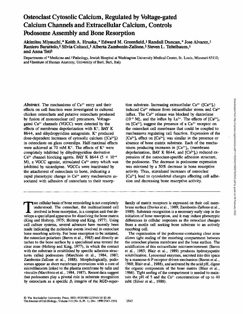
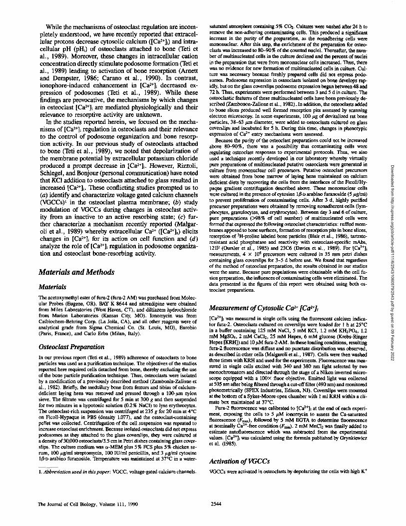
Effect of Membrane Depolarization on Osteoclast [Ca2÷1~ Basal [Cae+]~ of osteoclasts on glass coverslips in KRH was 123 + 9 (n = 41) (mean ± SE). The response of osteoclasts [Ca2+]~ to 100 m K ÷ is shown in Fig. 1. The effect of K ÷ on [Ca2+]i was directionally opposite to the response we previ- ously reported in osteoclasts attached to bone particles (Teti et al., 1989). High K ÷ induced an immediate increase in [Cae+]~ and a second slower rise to a new steady state of 267 nM within 300 s. High K ÷ increased [Ca2+]i in a similar manner in 83 % of examined cells attached to glass cover- slips (n = 18). The other 17% of cells demonstrated a de- crease in [Ca 2+] upon depolarization with K ÷. The increase in [Ca2+]~ from basal to peak levels achieved by various concentrations of K + in high K ÷ KRH was dose-dependent with a half-maximal effect achieved by 66 mM (Fig. 2). When cells were placed in a Ca-free KRH (CaC12 omitted from the buffer and 1 mM EGTA added), KC1 additions failed to elevate [Ca2+]i (not shown). This indicated that the increases in [Ca2+]i stimulated by high K ÷ were due to cal- cium entry from the extracellular fluid. The effects of high K + were almost completely inhibited by the dihydropyri- dine, nicardipine (Fig. 1 b).
The Effect of BAY K 8644 on [Caz+]~ BAY K 8644 is a dihydropyridine-derivative Ca 2+ channel agonist that increases opening frequency of dihydropyridine- sensitive Ca 2+ channels (Duncan and Misler, 1989; Gug- gino et al., 1989; Meier et al., 1988). After the addition of BAY K 8644 (5 × 10 ..6 M) in calcium containing KRH a
207
.Z
Bone Resorption .~ N
160 The effects of increasing [Ca2+]i, by activation of VGCC or changing .o. [Ca2+]e, on the bone-resorbing activity of osteoclast cultures was deter- mined. For this purpose, 2-d-old osteoclast cultures were incubated for 48 h with [3H]proline prelabeled devitalized rat or chicken bone particles (Blair et al., 1986) at a density of 400/~g of boneJ50,000 osteoclasts in 1.5 b cm ~o culture dishes. The cells were cultured in serum-free MEM in the presence of varying calcium concentrations, varying K + concentrations or by the addition of BAY K 8644. The experiments were carried out by (a) .= preincubating the cells for 30 rain with the experimental medium, then add- ~" 136 ing the bone particles, and (b) preincubating the cells for 24 h with the bone ¢~ 1 2 3
particles, then substituting the control medium with the experimental medium.
Bone resorption was evaluated by measuring the radioactivity present in the medium, which derived from osteoclast-mediated release of tritiated products of bone collagen degradation (Blair et al., 1986). Parallel samples, in absence of cells, were evaluated to measure the nonspecific 3H release, which was subtracted from the corresponding experimental values. The to- tal radioactivity present in the 400/~g of bone added to the cultures was measured after demineralization and solubilization of the organic matrix, as previously described (Blair et al., 1986), and the micrograms of bone
Nicardipi~o Hi K
l l
i i 100 S
Figure 1. Effect o f K + on osteoclast [Ca2+]i . ( a ) A fura-2 loaded single osteoclast , bathed in KRH, responded to substitution with high K + (100 m M ) K R H with a sustained increase in [Ca2+]i. (b) The effect o f pretreatment with nicardipine on high K + (100 mM) KRH induced increase in [Ca2+]~. Nicardipine (10 -7 M) inhibited the rise in [Ca2+]i after membrane depolarization.
Ca Regulates Osteoclast Adhesion and Resorption 2545 Miyauchi et al. 2+
Dow
nloaded from http://rupress.org/jcb/article-pdf/111/6/2543/1060579/2543.pdf by guest on 09 February 2022
200 (5)
15o
100
(10
~2 3o 50 1;0 [K*]o(mM)
Figure 2. Concentration dependence of the K+-induced changes in [Ca2+]i of single osteoc]asts. The maxima] increase in [Ca2+]i pro_ duced with increasing K + concentrations is plotted against K+o. The number of experiments is indicated in parentheses. Results are the mean + SE.
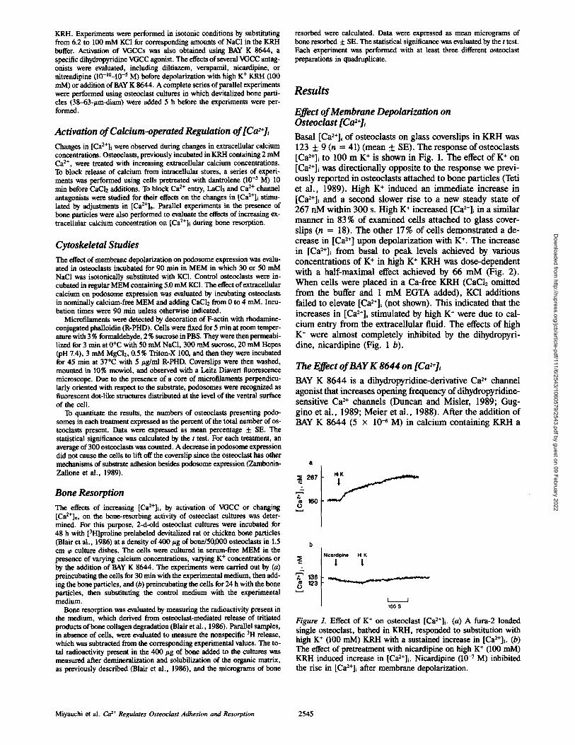
rise in [ C a 2 + ] i of 78 "1- 8 nM (n = 3) was observed, reach- ing plateau levels in 7-8 min (Fig. 3 a). BAY K 8644 (5 x 10 -6 M) had no effect on [Ca2÷]~ in a Ca~+-free medium (not shown), and the increase of [Ca2+]i induced by BAY K 8644 was completely abolished by nicardipine (10 -6 M) (Fig. 3 b). The effects of BAY K 8644 were slower in onset than high K ÷ KRH probably due to time for incorporation into plasma membranes in our system. The increase in [Ca~÷]+ produced by 5 x 10 -6 M BAY K was similar to that of 70 mM K ÷. These findings suggest that BAY K 8644 stimu- lated Ca 2+ influx through dihydropyridine-sensitive voltage- gated Ca 2+ channels.
Effects of Ca 2÷ Channel Blocking Agent on the K÷-induced Increase in [Ca2÷],
The effects of the dihydropyridine Ca 2+ channel blocker, m'cardipine 0 0 -7 M), on the K+-stimulated changes in [Ca~+]~
183
% lo5 O
&
O 130
Nicardipine Bay K 8644
l l
I • 100 S
Figure 3. Effect of the dihydropyridine VGCC agonist BAY K 8644 on osteoclast [ C a 2 + ] i . a illustrates the rise in [Ca2+]~ in a single os- teoclast treated with 5 x 10 -6 M BAY K 8644 (marked by an ar- row). The increase in [Ca2+]i reached a new steady state in 7-8 min. b illustrates the effect of pretreatment with 10-6 M nicardi- pine, which prevented BAY K 8644-induced increases in [Ca2+]~. Experiments shown in A and B are a pair of four similar experi- ments.
10o
o 80
o 60
~ 4o , Z ~ 2o
~ 0
0 • Diltiazem A ~ • Verapamil
\ & T \ \ T • Nicardipine ~ -- ~,~C) X Nit rendipine
i ~ i i L i
10 9 8 7 6 5
Ca Channel Blockers Conc. I-log M]
Figure 4. Effects of Ca 2+ channel antagonists on K+-induced eleva- tions in [ C a 2 + ] i o Dose-response curves are illustrated for os- teoclasts treated with nicardipine, nitrendipine (dihydropyridine derivatives), verapamil (phenylalkylamine), diltiazem 0aenzodi- azepine), or LaCI3 before the addition of 100 mM K ÷. The data represent the size of the maximal high K + (100 mM) induced ele- vation of [Ca2+]i in osteoclasts pretreated for 3 min with the corre- sponding VGCC antagonist. The data are expressed as mean + SE of three or four separate experiments.
are shown in Fig. 1 b. KRH with nicardipine (10 -7 M) was added for 2 min before high K + with nicardipine. K ÷ had no significant effect on [Ca2+]i in the presence of nicardipine. The K+-induced increase in [Ca2+]i, was restored after transfer to nicardipine-free KRH and a second addition of K +.
The effects of various Ca 2+ channel blockers on high K + KRH- (100 raM) induced elevations of [Ca2+]i were com- pared, as shown in Fig. 4. Dihydropyridine Ca 2+ channel blockers (nicardipine, nitrendipine at concentrations of 10-]°-10 -5 M (n = 3 or 4 of each concentration) inhibited the K+-induced [Ca2+]~ increases in a dose dependent manner. The half-maximal inhibition was observed at 5 × 10 -9 M. Verapamil was less potent than dihydropyridines, and diltia- zero was less effective than verapamil.
Effects of Changing Extracellular Calcium on [Ca2÷]j
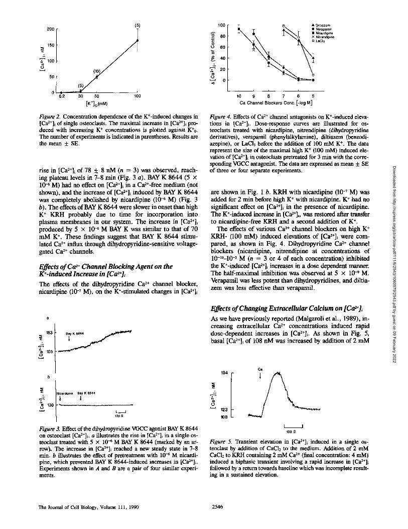
As we have previously reported (Malgaroli et al., 1989), in- creasing extracellular Ca 2+ concentrations induced rapid dose-dependent increases in [Ca2+]i. As shown in Fig. 5, basal [Ca2+]i of 108 nM was increased by addition of 2 mM
C a 194
..,
o 123
108
I I 100 S
Figure 5. Transient elevation in [Ca2+]i induced in a single os- teoclast by addition of CaClz to the medium. Addition of 2 mM Cae l2 to KRH containing 2 mM Ca 2+ (final concentration: 4 mM) induced a biphasic transient involving a rapid increase in [Ca~+]i followed by a return towards baseline which was incomplete result- ing in a sustained elevation.
The Journal of Cell Biology, Volume 111, 1990 2546
Dow
nloaded from http://rupress.org/jcb/article-pdf/111/6/2543/1060579/2543.pdf by guest on 09 February 2022
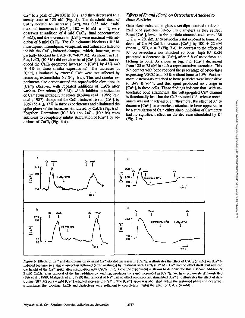
Ca 2+ to a peak of 194 nM in 80 s, and then decreased to a steady state at 123 nM (Fig. 5). The threshold dose of CaCl2 needed to increase [Ca2+]~ was 0.25 mM. Half- maximal increases in [Ca2+]i, 182 -t- 16 nM, n = 7, were observed at addition of 4 mM CaCI2 (final concentration 6 mM), and the increases in [Ca2+]~ were maximal with ad- dition of 8 mM CaCI2. The Ca 2+ channel blockers (10 -5 M nicardipine, m'trendipine, verapamil, and diltiazem) failed to inhibit the CaCl2-induced changes, which, however, were partially blocked by LaCI3 (10-4-10 -5 M). As shown in Fig. 6 a, LaCla (10 -~ M) did not alter basal [Ca2+]i levels, but re- duced the CaC12-prompted increase in [Ca2+]i by 43% (40 + 4% in three similar experiments). The increases in [Ca2+]i stimulated by external Ca 2+ were not affected by removing extracellular Na (Fig. 6 b). This and similar ex- periments also demonstrated the reproducible elevations in [Ca2÷]i observed with repeated additions of CaC12 after washes. Dantrolene (10 -5 M), which inhibits mobilization of Ca 2+ from intracellular stores (Kojima et al., 1985; Reid et al., 1987), dampened the CaC12 induced rise in [Ca2+]~ by 80% (55.4 + 17% in three experiments) and eliminated the spike phase of the increases stimulated by CaC12 (Fig. 6 c). Together, Dantrolene (104 M) and LaCI3 (10-4 M) were sufficient to completely inhibit stimulation of [Ca2+]~ by ad- ditions of CaC12 (Fig. 6 d).
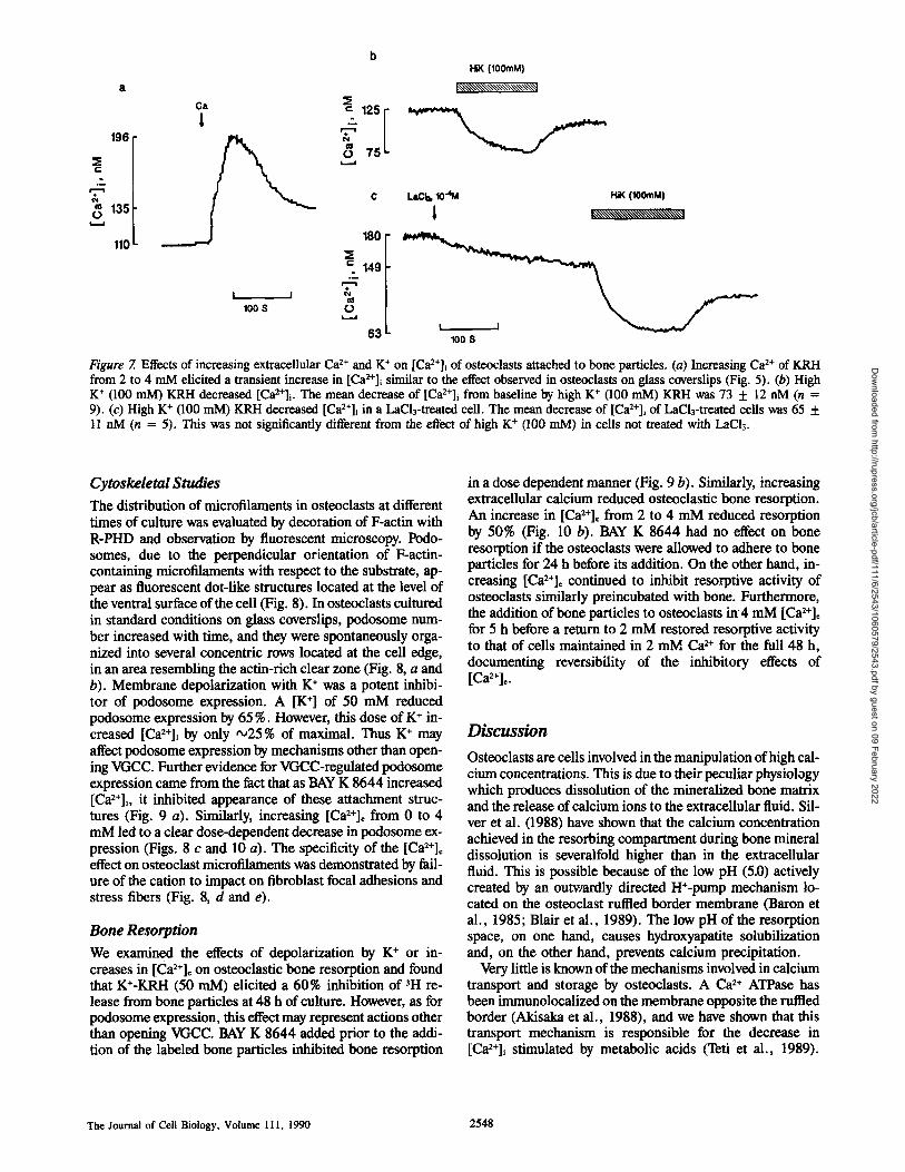
Effects of K + and [Ca2÷], on Osteoclasts Attached to Bone Particles
Osteoclasts cultured on glass coverslips attached to devital- ized bone particles (38-63 #m diameter) as they settled. Basal [Ca2+]i levels in the particle-attached cells were 136 + 7, n = 28, similar to osteoclasts not exposed to bone. Ad- dition of 2 mM CaC12 increased [Ca2+], by 100 + 22 nM (mean + SE), n = 7 (Fig. 7 a). In contrast to the effects of K + on osteoclasts not attached to bone, high K + KRH prompted a decrease in [Ca2+]~ after 5 h of osteoclasts at- taching to bone. As shown in Fig. 7 b, [Ca2÷]i decreased from 125 to 75 nM in such a representative osteoclast. This 5-h contact with bone reduced the percentage of osteoclasts expressing VGCC from 83 % without bone to 10%. Further- more, osteoclasts attached to bone particles were insensitive to BAY K 8644, and this agent produced no change in [Ca2+]~ in these cells. These findings indicate that, with os- teoclastic bone attachment, the voltage-gated Ca 2+ channel is functionally lost, but the Ca2+-induced Ca 2+ release mech- anism was not inactivated. Furthermore, the effect of K + to decrease [Ca2+]i in osteoclasts attached to bone appeared to be a stimulation of Ca 2+ efflux since inhibition of Ca 2+ entry had no significant effect on the decrease stimulated by K + (Fig. 7 c).
. ~ 160 III o
203 180
T cll o
82 76
Cat WUh CI Wash
194 | | | |
115 LsClail0"4M
I I 100 S
C~ W u h Ca W u h
I I I _I
Ne fr~e PB$
100 f
t ! ! e,oo /V.-.,,o. 153 i. -., ~
100 S
d Cl Wish
! 1
LaO3, ~T4M !
/ I !
100
C l
1
Figure 6. Effects of La 3+ and dantrolene on external Ca2+-elicited increases in [Ca2+]i. a illustrates the effect of CaC12 (2 mM) on [Ca2+]~ - induced biphasic in a single osteoclast followed (after washings) by treatment with LaCI3 (10-4 M). La 3~" had no effect itself, but reduced the height of the Ca 2+ spike after stimulation with CaC12. In b, a control experiment is shown to demonstrate that a second addition of 2 mM CaC12, after removal of the first addition by washing, produces the same increment in [Ca2+]i. We have previously demonstrated (Teti et al., 1989; Malgaroli et al., 1989) that removal of Na + had no effect on osteoclast stimulated [Ca2+]i. c illustrates the effect of dan- trolene (10 -5 M) on a 4 mM [Ca2+]e-elicited increase in [Ca2+]i. The [Ca2+]i spike was abolished, while the sustained phase still occurred. d illustrates that together, LaCI3 and dentrolene were sufficient to completely inhibit the effect of CaCI2 (4 mM).
+ Miyauchi et al. Ca z Regulates Osteoclast Adhesion and Resorption 2547
Dow
nloaded from http://rupress.org/jcb/article-pdf/111/6/2543/1060579/2543.pdf by guest on 09 February 2022
b HiK (100mM)
k~',~\\\\\~\\\\\\'~,~\~
196 ~m-
"~135
110
63 L 100 S
Figure 7. Effects of increasing extracellular Ca 2+ and K + on [Ca2+]i of osteoclasts attached to bone particles. (a) Increasing Ca 2+ of KRH from 2 to 4 mM elicited a transient increase in [Ca2+]i similar to the effect observed in osteoclasts on glass coverslips (Fig. 5). (b) High K + (100 mM) KRH decreased [Ca2+]i. The mean decrease of [Ca2+]~ from baseline by high K + (100 raM) KRH was 73 + 12 nM (n = 9). (c) High K + (100 mM) KRH decreased [Ca2+]i in a LaCl3-treated cell. The mean decrease of [Ca2+]i of LaCl3-treatexl cells was 65 + 11 nM (n = 5). This was not significantly different from the effect of high K + (100 mM) in cells not treated with LaCI3.
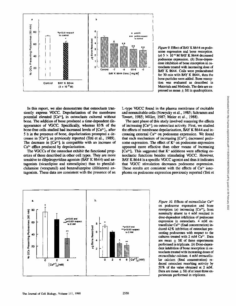
Cytoskeletal Studies The distribution of microfilaments in osteoclasts at different times of culture was evaluated by decoration of F-actin with R-PHD and observation by fluorescent microscopy. Podo- somes, due to the perpendicular orientation of F-actin- containing microfilaments with respect to the substrate, ap- pear as fluorescent dot-like structures located at the level of the ventral surface of the cell (Fig. 8). In osteoclasts cultured in standard conditions on glass coverslips, podosome num- ber increased with time, and they were spontaneously orga- nized into several concentric rows located at the cell edge, in an area resembling the actin-rich clear zone (Fig. 8, a and b). Membrane depolarization with K ÷ was a potent inhibi- tor of podosome expression. A [K ÷] of 50 mM reduced podosome expression by 65 %. However, this dose of K ÷ in- creased [Ca2+]i by only ,~25 % of maximal. Thus K ÷ may affect podosome expression by mechanisms other than open- ing VGCC. Further evidence for VGCC-regulated podosome expression came from the fact that as BAY K 8644 increased [Ca2+]i, it inhibited appearance of these attachment struc- tures (Fig. 9 a). Similarly, increasing [Ca2+]~ from 0 to 4 mM led to a clear dose-dependent decrease in podosome ex- pression (Figs. 8 c and 10 a). The specificity of the [Ca2+], effect on osteoclast microfilaments was demonstrated by fail- ure of the cation to impact on fibroblast focal adhesions and stress fibers (Fig. 8, d and e).
Bone Resorption We examined the effects of depolarization by K + or in- creases in [Ca2+], on osteoclastic bone resorption and found that K+-KRH (50 raM) elicited a 60 % inhibition of 3H re- lease from bone particles at 48 h of culture. However, as for podosome expression, this effect may represent actions other than opening VGCC. BAY K 8644 added prior to the addi- tion of the labeled bone particles inhibited bone resorption
in a dose dependent manner (Fig. 9 b). Similarly, increasing extracellular calcium reduced osteoclastic bone resorption. An increase in [Ca2+]e from 2 to 4 mM reduced resorption by 50% (Fig. 10 b). BAY K 8644 had no effect on bone resorption if the osteoclasts were allowed to adhere to bone particles for 24 h before its addition. On the other hand, in- creasing [Ca2+]o continued to inhibit resorptive activity of osteoclasts similarly preincubated with bone. Furthermore, the addition of bone particles to osteoclasts in4 mM [Ca2+]° for 5 h before a return to 2 mM restored resorptive activity to that of cells maintained in 2 mM Ca 2÷ for the full 48 h, documenting reversibility of the inhibitory effects of [Ca2+]~.
Discussion
Osteoclasts are cells involved in the manipulation of high cal- cium concentrations. This is due to their peculiar physiology which produces dissolution of the mineralized bone matrix and the release of calcium ions to the extracellular fluid. Sil- ver et al. (1988) have shown that the calcium concentration achieved in the resorbing compartment during bone mineral dissolution is severalfold higher than in the extracellular fluid. This is possible because of the low pH (5.0) actively created by an outvaurdly directed H÷-pump mechanism lo- cated on the osteoclast ruffled border membrane (Baron et al., 1985; Blair et al., 1989). The low pH of the resorption space, on one hand, causes hydroxyapatite solubilization and, on the other hand, prevents calcium precipitation.
Very little is known of the mechanisms involved in calcium transport and storage by osteoclasts. A Ca 2+ ATPase has been immunolocalized on the membrane opposite the ruffled border (Akisaka et al., 1988), and we have shown that this transport mechanism is responsible for the decrease in [Ca2+]i stimulated by metabolic acids ('left et al., 1989).
The Journal of Cell Biology, Volume 111, 1990 2548
Dow
nloaded from http://rupress.org/jcb/article-pdf/111/6/2543/1060579/2543.pdf by guest on 09 February 2022
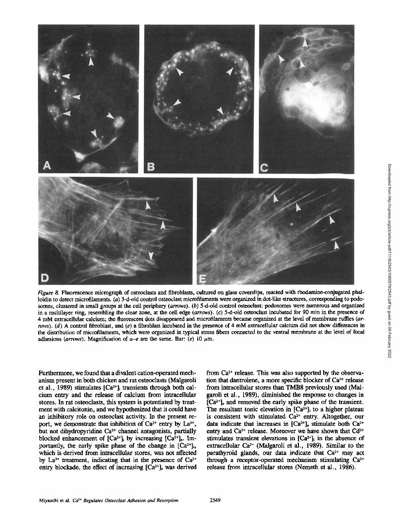
Figure 8. Fluorescence micrograph of osteoclasts and fibroblasts, cultured on glass coverslips, reacted with rhodamine-conjugated phai- loidin to detect microfilaments. (a) 3-d-old control osteoclast microfilaments were organized in dot-like structures, corresponding to podo- somes, clustered in small groups at the cell periphery (arrows). (b) 5-d-old control osteoclast: podosomes were numerous and organized in a multilayer ring, resembling the clear zone, at the cell edge (arrows). (c) 5-d-old osteoclast incubated for 90 min in the presence of 4 mM extracellular calcium; the fluorescent dots disappeared and microfilaments became organized at the level of membrane ruffles (ar- rows). (d) A control fibroblast, and (e) a fibroblast incubated in the presence of 4 mM extracellular calcium did not show differences in the distribution of microfilaments, which were organized in typical stress fibers connected to the ventral membrane at the level of focal adhesions (arrows). Magnification of a-e are the same. Bar: (e) 10 t~m.
Furthermore, we found that a divalent cation-operated mech- anism present in both chicken and rat osteoclasts (Malgaroli et al., 1989) stimulates [Ca2+]i transients through both cal- cium entry and the release of calcium from intracellular stores. In rat osteoclasts, this system is potentiated by treat- ment with calcitonin, and we hypothesized that it could have an inhibitory role on osteoclast activity. In the present re- port, we demonstrate that inhibition of Ca ~+ entry by La 3+, but not dihydropyridine Ca 2+ channel antagonists, partially blocked enhancemem of [Ca2+]i by increasing [Ca2+]c. Im- portantly, the early spike phase of the change in [Ca2+]i, which is derived from intracellular stores, was not affected by La 3+ treatment, indicating that in the presence of Ca x+ entry blockade, the effect of increasing [Ca2+]c was derived
from Ca 2+ release. This was also supported by the observa- tion that dantrolene, a more specific blocker of Ca ~+ release from intracellular stores than TMB8 previously used (Mal- garoli et al., 1989), diminished the response to changes in [Ca2+]c and removed the early spike phase of the transient. The resultant tonic elevation in [Ca~+]i to a higher plateau is consistent with stimulated Ca 2+ entry. Altogether, our data indicate that increases in [Ca2+], stimulate both Ca 2+ entry and Ca 2+ release. Moreover we have shown that Cd 2+ stimulates transient elevations in [Ca2+]i in the absence of extracellular Ca 2+ (Malgaroli et al., 1989). Similar to the parathyroid glands, our data indicate that Ca 2+ may act through a receptor-operated mechanism stimulating Ca 2+ release from intraceUular stores (Nemeth et al., 1986).
Miyauchi et al. Ca 2+ Regulates Osteoclast Adhesion and Resorption 2549
Dow
nloaded from http://rupress.org/jcb/article-pdf/111/6/2543/1060579/2543.pdf by guest on 09 February 2022
|
E
o o 6 0
o
~ 4o
~ 20 o
F *p<0.01 respect
to control
Control BAY K 8644 (5 x 10e M)
5
JO 5 4
~ 3
1l Control
~t p<0.05 t ~t p<0.01 respect to control
-7 -6 5X-6 -5 BAY K 8644 Conc. I-log M]
Figure 9. Effect of BAY K 8644 on podo- some expression and bone resorption. (a) 5 x 10⁴ M BAY K 8644 decreased podosome expression. (b) Dose-depen- dent inhibition of bone resorption in os- teoelasts treated with increasing dose of BAY K 8644. Ceils were preineubated for 30 rain with BAY K 8644, then the bone particles were added. Bone resorp- tion was evaluated as described in Materials and Methods. The data are ex- pressed as mean + SE in quadruplicate.
In this report, we also demonstrate that osteoclasts tran- siently express VGCC. Depolarization of the membrane potential elevated [Ca2+]~ in osteoclasts cultured without bone. The addition of bone produced a time-dependent dis- appearance of VGCC. Specifically, whereas 83% of the bone-free cells studied had increased levels of [Ca2+]i, after 5 h in the presence of bone, depolarization prompted a de- crease in [Ca2+]i as previously reported ('left et al., 1989). The decrease in [Ca2+]i is compatible with an increase of C a 2+ ef t ]ux produced by depolarization.
The VGCCs of the osteoclast exhibit the functional prop- erties of those described in other cell types. They are more sensitive to dihydropyridine agonists (BAY K 8644) and an- tagonists (nicardipine and nitrendipine) than to phenilal- chilamine (verapamiI) and benzodiazepine (diltiazem) an- tagonists. These data are consistent with the presence of an
L-type VGCC found in the plasma membrane of excitable and nonexcitable cells (Nowycky et al., 1985; Schramm and Towart, 1985; Miller, 1987; Meier et al., 1988).
The next phase of this study involved assessing the effects of increasing [Ca2÷]i on osteoclast activity. First, we studied the effects of membrane depolarization, BAY K 8644 and in- creasing external Ca 2+ on podosome expression. We found that each mechanism of increasing [Ca2+]i decreased podo- some expression. The effect of K + on podosome expression appeared more effective than other means of increasing [Ca2+]i. This suggested that K + additions were affecting os- teoclastic functions besides stimulating VGCC. However, BAY K 8644 is a specific VGCC agonist and thus it indicates that VGCC stimulation decreases podosome expression. These results are consistent with the effects of Ca 2+ iono- phores on podosome expression previously reported (Teti et
80 o o
40 c
~. 20
o
2
[Ca2*]o(mM)
, p<0.05 mind • • p<0.00l respect
to2mM
4
b,
8
i i *p<O.01 end **p<O.OOl respect
6 to2mM
c Q 4 • o
~2
0 2 4 6 [Ca2°]o(mM)
Figure 10. Effects of extracellulax Ca 2+ on podosome expression and bone resorption (a) increasing [Ca2+]e from nominally absent to 4 mM resulted in dose-dependent inhibition of podosome expression in osteoclasts. 4 mM ex- tracellular Ca 2+ (final concentration) in- duced 42 % inhibition of osteoclast pre- senting podosomes with respect to the cultures treated with 2 mM Ca 2+. Data are mean + SE of three experiments performed in triplicate. (b) Dose-depen- dent inhibition of bone resorption in os- teoclasts treated with increasing doses of extraeellular calcium. 4 mM extracellu- lax calcium (final concentration) re- dueed osteoelast resorbing activity by 50% of the value obtained at 2 raM. Data are mean + SE of at least three ex- periments performed in triplicate.
The Journal of Cell Biology, Volume 111, 1990 2550
Dow
nloaded from http://rupress.org/jcb/article-pdf/111/6/2543/1060579/2543.pdf by guest on 09 February 2022
al., 1989), and indicate that podosomes are a calcium- regulated adhesion structure.
The main candidate for mediating the effect of [Ca2÷]i on podosomes is gelsolin, a calcium-dependent actin regulating protein present in the osteoclast podosomes (Marchisio et al., 1987). Gelsolin, after complexing calcium, is capable of fragmenting pre-existing microfilaments and nucleating new ones, thereby contributing to the rearrangement of the microfilament network of the cell (Yin and Stossel, 1979). Furthermore, gelsolin presents a site for binding membrane phosphatidylinositol 4,5-bisphosphate, which inhibits its activity (Janmey and Matsudaira, 1988; Yin et al., 1988). We have only preliminary information concerning the me- tabolism of polyphosphoinositides in osteoclasts, suggesting that the PIP2-gelsolin-calcium complex could contribute to their cytoskeletal organization and adhesion properties.
Finally, we studied the effect of VGCC-dependent and [Ca2+]e-elicited [Ca2+]~ increase on osteoclastic bone resorp- tion. We found a reduction of bone collagen degradation by osteoclasts exposed to BAY K 8644 or increases in [Ca2+]e. These results suggest that increases in [Ca2+]~ negatively regulate osteoclast resorptive activity. They also demon- strated a direct correlation between reduced podosome ex- pression and decreased bone resorption.
It therefore appears that [Ca2+],-stimulated increase in cytosolic calcium may be a mechanism for inhibiting avian osteoclasts, an event reminiscent of the cations ability to block parathyroid hormone secretion (Schoback et al., 1983; Nemeth and Scarpa, 1986; Wallfelt et al., 1988). Cal- citonin synergistically augments [Ca2+]o enhanced [Ca2+]~ (Margaroli et al., 1989), further indicating that the Ca 2+ signal may be a regulated osteoclast function. The inhibition of osteoclast adhesion and bone resorption by [Ca2+],, through podosome disassembly is a potential feedback mechanism for signaling to the cell that the products of bone resorption have accumulated at the cell-matrix interface. In- hibition of adhesion, leading to leakage of the resorptive microenvironment could lead to release of these products into the extracellular fluid.
The role of the VGCC in bone resorption is more complex at this point than the modulation by [CaX+],. The decrease in bone resorptive activity with BAY K 8644 pretreatment can be explained by the inhibition of podosome formation. However, osteoclasts allowed to attach to bone and begin resorption were resistant to BAY K 8644, consistent with the observed disappearance of functional VGCC after addition of bone particles. The possibility exists that, during the cell's motile phase, VGCC are expressed on the plasma membrane of osteoclasts and promote movement. Activation of these cells by BAY K 8644 would decrease their ability to promote their adhesive capacity, express podosomes, and resorb bone.
Addendum
While this manuscript was in review, Zaidi et al. (1989) reported that extracellular Ca 2+ increases [Ca2+]i in neonatal rat long bone osteoclasts and inhibits bone resorption in agreement with the results reported herein and our previous report (Malgaroli et al., 1989).
We thank Dr. Harry Blair, for assistance with the osteoclast preparation, and Pat Harris and Betty Ytzaina for preparation of the manuscript.
This work was supported by National Institutes of Health grants AR- 39561 and AR-32087 (K. A. Hruska); AM-32788 (S. L. Teitelbaum), a National Aeronautics and Space Administration research associate award (E. M. Greenfield), grants from the Shriners Hospital for Crippled Chil- dren (St. Louis Unit), and grants from Ministero della Publica Instruzione (A. Teti and A. Zambonin-Zallone).
Received for publication 26 June 1990 and in revised form 27 August 1990.
References
Akisaka, T., T. Yamamoto, and C. V. Gay. 1988. Ultracytochemical investiga- tion of calcium-activated adenosine triphosphatase (Ca2÷-ATPase) in chick tibia. J. Bone Min. Res. 3:19-25.
Arnett, T. R., and D. W. Dempster. 1986. Effect ofpH on bone resorption by rat osteoelasts in vitro. Endocrinology. 119:119-124.
Baron, R., L. Neff, D. Louvard, and P. J. Courtoy. 1985. Cell-mediated ex- tracellular acidification and bone resorption: evidence for a low pH in resorb- ing lacunae and localization of a 100-KD lysosomal membrane protein on the osteoelast ruffled border. J. Cell. Biol. 101:2210-2222.
Baron, R., L. Neff, W. Brown, P. J. Courtoy, D. Louvard, and M. G. Far- quhar. 1988. Polarized secretion of lysosomal enzymes: co-distribution of cation-dependent mannose-6-phosphate receptors and lysosomal enzymes along the osteoclast exocytic pathway. J. Cell Biol. 106:1863-1872.
Blair, H. C., A. J. Kahn, E. C. Crouch, J. J. Jeffrey, and S. L. Teitelbaum. 1986. Isolated osteoclasts resorb the organic and inorganic components of bone. J. Cell Biol. 102:1164-1172.
Blair, H. C., S. L. Teitelbaum, P. A. Schimke, J. D. Konsek, C. M. Koziol, and P. H. Sehlesinger. 1988. Receptor-mediated uptake of a marmose-6- phosphate bearing glycoprotein by isolated chicken osteoclasts. J. Cell Phys- iol. 137:476--482.
Blair, H. C., S. L. Teitelbanm, R. Ghiselli, and S. Gluck. 1989. Osteoclastic bone resorption by a polarized vacuolar proton pump. Science (Wash. DC). 245:855-857.
Carano, A., S. Teitelbanm, J. P. Konsek, P. H. Schlesinger, and H. C. Blair. 1990. Bisphosphonates directly inhibit the bone resorption activity of iso- lated avian osteoclasts in vitro. J. Clin. Invest. 85:456--461.
Davies, J., J. Warwick, N. ToRy, R. Philip, M. Helfrich, and M. Horton. 1989. The osteoclast functional antigen, implicated in the regulation of bone resorption, is biochemically related to the vitronectin receptor. J. Cell Biol. 109:1817-1826.
Duncan, R. L., and S. Misler. 1989. Voltage-activated and stretch activated Ba 2÷ conducting channels in an osteoblast-like cell line (UMR-106). FEBS (Fed. Fur. Biochem. Soc.) Lett. 251:17-21.
Grynkiewicz, G., M. Poenie, and R. Y. Tsien. 1985. A new generation of Ca :÷ indicators with greatly improved fluorescence properties. J. Biol. Chem. 260:3440-3450.
Guggino, S. E., D. Lajeunesse, J. A. Wagner, and S. H. Snyder. 1989. Bone remodeling signaled by a dihydropyridine- and phenylalkylamine-sensitive calcium channel. Proc. Natl. Acad. Sci. USA. 86:2957-2960.
Holtrop, M. E., and G. J. King. 1977. The ultrastructure of osteoclast and its functional implications. Clin. Orthrop. Relat. Res. 123:177-196.
Janmey, P. A., and P. T. Matsudaira. 1988. Functional comparison of villin and gelsolin. Effects of Ca 2÷, KCI, and polyphosphoinositides. J. Biol. Chem. 263:16738-16743.
King, G. J., and M. E. Holtrop. 1975. Actin-like filaments in bone cells of cul- tored mouse calvaria as demonstrated by binding to heavy meromyosin. J. Cell Biol. 66:445--451.
Kojima, I., K. Kojima, and H. Rasmussen. 1985. Effects of ANG II and K + on Ca efflux and aldosterone production in adrenal glomerulosa cells. Am. J. Physiol. 348:E36-E43.
Malgaroli, A., D. Milani, J. Meldolesi, and T. Pozzan. 1987. Fura-2 measure- ment of cytosolic free Ca ~+ in monolayers and suspensions of various types of animal ceils. J. Cell Biol. 105:2145-2155.
Malgaroli, A., J. Meldolesi, A. Zambonin-Zallone, and A. Teti. 1989. Control of cytosolic free calcium in rat and chicken osteoclasts. The role of extracel- lular calcium and calcitonin. J. Biol. Chem. 264:14342-14347.
Marchisio, P. C., D. Ciriilo, L. Naldini, M. V. Primavera, A. Teti, and A. Zambonin-Zallone. 1984. Cell-substratum interaction of cultured avian os- teoclasts is mediated by specific adhesion structures. J. Cell Biol. 99:1696-1705.
Marchisio, P. C., D. Cirillo, A. Teti, A. Zambonin-Zallone, and G. Tarone. 1987. Rous sarcoma virus-transformed fibroblasts and cells of monocytic origin display a peculiar dot-like organization of cytoskeletal proteins in- volved in mierofilament-membrane interactions. Exp. Cell Res. 169:202- 214.
Meier, K., W. Knepel, and C. Sch6fl. 1988. Potassium depolarization elevates cytosolic free calcium concentration in rat anterior pituitary cells through 1,4-dihydropyridine-sensitive, ~0-conotoxin-insensitive calcium channels. Endocrinology. 122:2764-2770.
Miller, R. J. 1987. Multiple calcium channels and neuronal function. Science (Wash. DC). 235:46-52.
Nemeth, E. F., and A. Scarpa. 1986. Cytosolic Ca 2+ and regulation in
Miyauchi et al. Ca z÷ Regulates Osteoclast Adhesion and Resorption 2551
Dow
nloaded from http://rupress.org/jcb/article-pdf/111/6/2543/1060579/2543.pdf by guest on 09 February 2022
parathyroid cells. FEBS (Fed. Fur. Biochem. Soc.) Lett. 203:15-19. Nemeth, E. F., J. Wallace, and A. Scarpa. 1986. Stimulus-secretion coupling
in bovine parathyroid cells. J. BioL Chem. 261:2668-2674. Nowycky, M. C., A. P. Fox, and R. Y. Tsien. 1985. Three types of neuronal
calcium channel with different calcium agonist sensitivity. Nature (Lond.). 316:440--443.
Oursler, M. J., L. V. Bell, B. Clevinger, and P. Osdsby. 1985. Identification of osteoclast-specific antibodies. J. Cell Biol. 100:1592-1600.
Reid, I. R., R. Civitelli, L. R. Halstead, L. V. Avioli, and K. Hruska. 1987. Parathyroid hormone acutely elevates intracellular calcium in osteoblastlike cells. Am. J. Physiol. 253:E45-ES1.
Schoback, D., J. Thareher, R. Leombruno, and E. M. Brown. 1983. Effects of extracellular Ca 2+ and Mg 2÷ on cytosolic Ca 2÷ and PTH release in dis- persed bovine parathyroid cells. Endocrinology. 113:4324-4326.
Schramm, M., and R. Towart. 1985. Modulation of calcium channel function by drugs. Life Sci. 37:1843-1860.
Silver, I. A., R. J. Murrills, and D. J. Etherington. 1988. Microelectrode studies on the acid microenvironment beneath adherent macrophages and os- teoclasts. Exp. Cell Res. 175:266-276.
Teti, A., G. Volleth, A. Carano, and A. Zambonin-Zallone. 1988. The effects of parathyroid hormone or 1,25-dihydroxyvitamin D3 on monocyte- osteoelast fusion. Calcif Tissue Int. 42:302-308.
Teti, A., H. C. Blair, P. Schlesinger, M. Grano, A. Zambonin-Zallone, A. J. Kahn, S. L. Teitelbaum, and K. A. Hruska. 1989. Extracellular protons acidify osteoelasts, reduce cytosolic calcium and promote expression of cell-
matrix attachment structures. J. Clin. Invest. 84:773-780. Wallfelt, C., E. Lindh, R. Larsson, H. Johansson, J. Rastud, G. Akerstrom,
and E. Gylfe. 1988. Kinetik evidence for cytoplasmic calcium as an inhibi- tory messenger in parathyroid hormone release. Biochim. Biophys. Acta. 969:257-262.
Yin, H. L., and T. P. Stossel. 1979. Control of cytoplasmic actin gel-sol trans- formation by gelsolin, a calcium-dependent regulatory protein. Nature (Lond.). 281:581-586.
Yin, H. L., K. Iida, and P. A. Janmey. 1988. Identification of a polyphospboinositide-modulated domain in gelsolin which binds to the sides of actin filaments. J. Cell Biol. 106:805-812.
Zaidi, M., H. K. Datta, A. Patchell, B. Moonga, and I. Maelntyre. 1989. Calcium-activated intraceUular calcium elevation: a novel mechanism of os- teoclast regulation. Biochem. Biophys. Res. Commun. 163:1461-1465.
Zambonin-Zallone, A., A. Tefi, and M. V. Primavera. 1982. Isolated os- teoclasts in primary culture: first observations on structure and survival in culture media. Anat. Embryol. 165:405-413.
Zambonin-Zallone, A., A. Teti, A. Carano, and P. C. Marchisio. 1988. The distribution of podosomes in ost~oelasts cultured on bone laminae: effect of retinol. J. Bone Min. Res. 3:517-523.
Zambonin-Zallone, A., A. Teti, M. Grano, A. Rubinacci, M. Abbadini, M. Gaboli, and P. C. Marchisio. 1989. Immunocytochemieal distribution of ex- tracellular matrix receptors in human osteoclasts: a B3 integrin is colocalized with vinculin and talin in the podosomes of ost~oclastoma giant cells. Exp. Cell Res. 182:645-652.
The Journal of Cell Biology, Volume 111, 1990 2552
Dow
nloaded from http://rupress.org/jcb/article-pdf/111/6/2543/1060579/2543.pdf by guest on 09 February 2022


![Cytosolic [Ca]](https://img.pdfslide.us/doc/110x75/56814e3f550346895dbbac79/cytosolic-ca.jpg)