Embed Size (px)
Citation preview

682 Research Article
IntroductionDysregulation of osteoblast and osteoclast activities and consequentloss of bone homeostasis are associated with a range of pathologicaldiseases including osteoporosis (Karsenty and Wagner, 2002).Previous studies have shown that insulin and insulin-like growthfactor 1 (IGF-1) have anabolic effects on osteoblasts through serine-threonine Akt kinase (AKT) activation to maintain bone mass andturnover (Fujita et al., 2004; Fulzele et al., 2007; Scheid andWoodgett, 2001; Thomas et al., 1996). Human beings and animalswith diabetes frequently exhibit impaired bone mineralization anddefective bone strength (Thrailkill et al., 2005). However, themolecular mechanisms underlying the osteoblastic responses to IGF-1 and insulin during osteoblast development and function have notbeen fully investigated.
The growth factor receptor-bound protein 2 (Grb-2)-associatedbinder (Gab) proteins are scaffolding adapter molecules,consisting of Gab1, Gab2 and Gab3, which are engaged in signalrelay from cytokine and growth factor receptors (Liu andRohrschneider, 2002). On stimulation, Gab proteins undergo rapidtyrosine phosphorylation, creating multiple docking sites torecruit and activate Src homology-2 (SH2) domain-containingproteins such as Shp2 (Src homology 2 containing tyrosinephosphatase) and phosphoinositide 3-kinase (PI3K) (Holgado-Madruga et al., 1996; Takahashi-Tezuka et al., 1998). For Gab1and Gab2, the Gab-SHP2 interaction is considered to be anessential component for extracellular signal-regulated kinase(ERK) activation, whereas the association between Gab and thep85 subunit of PI3K is important for mediating the PI3K-AKT
signaling pathway (Gu and Neel, 2003). Reports on Gabsknockout mice have clearly demonstrated the crucial roles of theGab proteins during development and in maintenance of tissuehomeostasis (Bard-Chapeau et al., 2005; Bentires-Alj et al., 2006;Cai et al., 2002; Gu et al., 2001; Itoh et al., 2000; Nakaoka etal., 2007; Nishida et al., 2002; Sachs et al., 2000; Wada et al.,2005). Gab1 is ubiquitously expressed within various tissue celltypes, and plays a crucial role in transmitting key signals thatcontrol a diverse set of biological responses (Bard-Chapeau etal., 2005; Cai et al., 2002; Holgado-Madruga et al., 1997;Ingham et al., 2001; Itoh et al., 2000; Sachs et al., 2000;Yamasaki et al., 2003). Disruption of the Gab1 gene in miceresults in embryonic lethality caused by impaired developmentof the heart, placenta and skin (Itoh et al., 2000). Gab2 knockoutmice exhibit impaired allergic responses and osteopetrosis causedby defective osteoclastogenesis (Gu et al., 2001; Wada et al.,2005). Although crucial roles of Gab1 involved in the signaltransduction of many growth factors have been proposed, thefunction of Gab1 in postnatal bone metabolism remains poorlyunderstood.
In this report, we show that Gab1 is required for osteoblastactivity and bone homeostasis. Osteoblast-specific disruption ofGab1 led to low trabecular bone mass with decreased boneformation and impaired bone mechanical properties. Furthermore,Gab1 deficiency is associated with declined mineralization andincreased susceptibility to apoptosis in osteoblasts. We also showthat activation of AKT and ERK in response to IGF-1 and insulinis attenuated in Gab1-deficient osteoblasts.
Osteoblastic molecular scaffold Gab1 is required formaintaining bone homeostasisTujun Weng1, Fengfeng Mao1, Youliang Wang1, Qiang Sun1, Ruixin Li2, Guan Yang1, Xizheng Zhang2,Jincai Luo3, Gen-Sheng Feng4 and Xiao Yang1,5,*1State Key Laboratory of Proteomics, Genetic Laboratory of Development and Disease, Institute of Biotechnology, Beijing 100071, P.R. China2Institute of Medical Equipment, Academy of Tianjin Medical Science, Tianjin 300161, P.R. China3The Laboratory of Vascular Biology, Institute of Molecular Medicine, Peking University, Beijing 100871, P.R. China4Department of Pathology, School of Medicine, and Section of Molecular Biology, Division of Biological Sciences, University of California atSan Diego, La Jolla, California 92037, USA5Model Organism Division, E-institutes of Shanghai Universities, Shanghai Jiaotong University, P.R. China*Author for correspondence ([email protected])
Accepted 27 November 2009Journal of Cell Science 123, 682-689 © 2010. Published by The Company of Biologists Ltddoi:10.1242/jcs.058396
SummaryThe Grb2-associated binder 1 (Gab1), which serves as a scaffolding adaptor protein, plays a crucial role in transmitting key signalsthat control cell growth, differentiation and function from multiple receptors. However, its biological role in osteoblast activity andpostnatal bone metabolism remains unclear. To elucidate the in vivo function of Gab1 in postnatal bone remodeling, we generatedosteoblast-specific Gab1 knockout mice. Disruption of Gab1 expression in osteoblasts led to decreased trabecular bone mass with areduced bone formation rate and a decreased bone resorption. Bones from Gab1 mutants also exhibited inferior mechanical properties.Moreover, primary osteoblasts from Gab1 mutant mice demonstrated markedly suppressed osteoblast mineralization, increasedsusceptibility to apoptosis and decreased expression of receptor activator of NF-B ligand (RANKL). Activation of serine-threonineAkt kinase and extracellular signal-regulated kinase in response to insulin and insulin-like growth factor 1 was attenuated inGab1 mutant osteoblasts. Our results show that Gab1-mediated signals in osteoblasts are crucial for normal postnatal bonehomeostasis.
Key words: Gab1, Osteoblast, Bone mass, AKT, ERK, Apoptosis
Jour
nal o
f Cel
l Sci
ence

683Function of Gab1 in Osteoblasts
ResultsTargeted ablation of Gab1 in osteoblasts causesdecreased bone massTo elucidate the physiological role of Gab1 in osteoblast functionand bone homeostasis, we generated an osteoblast-specific Gab1knockout mouse line by breeding the Gab1flox/flox mouse (Wada etal., 2005) with the Osteocalcin-Cre transgenic mouse (OC-Cre) (Tanet al., 2007). The Gab1flox/flox;OC-Cre mice (Gab1 cKO) were bornnormally and were fertile. Cre-mediated deletion of exon 3 of Gab1in different tissues isolated from a Gab1flox/flox;OC-Cre mouserevealed that an efficient excision occurred exclusively in calvaria,femurs and spine, all of which were bone tissues containingosteoblasts (supplementary material Fig. S1A,B).
X-ray and three-dimensional microcomputed tomography (micro-CT) scanning analyses of vertebrae and femora revealed that maleGab1 mutant mice exhibited lower trabecular bone mass than theirwild-type littermates at 6.5 months of age (Fig. 1A-C). Corticalbone thickness, the most important parameter for biomechanicalstrength, was reduced from 268.7±54.9 m in controls to 199.9±32.5m in Gab1 mutant mice at 6.5 months of age (Fig. 1D). Bonemineral density (BMD) of femurs from male mutant mice was about20% lower than that of controls at 2 months and 6.5 months of age(Fig. 1E). Consistently, BMD of female mutant femurs exhibited asignificant decrease as compared with sex-matched controls at 2months and 15 months (Fig. 1F). Micro-CT analysis in trabeculaefrom female femurs also showed decreased bone mass at 2 monthsof age (supplementary material Fig. S2A-H). Detailed histologicalanalysis of tibias revealed decreased bone mass in mutants at theage of 2 months (Fig. 1G). We also generated another osteoblast-specific Gab1 knockout mouse by breeding the Gab1flox/flox mousewith the Collagen1a1-Cre transgenic mouse (Col1a1-Cre) (Zha etal., 2008). The Gab1flox/flox;Col1a1-Cre mice exhibited similarphenotypes as Gab1flox/flox;OC-Cre mice (supplementary materialFig. S3A-F). The results showed that targeted disruption of Gab1in osteoblasts resulted in osteopenia in mutant mice.
Gab1 deficiency leads to decreased bone formationTo determine the cellular basis underlying the bone mass loss inGab1 mutants, we performed bone histomorphometric analysis of
the proximal tibias of male mice at 2 months of age. von Kossastaining showed a significantly decreased bone volume in the mutantmice (Fig. 2A-D). The ratio of trabecular bone volume to tissuevolume (BV/TV) in tibias of Gab1-deficient mice was 33% lowerthan that of controls (Fig. 2G). The decreased bone volumes werecorrelated with decreased trabecular number (TbN) and increasedtrabecular separation (Tb.Sp) in mutants compared with controls(Fig. 2H,I). In addition, trabecular thickness was decreased in mutantmice (Fig. 2J). Number of osteoblasts per tissue area (N.Ob/T.Ar)and number of osteoblasts per bone perimeter (N.Ob/B.Pm) weresignificantly decreased in Gab1 mutants (Fig. 2K,L). Doublecalcein labeling revealed a dramatically declined bone formationrate (BFR) in 2-month-old mutant mice as compared with controls(Fig. 2E,F). The ratio of bone formation rate to bone surface(BFR/BS) and the mineral appositional rate (MAR) were alsomarkedly reduced in mutants (Fig. 2M,N). These results suggestedthat loss of osteoblastic Gab1 resulted in impaired osteoblastfunction.
Loss of Gab1 in osteoblasts causes reduced osteoclastactivityDecreased osteoblast activity might result in the reduction ofosteoclast function. As expected, we found that Gab1-deficientmice had less numbers of tartrate-resistant acid phosphatase(TRAP)-positive osteoclasts on tibia sections at 2 months of age(Fig. 3A-D). Bone histomorphometric analyses revealed that allbone resorption parameters, the percentage of bone surfacecovered by mature osteoclasts (Oc.S/BS), the number of matureosteoclasts in tissue area (N.Oc/T.Ar) and the number of matureosteoclasts (N.Oc/B.Pm) were significantly decreased in Gab1mutant mice (Fig. 3E-G). Real-time PCR analysis showed thatexpression of genes encoding TRAP, cathepsin K (CathK) andmatrix metalloproteinase 9 (MMP-9), lysosomal enzymes essentialfor osteoclastic bone resorption, were markedly reduced in femoralbones of Gab1 mutant mice (Fig. 3H-J). Co-culture of calvarialosteoblasts and a common osteoclast progenitor population derivedfrom the wild-type spleen showed that TRAP-positive cells weresignificantly reduced, when osteoblasts were derived from Gab1mutant mice as compared with those from controls (Fig. 3K,L).
Fig. 1. Disruption of Gab1 in osteoblasts leads toosteopenia. (A)Radiographic images of control (Gab1flox/flox)and Gab1 cKO (Gab1flox/flox;OC-Cre) spine (upper) and tibia(lower) from 6.5-month-old mice. (B,C)3D microCT imagesof femurs. (D)Cortical micro-CT in the midshaft on femurfrom control and Gab1 cKO mice. (E)Femur BMD wasremarkably decreased in both 2-month-old and 6.5-month-oldmale Gab1 mutant mice. (F)Femur BMD was decreased inboth 2-month-old and 15-month-old female Gab1 mutantmice. In E and F, *P<0.05, **P<0.01. All values are mean ±s.d. from five control (black bars) or Gab1 mutant mice(white bars). (G)Histological analysis of tibias bone in Gab1mutant and control mice. Scale bar: 300m.
Jour
nal o
f Cel
l Sci
ence

684
Furthermore, expression of the receptor activator of NF-B ligand(RANKL) was markedly downregulated in Gab1 mutantosteoblasts, whereas the expression of osteoprotegerin (OPG) wasnot changed (Fig. 3M,N). Quantitative analysis of mouse serumRANKL concentrations also showed a reduction in the Gab1mutant mice as compared with controls (Fig. 3O). These resultsindicated that the osteoclastogenic activity of the Gab1 mutantosteoblasts was significantly decreased.
Journal of Cell Science 123 (5)
Loss of Gab1 in osteoblasts results in inferiorbiomechanical propertiesThe mechanical properties of bone are determined by the amountand structure of bone. To investigate the role of Gab1 in themacromechanical properties of bone, we performed a three-pointbending test whereby right femoral mid-diaphyses were deflectedto failure by a loaded bar from above (Fig. 4A). Femurs from Gab1-deficienct mice exhibited altered mechanical properties, indicated
Fig. 2. Decreased bone formation in Gab1 mutant mice.(A,B)von Kossa staining of the proximal tibia sections of Gab1cKO (B) and control male mice (A). (C,D)Toluidine bluestaining showed a decrease in the number of Gab1 cKOosteoblasts. (E,F)Representative calcein-labeled sections of tibiafrom control (E) and Gab1 cKO male mice (F).(G-N)Quantitative histomorphometric measurements wereperformed on the spongiosa at the proximal tibias of 2-month-oldmale mice. (G)BV/TV, bone volume/tissue volume; (H) Tb.Sp,trabecular separation; (I) TbN, trabecular number; (J) Tb.Th,trabecular thickness; (K) N.Ob/T.Ar, osteoblast number/tissuearea; (L) N.Ob/B.Pm, osteoblast number/bone perimeter; (M)BFR/BS, bone formation rate/bone surface; (N) MAR, mineralapposition rate. All values are mean ± s.d. from five control(black bars) or Gab1 cKO mice (white bars). *P<0.05,**P<0.01. Scale bars: 600m (A,B); 50m (C-D); 100m(E-F).
Fig. 3. Deletion of Gab1 in osteoblasts results in decreased bone resorption. (A-D)Representative images of TRAP-stained proximal tibias of 2-month-oldcontrol (A,C) and Gab1 cKO male mice (B,D). C and D show higher magnification. (E-G)Bone histomorphometry shows that Gab1 mutant mice have lowerratios of osteoclast surface to bone surface (Oc.S/BS, E), number of mature osteoclasts in tissue area (N.Oc/T.Ar, F) and number of mature osteoclasts in boneperimeter (N.Oc/B.Pm, G) on the spongiosa at the proximal tibias. (H-J)Real-time PCR analyses of genes encoding CathK (H), TRAP (I) and MMP-9 (J) incontrol and Gab1 mutant femoral bone mRNA of 2-month-old mice. (K,L)TRAP staining was performed following co-culture of osteoclast progenitor cellsderived from the wild-type spleen with control (K) or Gab1 mutant primary calvarial osteoblasts (L). (M,N)Gene expression of OPG (M) and RANKL (N) wasexamined by real-time PCR using the RNA isolated from the calvarial osteoblast cultures at day 7. (O)Detection of RANKL in the serum by ELISA revealed areduction of RANKL in the mutant mice. All values in E-J are mean ± s.d. from five control (black bars) or Gab1 cKO mice (white bars). Values in M and N fromcontrol (black bars) or Gab1 mutant primary calvarial osteoblasts (white bars) are at least repeated three times. Values in O are from six wild-type or Gab1 cKOmice. *P<0.05, **P<0.01. Scale bars: 600m (A,B); 150m (C,D); 100m (K,L).
Jour
nal o
f Cel
l Sci
ence

685Function of Gab1 in Osteoblasts
by the load-deflection diagram (Fig. 4B). Young’s elastic modulusin mutant mice exhibited a 41% decrease relative to that of wild-type mice (Fig. 4C). Femurs from 2-month-old Gab1 mutant miceshowed a 42% decrease in bone strength (maximum load) ascompared with wild-type controls (Fig. 4D), consistent with thedecreased cortical bone area. In addition, stiffness and energy-to-failure were reduced significantly (by 49% and 50%, respectively)relative to wild-type controls (Fig. 4E,F). Ultimate stress (the stresscausing bone fracture) of Gab1-deficient bone decreased by nearly50% compared with controls (Fig. 4G). These results demonstratedthat loss of osteoblastic Gab1 eventually affected the intrinsic bonemechanical properties.
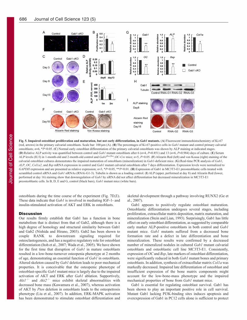
Gab1 deficiency suppresses osteoblast proliferation andmineralizationTo explore the cellular mechanism underlying the osteopeniaphenotype of Gab1 mutant mice, primary osteoblast proliferationand function were measured in in vitro culture systems. The Gab1mutant calvarial osteoblasts isolated from Gab1flox/flox;Col1a1-Cremice showed a decreased percentage of Ki-67-positive osteoblastsas compared with that of controls (35.6±7.87% in wild type versus21.1±1.8% in mutants) (Fig. 5A,B). Early osteoblast differentiationwas evaluated by alkaline phosphotase (ALP) staining. In order toavoid the effect of reduced proliferation, we analyzed osteoblastdifferentiation at high density in cultures. ALP staining and relativeALP activity in cell extracts were unchanged in mutants comparedwith controls at all stages examined (Fig. 5C,D). Furthermore, theamount of ALP in serum revealed no obvious difference betweenGab1 mutant mice and control mice (Fig. 5E). These data suggestedthat Gab1 was not necessary for early osteoblast differentiation.However, when late differentiation of osteoblast was assayed byAlizarin Red staining and von Kossa staining, the deposition ofmineralized extracellular matrix (bone nodules) was reduced inGab1 mutants at day 18 in culture (Fig. 5F). Next, in order to identifycandidate genes that might be responsible for the impaired osteoblastmaturation, the expression of osteoblast marker genes was analyzedby real-time PCR in cultures at day 7. Expression levels oftranscripts for collagen type 1a (Col1a), osteocalcin (OC) and bonesialoprotein (Bsp) were significantly reduced, whereas mRNAexpression of ALP was not changed (Fig. 5G). In 2-month-old totalbone extracts, the reduction of Col1a, OC and Bsp were alsoconfirmed by real-time PCR (data not shown), which was inagreement with reduced bone mineralization in vivo. We alsoperformed in situ hybridization on bone sections from 2-month-oldmice and immunofluorescence staining on primary osteoblasts to
confirm that the expression of Col1a was indeed decreased in Gab1mutants (supplementary material Fig. S4A-D). In addition, westudied whether silencing of Gab1 in a preosteoblastic cell line couldmimic the defective mineralization phenotype of osteoblasts derivedfrom Gab1 mutant mice. Western blot suggested that Gab1 wasknocked down efficiently in MC3T3-E1 cells (Fig. 5H). We foundthat interference of Gab1 reduced cell proliferation (data notshown), decreased bone nodule formation but did not influence earlydifferentiation (Fig. 5I). These results imply that Gab1 is essentialfor late differentiation and mineralization of osteoblasts.
Loss of Gab1 in osteoblasts increases susceptibility toapoptosisTo investigate the role of Gab1 in mediating survival signals toosteoblasts, osteoblastic apoptosis was detected using the TUNEL(terminal dUTP nick-end labeling) assay and Annexin-V flowcytometry analysis. The rate of apoptotic cells was significantlyincreased in cultured primary Gab1 mutant osteoblasts relative tocontrol cells, as revealed by the TUNEL assay (Fig. 6A,B). Inaddition, the percentage of Annexin-V-positive osteoblasts wassignificantly increased under serum-free induction (Fig. 6C,D). Toconfirm the anti-apoptotic role of Gab1, we performed the TUNELassay on Gab1-silenced MC3T3E1-S14 preosteoblastic cells.Consistently, Gab1-silenced preosteoblasts also showed enhancedsusceptibility to serum-deprivation-induced cell apoptosis (Fig.6E,F). These results suggested that Gab1 deficiency in osteoblastsenhanced their susceptibility to apoptosis.
Impairment of AKT and ERK activation in Gab1-deficientosteoblastsIt has been shown that Gab1 serves as a scaffolding proteindownstream of several receptor protein-tyrosine kinases, includingthe insulin and epidermal growth factor (EGF) receptors. Onstimulation of receptors, Gab proteins recruit the p85 subunit ofPI3K and Shp2, resulting in activation of both AKT and ERK,respectively (Cunnick et al., 2000; Ingham et al., 2001; Koyama etal., 2008). So, we next examined whether AKT and ERK signaltransduction cascades were affected by Gab1 deficiency. We foundthat phosphorylation of AKT and ERK significantly decreased inprimary mutant osteoblasts isolated from calvaria (Fig. 7A).Phosphorylation of AKT and ERK stimulated by IGF-1 and insulinin Gab1-deficient cells was in all cases lower than that of controls(Fig. 7B,C). By contrast, the levels of activation of AKT and ERKinduced by basic fibroblast growth factor (bFGF) and EGF werenot significantly different between Gab1-deficient and control
Fig. 4. Mechanical testing of femur shaft shows decreased bonestrength in Gab1 cKO mice. (A)Diagram of three-point bendingtest of femurs. (B)Representative image of load-deflection diagramdemonstrating the differences in the mechanical properties of bonefrom control and Gab1 cKO mice. (C-G)Impairment ofbiochemical properties in Gab1 cKO mice is indicated by indicesof bone strength (Young’s elastic modulus, maximum load,stiffness, energy-to-failure, ultimate stress). All values in C-G aremean ± s.d. from six control (black bars) or Gab1 cKO male mice(white bars). *P<0.05, **P<0.01.
Jour
nal o
f Cel
l Sci
ence
686
osteoblasts during the time course of the experiment (Fig. 7D,E).These data indicate that Gab1 is involved in mediating IGF-1- andinsulin-stimulated activation of AKT and ERK in osteoblasts.
DiscussionOur results firmly establish that Gab1 has a function in bonemetabolism that is distinct from that of Gab2, although there is ahigh degree of homology and structural similarity between Gab1and Gab2 (Nishida and Hirano, 2003). Gab2 has been shown tocouple RANK to downstream signaling required forosteoclastogenesis, and has a negative regulatory role for osteoblastdifferentiation (Itoh et al., 2007; Wada et al., 2005). We have shownfor the first time that disruption of Gab1 in mature osteoblastsresulted in a low-bone-turnover osteopenia phenotype at 2 monthsof age, demonstrating an essential function of Gab1 in osteoblasts.Altered skeleton caused by Gab1 deletion leads to poor mechanicalproperties. It is conceivable that the osteopenic phenotype ofosteoblast-specific Gab1 mutant mice is largely due to the impairedactivation of AKT and ERK after Gab1 ablation. Supportively,Akt1–/– and Akt2–/– mice exhibit skeletal abnormalities withdecreased bone mass (Kawamura et al., 2007), whereas activationof AKT by Pten deletion in osteoblasts leads to the osteopetrosisphenotype (Liu et al., 2007). In addition, ERK-MAPK activationhas been demonstrated to stimulate osteoblast differentiation and
Journal of Cell Science 123 (5)
skeletal development through a pathway involving RUNX2 (Ge etal., 2007).
Gab1 appears to positively regulate osteoblast maturation.Osteoblastic differentiation undergoes several stages, includingproliferation, extracellular matrix deposition, matrix maturation, andmineralization (Stein and Lian, 1993). Surprisingly, Gab1 has littleeffect on early osteoblast differentiation, as suggested by comparableearly marker ALP-positive osteoblasts in both control and Gab1mutant mice. Gab1 mutants suffered from a decreased boneformation rate and a defect in late differentiation and bonemineralization. These results were confirmed by a decreasednumber of mineralized nodules in cultured Gab1 mutant calvarialosteoblasts and osteoblastic cell line MC3T3-E1. Consistently,expression of OC and Bsp, late markers of osteoblast differentiation,were significantly reduced in both Gab1 mutant bones and primaryosteoblasts. In addition, synthesis of extracellular matrix Col1a wasmarkedly decreased. Impaired late differentiation of osteoblast andinsufficient expression of the bone matrix components mightaccount for the low-bone-mass phenotype and the impairedmechanical properties of bone from Gab1 mutant mice.
Gab1 is essential for regulating osteoblast survival. Gab1 hasbeen shown to play an important positive role in cell survival.Mutant Gab1 lacking PI3K-binding sites induces apoptosis andoverexpression of Gab1 in PC12 cells alone is sufficient to protect
Fig. 5. Impaired osteoblast proliferation and maturation, but not early differentiation, in Gab1 mutants. (A)Fluorescent immunohistochemistry of Ki-67(red, arrows) in the primary calvarial osteoblasts. Scale bar: 100m (A). (B)The percentages of Ki-67-positive cells in Gab1 mutant and control primary calvarialosteoblasts; n6, *P<0.05. (C)Normal early osteoblast differentiation of the primary calvarial osteoblasts was shown by ALP staining at indicated stages.(D)Relative ALP activity was quantified between control and Gab1 mutant osteoblasts after 6 (n4, P0.851) and 13 (n6, P0.984) days of culture. (E)SerumALP levels (IU/l) in 1-month-old and 2-month-old control and Gab1flox/flox;OC-Cre mice; n5, P>0.05. (F)Alizarin Red (left) and von Kossa (right) staining of thecalvarial osteoblast cultures demonstrates the impaired maturation of osteoblasts (mineralization) in Gab1-deficient mice. (G)Real-time PCR analysis of Gab1,ALP, OC, Col1a2, and Bsp mRNA expression in control and Gab1 mutant calvarial osteoblasts after 7 days differentiation. Expression levels were normalized toGAPDH expression and are presented as relative expression; n3, *P<0.05, **P<0.01. (H)Expression of Gab1 in MC3T3-E1 preosteoblastic cells treated withscrambled control siRNA and Gab1 siRNAs (RNAi-G1-3). Tubulin is shown as a loading control. (I)ALP (upper, performed at day 8) and Alizarin Red (lower,performed at day 16) staining show that downregulation of Gab1 by siRNA did not affect differentiation but decreased mineralization in MC3T3-E1preosteoblastic cells. In B, D, E and G, control (black bars), Gab1 mutant mice (white bars).Jo
urna
l of C
ell S
cien
ce

687Function of Gab1 in Osteoblasts
cells from apoptosis (Holgado-Madruga et al., 1997; Korhonen etal., 1999). The Shp2 interaction with Gab1 has been well studied,and deletion of Shp2 in trophoblast stem cells leads to increasedapoptosis (Yang et al., 2006). Inhibition of PI3K and AKT and ofMAPK and ERK activities strongly increased osteoblast apoptosisunder serum-free conditions (Liang et al., 2008). In this study, weshowed that both primary Gab1-deficient osteoblasts and Gab1knockdown pre-osteoblastic cells exhibited increased apoptosis,providing evidence to demonstrate that Gab1 played a protectiverole in osteoblast apoptosis.
More importantly, disruption of adaptor Gab1 in osteoblastsresulted in decreased expression of RANKL, which in turn affectedosteoclast differentiation. The OPG-RANKL system has long beenknown as a key regulator that couples the function of osteoblastsand osteoclasts (Kearns et al., 2008). Recently, it has been shownthat the Wnt signaling pathway in osteoblasts determinesosteoclastgenesis through regulating expression of OPG (Boyce etal., 2005; Glass et al., 2005; Holmen et al., 2005), whereasHedgehog signaling in osteoblasts promotes osteoclast formationby upregulating parathyroid hormone-related protein (PTHrP) andRANKL expression (Mak et al., 2008). We, and others, have alsorevealed that TGF- signaling plays a crucial role in the regulationof the RANKL-OPG axis (Hofbauer et al., 1998; Murakami et al.,1998; Okamoto et al., 2006; Tan et al., 2007). Here, we havedemonstrated that, in mature osteoblasts, signals mediated by Gab1are required for osteoclastgenesis through positive regulation ofRANKL expression.
Our results suggest that adaptor Gab1 might participate inmediating IGF-1- and insulin-triggered AKT and ERK activationin osteoblasts. IGF-1 and insulin have been shown to play importantroles in the anabolic regulation of bone metabolism through effectson collagen synthesis (Kream et al., 1985; Rosen and Luben, 1983;Thomas et al., 1996) and ALP production (Canalis, 1983). Micewith osteoblast-specific deletion of IGF-1 receptor exhibitedosteopenia phenotype with normal size and weight (Zhang et al.,2002). Insulin receptor substrate-1 (Irs1)-deficient mice show lowturnover osteopenia, with the impairment of osteoblast proliferationand differentiation, and support of osteoclastogenesis (Ogata et al.,2000). Our Gab1 mutant phenotypes are similar to those of micedeficient in IGF-1 receptor and IRS-1, described above.Overexpression of Gab1 in Irs1-deficient fibroblasts can partiallyrestore biological effects mediated by IGF-1 signaling (Winnay etal., 2000). In the current study, we found that the phosphorylationlevels of AKT and ERK stimulated by IGF-1 or insulin, but not bybFGF or EGF, were significantly diminished in Gab1-deficientosteoblasts as compared with controls, suggesting that decreasedbone mass in Gab1 mutant mice is possibly due to the reductionin sustained activation of AKT and ERK of osteoblasts in responseto IGF-1 and insulin.
In summary, we have provided first genetic evidencedemonstrating that osteoblastic Gab1 is essential for maintainingnormal postnatal bone homeostasis and bone mechanical property.We show that selective deletion of Gab1 in osteoblasts results indecreased proliferation, defective mineralization and increasedapoptosis. Moreover, differentiation of osteoclasts is also decreased
Fig. 6. Disruption of Gab1 is associated with increased apoptosis.(A)TUNEL assay of primary calvarial osteoblasts isolated from control andGab1-deficient mice. The primary osteoblasts were cultured in a serum-freemedium for 24 hours before analysis. (B)The percentages of TUNEL-positivecells in control and mutant primary calvarial osteoblasts is shown; n3,**P<0.01. (C)Flow cytometric analyses of Annexin-V expression in primaryosteoblasts isolated from control and Gab1 mutant mice. A representativeexperiment is shown. (D)Quantification shows that the percentage ofAnnexin-V-positive cells is increased in Gab1 mutant osteoblasts. *P<0.05.(E)TUNEL assay shows that downregulation of Gab1 by siRNA increasedapoptosis in MC3T3-E1 preosteoblastic cells. (F)The percentages of TUNEL-positive cells in Gab1 knockdown and control MC3T3-E1 preosteoblastic cellsis shown; n5, **P<0.01. Scale bar: 100m (A,E).
Fig. 7. Impairment of AKT and ERK activation inGab1-deficient osteoblasts. (A)Expression of AKTand ERK phosphorylation was determined by westernblotting. -actin is shown as a loading control.(B-E)Reduced activation of AKT and ERK kinase isdetected in Gab1-deficient osteoblastic cells inresponse to IGF-1 (B) and insulin (C), but not bFGF(D) and EGF (E). Wild-type and Gab1-deficientprimary osteoblasts were serum-starved and thenstimulated for 5, 10 and 30 minutes with 10 nMIGF-1, 100 nM insulin, 1nM bFGF or 100 ng/ml EGF.
Jour
nal o
f Cel
l Sci
ence
688 Journal of Cell Science 123 (5)
due to impaired capacity of Gab1 mutant osteoblasts in supportingosteoclastogenesis. Mechanistically, we show that the activation ofAKT and ERK is significantly attenuated in Gab1-deficientosteoblasts in response to IGF-1 and insulin.
Materials and MethodsGeneration of osteoblast-specific Gab1 knockout miceMice lacking Gab1 in ostoblasts were generated by breeding homozygous carryingthe floxed Gab1 alleles (Gab1flox/flox) (Bard-Chapeau et al., 2005) with OC-Cre (Tanet al., 2007) or Col1a1-Cre (Zha et al., 2008) transgenic mice. Gab1flox/flox;OC-Creor Gab1flox/flox;Col1a1-Cre mice were obtained by breeding the Gab1flox/flox mice withGab1flox/flox;OC-Cre or Gab1flox/flox;Col1a1-Cre mice. The Gab1flox/flox littermates wereused as controls. Animals were handled in accordance with institutional guidelines.Genotyping for Gab1 locus and Cre transgene were detected by PCR analysis ongenomic DNA extracted from mouse tails using primers described previously (Bard-Chapeau et al., 2005; Tan et al., 2007). Primers used to detect the specific deletionof exon 3 of Gab1 in the bone by PCR were described previously (Bard-Chapeau etal., 2005).
Bone mineral density measurements and micro-CTRadiographic analyses were carried out using a soft X-ray system (Contour Plus).BMD of the left femur was measured by dual energy X-ray absorptiometry with aPiximus Mouse Densitometer (GE Lunar Medical System). The femurs were scannedand reconstructed with 8 m isotropic voxel size on a microcomputed tomographyanalysis (micro-CT) system (eXplore Locus SP, GE Medical Systems). Thereconstructed 3D imagines of femurs were analyzed using Microviewer (GE MedicalSystems). A fixed threshold (1600) was used to separate the bone and marrow phases.Specifically, trabecular bone architecture was analyzed at the distal femoralmetaphysis, whereas cortical bone morphology was evaluated at the femoral midshaft.
Histological and histomorphometric analysisTissues were fixed in 10% formalin overnight, decalcified in 15% EDTA-PBS andembedded in paraffin. Sections 6 m thick were cut and stained with hematoxylineosinand TRAP according to standard methods. For in vivo fluorescent labeling, 2-month-old animals were injected with calcein (20 mg/kg body weight) intraperitoneally atdays 7 and 2 before tissue collection. After dehydration, the undecalcified tibiae wereembedded in methylmethacrylate, and 5-m sections were cut and stained with vonKossa stain or toluidine blue. Histomorphometric analyses were performed usingOsteoMeasure (OsteoMetrics, Decatur, GA) bone analysis software. The regions ofinterest for trabecular bone data collection were measured in an area 1.5 mm in length,from 0.3 mm below the growth plate of the proximal tibias. All histomorphometricparameters are reported in accordance with standard criteria (Parfitt et al., 1987).
Femur biomechanical testingTo test for differences in bone failure properties between Gab1 mutant and controlmice, femur from 2-month-old mice were rehydrated at room temperature inphosphate buffered saline (PBS) and femoral biomechanical properties assessed bythree-point bending. Strength tests were performed at the right femur midshaft witha displacement rate of 6 mm/minute (span length, 8 mm) using a mechanical testingmachine (model 5865; Instron, Norwood, MA). Whole-bone mechanical properties,including maximum load, stiffness, energy-to-failure and Young’s elastic modulus,were determined using load-deflection diagrams.
Primary cell culturePrimary osteoblasts were isolated from calvariae of 3-day-old neonatalGab1flox/flox;Col1a1-Cre mice by serial digestion using 0.1% collagenase I (Gibco)as previously described (Tan et al., 2007). Cells were grown in -MEM containing10% FBS until confluent. Cells were then re-plated for differentiation at 8�104 cellsper well, or for proliferation at 2�104 cells per well in a 24-well plate. Osteoblastswere grown on the glasses in a 24-well plate and the Ki-67 (Abcam) assay used todetect proliferation. For differentiation, medium was supplemented with 50 mg/mlL-ascorbic acid and 10 mM -glycerophosphate at day 2 of culture. Staining of culturesfor ALP activity was conducted using Sigma Kit 86R. Bone nodules were identifiedmorphologically by staining with Alizarin Red S (Sigma-Aldrich) and von Kossastain. Osteoblast-osteoclast co-culture experiments were performed as described(Jochum et al., 2000). Briefly, calvarial osteoblasts (5�104 per 24-well culture dish)were co-cultured with nonadherent monocyte/macrophage progenitor cells derivedfrom wild-type spleen (5�105 per 24-well culture dish) in medium supplementedwith 10 nM 1,25-dihydroxy vitamin D3. TRAP staining was performed after 8 daysof culture.
Cell death analysesTo determine the osteoblast apoptosis, primary osteoblasts and MC3T3-E1 cell linewere grown on glasses in a 24-well plate and then the culture medium changed to aserum-free medium for 24 hours. Apoptotic osteoblasts were then further identifiedby an in situ cell-death detection kit (Promega) according to the manufacturer’sinstructions. FITC-labeled Annexin-V staining was used to confirm the presence ofapoptosis. Osteoblasts were plated on 100-mm plates (1�106 per plate) and cultured
for 1 day, and then cultured in serum-free medium for 24 hour before harvesting forAnnexin-V–FITC staining. After staining with Annexin-V–FITC, osteoblasts wereanalyzed by FACS and 20,000 cells were counted.
RNA interference in MC3T3-E1To inhibit the expression of Gab1 in MC3T3-E1 pre-osteoblasts, threeoligoribonucleotides specifically targeting mouse Gab1 were synthesized byInvitrogen. The coding sequences were: RNAi-G1, 5�-CCTAACAGAACC -CTCTTTG-3�; RNAi-G2, 5�-ATGATGTATGACTGCCCAC-3�; and RNAi-G3,5�-AGCCACATCCAACTCATGA-3�. The targeted oligoribonucleotides wereintroduced into pSUPERretro Vector, and retroviral vector pSUPERretro-Gab1 wastransfected into PLAT-E cells with FuGene6 (Roche, Basel, Switzerland) to bepackaged. The infection of MC3T3-E1 by retrovirus was described previously (Sunet al., 2008).
RNA isolation and real-time PCRTotal RNA was isolated from mouse bones and osteoblasts using the TRizol reagent(Invitrogen) according to the manufacturer’s instructions. Total RNA (5 g) wasreverse transcribed to cDNA with the use of the first-strand cDNA synthesis kit(Invitrogen). Quantitative PCR was performed to measure the relative mRNA levelsusing the LightCycler system (Roche) with SYBR Green. The primers have beendescribed previously (Tan et al., 2007).
Western blot analysisPrimary osteoblasts were plated on 60-mm plates (5�105 per plate) and cultured for2 days. Culture medium was changed to -MEM without serum for 6 hours and thenstimulated with insulin (100 nM), IGF-1 (10 nM), EGF (100 ng/ml) or bFGF (1 nM).Total cell proteins were extracted by sonication in RIPA buffer containing proteaseinhibitors (Roche). After measurement of the protein content according to the BCAprotein assay kit (Thermo Scientific, Rockford, IL), protein samples were separatedon SDS-PAGE gels and transferred to polyvinylidene difluoride (PVDF) membranes(Millipore). Primary antibodies used included: Gab1, phosphorylated ERK,phosphorylated AKT, total ERK, total AKT (Cell Signaling), -actin (Sigma) andtubulin (Santa Cruz Biotechnology).
In situ hybridization and immunofluorescenceIn situ hybridization was performed as previously described (Yang et al., 2008). Forimmunofluorescence, primary osteoblasts cultured for 7 days were fixed in 4% PFAfor 15 minutes, washed in PBS containing 0.5% Triton X-100, blocked for 1 hour atroom temperature using goat serum, and incubated with collegen antibody (1:500;Abcam) for 2 hours at room temperature. After washing, FITC-conjugated goat anti-rabbit antibody was used to detect the primary antibody. For nuclear staining,osteoblasts were treated with DAPI (Sigma).
ELISA assaySera were collected from 2-month-old female mice and RANKL levels determinedusing the Quantikine mouse RANKL ELISA kit (R&D Systems).
Statistical analysisAll results were expressed as mean ± standard deviation. All statistical analyses wereperformed using the SPSS software package. In the Student’s t-test, P<0.05 wasaccepted as significant.
We thank Bin Zhao for undecalcified sections, Xiwen Xiong andChuwen Lin for BMD detections and Jun Wang for helpful instructionin microCT assays. This work was supported by Chinese National KeyProgram on Basic Research (2005CB522506; 2006CB943501;2006BAI23B01-3), Key Project for Drug Discovery and Developmentin China (2009ZX09501-027), Key Project for Infectious Diseases inChina (2008ZX10002-016), National Natural Science Foundation ofChina (30671030, 30871396, 90607004) and E-Institutes of ShanghaiMunicipal Education Commission (E03003).
Supplementary material available online athttp://jcs.biologists.org/cgi/content/full/123/5/682/DC1
ReferencesBard-Chapeau, E. A., Hevener, A. L., Long, S., Zhang, E. E., Olefsky, J. M. and Feng,
G. S. (2005). Deletion of Gab1 in the liver leads to enhanced glucose tolerance andimproved hepatic insulin action. Nat. Med. 11, 567-571.
Bentires-Alj, M., Gil, S. G., Chan, R., Wang, Z. C., Wang, Y., Imanaka, N., Harris,L. N., Richardson, A., Neel, B. G. and Gu, H. (2006). A role for the scaffolding adapterGAB2 in breast cancer. Nat. Med. 12, 114-121.
Boyce, B. F., Xing, L. and Chen, D. (2005). Osteoprotegerin, the bone protector, is asurprising target for beta-catenin signaling. Cell Metab. 2, 344-345.
Cai, T., Nishida, K., Hirano, T. and Khavari, P. A. (2002). Gab1 and SHP-2 promoteRas/MAPK regulation of epidermal growth and differentiation. J. Cell Biol. 159, 103-112.
Jour
nal o
f Cel
l Sci
ence

689Function of Gab1 in Osteoblasts
Canalis, E. (1983). Effect of hormones and growth factors on alkaline phosphatase activityand collagen synthesis in cultured rat calvariae. Metabolism 32, 14-20.
Cunnick, J. M., Dorsey, J. F., Munoz-Antonia, T., Mei, L. and Wu, J. (2000).Requirement of SHP2 binding to Grb2-associated binder-1 for mitogen-activated proteinkinase activation in response to lysophosphatidic acid and epidermal growth factor. J.Biol. Chem. 275, 13842-13848.
Fujita, T., Azuma, Y., Fukuyama, R., Hattori, Y., Yoshida, C., Koida, M., Ogita, K.and Komori, T. (2004). Runx2 induces osteoblast and chondrocyte differentiation andenhances their migration by coupling with PI3K-Akt signaling. J. Cell Biol. 166, 85-95.
Fulzele, K., DiGirolamo, D. J., Liu, Z., Xu, J., Messina, J. L. and Clemens, T. L. (2007).Disruption of the insulin-like growth factor type 1 receptor in osteoblasts enhances insulinsignaling and action. J. Biol. Chem. 282, 25649-25658.
Ge, C., Xiao, G., Jiang, D. and Franceschi, R. T. (2007). Critical role of the extracellularsignal-regulated kinase-MAPK pathway in osteoblast differentiation and skeletaldevelopment. J. Cell Biol. 176, 709-718.
Glass, D. A., 2nd, Bialek, P., Ahn, J. D., Starbuck, M., Patel, M. S., Clevers, H., Taketo,M. M., Long, F., McMahon, A. P., Lang, R. A. et al. (2005). Canonical Wnt signalingin differentiated osteoblasts controls osteoclast differentiation. Dev. Cell 8, 751-764.
Gu, H. and Neel, B. G. (2003). The “Gab” in signal transduction. Trends Cell Biol. 13,122-130.
Gu, H., Saito, K., Klaman, L. D., Shen, J., Fleming, T., Wang, Y., Pratt, J. C., Lin,G., Lim, B., Kinet, J. P. et al. (2001). Essential role for Gab2 in the allergic response.Nature 412, 186-190.
Hofbauer, L. C., Dunstan, C. R., Spelsberg, T. C., Riggs, B. L. and Khosla, S. (1998).Osteoprotegerin production by human osteoblast lineage cells is stimulated by vitaminD, bone morphogenetic protein-2, and cytokines. Biochem. Biophys. Res. Commun. 250,776-781.
Holgado-Madruga, M., Emlet, D. R., Moscatello, D. K., Godwin, A. K. and Wong, A.J. (1996). A Grb2-associated docking protein in EGF- and insulin-receptor signalling.Nature 379, 560-564.
Holgado-Madruga, M., Moscatello, D. K., Emlet, D. R., Dieterich, R. and Wong, A.J. (1997). Grb2-associated binder-1 mediates phosphatidylinositol 3-kinase activationand the promotion of cell survival by nerve growth factor. Proc. Natl. Acad. Sci. USA94, 12419-12424.
Holmen, S. L., Zylstra, C. R., Mukherjee, A., Sigler, R. E., Faugere, M. C., Bouxsein,M. L., Deng, L., Clemens, T. L. and Williams, B. O. (2005). Essential role of beta-catenin in postnatal bone acquisition. J. Biol. Chem. 280, 21162-21168.
Ingham, R. J., Santos, L., Dang-Lawson, M., Holgado-Madruga, M., Dudek, P.,Maroun, C. R., Wong, A. J., Matsuuchi, L. and Gold, M. R. (2001). The Gab1 dockingprotein links the b cell antigen receptor to the phosphatidylinositol 3-kinase/Aktsignaling pathway and to the SHP2 tyrosine phosphatase. J. Biol. Chem. 276, 12257-12265.
Itoh, M., Yoshida, Y., Nishida, K., Narimatsu, M., Hibi, M. and Hirano, T. (2000).Role of Gab1 in heart, placenta, and skin development and growth factor- and cytokine-induced extracellular signal-regulated kinase mitogen-activated protein kinase activation.Mol. Cell. Biol. 20, 3695-3704.
Itoh, S., Yoshitake, F., Narita, H., Ishihara, K. and Ebisu, S. (2007). Gab2 plays distinctroles in bone homeostasis at different time points. J. Bone Miner. Metab. 25, 81-85.
Jochum, W., David, J. P., Elliott, C., Wutz, A., Plenk, H., Jr, Matsuo, K. and Wagner,E. F. (2000). Increased bone formation and osteosclerosis in mice overexpressing thetranscription factor Fra-1. Nat. Med. 6, 980-984.
Karsenty, G. and Wagner, E. F. (2002). Reaching a genetic and molecular understandingof skeletal development. Dev. Cell 2, 389-406.
Kawamura, N., Kugimiya, F., Oshima, Y., Ohba, S., Ikeda, T., Saito, T., Shinoda, Y.,Kawasaki, Y., Ogata, N., Hoshi, K. et al. (2007). Akt1 in osteoblasts and osteoclastscontrols bone remodeling. PLoS ONE 2, e1058.
Kearns, A. E., Khosla, S. and Kostenuik, P. J. (2008). Receptor activator of nuclearfactor kappaB ligand and osteoprotegerin regulation of bone remodeling in health anddisease. Endocr. Rev. 29, 155-192.
Korhonen, J. M., Said, F. A., Wong, A. J. and Kaplan, D. R. (1999). Gab1 mediatesneurite outgrowth, DNA synthesis, and survival in PC12 cells. J. Biol. Chem. 274, 37307-37314.
Koyama, T., Nakaoka, Y., Fujio, Y., Hirota, H., Nishida, K., Sugiyama, S., Okamoto,K., Yamauchi-Takihara, K., Yoshimura, M., Mochizuki, S. et al. (2008). Interactionof scaffolding adaptor protein Gab1 with tyrosine phosphatase SHP2 negatively regulatesIGF-I-dependent myogenic differentiation via the ERK1/2 signaling pathway. J. Biol.Chem. 283, 24234-24244.
Kream, B. E., Smith, M. D., Canalis, E. and Raisz, L. G. (1985). Characterization ofthe effect of insulin on collagen synthesis in fetal rat bone. Endocrinology 116, 296-302.
Liang, M., Russell, G. and Hulley, P. A. (2008). Bim, Bak, and Bax regulate osteoblastsurvival. J. Bone Miner. Res. 23, 610-620.
Liu, X., Bruxvoort, K. J., Zylstra, C. R., Liu, J., Cichowski, R., Faugere, M. C.,Bouxsein, M. L., Wan, C., Williams, B. O. and Clemens, T. L. (2007). Lifelong
accumulation of bone in mice lacking Pten in osteoblasts. Proc. Natl. Acad. Sci. USA104, 2259-2264.
Liu, Y. and Rohrschneider, L. R. (2002). The gift of Gab. FEBS Lett. 515, 1-7.Mak, K. K., Bi, Y., Wan, C., Chuang, P. T., Clemens, T., Young, M. and Yang, Y.
(2008). Hedgehog signaling in mature osteoblasts regulates bone formation andresorption by controlling PTHrP and RANKL expression. Dev. Cell 14, 674-688.
Murakami, T., Matoba, H., Kuga, Y., Ozawa, S., Kubota, K. and Yoshida, S. (1998).Hyponatremia in a patient with chronic inflammatory disease. Intern. Med. 37, 792-795.
Nakaoka, Y., Nishida, K., Narimatsu, M., Kamiya, A., Minami, T., Sawa, H., Okawa,K., Fujio, Y., Koyama, T., Maeda, M. et al. (2007). Gab family proteins are essentialfor postnatal maintenance of cardiac function via neuregulin-1/ErbB signaling. J. Clin.Invest. 117, 1771-1781.
Nishida, K. and Hirano, T. (2003). The role of Gab family scaffolding adapter proteinsin the signal transduction of cytokine and growth factor receptors. Cancer Sci. 94, 1029-1033.
Nishida, K., Wang, L., Morii, E., Park, S. J., Narimatsu, M., Itoh, S., Yamasaki, S.,Fujishima, M., Ishihara, K., Hibi, M. et al. (2002). Requirement of Gab2 for mastcell development and KitL/c-Kit signaling. Blood 99, 1866-1869.
Ogata, N., Chikazu, D., Kubota, N., Terauchi, Y., Tobe, K., Azuma, Y., Ohta, T.,Kadowaki, T., Nakamura, K. and Kawaguchi, H. (2000). Insulin receptor substrate-1in osteoblast is indispensable for maintaining bone turnover. J. Clin. Invest. 105, 935-943.
Okamoto, M., Murai, J., Yoshikawa, H. and Tsumaki, N. (2006). Bone morphogeneticproteins in bone stimulate osteoclasts and osteoblasts during bone development. J. BoneMiner. Res. 21, 1022-1033.
Parfitt, A. M., Drezner, M. K., Glorieux, F. H., Kanis, J. A., Malluche, H., Meunier,P. J., Ott, S. M. and Recker, R. R. (1987). Bone histomorphometry: standardizationof nomenclature, symbols, and units. Report of the ASBMR HistomorphometryNomenclature Committee. J. Bone Miner. Res. 2, 595-610.
Rosen, D. M. and Luben, R. A. (1983). Multiple hormonal mechanisms for the controlof collagen synthesis in an osteoblast-like cell line, MMB-1. Endocrinology 112, 992-999.
Sachs, M., Brohmann, H., Zechner, D., Muller, T., Hulsken, J., Walther, I., Schaeper,U., Birchmeier, C. and Birchmeier, W. (2000). Essential role of Gab1 for signalingby the c-Met receptor in vivo. J. Cell Biol. 150, 1375-1384.
Scheid, M. P. and Woodgett, J. R. (2001). PKB/AKT: functional insights from geneticmodels. Nat. Rev. Mol. Cell. Biol. 2, 760-768.
Stein, G. S. and Lian, J. B. (1993). Molecular mechanisms mediatingproliferation/differentiation interrelationships during progressive development of theosteoblast phenotype. Endocr. Rev. 14, 424-442.
Sun, Q., Zhang, Y., Yang, G., Chen, X., Zhang, Y., Cao, G., Wang, J., Sun, Y., Zhang,P., Fan, M. et al. (2008). Transforming growth factor-beta-regulated miR-24 promotesskeletal muscle differentiation. Nucleic Acids Res. 36, 2690-2699.
Takahashi-Tezuka, M., Yoshida, Y., Fukada, T., Ohtani, T., Yamanaka, Y., Nishida,K., Nakajima, K., Hibi, M. and Hirano, T. (1998). Gab1 acts as an adapter moleculelinking the cytokine receptor gp130 to ERK mitogen-activated protein kinase. Mol. Cell.Biol. 18, 4109-4117.
Tan, X., Weng, T., Zhang, J., Wang, J., Li, W., Wan, H., Lan, Y., Cheng, X., Hou, N.,Liu, H. et al. (2007). Smad4 is required for maintaining normal murine postnatal bonehomeostasis. J. Cell Sci. 120, 2162-2170.
Thomas, D. M., Hards, D. K., Rogers, S. D., Ng, K. W. and Best, J. D. (1996). Insulinreceptor expression in bone. J. Bone Miner. Res. 11, 1312-1320.
Thrailkill, K. M., Lumpkin, C. K., Jr, Bunn, R. C., Kemp, S. F. and Fowlkes, J. L.(2005). Is insulin an anabolic agent in bone? Dissecting the diabetic bone for clues. Am.J. Physiol. Endocrinol. Metab. 289, E735-E745.
Wada, T., Nakashima, T., Oliveira-dos-Santos, A. J., Gasser, J., Hara, H., Schett, G.and Penninger, J. M. (2005). The molecular scaffold Gab2 is a crucial component ofRANK signaling and osteoclastogenesis. Nat. Med. 11, 394-399.
Winnay, J. N., Bruning, J. C., Burks, D. J. and Kahn, C. R. (2000). Gab-1-mediatedIGF-1 signaling in IRS-1-deficient 3T3 fibroblasts. J. Biol. Chem. 275, 10545-10550.
Yamasaki, S., Nishida, K., Yoshida, Y., Itoh, M., Hibi, M. and Hirano, T. (2003). Gab1is required for EGF receptor signaling and the transformation by activated ErbB2.Oncogene 22, 1546-1556.
Yang, G., Sun, Q., Teng, Y., Li, F., Weng, T. and Yang, X. (2008). PTEN deficiencycauses dyschondroplasia in mice by enhanced hypoxia-inducible factor 1alpha signalingand endoplasmic reticulum stress. Development 135, 3587-3597.
Yang, W., Klaman, L. D., Chen, B., Araki, T., Harada, H., Thomas, S. M., George, E.L. and Neel, B. G. (2006). An Shp2/SFK/Ras/Erk signaling pathway controls trophoblaststem cell survival. Dev. Cell 10, 317-327.
Zha, L., Hou, N., Wang, J., Yang, G., Gao, Y., Chen, L. and Yang, X. (2008).Collagen1alpha1 promoter drives the expression of Cre recombinase in osteoblasts oftransgenic mice. J. Genet. Genomics 35, 525-530.
Zhang, M., Xuan, S., Bouxsein, M. L., von Stechow, D., Akeno, N., Faugere, M. C.,Malluche, H., Zhao, G., Rosen, C. J., Efstratiadis, A. et al. (2002). Osteoblast-specificknockout of the insulin-like growth factor (IGF) receptor gene reveals an essential roleof IGF signaling in bone matrix mineralization. J. Biol. Chem. 277, 44005-44012.
Jour
nal o
f Cel
l Sci
ence


![[68Ga]PSMA-HBED-CC Uptake in Osteolytic, Osteoblastic, and ... · Conclusions: [68Ga]PSMA-HBED-CC uptake is higher in osteolytic and bone marrow metastases compared to osteoblastic](https://img.pdfslide.us/doc/110x75/607572caf32e2d79681dbd86/68gapsma-hbed-cc-uptake-in-osteolytic-osteoblastic-and-conclusions-68gapsma-hbed-cc.jpg)