Embed Size (px)
Citation preview
CHAPTER 2
*Galit Lahav—Department of Systems Biology, Harvard Medical School, Boston, MA 02115,USA. Email: [email protected]
Cellular Oscillatory Mechanisms, edited by Miguel Maroto and Nicholas A.M. Monk.©2008 Landes Bioscience and Springer Science+Business Media.
Oscillations by the p53-Mdm2Feedback LoopGalit Lahav*
Abstract
The p53 network is perhaps the most important pathway involved in preventing theinitiation of cancer. p53 levels and activity are upregulated in response to various stressesincluding DNA damage, hypoxia, and oncogene activation. Active p53 initiates differ-
ent transcriptional programs that result in cell cycle arrest, cellular senescence or apoptosis. p53also activates the transcription of Mdm2, which in turns target p53 for degradation, thereforecreating a negative feedback loop on p53. Previous studies showed that the level of p53 in-creased dramatically after exposure to damaging radiation, then declined in a series of dampedoscillations. Recent quantitative studies examined p53 responses in individual living cells, us-ing time-lapse fluorescent microscopy and showed that—on an individual cell level—the oscil-lations are not damped. Instead, one cell may have only one pulse of p53, while its neighbormay show several repeated pulses. As the amount of irradiation increased, the percentage ofcells showing a high number of p53 pulses also increased. The mean height and width of thepulses was constant and did not depend on the damage level. These observations opened newquestions regarding the mechanism and function of p53 oscillatory dynamics. In this chapter Iwill review the different models that have been suggested for p53 oscillations, including pro-posed reasons for variation between cells, and will discuss potential functions for oscillatorydynamics in the p53 signaling pathway and in stress responses in general.
IntroductionThe tumor suppressor protein p53 is the protein most frequently inactivated in human
cancer.1 More than half of all human cancers contain mutations in the p53 gene, and in almostall cancers the p53 regulatory circuit is functionally inactivated.2 The protein is known as the“guardian of the genome” because it is activated when cells are under stress. For example, whencells suffer DNA damage (as skin cells do when they are exposed to excessive radiation), boththe level of p53 and its transcriptional activity are increased. This may cause the cell to delayDNA replication to give extra time to repair the DNA. Sometimes, however, p53 triggers a celldeath pathway instead, preventing the chance that the damaged cell may later become cancer-ous. If p53 is not functional, the cell cycle might continue unrestrained, leading to uncon-trolled cell proliferation and cancer.
Under normal, unstressed conditions p53 is kept at low levels, primarily through a mecha-nism in which the negative regulator Mdm2 targets p53 to degradation. Mdm2 is one of p53’starget genes and thus any increase of p53 normally leads to an increase in Mdm2 levels, which
29Oscillations by the p53-Mdm2 Feedback Loop
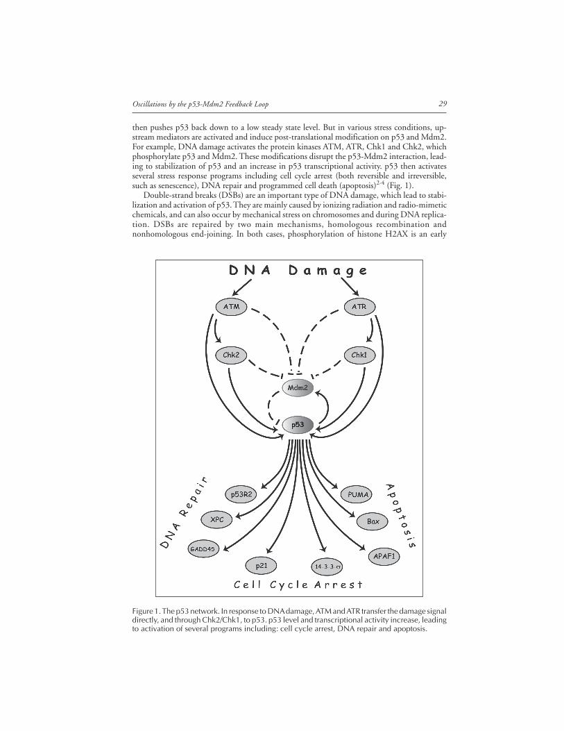
then pushes p53 back down to a low steady state level. But in various stress conditions, up-stream mediators are activated and induce post-translational modification on p53 and Mdm2.For example, DNA damage activates the protein kinases ATM, ATR, Chk1 and Chk2, whichphosphorylate p53 and Mdm2. These modifications disrupt the p53-Mdm2 interaction, lead-ing to stabilization of p53 and an increase in p53 transcriptional activity. p53 then activatesseveral stress response programs including cell cycle arrest (both reversible and irreversible,such as senescence), DNA repair and programmed cell death (apoptosis)2-4 (Fig. 1).
Double-strand breaks (DSBs) are an important type of DNA damage, which lead to stabi-lization and activation of p53. They are mainly caused by ionizing radiation and radio-mimeticchemicals, and can also occur by mechanical stress on chromosomes and during DNA replica-tion. DSBs are repaired by two main mechanisms, homologous recombination andnonhomologous end-joining. In both cases, phosphorylation of histone H2AX is an early
Figure 1. The p53 network. In response to DNA damage, ATM and ATR transfer the damage signaldirectly, and through Chk2/Chk1, to p53. p53 level and transcriptional activity increase, leadingto activation of several programs including: cell cycle arrest, DNA repair and apoptosis.
Cellular Oscillatory Mechanisms30
event that leads to recruitment of the ATM (ataxia-telangiectasia mutated) protein, as well asother components of a sensor/repair pathway. DSBs induce rapid autophosphorylation ofATM, and increase ATM kinase activity.5 Active ATM-P phosphorylates several substrates,including p53 and Mdm2, leading to disassociation of the p53-Mdm2 complex, and hencethe stabilization of p53 and an increase in p53 protein levels. ATM also phosphorylates thecheckpoint kinase Chk2, which directly phosphorylates p53 and Mdm2, further contributingto p53 stabilization.
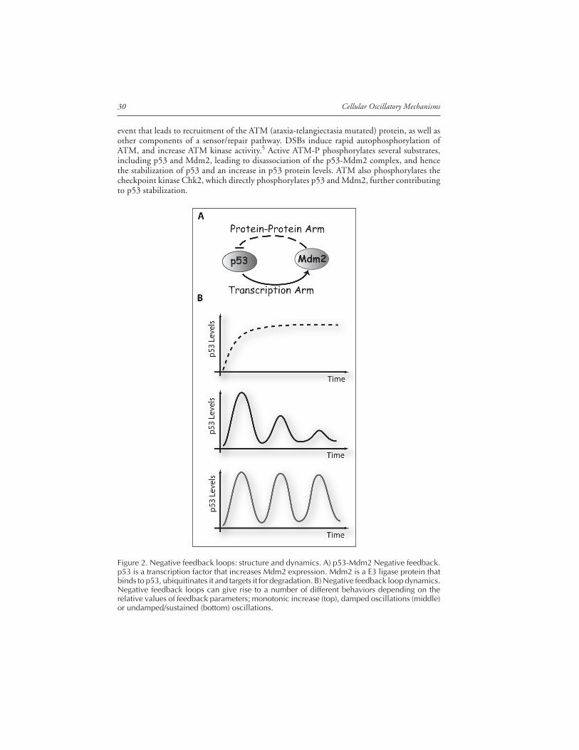
Figure 2. Negative feedback loops: structure and dynamics. A) p53-Mdm2 Negative feedback.p53 is a transcription factor that increases Mdm2 expression. Mdm2 is a E3 ligase protein thatbinds to p53, ubiquitinates it and targets it for degradation. B) Negative feedback loop dynamics.Negative feedback loops can give rise to a number of different behaviors depending on therelative values of feedback parameters; monotonic increase (top), damped oscillations (middle)or undamped/sustained (bottom) oscillations.
31Oscillations by the p53-Mdm2 Feedback Loop
The p53-Mdm2 Negative Feedback LoopThe core regulatory circuit of p53 consists of p53 and the E3 ligase protein, Mdm2. p53
and Mdm2 form a negative feedback loop, in which p53 positively regulates Mdm2 by activat-ing Mdm2 transcription,6,7 and Mdm2 negatively regulates p53 by promoting its ubiquitinationand degradation8,9 (Fig. 2A). The main mechanism for p53 stabilization in response to stresssignals is a reduction in its interaction with Mdm2, causing an increase in p53 levels.10 Thiscore negative feedback loop is embedded inside a network of additional interactions, many ofwhich are well characterized.11 For example, there are at least two additional target genes thatcreate a negative feedback loop with p53: Cop1 and Pirh2.12,13 Both are ubiquitin ligases thatpromote p53 ubiquitination and proteasomal degradation.
Negative feedback loops, such as that between p53 and Mdm2, are motifs found far moreoften than predicted by chance in biological networks.14 The p53/Mdm2 loop is a hybrid,composed of interactions on two different timescales: a slow positive transcriptional arm and afast negative protein-interaction arm (Fig. 2A). This type of loop can give rise to a number ofdifferent behaviors depending on the relative values of feedback parameters such as the rate atwhich the gene encoding the inhibitor is expressed, or the rate at which the inhibitor and thetranscription factor form an inactive complex. A loop with the same logical structure can giverise to a monotonic increase in p53 levels in response to DNA damage, damped oscillations orundamped (sustained) oscillations. If the time delay between the increase in p53 and the in-crease in Mdm2 transcription is long, oscillatory behavior is expected.15-18 If, on the otherhand, the basal degradation rates of the proteins are high, this would tend to damp out theoscillations (Fig. 2B).
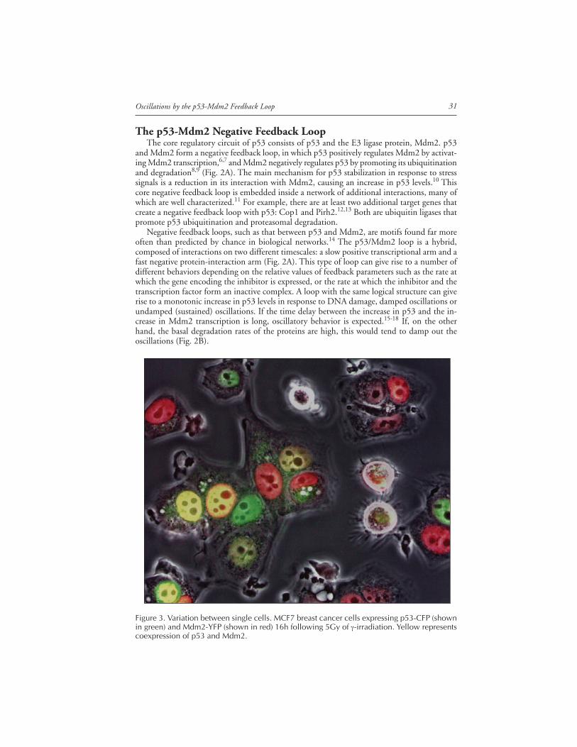
Figure 3. Variation between single cells. MCF7 breast cancer cells expressing p53-CFP (shownin green) and Mdm2-YFP (shown in red) 16h following 5Gy of γ-irradiation. Yellow representscoexpression of p53 and Mdm2.
Cellular Oscillatory Mechanisms32
Oscillations of p53 and Mdm2Experimental studies in populations of cultured cells have shown that p53 and Mdm2
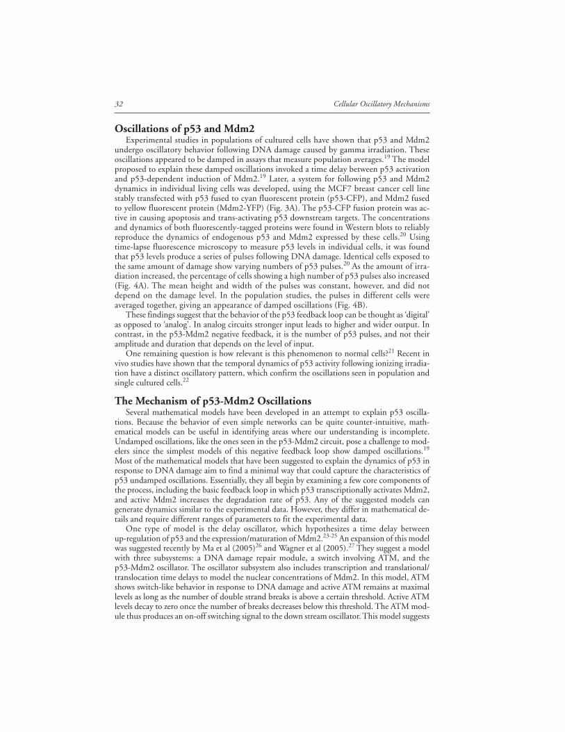
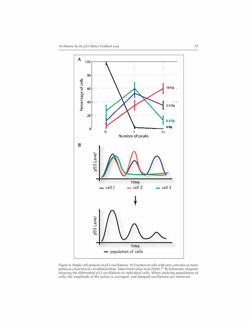
undergo oscillatory behavior following DNA damage caused by gamma irradiation. Theseoscillations appeared to be damped in assays that measure population averages.19 The modelproposed to explain these damped oscillations invoked a time delay between p53 activationand p53-dependent induction of Mdm2.19 Later, a system for following p53 and Mdm2dynamics in individual living cells was developed, using the MCF7 breast cancer cell linestably transfected with p53 fused to cyan fluorescent protein (p53-CFP), and Mdm2 fusedto yellow fluorescent protein (Mdm2-YFP) (Fig. 3A). The p53-CFP fusion protein was ac-tive in causing apoptosis and trans-activating p53 downstream targets. The concentrationsand dynamics of both fluorescently-tagged proteins were found in Western blots to reliablyreproduce the dynamics of endogenous p53 and Mdm2 expressed by these cells.20 Usingtime-lapse fluorescence microscopy to measure p53 levels in individual cells, it was foundthat p53 levels produce a series of pulses following DNA damage. Identical cells exposed tothe same amount of damage show varying numbers of p53 pulses.20 As the amount of irra-diation increased, the percentage of cells showing a high number of p53 pulses also increased(Fig. 4A). The mean height and width of the pulses was constant, however, and did notdepend on the damage level. In the population studies, the pulses in different cells wereaveraged together, giving an appearance of damped oscillations (Fig. 4B).
These findings suggest that the behavior of the p53 feedback loop can be thought as ‘digital’as opposed to ‘analog’. In analog circuits stronger input leads to higher and wider output. Incontrast, in the p53-Mdm2 negative feedback, it is the number of p53 pulses, and not theiramplitude and duration that depends on the level of input.
One remaining question is how relevant is this phenomenon to normal cells?21 Recent invivo studies have shown that the temporal dynamics of p53 activity following ionizing irradia-tion have a distinct oscillatory pattern, which confirm the oscillations seen in population andsingle cultured cells.22
The Mechanism of p53-Mdm2 OscillationsSeveral mathematical models have been developed in an attempt to explain p53 oscilla-
tions. Because the behavior of even simple networks can be quite counter-intuitive, math-ematical models can be useful in identifying areas where our understanding is incomplete.Undamped oscillations, like the ones seen in the p53-Mdm2 circuit, pose a challenge to mod-elers since the simplest models of this negative feedback loop show damped oscillations.19
Most of the mathematical models that have been suggested to explain the dynamics of p53 inresponse to DNA damage aim to find a minimal way that could capture the characteristics ofp53 undamped oscillations. Essentially, they all begin by examining a few core components ofthe process, including the basic feedback loop in which p53 transcriptionally activates Mdm2,and active Mdm2 increases the degradation rate of p53. Any of the suggested models cangenerate dynamics similar to the experimental data. However, they differ in mathematical de-tails and require different ranges of parameters to fit the experimental data.
One type of model is the delay oscillator, which hypothesizes a time delay betweenup-regulation of p53 and the expression/maturation of Mdm2.23-25 An expansion of this modelwas suggested recently by Ma et al (2005)26 and Wagner et al (2005).27 They suggest a modelwith three subsystems: a DNA damage repair module, a switch involving ATM, and thep53-Mdm2 oscillator. The oscillator subsystem also includes transcription and translational/translocation time delays to model the nuclear concentrations of Mdm2. In this model, ATMshows switch-like behavior in response to DNA damage and active ATM remains at maximallevels as long as the number of double strand breaks is above a certain threshold. Active ATMlevels decay to zero once the number of breaks decreases below this threshold. The ATM mod-ule thus produces an on-off switching signal to the down stream oscillator. This model suggests
33Oscillations by the p53-Mdm2 Feedback Loop
B
Figure 4. Single cell analysis of p53 oscillations. A) Fraction of cells with zero, one two or morepulses as a function of γ-irradiation dose. Taken from Lahav et al (2004).20 B) Schematic diagramshowing the differential p53 oscillations in individual cells. When studying populations ofcells, the amplitude of the pulses is averaged, and damped oscillations are observed.
A
Cellular Oscillatory Mechanisms34
that the levels of active ATM-P should be high throughout the period when DNA damage ispresent, then actively switched off when the DNA is completely repaired.26
An alternative network structure that could explain the undamped oscillations invokes ad-ditional intermediate components for p53 and Mdm2, and an additional positive feedbackloop on p53.28 Combinations of negative and positive feedback loops are found in other bio-logical systems that are known to produce oscillations, such as circadian clocks and the cellcycle oscillator. In the case of the cell cycle it was shown experimentally that positive feedbackon Cdc2 activity is required to properly activate the negative feedback loop and that eliminat-ing or shortening the positive feedback damped the oscillations of Cdc2 activity in Xenopuseggs extracts.29 It is possible therefore that positive feedback loop(s) on p53, as was suggested inCiliberto et al (2005)28 are also essential for its oscillatory behavior.
There are several known positive feedbacks on p53 that might be crucial for its oscillatorybehavior: one is via activation of PTEN, a target of p53 that down-regulates Mdm2 and thusprevents Mdm2 negative regulation of p53.11,28,30 PTEN is not induced in MCF7 cells aftergamma irradiation as was shown by us and others, implying that the positive feedback on p53through PTEN is not active in MCF7 cells and therefore is not required for p53 oscillations. Itis still possible, however, that feedback regulation through PTEN may operate via posttransla-tional modifications that do not require PTEN upregulation. Other known positive feedbackson p53—through dapk1,31 c-Ha-Ras32 and DDR133—involve the activation of p19ARF, whichinhibits Mdm2. Since MCF7 cells do not have the ARF gene, it is unlikely that these loops areessential for the p53 oscillatory behavior that was observed in these cells. An additional poten-tial positive feedback on p53 is through p21. p21 is a transcriptional target of p53 that inhibitsthe complex cyclinE-cdk2. This inhibition increases the formation of an Rb-Mdm2 complexthat enhances activation of p53.11 Additional experiments are required to determine whetherthis or other positive feedback loops on p53 are required for its oscillatory behavior; it is pos-sible that one or more of the known feedback loops, or a so-far-unknown feedback loop isimportant in this behavior, but it is also possible that these loops are not connected to theoscillatory behavior.
A new model, which was proposed recently, suggests that a combination of two negativefeedback loops explain the observed oscillations, one directly on p53 through Mdm2 and alonger one on an upstream regulator of p53.34 This model suggests that a protein downstreamof p53 inhibits the activity of a protein upstream of p53 that is involved in detecting DNAdamage, and that the apparent oscillations are in fact repeated p53 pulses resulting from re-peated examinations of the damaged DNA. This model explicitly considers the dynamics ofelements upstream to p53 in the DNA damage signal response, and predicts that such elements(e.g., ATM-P or Chk2-P)5,35-37 may also show repeated pulses.
There are at least two potential target genes of p53 that might be involved in the postulatedfeedback loop inhibiting up-regulators of p53. One is the protein cyclin G, which was shownto recruit protein phosphatase 2A (PP2A)38 to dephosphorylate Mdm2 and ATM.39,40 Breastcancer cells such as MCF7 are known to have high constant levels of cyclin G mRNA andprotein in unstressed conditions,41 yet still show p53 oscillations. This implies that the feed-back involving cyclin G upregulation is not essential for p53 oscillations in MCF7 cells. Thesecond candidate is the phosphatase Wip1 (PPM1D). Wip1 levels show a pulse during the first6 hours in response to gamma irradiation,42 a behavior that is predicted for the putative inhibi-tor. Wip1 is also a downstream target of p53. It is not known whether Wip1 directly interactswith ATM, but there is evidence that Wip1 can reverse the phosphorylation status of bothp5343 and Chk2.44 In addition, the Ser1981 phosphorylation site in ATM-P is a strong candi-date for a Wip1 target sequence, based on sequence homology.44 These data are consistent withthe notion that Wip1 may feed back on either or both of ATM and Chk2.
Additional experiments together with repeated iterative simulation and model-building arerequired to test the validity of any of these models, in order to have a clear comprehensivepicture that robustly explains p53 oscillations.
35Oscillations by the p53-Mdm2 Feedback Loop
Variability in the Response of Individual CellsTwo types of variability in the p53 pulses in individual cells can be observed: one is in the
number of pulses, the other in the shape of each pulse (both amplitude and duration). Aplausible hypothesis for variation in the number of p53 pulses is that it may be determined bythe variable number of double-strand breaks (DSBs) in the DNA of each cell. Cells with manyDSBs might oscillate for longer, as they try again and again to repair their DNA. If this is thecase, the number of p53 pulses would correlate with the amount of DNA damage an indi-vidual cell has suffered, and the variable number of pulses would offer a potential mechanismfor “counting” the number of attempts a cell has made to repair damage.
The canonical marker for double strand breaks is phosphorylated H2AX (γ-H2AX) whichbinds to the sites of damage. Clear foci of γ-H2AX, which are thought to represent individualDNA breaks, can be detected with antibody staining. The variation in the number of foci seenafter damage45 is consistent with the hypothesis that the difference in the number of p53 pulsesseen in single cells may reflect a difference in the amount of DNA damage.
The variability in the shape of the oscillations appears to be mainly in the amplitude, whilethe duration is relatively constant. Detailed analysis revealed that these variations appear tostem from fluctuations in the protein production rates.34 Although the cell populations stud-ied are clonal, some of this variation may be due to cell-to-cell differences that are caused by theknown genomic instability of cancer cells in culture.
The Potential Function of p53 OscillationsOscillations are found in diverse systems in biology such as the segmentation clock, cell
cycle regulation and circadian rhythms. Since these cellular systems need to change in a cyclicalpattern over time, it seems useful for them to have feedback parameters that give rise to oscilla-tory behavior. In the case of cellular stress, however, the need for oscillations is much lessobvious. The first question to answer is whether the apparent oscillations are indeed oscilla-tions, or repeated pulses that are individually and repetitively triggered.
If the p53 system does indeed oscillate, it is possible that the downstream effect of p53activation is determined by the frequency of the oscillations. This is known to be the case forintracellular Ca2+ oscillations and for the pulsatile release of cyclic adenosine monophosphate(cAMP) signals in Dictyostelium amoebae. In addition, most hormones are secreted in a pulsa-tile rather than continuous manner, and the temporal pattern in which a hormone appears isoften more important than its concentration. Changing the frequency of p53 oscillations andtesting the effect of such changes on the expression of its target genes will help reveal whetherthis is also the case for the p53 system. An alternative explanation is that the oscillations maysimply exist to allow DNA repair without the risk of irreversible consequences of continuedexcessive p53 levels.
If the apparent oscillations are instead repeatedly triggered individual pulses, their purposemight be more clear: in each p53 pulse the DNA repair machinery is launched and the intervalbetween pulses is used to reevaluate the cell state by checking whether damage remains.46
In either case, it is possible that the number of pulses (or oscillations) may convey informa-tion. For example, the post-translational modifications on p53 may change over time as re-peated pulses occur. Although gaps still exist in our knowledge regarding the role of many p53posttranslational modifications, recent evidence showed that p53 carrying different modifica-tions may selectively activate certain p53 target genes. For example, phosphorylation on Ser46was found to be essential for activating transcription of genes in the apoptotic pathway, such asp53AIP1.47-49 We do not know precisely what causes p53 to be modified in one way ratherthan another, but it is possible that the modifications integrate a variety of upstream signals.The importance of the pattern of modifications might offer a different explanation for theneed for oscillations. After each pulse, if a decision to grow or die has not been made, thepost-translational modifications may need to be erased. The modified p53 produced in theprevious pulse is degraded, and new protein is synthesized from scratch in the next pulse.46 A
Cellular Oscillatory Mechanisms36
similar hypothesis may also explain the oscillatory dynamics observed in the NFκB-IκB feed-back loop. Nelson et al showed recently that NF-κB protein trapped in the nucleus could nolonger activate its target genes50 probably because of dephosphorylation of NF-κB in the nucleusby protein phosphatase A2, or deacetylation by histone deacetylase 3. Thus, it appears that theexport of NF-κB induced by IκBα is not in fact essential for turning off the transcriptionalresponse, but instead is required to bring NF-κB back to the cytoplasm, where it can receivenew signals.
Highly regulated oscillations with variable amplitude and precise timing were also recentlyfound in the SOS DNA damage response in E. coli.51 The presence of oscillations in this andthe NFκB system suggest that oscillations (or repeated pulses) may play a general role in stressor damage responses. Further work is required to determine whether the unusual temporaldynamics of p53 after DNA damage have an important role and whether oscillations in generalserve as a mechanism for integrating multiple signals over time.
One potential clue for the importance of oscillations comes from cells in which p53 oscilla-tions are naturally modified. Recent studies showed that a single nucleotide polymorphism inthe Mdm2 promoter (SNP309) leads to an increase in binding of the transcriptional activatorSp1, resulting in higher levels of Mdm2 RNA and protein.52,53 SNP309 has been shown to beassociated with accelerated tumor formation in humans and to cause defects in the activationof p53-dependent transcriptional programs.52 Cell lines carrying SNP309 allow p53up-regulation but do not show oscillations,26 providing correlative evidence that the oscilla-tions might be functionally important and possibly implying a role for p53 oscillations inpreventing cancer.
Conclusion and Key Questions in the FieldThe tumor suppressor p53 is perhaps one of the most intensely investigated proteins. Since
its discovery in 1979, almost 40,000 papers have been published on this topic, and yet it canstill surprise us. It is only recently that new technological developments have allowed detailedanalysis of p53 kinetics in individual cells. The discovery that single cells vary in the number ofp53 pulses show the limitations of population data in studying the dynamics of this system,and emphasize the importance of single cell studies to reveal protein behavior and connectingbehavior to molecular mechanism.
The series of oscillations in the p53 system open several new questions in the field: forexample, it is still unclear how p53 oscillations arise. Although several mathematical modelshave been suggested to explain p53’s oscillatory behavior we are still far from having a compre-hensive predictive picture of the mechanisms that generate these unusual dynamics. We also donot yet know how general this phenomenon may be—is it a consequence of changes that cellculture cells undergo, or do normal human cells, or uncultured cancer cells, also show periodicp53 pulses in response to DNA damage? Perhaps most importantly, it is still unknown what, ifany, is the function of p53 oscillations and whether they play a role in elucidating the rightcellular behavior in response to DNA damage. Even after a quarter century of study, p53 stillhas much to teach us.
AcknowledgementsI thank A. Loewer for help with illustration of figures and all the members of our laboratory
for useful discussions. I acknowledge support from the Smith Family New Investigator AwardProgram—the Medical Foundation.
References1. Levine AJ. p53, the cellular gatekeeper for growth and division. Cell 1997; 88(3):323-331.2. Jin S, Levine AJ. The p53 functional circuit. J Cell Sci 2001; 114(Pt 23):4139-4140.3. Vogelstein B, Lane D, Levine AJ. Surfing the p53 network. Nature 2000; 408(6810):307-310.4. Hofseth LJ, Hussain SP, Harris CC. p53: 25 years after its discovery. Trends Pharmacol Sci 2004;
25(4):177-181.
37Oscillations by the p53-Mdm2 Feedback Loop
5. Bakkenist CJ, Kastan MB. DNA damage activates ATM through intermolecular autophosphorylationand dimer dissociation. Nature 2003; 421(6922):499-506.
6. Barak Y, Juven T, Haffner R et al. Mdm2 expression is induced by wild type p53 activity. EMBOJ 1993; 12(2):461-468.
7. Wu X, Bayle JH, Olson D et al. The p53-mdm-2 autoregulatory feedback loop. Genes Dev 1993;7(7A):1126-1132.
8. Haupt Y, Maya R, Kazaz A et al. Mdm2 promotes the rapid degradation of p53. Nature 1997;387(6630):296-299.
9. Kubbutat MH, Jones SN, Vousden KH. Regulation of p53 stability by Mdm2. Nature 1997;387(6630):299-303.
10. Michael D, Oren M. The p53-Mdm2 module and the ubiquitin system. Semin Cancer Biol 2003;13(1):49-58.
11. Harris SL, Levine AJ. The p53 pathway: Positive and negative feedback loops. Oncogene 2005;24(17):2899-2908.
12. Dornan D, Wertz I, Shimizu H et al. The ubiquitin ligase COP1 is a critical negative regulator ofp53. Nature 2004; 429(6987):86-92.
13. Leng RP, Lin Y, Ma W et al. Pirh2, a p53-induced ubiquitin-protein ligase, promotes p53 degra-dation. Cell 2003; 112(6):779-791.
14. Yeger-Lotem E, Sattath S, Kashtan N et al. Network motifs in integrated cellular networks oftranscription-regulation and protein-protein interaction. Proc Natl Acad Sci USA 2004;101(16):5934-5939.
15. Tyson JJ. Monitoring p53’s pulse. Nat Genet 2004; 36(2):113-114.16. Tyson JJ, Chen KC, Novak B. Sniffers, buzzers, toggles and blinkers: Dynamics of regulatory and
signaling pathways in the cell. Curr Opin Cell Biol 2003; 15(2):221-231.17. Nelson DE, Ihekwaba AEC, Elliott M et al. Oscillations in NF-{kappa}B signaling control the
dynamics of gene expression. Science 2004; 306(5696):704-708.18. Hoffmann A, Levchenko A, Scott ML et al. The IkB-NF-kB signaling module: Temporal control
and selective gene activation. Science 2002; 298:1241-1245.19. Lev Bar-Or R, Maya R, Segel LA et al. Generation of oscillations by the p53-Mdm2 feedback
loop: A theoretical and experimental study. Proc Natl Acad Sci USA 2000; 97(21):11250-11255.20. Lahav G, Rosenfeld N, Sigal A et al. Dynamics of the p53-Mdm2 feedback loop in individual
cells. Nat Genet 2004; 36(2):147-150.21. Tyson JJ. Another turn for p53. Mol Syst Biol 2006; 2:0032.22. Hamstra DA, Bhojani MS, Griffin LB et al. Real-time evaluation of p53 oscillatory behavior in
vivo using bioluminescent imaging. Cancer Res 2006; 66(15):7482-7489.23. Mihalas GI, Simon Z, Balea G et al. Possible oscillatory behavior in p53-Mdm2 interaction com-
puter simulation. Journal of Biological Systems 2000; 8:21-29.24. Monk NAM. Oscillatory expression of Hes1, p53, and NF-κB driven by transcriptional time de-
lays. Curr Biol 2003; 13(16):1409-1413.25. Tiana G, Jensen MH, Sneppen K. Time delay is a key to apoptosis induction in the p53 network.
European Physical Journal B 2002; 29:135-140.26. Ma L, Wagner J, Rice JJ et al. A plausible model for the digital response of p53 to DNA damage.
Proc Natl Acad Sci USA 2005; 102(40):14266-14271.27. Wagner J, Ma L, Rice JJ et al. p53-Mdm2 loop controlled by a balance of its feedback strength
and effective dampening using ATM and delayed feedback. IEEE Proc.-Syst Biol 2005;152(3):109-118.
28. Ciliberto A, Novak B, Tyson JJ. Steady states and oscillations in the p53/Mdm2 network. CellCycle 2005; 4(3):488-493.
29. Pomerening JR, Kim SY, Ferrell Jr JE. Systems-level dissection of the cell-cycle oscillator: Bypass-ing positive feedback produces damped oscillations. Cell 2005; 122(4):565-578.
30. Mayo LD, Dixon JE, Durden DL et al. PTEN protects p53 from Mdm2 and sensitizes cancer cellsto chemotherapy. J Biol Chem 2002; 277(7):5484-5489.
31. Martoriati A, Doumont G, Alcalay M et al. dapk1, encoding an activator of a p19ARF-p53-mediatedapoptotic checkpoint, is a transcription target of p53. Oncogene 2005; 24(8):1461-1466.
32. Deguin-Chambon V, Vacher M, Jullien M et al. Direct transactivation of c-Ha-Ras gene by p53:Evidence for its involvement in p53 transactivation activity and p53-mediated apoptosis. Oncogene2000; 19(51):5831-5841.
33. Ongusaha PP, Kim JI, Fang L et al. p53 induction and activation of DDR1 kinase counteractp53-mediated apoptosis and influence p53 regulation through a positive feedback loop. EMBO J2003; 22(6):1289-1301.
Cellular Oscillatory Mechanisms38
34. Geva-Zatorsky N, Rosenfeld N, Itzkovitz S et al. Oscillations and variability in the p53 system.Molecular Systems Biology 2006; 2:E1-E13.
35. Canman CE, Lim DS, Cimprich KA et al. Activation of the ATM kinase by ionizing radiation andphosphorylation of p53. Science 1998; 281(5383):1677-1679.
36. Canman CE, Lim DS. The role of ATM in DNA damage responses and cancer. Oncogene 1998;17(25):3301-3308.
37. Khosravi R, Maya R, Gottlieb T et al. Rapid ATM-dependent phosphorylation of MDM2 pre-cedes p53 accumulation in response to DNA damage. Proc Natl Acad Sci USA 1999;96(26):14973-14977.
38. Okamoto K, Kamibayashi C, Serrano M et al. p53-dependent association between cyclin G andthe B’ subunit of protein phosphatase 2A. Mol Cell Biol 1996; 16(11):6593-6602.
39. Ohtsuka T, Jensen MR, Kim HG et al. The negative role of cyclin G in ATM-dependent p53activation. Oncogene 2004; 23(31):5405-5408.
40. Goodarzi AA, Jonnalagadda JC, Douglas P et al. Autophosphorylation of ataxia-telangiectasia mu-tated is regulated by protein phosphatase 2A. EMBO J 2004; 23(22):4451-4461.
41. Reimer CL, Borras AM, Kurdistani SK et al. Altered regulation of cyclin G in human breastcancer and its specific localization at replication foci in response to DNA damage in p53+/+ cells.J Biol Chem 1999; 274(16):11022-11029.
42. Fiscella M, Zhang H, Fan S et al. Wip1, a novel human protein phosphatase that is induced inresponse to ionizing radiation in a p53-dependent manner. Proc Natl Acad Sci USA 1997;94(12):6048-6053.
43. Lu X, Nannenga B, Donehower LA. PPM1D dephosphorylates Chk1 and p53 and abrogates cellcycle checkpoints. Genes Dev 2005; 19(10):1162-1174.
44. Fujimoto H, Onishi N, Kato N et al. Regulation of the antioncogenic Chk2 kinase by the onco-genic Wip1 phosphatase. Cell Death Differ 2005.
45. Rothkamm K, Lobrich M. Evidence for a lack of DNA double-strand break repair in human cellsexposed to very low X-ray doses. Proc Natl Acad Sci USA 2003; 100(9):5057-5062.
46. Lahav G. The strength of indecisiveness: Oscillatory behavior for better cell fate determination. SciSTKE 2004; 2004(264):pe55.
47. Komiyama S, Taniguchi S, Matsumoto Y et al. Potentiality of DNA-dependent protein kinase tophosphorylate Ser46 of human p53. Biochem Biophys Res Commun 2004; 323(3):816-822.
48. Oda K, Arakawa H, Tanaka T et al. p53AIP1, a potential mediator of p53-dependent apoptosis,and its regulation by Ser-46-phosphorylated p53. Cell 2000; 102(6):849-862.
49. Mayo LD, Seo YR, Jackson MW et al. Phosphorylation of human p53 at serine 46 determinespromoter selection and whether apoptosis is attenuated or amplified. J Biol Chem 2005;280(28):25953-25959.
50. Nelson DE, See V, Nelson G et al. Oscillations in transcription factor dynamics: A new way tocontrol gene expression. Biochem Soc Trans 2004; 32(Pt 6):1090-1092.
51. Friedman N, Vardi S, Ronen M et al. Precise temporal modulation in the response of the SOSDNA repair network in individual bacteria. PLoS Biol 2005; 3(7):e238.
52. Bond GL, Hu W, Bond EE et al. A single nucleotide polymorphism in the MDM2 promoterattenuates the p53 tumor suppressor pathway and accelerates tumor formation in humans. Cell2004; 119(5):591-602.
53. Bond GL, Hu W, Levine A. A single nucleotide polymorphism in the MDM2 gene: From a mo-lecular and cellular explanation to clinical effect. Cancer Res 2005; 65(13):5481-5484.