Embed Size (px)
Citation preview
Bulletin of the NYU Hospital for Joint Diseases 2008;66(4):306-16306
Latifzai K, Sites BD, Koval KJ. Orthopaedic anesthesia: Part 2. Common techniques of regional anesthesia in orthopaedics. Bull NYU Hosp Jt Dis. 2008;66(4):306-16.
Orthopaedic Anesthesia Part 2. Common Techniques of Regional Anesthesia in Orthopaedics
Khushal Latifzai, B.A., Brian D. Sites, M.D., and Kenneth J. Koval, M.D.
Abstract
Anesthesia may be considered in terms of two categories: general and regional. The aim of general anesthesia is to induce analgesia, sedation, amnesia, suppression of autonomic reflexes, and relaxation of muscles. Regional anesthesia is more site-specific and is typically divided into three categories based on the location of injection: 1. a central neuraxial block is an injection of an anesthetic drug into the epidural or intrathecal space; 2. a periph-eral nerve block is an injection near the nerve or plexus supplying the area under operation; and 3. a field block is an injection into the adjoining tissues with subsequent diffusion into the surgical area (in orthopaedics, it is typically employed for minor procedures of the hand or foot). Of these three categories of regional anesthesia (i.e., neuraxial, peripheral, and field blocks), this article focuses on the latter two. Although neuraxial blocks com-prise an important part of regional anesthesia, they are typically performed by anesthesiologists in an operative setting for major procedures of the lower extremities. The intent of this article is to familiarize the orthopaedist with techniques that have implications for emergency rooms and other ambulatory settings in which regional techniques are sometimes favored over general alterna-tives because they entail less risk of systemic side effects and may involve more cost-effective use of resources.
Anesthesia may be considered in terms of two cat-egories: general and regional. The aim of general anesthesia is to induce analgesia, sedation, amne-
sia, suppression of autonomic reflexes, and relaxation of muscles. Regional anesthesia is more site-specific and is typically divided into three categories based on the loca-tion of injection: 1. central neuraxial block is injection of an anesthetic drug into the epidural or intrathecal space; 2. peripheral nerve block is injection near the nerve or plexus supplying the area under operation; and 3. field block is injection into the adjoining tissues, with subsequent diffu-sion into the surgical area (in orthopaedics, it is typically employed for minor procedures of the hand or foot). Of the three categories of regional anesthesia (i.e., neur-axial, peripheral, and field blocks), this article will focus on the latter two. Although neuraxial blocks comprise an important part of regional anesthesia, they are typically performed by anesthesiologists in an operative setting for major procedures of the lower extremities. The intent of this article is to familiarize the orthopaedist with techniques that have implications for the emergency room and other ambula-tory settings, in which regional techniques are sometimes favored over general alternatives, as they entail less risk for systemic side effects and may involve more cost-effective use of resources.
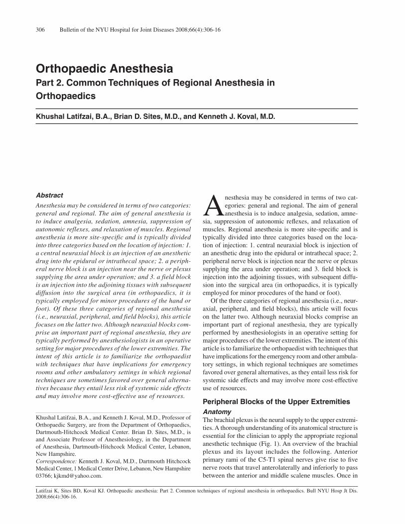
Peripheral Blocks of the Upper ExtremitiesAnatomy The brachial plexus is the neural supply to the upper extremi-ties. A thorough understanding of its anatomical structure is essential for the clinician to apply the appropriate regional anesthetic technique (Fig. 1). An overview of the brachial plexus and its layout includes the following. Anterior primary rami of the C5-T1 spinal nerves give rise to five nerve roots that travel anterolaterally and inferiorly to pass between the anterior and middle scalene muscles. Once in
Khushal Latifzai, B.A., and Kenneth J. Koval, M.D., Professor of Orthopaedic Surgery, are from the Department of Orthopaedics, Dartmouth-Hitchcock Medical Center. Brian D. Sites, M.D., is and Associate Professor of Anesthesiology, in the Department of Anesthesia, Dartmouth-Hitchcock Medical Center, Lebanon, New Hampshire. Correspondence: Kenneth J. Koval, M.D., Dartmouth Hitchcock Medical Center, 1 Medical Center Drive, Lebanon, New Hampshire 03766; [email protected].
307Bulletin of the NYU Hospital for Joint Diseases 2008;66(4):306-16
the interscalene space, the roots merge into the superior (C5-C6), middle (C7), and inferior (C8-T1) trunks. Each one of these three trunks separates into an anterior and posterior
division (for a total of six divisions) at the level of the first rib (in some texts, the anterior and posterior divisions of each trunk are referred to as the superior and inferior divi-sions, respectively). The six divisions continue into the axilla, where they unite in various combinations to yield a total of three cords that are named in accordance with their relationship to the second part of the axillary artery. Details of this division-to-cord transition are as follows: the anterior divisions of the superior and middle trunks combine to produce the lateral cord; the posterior divisions of all three trunks unite, forming the posterior cord; and the anterior division of the inferior trunk continues on as the medial cord. The next transition, from the three cords to peripheral branches, occurs at the lateral border of the pectoralis minor. The lateral cord contributes to the formation of two peripher-al nerves: it alone gives rise to the musculocutaneous nerve, and it also combines with the medial cord to establish the median nerve. In addition to its contribution to the median nerve, the medial cord generates the ulnar, antebrachial, and medial brachial cutaneous nerves. And finally, the posterior cord splits into the axillary and radial nerves. Important departures from the pattern of transition of the brachial plexus thus outlined are several branches that emanate directly from the roots (instead of cords). These
Figure 1 Anatomy of the brachial plexus. (Reproduced from: Campbell’s Operative Orthopaedics. (10th ed). Copyright © 2003 Mosby, Inc., with permission.)
Table 1 Plexus Blocks of the Upper Extremities
Technique Neural Target/Indications Comments
Interscalene Block Target: Root-to-trunk Injection Volume: 10-15 mL transition (interscalene space). (ultrasound); otherwise, 20-40 mL. Indications: Proximal procedures of arm; requires supplementation with ulnar nerve block for procedures of forearm and hand.
Supraclavicular Block Target: Trunk-to-division Injection Volume: 10-15 mL (ultrasound transition. highly desirable; if unavailable, use Indications: Procedures of infraclavicular block). the arm, elbow, or forearm.
Infraclavicular Block Target: Posterior, lateral, and Injection Volume: 7-11 mL per neural medial cords. cord (ultrasound); otherwise, one 20-30 mL) injection. Indications: Procedures of the Compared with axillary block, has elbow, forearm, and hand. advantage of not requiring supplemental injection of musculocutaneous nerve. If ultrasound and nerve stimulation are unavailable unavailable, forego above advantage, and use axillary block.
Axillary Block Target: Radial, ulnar, and median Injection Volume: 5-8 mL per nerve nerves. (ultrasound). If ultrasound is unavailable, Indications: Procedures of elbow, several options still exist: 35-40 mL (single forearm, and hand. injection); 10 mL (near each of the three nerves); two 20 mL injections (transarterial approach); or 40-50 mL (fascial click method). Compared with infraclavicular block, can be performed in the absence of ultrasound or nerve stimulation. Requires supplemental injection of musculocutaneous nerve.
Bulletin of the NYU Hospital for Joint Diseases 2008;66(4):306-16308
branches primarily supply motor functions to muscles of the axial skeleton (e.g., serratus anterior, rhomboids, etc.). However, the suprascapular nerve (C5-C6) provides sensa-tion to the shoulder joint in addition to its motor functions. Of the brachial plexus blocks, the interscalene approach is the only one to anesthetize the suprascapular nerve. Therefore, it is the most appropriate block for procedures involving the shoulder.
Interscalene BlockBest suited for procedures involving the most proximal as-pects of the arm (e.g., the shoulder), this block is directed at the root-to-trunk transition of the brachial plexus occurring in the interscalene space, at the level of the C6 vertebra. The result is a blockade of the superior and middle trunks (C5-C7), as well as the suprascapular nerve (C5-C6). Be-cause the inferior trunk (C8-T1) is not entirely anesthetized with this approach, it is not indicated for procedures of the forearm and hand unless supplemented with an ulnar nerve block (Table 1).
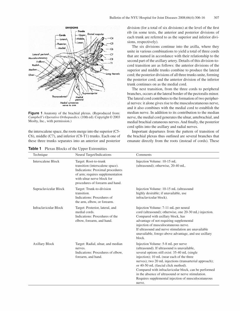
Supraclavicular BlockApplied at the level of the supraclavicular fossa, this ap-proach targets the transition of the three brachial plexus trunks into their respective divisions. The supraclavicular block is indicated for procedures of the arm, elbow, or forearm. Because of the proximity of the cervical pleura, ultrasound visualization is highly recommended (Fig. 2). If ultrasound tools are unavailable, then the infraclavicular approach should be considered, because the latter poses less risk of a pneumothorax (Table 1).1
Infraclavicular BlockApplied at the level of three cords (medial, lateral, and posterior) of the brachial plexus, this approach is indicated for procedures involving areas from the distal arm to the hand. Blockade of the musculocutaneous nerve gives this
approach an advantage over the axillary block. However, in the absence of ultrasound, while this technique relies on a nerve stimulator to guide the needle, the axillary block can be employed using a palpable vascular landmark, making the latter a simpler technique to administer (Table 1).
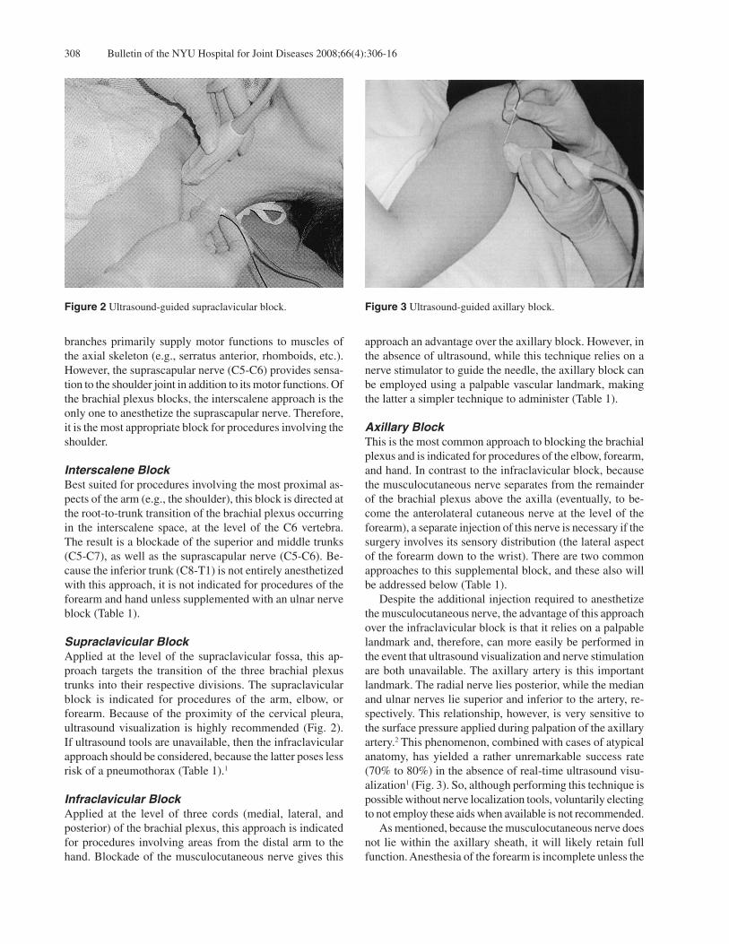
Axillary Block This is the most common approach to blocking the brachial plexus and is indicated for procedures of the elbow, forearm, and hand. In contrast to the infraclavicular block, because the musculocutaneous nerve separates from the remainder of the brachial plexus above the axilla (eventually, to be-come the anterolateral cutaneous nerve at the level of the forearm), a separate injection of this nerve is necessary if the surgery involves its sensory distribution (the lateral aspect of the forearm down to the wrist). There are two common approaches to this supplemental block, and these also will be addressed below (Table 1). Despite the additional injection required to anesthetize the musculocutaneous nerve, the advantage of this approach over the infraclavicular block is that it relies on a palpable landmark and, therefore, can more easily be performed in the event that ultrasound visualization and nerve stimulation are both unavailable. The axillary artery is this important landmark. The radial nerve lies posterior, while the median and ulnar nerves lie superior and inferior to the artery, re-spectively. This relationship, however, is very sensitive to the surface pressure applied during palpation of the axillary artery.2 This phenomenon, combined with cases of atypical anatomy, has yielded a rather unremarkable success rate (70% to 80%) in the absence of real-time ultrasound visu-alization1 (Fig. 3). So, although performing this technique is possible without nerve localization tools, voluntarily electing to not employ these aids when available is not recommended. As mentioned, because the musculocutaneous nerve does not lie within the axillary sheath, it will likely retain full function. Anesthesia of the forearm is incomplete unless the
Figure 2 Ultrasound-guided supraclavicular block. Figure 3 Ultrasound-guided axillary block.
309Bulletin of the NYU Hospital for Joint Diseases 2008;66(4):306-16
musculocutaneous nerve is also blocked, either at the antecu-bital fossa or the coracobrachialis muscle.3 Anesthesia at the antecubital fossa is described in the section on elbow blocks. As for the midhumeral alternative, 5 mL of local anesthetic solution may be injected into the coracobrachialis muscle (above the axial artery, but beneath the biceps muscle).
Peripheral Blocks at the Elbow, Wrist, and DigitsBlockade at either the elbow or wrist anesthetizes the pe-ripheral branches of the brachial plexus supplying the hand. Although a more proximal blockade (e.g., the axillary block) can serve the same purpose, the more peripheral alternatives are available if infection impedes access to the brachial plexus, the patient suffers from coagulation abnormalities, bilateral surgery is indicated, the patient has a difficult anatomy, or if blockade of the brachial plexus requires
further supplementation (Table 2).
Innervation of the HandThe median nerve provides sensory innervation to areas lat-eral to the radial half of the ring finger on the palmar surface of the hand, as well as to the nail beds of these digits on the dorsal surface. The median nerve also supplies the muscles of the thenar eminence, the lumbricales of the first and second digits, as well as the wrist flexors in the forearm. The radial nerve carries sensation from the area lateral to the radial half of the ring finger on the dorsum of the hand (excluding the nail beds, which are the domain of the median nerve). It also provides motor function to the extensor muscles of the elbow, as well as the extrinsic wrist and finger extensors. The ulnar nerve has a sensory distribution that includes the area medial to the ulnar half of the ring finger and a motor distribution that includes the intrinsic muscles of the hand (with the exception of the thenars and the first and second lumbricals, which are supplied by the median nerve).
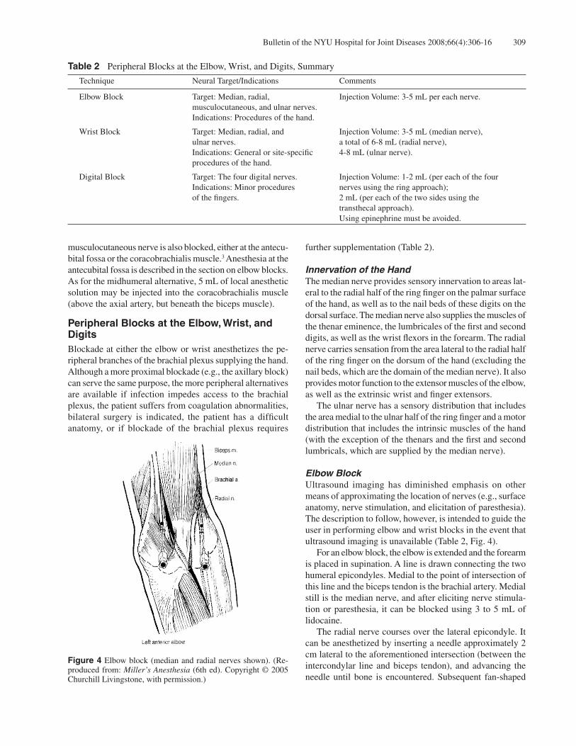
Elbow Block Ultrasound imaging has diminished emphasis on other means of approximating the location of nerves (e.g., surface anatomy, nerve stimulation, and elicitation of paresthesia). The description to follow, however, is intended to guide the user in performing elbow and wrist blocks in the event that ultrasound imaging is unavailable (Table 2, Fig. 4). For an elbow block, the elbow is extended and the forearm is placed in supination. A line is drawn connecting the two humeral epicondyles. Medial to the point of intersection of this line and the biceps tendon is the brachial artery. Medial still is the median nerve, and after eliciting nerve stimula-tion or paresthesia, it can be blocked using 3 to 5 mL of lidocaine. The radial nerve courses over the lateral epicondyle. It can be anesthetized by inserting a needle approximately 2 cm lateral to the aforementioned intersection (between the intercondylar line and biceps tendon), and advancing the needle until bone is encountered. Subsequent fan-shaped
Table 2 Peripheral Blocks at the Elbow, Wrist, and Digits, Summary
Technique Neural Target/Indications Comments
Elbow Block Target: Median, radial, Injection Volume: 3-5 mL per each nerve. musculocutaneous, and ulnar nerves. Indications: Procedures of the hand.
Wrist Block Target: Median, radial, and Injection Volume: 3-5 mL (median nerve), ulnar nerves. a total of 6-8 mL (radial nerve), Indications: General or site-specific 4-8 mL (ulnar nerve). procedures of the hand.
Digital Block Target: The four digital nerves. Injection Volume: 1-2 mL (per each of the four Indications: Minor procedures nerves using the ring approach); of the fingers. 2 mL (per each of the two sides using the transthecal approach). Using epinephrine must be avoided.
Figure 4 Elbow block (median and radial nerves shown). (Re-produced from: Miller’s Anesthesia (6th ed). Copyright © 2005 Churchill Livingstone, with permission.)
Bulletin of the NYU Hospital for Joint Diseases 2008;66(4):306-16310
injection of 3 to 5 mL of local anesthetic solution effectively blocks the radial nerve. To anesthetize the musculocutaneous nerve (perhaps as a supplement to the axillary block), the needle should be positioned 1 cm lateral and proximal to the same intersec-tion-point used previously, and 3 to 5 mL of local anesthetic solution may then be deposited superficially. The ulnar nerve traverses the medial epicondyle on the posterior aspect of the elbow. Injection at this location, perhaps as a consequence of insulation by fibrous tissue, carries a theoretically higher risk of nerve injury. An al-ternative injection site is at a point 1 to 2 cm proximal to a line connecting the medial epicondyle and the olecranon. Typically, 3 to 5 mL of lidocaine is employed in a fan-like manner, very superficially. No paresthesia should be evoked using this approach.
Wrist Block Based on the location of the planned surgical procedure, the clinician may opt for a nerve block at the wrist. For example, blocking the median nerve may be appropriate in cases involving multiple finger fractures or nail bed lacera-tions on the lateral aspect of the hand. An ulnar nerve block may be warranted when the injury (e.g., lacerations or fifth metacarpal fracture) is on the medial side of the hand. Lac-erations of the thumb or dorsum of the hand are indications for a radial nerve block (Table 2). To block the median nerve, the forearm is supinated, and the needle is inserted roughly 2 cm proximal to the wrist crease, between the flexor carpi radialis and palmaris longus tendons. In patients lacking a palmaris longus tendon, the median nerve is approximately 1 cm ulnar to the flexor carpi radialis tendon. The needle should be inserted vertically and advanced to a depth of roughly 1 cm, until the flexor retinacu-lum is pierced (indicated by a slight “pop”). At this point, 3 to 5 mL of lidocaine solution may be injected. Because the median nerve is housed immediately deep to the retinaculum, it is recommended that injection begin deep and continue as the needle is being retracted. Doing the opposite may result in deposition of solution above the retinaculum. Due to the branching fibers of the radial nerve, blocking
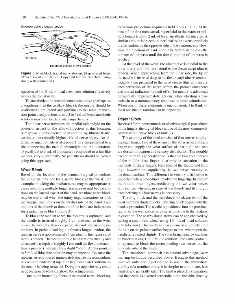
its various projections requires a field block (Fig. 5). At the base of the first metacarpal, superficial to the extensor pol-licis longus tendon, 2 mL of local anesthetic are injected. A similar amount is injected superficial to the extensor pollicis brevis tendon, on the opposite side of the anatomic snuffbox. Smaller injections of 1 mL should be administered over the dorsum of the wrist until the dorsal midline of the wrist is reached. At the level of the wrist, the ulnar nerve is medial to the ulnar artery and both are lateral to the flexor carpi ulnaris tendon. When approaching from the ulnar side, the tip of the needle is inserted deep to the flexor carpi ulnaris tendon, roughly 6 cm proximal to the wrist crease (this will ensure anesthetization of the nerve before the palmar cutaneous and dorsal radiations branch off). The needle is advanced horizontally approximately 1.5 cm, while eliciting a par-esthesia or a motor/sensory response to nerve stimulation. When one of these endpoints is encountered, 4 to 8 mL of local anesthetic solution can be deposited.
Digital BlockReserved for minor traumatic or elective surgical procedures of the fingers, the digital block is one of the most commonly administered nerve blocks (Table 2). The anatomy of the hand consists of four nerves supply-ing each finger. Two of these run on the volar aspect of each finger and supply the volar surface of that digit; and two are dorsal in location and sensory distribution. The notable exception to this generalization is that the two volar nerves of the middle three fingers also provide sensation to the nail beds of these fingers. Nail beds of the thumb and fifth digit, however, are supplied by the two nerves running on the dorsal surface. This difference in sensory distribution is important when procedures involve the fingertip. In case of the middle three fingers, medicating the two volar nerves will suffice; whereas, in case of the thumb and fifth digit, anesthetizing all four nerves is necessary. The ring block and the transthecal block are two of the most common digital blocks. The ring block begins with the hand in pronation. The needle is positioned into the proximal region of the web space, as close as possible to the phalanx in question. The nearby dorsal nerve can be anesthetized by raising a small skin wheal using 1.0 mL of local solution (1% lidocaine). The needle is then advanced anteriorly until the skin on the palmar surface begins to tent, whereupon the needle is retracted slightly. The volar branch nearby can thus be blocked using 1 to 2 mL of solution. The same protocol is repeated to block the corresponding two nerves on the opposite side of the finger. The transthecal approach has several advantages over the ring technique described above. Because this method involves only one injection and is not in the immediate vicinity of a terminal artery, it is simpler to administer, less painful, and generally safer. The hand is placed in supination, and the needle is inserted perpendicular to the skin, directly
Figure 5 Wrist block (radial nerve shown). (Reproduced from: Miller’s Anesthesia. (6th ed). Copyright © 2005 Churchill Living-stone, with permission.)
311Bulletin of the NYU Hospital for Joint Diseases 2008;66(4):306-16
over the metacarpal head. After making contact with bone, the needle is withdrawn slightly and maneuvered toward the two palmar nerves on either side. About 2 mL of local solution is injected at the two locations. When using either technique, co-administration of epi-nephrine with the local anesthetic must be strictly avoided. This can lead to spasm of the end arteries supplying the digits, potentially resulting in ischemia and even necrosis of areas more distal.
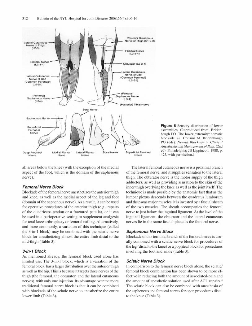
Peripheral Blocks of the Lower ExtremitiesAnatomyNeural supply to the lower extremities is derived from two main plexuses: the lumbar plexus, which is comprised of the anterior rami of the L1-L4 spinal nerves, and the sacral plexus, which is made-up of the anterior rami of the S1-S4 spinal nerves (Fig. 6). In general terms, the L2-L4 components of the lumbar plexus supply the thigh. More specifically, anterior divi-sions of L2-L4 comprise the obturator nerve, which then innervates the medial aspects of the thigh; whereas the posterior divisions give rise to the femoral nerve (L2-L4) and the lateral femoral cutaneous nerves (L2-L3), which supply the anterior and lateral parts of the thigh, respec-tively. Once the femoral nerve passes under the inguinal liga-ment, it enters the thigh and immediately divides into several
branches. Those descending anteriorly provide sensation to the anterior thigh from the inguinal ligament to the knee, while branches lying more posterior provide motor function to the extensors of the leg. One very important terminal sen-sory branch of the femoral nerve is the saphenous nerve. The saphenous nerve descends into the leg along the medial as-pect of the knee, between the sartorius and gracilis tendons. At this point, the saphenous nerve becomes subcutaneous, and forms branches that innervate the medial aspect of the leg and foot. Again, in general terms, both the lumbar and the sacral plexuses contribute to the formation of the sciatic nerve (L4-S3). More specifically, ventral branches of L4-S3 com-prise that aspect of the sciatic nerve that will become the tibial nerve, whereas dorsal branches of these same rami form that part that will become the common peroneal nerve. Immediately after originating from the above mentioned rami, the sciatic nerve traverses the greater sciatic fora-men (along with the posterior cutaneous nerves, S1-S3), and continues under the piriformis muscle, between the greater trochanter and the ischial tuberosity. At the lower edge of the gluteus maximus muscle, the nerve is at its most superficial before descending to the popliteal fossa, where it divides into the tibial and common peroneal nerves. The tibial nerve is medial and anterior, whereas the common peroneal lies more posterior and lateral. Along its course, the sciatic nerve innervates the posterior thigh, as well as
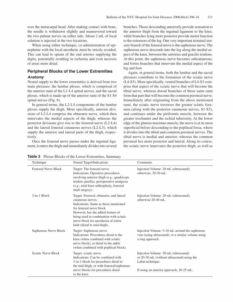
Table 3 Plexus Blocks of the Lower Extremities, Summary
Technique Neural Target/Indications Comments
Femoral Nerve Block Target: The femoral nerve. Injection Volume: 20 mL (ultrasound); Indications: Operative procedures otherwise, 20-30 mL. involving anterior thigh (e.g., quadriceps tendon, patella), perioperative analgesia (e.g., total knee arthroplasty, femoral shaft surgery).
3-in-1 Block Target: Femoral, obturator, and lateral Injection Volume: 20 mL (ultrasound); cutaneous nerves. otherwise 20-40 mL. Indications: Same as those mentioned for femoral nerve block. However, has the added feature of being used in combination with sciatic nerve block for anesthesia of entire limb (distal to mid-thigh).
Saphenous Nerve Block Target: Saphenous nerve. Injection Volume: 5-10 mL around the saphenous Indications: Procedures distal to the vein (using ultrasound); or a similar volume using knee (when combined with sciatic a ring approach. nerve block), or distal to the ankle (when combined with popliteal block).
Sciatic Nerve Block Target: sciatic nerve. Injection Volume: 20 mL (ultrasound) Indications: Can be combined with or 20-30 mL (without ultrasound) using the 3-in-1 block for procedures distal to Labat technique. the mid-thigh, or with femoral/saphenous nerve blocks for procedures distal If using an anterior approach, 20-25 mL. to the knee.
Bulletin of the NYU Hospital for Joint Diseases 2008;66(4):306-16312
all areas below the knee (with the exception of the medial aspect of the foot, which is the domain of the saphenous nerve).
Femoral Nerve BlockBlockade of the femoral nerve anesthetizes the anterior thigh and knee, as well as the medial aspect of the leg and foot (domain of the saphenous nerve). As a result, it can be used for operative procedures of the anterior thigh (e.g., repairs of the quadriceps tendon or a fractured patella), or it can be used in a perioperative setting to supplement analgesia for total knee arthroplasty or femoral nailing. Alternatively, and more commonly, a variation of this technique (called the 3-in-1 block) may be combined with the sciatic nerve block for anesthetizing almost the entire limb distal to the mid-thigh (Table 3).
3-in-1 BlockAs mentioned already, the femoral block used alone has limited use. The 3-in-1 block, which is a variation of the femoral block, has a larger distribution over the anterior thigh as well as the hip. This is because it targets three nerves of the thigh (the femoral, the obturator, and the lateral cutaneous nerves), with only one injection. Its advantage over the more traditional femoral nerve block is that it can be combined with blockade of the sciatic nerve to anesthetize the entire lower limb (Table 3).
The lateral femoral cutaneous nerve is a proximal branch of the femoral nerve, and it supplies sensation to the lateral thigh. The obturator nerve is the motor supply of the thigh adductors, as well as providing sensation to the skin of the inner thigh overlying the knee as well as the joint itself. The technique is made possible by the anatomic fact that as the lumbar plexus descends between the quadratus lumborum and the psoas major muscles, it is invested by a fascial sheath of the two muscles. The sheath accompanies the femoral nerve to just below the inguinal ligament. At the level of the inguinal ligament, the obturator and the lateral cutaneous nerves lie in the same fascial plane as the femoral nerve.
Saphenous Nerve BlockBlockade of this terminal branch of the femoral nerve is usu-ally combined with a sciatic nerve block for procedures of the leg (distal to the knee) or a popliteal block for procedures involving the foot and ankle (Table 3).
Sciatic Nerve BlockIn comparison to the femoral nerve block alone, the sciatic/femoral block combination has been shown to be more ef-fective in reducing both the amount of associated-pain and the amount of anesthetic solution used after ACL repairs.4 The sciatic block can also be combined with anesthesia of the saphenous and femoral nerves for open procedures distal to the knee (Table 3).
Figure 6 Sensory distribution of lower extremities. (Reproduced from: Briden-baugh PO. The lower extremity: somatic blockade. In: Cousins M, Bridenbaugh PO (eds): Neural Blockade in Clinical Anesthesia and Management of Pain. (2nd ed). Philadelphia: JB Lippincott, 1988, p. 425, with permission.)
313Bulletin of the NYU Hospital for Joint Diseases 2008;66(4):306-16
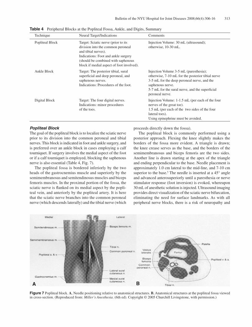
Popliteal BlockThe goal of the popliteal block is to localize the sciatic nerve prior to its division into the common peroneal and tibial nerves. This block is indicated in foot and ankle surgery, and is preferred over an ankle block in cases employing a calf tourniquet. If surgery involves the medial aspect of the foot or if a calf tourniquet is employed, blocking the saphenous nerve is also essential (Table 4, Fig. 7). The popliteal fossa is bordered inferiorly by the two heads of the gastrocnemius muscle and superiorly by the semimembranosus and semitendinosus muscles and biceps femoris muscles. In the proximal portion of the fossa, the sciatic nerve is flanked on its medial aspect by the popli-teal vein, and anteriorly by the popliteal artery. It is here that the sciatic nerve branches into the common peroneal nerve (which descends laterally) and the tibial nerve (which
proceeds directly down the fossa). The popliteal block is commonly performed using a posterior approach. Flexing the knee slightly makes the borders of the fossa more evident. A triangle is drawn; the knee crease serves as the base, and the borders of the semimembranosus and biceps femoris are the two sides. Another line is drawn starting at the apex of the triangle and ending perpendicular to the base. Needle placement is approximately 1.0 cm lateral to the mid-line, and 7-10 cm superior to the base.5 The needle is inserted at a 45° angle and advanced anterosuperiorly until a paresthesia or nerve stimulator response (foot inversion) is evoked, whereupon 30 mL of anesthetic solution is injected. Ultrasound imaging provides direct visualization of the sciatic nerve bifurcation, eliminating the need for surface landmarks. As with all peripheral nerve blocks, there is a risk of neuropathy and
Table 4 Peripheral Blocks at the Popliteal Fossa, Ankle, and Digits, Summary
Technique Neural Target/Indications Comments
Popliteal Block Target: Sciatic nerve (prior to its Injection Volume: 30 mL (ultrasound); division into the common peroneal otherwise, 10-30 mL. and tibial nerves). Indications: Foot and ankle surgery (should be combined with saphenous block if medial aspect of foot involved).
Ankle Block Target: The posterior tibial, sural Injection Volume 3-5 mL (paresthesia); superficial and deep peroneal, and otherwise, 7-10 mL for the posterior tibial nerve saphenous nerves. 3-5 mL for the deep peroneal nerve, and the Indications: Procedures of the foot. saphenous nerve. 5-7 mL for the sural nerve, and the superficial peroneal nerve.
Digital Block Target: The four digital nerves. Injection Volume: 1-1.5 mL (per each of the four Indications: minor procedures nerves of the great toe). of the toes. 1.5 mL (per each of the two sides of the four lateral toes). Using epinephrine must be avoided.
Figure 7 Popliteal block. A, Needle positioning relative to anatomical structures. B, Anatomical structures at the popliteal fossa viewed in cross-section. (Reproduced from: Miller’s Anesthesia. (6th ed). Copyright © 2005 Churchill Livingstone, with permission.)
BA
Bulletin of the NYU Hospital for Joint Diseases 2008;66(4):306-16314
intravascular injection.
Ankle BlockAnkle blocks are indicated for open procedures of the foot that do not require a tourniquet. Below is a brief descrip-tion of the peripheral sensory distributions at the level of the foot (Table 4).
Innervation of the Foot:Five nerves descend past the ankle and four of these stem from the sciatic nerve. The common peroneal and tibial nerves branch off the sciatic nerve in the popliteal fossa. At or slightly below the head of the fibula, the common peroneal nerve gives rise to its superficial and deep branches that eventually descend to the foot. The tibial nerve produces two of its own branches, the posterior tibial and sural nerves, at or slightly below the tibial mid-shaft. The two branches traverse the ankle on either side of the Achilles tendon: the sural nerve lies laterally, while the posterior tibial is medial. The last of the five nerves, the saphenous nerve, is a terminal branch of the femoral nerve and it supplies the proximal, medial aspect of the foot.
Selectively Blocking Nerves of the FootThe posterior tibial nerve innervates the heel, sole, and plan-tar aspects of the toes. With the patient prone, the posterior tibial artery can be palpated (immediately posterior to the medial malleolus) and used as a landmark. Slightly posterior to this artery, at the level of the medial malleolus, a 3.75 cm 25-gauge needle may be inserted and advanced towards the medial malleolus. At a depth of 0.5-1.0 cm, paresthesia should be sought by moving the needle from side to side. If paresthesia is successfully evoked, 3-5 mL of solution can then be injected; otherwise, the needle should be forwarded until it encounters bone, and then withdrawn slightly before depositing 7-10 mL of solution. The sural nerve supplies the lateral foot and proximal parts of the sole. The nerve courses between the lateral malleolus and the Achilles tendon. With the patient prone, the needle is inserted lateral to the tendon (approximately 1 cm above the lateral malleolus) and advanced towards the malleolus while injecting 5-7 mL of anesthetic solution subcutaneously. To block the two peroneal nerves, as well as the saphenous nerve through the same needle-entry point, a line should first be drawn connecting the two malleoli on the dorsal aspect of the foot. Next, the anterior tibial artery is identified by palpating the area between the extensor hallucis longus tendon (which is evident on dorsiflexion of the first toe) and extensor digitorum longus muscle. After raising a skin wheal immediately lateral to the artery, the needle can be inserted perpendicular to the skin. The deep peroneal nerve supplies the skin between the first and second toes, as well as the short extensors of the toes. To anesthetize this nerve, the needle is advanced under
the extensor hallucis longus tendon until it encounters the tibia (usually at a depth of 1 cm). At this point, 3-5 mL of solution can be injected. The saphenous nerve supplies the medial foot, and to anesthetize this area, the needle should be maneuvered medially while still inside and another 3-5 mL of solution deposited. The superficial peroneal nerve innervates the dorsum of the foot with the exception of the first interdigital cleft. Having just anesthetized the deep peroneal and saphenous nerves, the needle is withdrawn from areas medial to the tibia, and maneuvered laterally (through the same skin inser-tion point) towards the superficial peroneal nerve (located midway between the extensor hallucis longus tendon and the lateral malleolus). Once positioned, 5-7 mL of solution is injected subcutaneously.
Digital Block The neural anatomy of the toes is akin to that of the fingers, four nerves supplying each digit. For each toe, two nerves are plantar in location and sensory distribution, and two are dorsal (Table 4). The technique used to block the first digit of the foot varies slightly from that used for the lateral four toes. For the great toe, a 25-gauge (or smaller) needle is positioned perpendicular to the skin, dorsolateral to the proximal phalanx. It is advanced to the subcutaneous tissue on the plantar surface of the toe, at which point 1 to 1.5 mL of 1% lidocaine is injected. As the needle is withdrawn, a similar volume is deposited near the dorsal of the two nerves on the lateral aspect of this toe. The needle is then withdrawn completely and reinserted on the contralateral side of the great toe, with this protocol repeated (injecting 4 to 6 mL of solution in all). Details of anesthetizing the four lateral toes differ only slightly from the technique just described. Again, the point of needle insertion is the base of the toe on the dorsal surface. The endpoint for needle advancement, in this case, is tenting of the skin on the plantar surface of the toe. The solution (1.5 mL) is injected as the needle is slowly withdrawn. This is repeated on the opposite side of the same toe, for a total of 3.0 mL of solution. As with digital block of the fingers, co-administration of epinephrine must be strictly avoided as this can cause spasm of the end arteries supplying the digits, potentially resulting in ischemia and even necrosis of areas more distal.
Bier Block and Hematoma BlockBier BlockAlso known as intravenous regional anesthesia, the Bier block is a safe and very simple method of anesthetizing areas distal to the elbow or knee and is indicated for opera-tive procedures that will be completed in under 30 minutes. Contraindications include uncontrolled hypertension, severe peripheral vascular disease, and preexisting soft tissue dam-
315Bulletin of the NYU Hospital for Joint Diseases 2008;66(4):306-16
age that has potential for further exacerbation. Unlike previ-ously described approaches, this method does not selectively target individual nerves. Instead, the Bier block relies on local infiltration and diffusion through the tissue to provide anesthesia to an entire region (Table 5). The Bier approach has two variations, differing primarily in the number of tourniquets employed. Both approaches begin similarly: an intravenous cannula is placed in the nonoperative limb, through which fluids and other agents are administered and allowed to circulate systemically. The double-tourniquet approach calls for two tourniquets being placed on the operative side around the proximal regions of the limb. Next, an intravenous cannula is placed into a vein in the operative hand or foot. The limb is then elevated and wrapped with an Esmarch bandage to exsanguinate the extremity. If the injury does not allow for such wrapping, then there is the somewhat less effective option of elevat-ing the limb for at least 3 minutes. If the wrapping option is employed, then the bandage should remain in place until the cuff is reinflated to a pressure of 250 mmHg (in children, 50 mmHg above the systolic will usually suffice). In terms of dosing, for upper extremity procedures, 3 to 3.5 mg/kg (40 to 50 mL of 0.5% solution) of lidocaine (without epinephrine), is the limit. For procedures of the lower extremity, increasing the volume to 50 to 80 mL and decreasing the concentration to 0.25% lidocaine is ideal. The solution should be dispensed slowly, and the effect should be expected in approximately 15 minutes. Once anesthesia takes effect, the catheter should be retracted and the site taped to avoid leakage. The advantage of the double tourniquets is that if pain develops, one has the option of inflating the distal cuff over-lying anesthetized skin and then deflating the proximal cuff that was causing discomfort. There is evidence, however, that if a single wide cuff is employed, then the required infla-tion pressure is lower than the double-cuff alternative and resultant pressure-related neurologic damage may also be limited. Regardless of approach, the limitation of this block lies in the patient’s ability to tolerate the tourniquet. Typi-
cally, patients experience significant tourniquet discomfort at 45 minutes. Co-administration of a low dose of ketamine (0.1 mg/kg) has been shown to decrease this discomfort.6 To avoid systemic toxicity, cuff deflation should be car-ried out slowly and sequentially. This is especially true if the time elapsed since the administration of lidocaine is under 30 minutes, as this period may not be sufficient to allow for adequate drug-tissue binding. In such cases, it may be prudent to wait as long as 30 minutes before attempting to deflate. When ready, the cuff should be deflated for 5 seconds and then reinflated for 1 to 2 minutes. This pattern should be repeated at least three times. Another way to decrease the risk of systemic toxicity is to progressively deflate in three 10-second intervals, separated by 1 minute reinflation periods.7 Undesired effects in applying the Bier technique include tourniquet associated pain, pain during exsanguination given the injury, and systemic toxicity due to early cuff-deflation. Substitution of lidocaine with bupivacaine is advised against, due to risk of cardiovascular collapse.
Hematoma BlockThis approach is best suited for closed reduction of wrist and ankle fractures.8,9 Advantages of this block are that the patient tends to retain the ability to verbally communicate during the procedure and recovers quickly following cast-ing.10 Studies have indicated that less pain is associated with this approach than with intravenous sedation,11,12 although results have been less favorable in comparison to the Bier block (Table 5).13
One means to this approach is to premedicate the patient using an oral dose of 0.2 mg/kg of oxycodone, 30 to 60 minutes prior to placement of the block. After the fracture site has been appropriately sterilized and a skin wheal raised, a large bore needle (e.g., 21 gauge) can be used to aspirate the hematoma fluid and replace it with 10 to 15 mL of 1% buffered lidocaine (without epinephrine). Allowing 5 to 7 minutes before performing reduction maneuvers will allow the local anesthetic to diffuse to the nerve fibers conducting
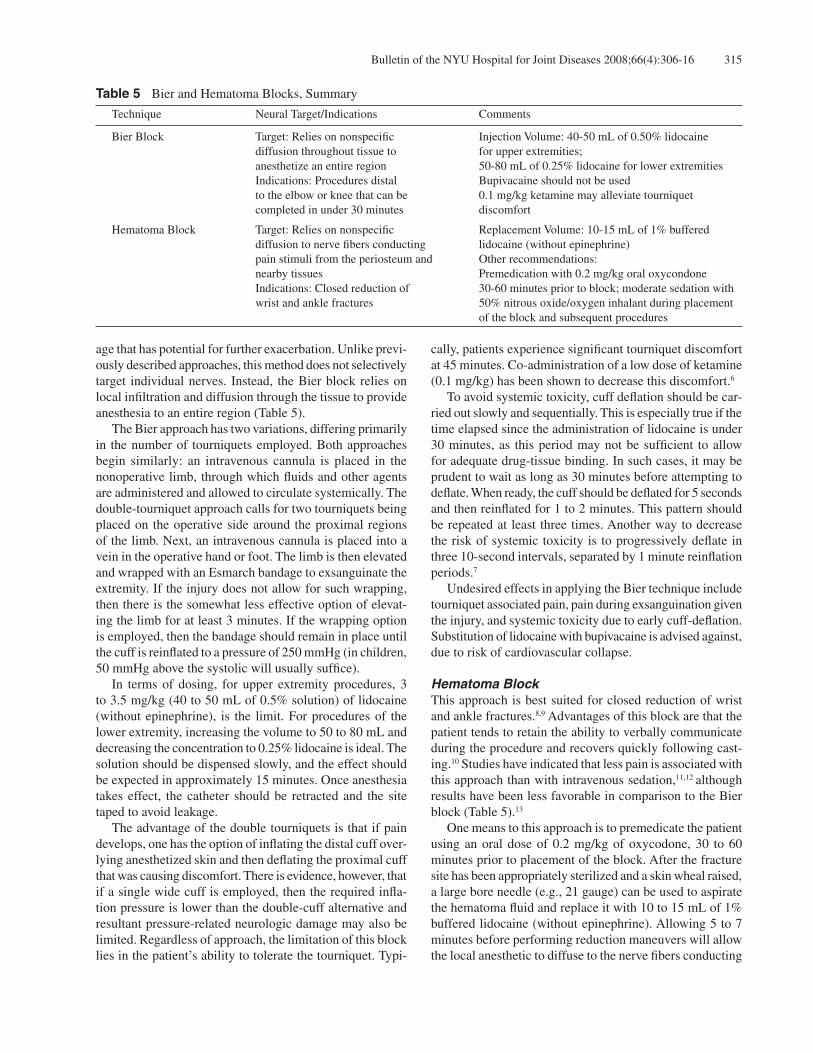
Table 5 Bier and Hematoma Blocks, Summary
Technique Neural Target/Indications Comments
Bier Block Target: Relies on nonspecific Injection Volume: 40-50 mL of 0.50% lidocaine diffusion throughout tissue to for upper extremities; anesthetize an entire region 50-80 mL of 0.25% lidocaine for lower extremities Indications: Procedures distal Bupivacaine should not be used to the elbow or knee that can be 0.1 mg/kg ketamine may alleviate tourniquet completed in under 30 minutes discomfort
Hematoma Block Target: Relies on nonspecific Replacement Volume: 10-15 mL of 1% buffered diffusion to nerve fibers conducting lidocaine (without epinephrine) pain stimuli from the periosteum and Other recommendations: nearby tissues Premedication with 0.2 mg/kg oral oxycondone Indications: Closed reduction of 30-60 minutes prior to block; moderate sedation with wrist and ankle fractures 50% nitrous oxide/oxygen inhalant during placement of the block and subsequent procedures
Bulletin of the NYU Hospital for Joint Diseases 2008;66(4):306-16316
pain stimuli from the periosteum and nearby tissues. Moder-ately sedating the patient using a 50% nitrous oxide/oxygen inhalation mixture during placement of the block and the subsequent reduction process can further reduce the pain associated with these procedures.14 Effects of the inhalation mixture can be reversed by simply removing the mask fol-lowing casting.
Disclosure StatementNone of the authors have a financial or proprietary interest in the subject matter or materials discussed, including, but not limited to, employment, consultancies, stock ownership, honoraria, and paid expert testimony.
References1. Marhofer P, Greher M, Kapral S. Ultrasound guidance in
regional anaesthesia. Br J Anaesth. 2005;94(1):7-17.2. Retzl G, Kapral S, Greher M, Maritz W. Ultrasonographic
findings of the axillary part of the brachial plexus. Anesth Analg. 2001;92:1271-5.
3. deJong RH. Axillary block of the brachial plexus. Anesthesiol-ogy. 1961;22:215.
4. Williams BA, Kentor ML, Vogt MT, et al. Femoral-sciatic nerve blocks for complex outpatient knee surgery are associ-ated with less postoperative pain before same-day discharge: a review of 1,200 consecutive cases from the period 1996-1999. Anesthesiology. 2003;98(5):1206-13.
5. Vloka JD, Hadzic A, April E, Thys DM. The division of the sciatic nerve in the popliteal fossa: Anatomical implications for popliteal nerve blockade. Anesth Analg. 2001;92:215.
6. Gorgias NK, Maidatsi PG, Kyriakidis AM, et al. Clonidine versus ketamine to prevent tourniquet pain during intravenous regional anesthesia with lidocaine. Reg Anesth Pain Med. 2001;Nov-Dec;26(6):512-7.
7. Sukhani R, Garcia CJ, Munhall RJ, et al. Lidocaine disposition following intravenous regional anesthesia with different tour-niquet deflation techniques. Anesth Analg. 1989;68:633.
8. Aliota RJ, Furia JP, Marquardt JD. Hematoma block for ankle fractures: a safe and efficacious technique for manipulation. J Orthop Trauma. 1995;9(2):113-6.
9. Case RD. Haematoma block—a safe method of reducing Colles’ fractures. Injury. 1985 Jul;16(7):469-70.
10. Kennedy RM, Luhmann JD, Luhmann SJ. Emergency Depart-ment management of pain and anxiety related to orthopedic fracture care. A guide to analgesic techniques and procedural sedation in children. Pediatr Drugs. 2004;6(1):11-31.
11. Furia JP, Alioto RJ, Marquardt JD. The efficacy and safety of the hematoma block for fracture reduction in closed isolated fractures. Orthopedics. 1997;20:423-6.
12. Singh GK, Manglik RK, Lakhtakia PK, et al. Analgesia for the reduction of Colles fracture: a comparison of hematoma block and intravenous sedation. Online J Curr Clin Trials. 1992;Oct 1;Doc No 23.
13. Abbaszadegan H, Jonsson U. Regional anesthesia preferable for Colles’ fracture: controlled comparison with local anes-thesia. Acta Orthop Scand. 1990;61(4):348-9.
14. Hennrikus WL, Shin AY, Klingelberger CE. Self-administered nitrous oxide and a hematoma block for analgesia in the out-patient reduction of fractures in children. J Bone Joint Surg Am. 1995 Mar;77(3):335-9.