Embed Size (px)
Citation preview

J Korean Acad Pediatr Dent 42(4) 2015ISSN (print) 1226-8496 ISSN (online) 2288-3819
319
Orthodontic Treatment of a Child with Short Root Anomaly : a Case Report
Jeongeun Lee, Jewoo Lee, Gayoung Shin, Soyoun An, Jihyun Song, Jiyoung Ra
Department of Pediatric Dentistry, College of Dentistry, Wonkwang University
Short root anomaly (SRA) is very rare, but can be problematic for physicians because patients with SRA are
more vulnerable to root resorption with orthodontic forces. During the mixed dentition period, it may be difficult
to diagnose generalized SRA. This article reports the treatment of an orthodontic patient with SRA at the early
mixed dentition stage. Despite local tooth loss, a relatively favorable outcome was obtained without excessive
root resorption. Ultimately, orthodontic therapy is possible for patients with generalized SRA, but precautions
should be taken to avoid complications, such as tooth loss or root resorption.
Key words : Short root anomaly, Mixed dentition, Orthodontic treatment, Root resorption, Tooth loss
Abstract
Corresponding author : Jiyoung RaDepartment of Pediatric Dentistry, College of Dentistry, Wonkwang University, 895 Moowang-ro, Iksan, Jeollabuk-do, 54538, KoreaTel: +82-63-850-6633 / Fax: +82-63-851-5324 / E-mail: [email protected] August 12, 2015 / Revised September 22, 2015 / Accepted September 11, 2015※This research was supported by Wonkwang University, in 2013.
http://dx.doi.org/10.5933/JKAPD.2015.42.4.319
Ⅰ. Introduction
In 1972, Lind1) discovered short root anomaly (SRA)
in a Swedish population, and defined the condition as
being characterized by an abnormally short and blunt
root shape, affecting mainly the maxillary central in-
cisors. SRA is typically diagnosed based on the crown-
to-root ratio when the root length is equal to or smaller
than the crown length1,2). The estimated prevalence of
SRA is 1.3~2.7%2). It shows familial occurrence, and
hereditary characteristics have been reported in studies
of parents and siblings2,3). Apajalahti et al.2) suggested
that SRA affected only the permanent teeth, based on
the absence of reports on SRA in the primary teeth. SRA
typically affects the maxillary central incisors, but some-
times the premolars1). Rarely, generalized SRA may af-
fect the entire dentition4-6).
Orthodontic forces can occasionally result in root re-
sorption, and the risk increases with a short root1,7-9).
Orthodontic therapy often begins in the mixed dentition
period in growing children. Unfortunately, root formation
is incomplete at this stage, which makes diagnosis diffi-
cult with radiographs alone.
Here, we present a case involving orthodontic treat-
ment performed in a patient at the early mixed dentition
stage, when the identification of SRA is most challeng-
ing. This report highlights the importance of the early
detection of SRA and precautions needed during ortho-
dontic procedures. Informed consent was obtained from
the patient’s parents for publication of this report.
J Korean Acad Pediatr Dent 42(4) 2015
320
Ⅱ. Case report
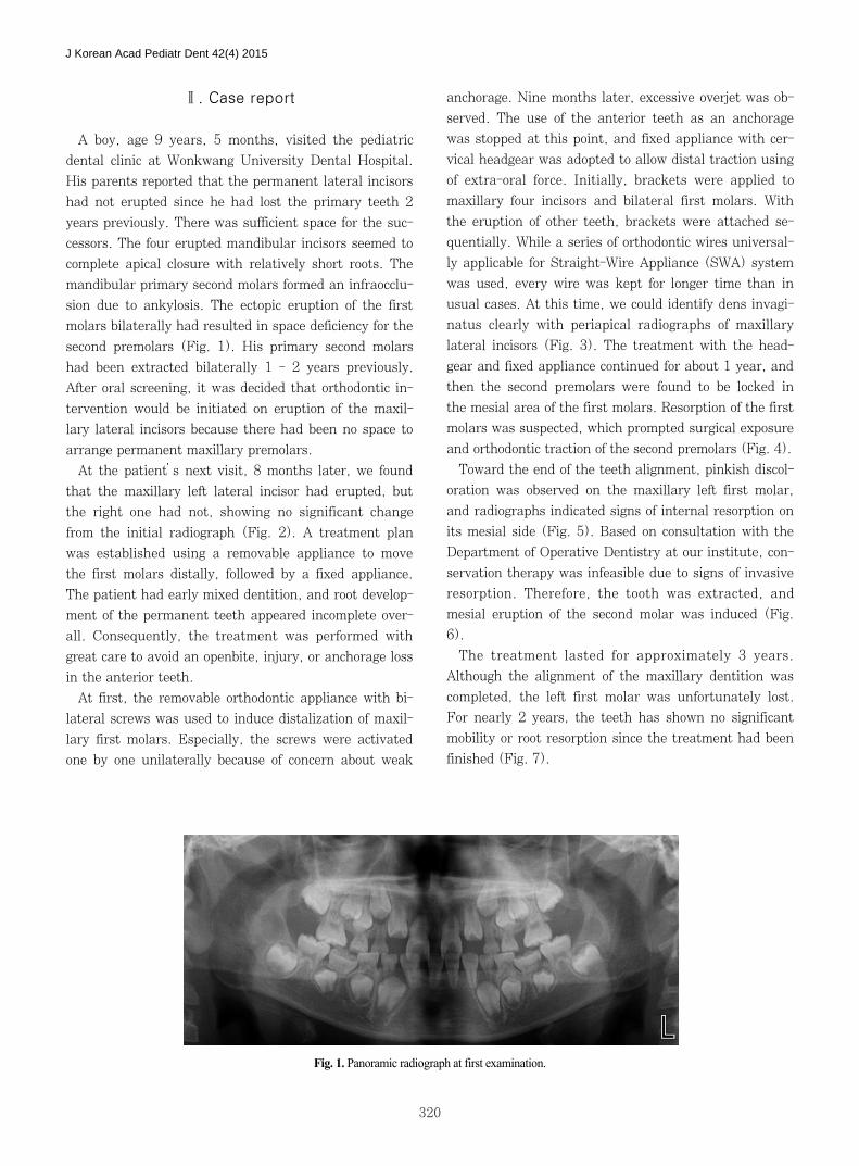
A boy, age 9 years, 5 months, visited the pediatric
dental clinic at Wonkwang University Dental Hospital.
His parents reported that the permanent lateral incisors
had not erupted since he had lost the primary teeth 2
years previously. There was sufficient space for the suc-
cessors. The four erupted mandibular incisors seemed to
complete apical closure with relatively short roots. The
mandibular primary second molars formed an infraocclu-
sion due to ankylosis. The ectopic eruption of the first
molars bilaterally had resulted in space deficiency for the
second premolars (Fig. 1). His primary second molars
had been extracted bilaterally 1 - 2 years previously.
After oral screening, it was decided that orthodontic in-
tervention would be initiated on eruption of the maxil-
lary lateral incisors because there had been no space to
arrange permanent maxillary premolars.
At the patient’s next visit, 8 months later, we found
that the maxillary left lateral incisor had erupted, but
the right one had not, showing no significant change
from the initial radiograph (Fig. 2). A treatment plan
was established using a removable appliance to move
the first molars distally, followed by a fixed appliance.
The patient had early mixed dentition, and root develop-
ment of the permanent teeth appeared incomplete over-
all. Consequently, the treatment was performed with
great care to avoid an openbite, injury, or anchorage loss
in the anterior teeth.
At first, the removable orthodontic appliance with bi-
lateral screws was used to induce distalization of maxil-
lary first molars. Especially, the screws were activated
one by one unilaterally because of concern about weak
anchorage. Nine months later, excessive overjet was ob-
served. The use of the anterior teeth as an anchorage
was stopped at this point, and fixed appliance with cer-
vical headgear was adopted to allow distal traction using
of extra-oral force. Initially, brackets were applied to
maxillary four incisors and bilateral first molars. With
the eruption of other teeth, brackets were attached se-
quentially. While a series of orthodontic wires universal-
ly applicable for Straight-Wire Appliance (SWA) system
was used, every wire was kept for longer time than in
usual cases. At this time, we could identify dens invagi-
natus clearly with periapical radiographs of maxillary
lateral incisors (Fig. 3). The treatment with the head-
gear and fixed appliance continued for about 1 year, and
then the second premolars were found to be locked in
the mesial area of the first molars. Resorption of the first
molars was suspected, which prompted surgical exposure
and orthodontic traction of the second premolars (Fig. 4).
Toward the end of the teeth alignment, pinkish discol-
oration was observed on the maxillary left first molar,
and radiographs indicated signs of internal resorption on
its mesial side (Fig. 5). Based on consultation with the
Department of Operative Dentistry at our institute, con-
servation therapy was infeasible due to signs of invasive
resorption. Therefore, the tooth was extracted, and
mesial eruption of the second molar was induced (Fig.
6).
The treatment lasted for approximately 3 years.
Although the alignment of the maxillary dentition was
completed, the left first molar was unfortunately lost.
For nearly 2 years, the teeth has shown no significant
mobility or root resorption since the treatment had been
finished (Fig. 7).
Fig. 1. Panoramic radiograph at first examination.

J Korean Acad Pediatr Dent 42(4) 2015
321
Fig. 3. During treatment; intraoral appliance for headgear and dens invaginatus of maxillary lateral incisors.
Fig. 2. Pretreatment panoramic radiograph and cast models.

J Korean Acad Pediatr Dent 42(4) 2015
322
Fig. 5. Internal root resorption of maxillary left first molar.
Fig. 4. Surgical exposure and orthodontic traction of second premolars.
Ⅲ. Discussion
This patient was unique in having idiopathic general-
ized SRA, which was not associated with any related
systemic disease or family history. Idiopathic SRA is rare
and difficult to predict. There have been several case re-
ports on idiopathic generalized SRA accompanied by mi-
crodontia, taurodontia, dens invaginatus, agenesis, an
ectopic canine, and obliteration of the pulp10,11). In this
patient, the initial radiographs showed ectopic eruption
and ankylosis, and indicated that the apical shape of the
incisors and first molars differed from that of the normal
immature permanent teeth. In addition, dens invagina-
tus of the maxillary lateral incisors were observed.
In a recent case report, tooth anomalies such as short
root formation, abnormal shape of crown or root, and hy-
poplasia of enamel were assumed to be caused by epige-
netic factors, such as damage, infection, medication ex-
posure during the first 2 years of life, or damage at
birth, especially damage affecting the central nervous
system12). These events tended to match the time of de-
velopment and damage to the teeth. However, our case
involved a systemic anomaly, which appeared uncon-
nected to such factors.

J Korean Acad Pediatr Dent 42(4) 2015
323
Fig. 6. After treatment; panoramic radiograph and intraoral photograph with a removable retainer.
Fig. 7. Follow-up examination of 19 months.

Several animal studies have identified various genes
associated with root development, including Msx2 (msh
homeobox 2), Sp6 (transcription factor), Shh (sonic
hedgehog), and Nog(noggin)13,14). In particular, the Nfic
(nuclear factor Ic) gene encodes a regulator that controls
root dentin formation and is involved in odontoblast dif-
ferentiation. The absence of this gene resulted in abnor-
mal short roots in Nfic-mutant mice6,14,15).
SRA has also been shown to be associated with other
syndromes and systemic diseases11,16). If the related fac-
tors mentioned above cannot be identified, the prediction
of SRA in mixed dentition showing immature permanent
teeth is very challenging. The patient’s parents knew of
no relevant family history and did not want to undergo
further medical evaluation. Since there was no further
evaluation, a potential relationship with a systemic dis-
ease or specific gene cannot be ruled out.
Teeth with short roots are at greater risk of root re-
sorption, caused by orthodontic forces7-9). One study of
the distribution of the orthodontic force using finite ele-
ment methods showed that significant stress concentra-
tion occurs in the middle area of short roots, generating
sufficient strength to trigger root resorption17).
Some clinicians believe that orthodontic therapy is still
possible for SRA patients, except in severe cases4,5). They
insist that the risk of resorption can be controlled with
clinical and radiological monitoring in most cases4,18).
Precautions should be taken at all stages of orthodontic
procedures. Lighter force needs to be applied because
the center of resistance is closer to the crown than that
of a normal tooth and the momentum/force ratio is easi-
ly affected4). Light, intermittent force may control re-
sorption19). To allow sufficient time to repair the resorp-
tion, longer intervals between the activation of force are
required20). Splints can be applied to ensure the stability
of local SRA teeth, if needed4,21). Furthermore, in local-
ized SRA, treatment methods that do not exert excessive
force on the affected teeth should be considered.
Orthopedic appliances, such as headgear or activators,
can be used if the timing is appropriate. There is a refer-
ential report of a patient with class II malocclusion who
underwent orthodontic treatment before the growth
spurt and a desirable outcome was achieved using head-
gear without further damage to the maxillary central in-
cisors in SRA21).
Unfortunately, our patient had generalized, not local-
ized, SRA so this strategy could not be used. A remov-
able appliance was used with the anterior teeth as an-
chors during the early stages of treatment, and it was
subsequently replaced by headgear to apply an extra-
oral force. In addition, light forces and a prolonged acti-
vation interval were applied as much as possible
throughout the treatment. Without the use of the head-
gear, other anchor teeth may have been lost, leading to
an unsuccessful result. Ultimately, this patient lost his
left first molar, but coped with the associated complica-
tions by compensatory mesial eruption of the second mo-
lar. Furthermore, root development did not progress
normally during the treatment period, and similar short
roots were observed in the untreated mandibular denti-
tion. This indicated a general state of arrested root for-
mation, rather than iatrogenic root resorption caused by
the orthodontic treatment.
Lind1) stated that SRA has not received sufficient at-
tention for several reasons. He mentioned that it could
easily be misdiagnosed as root resorption because of a
lower prevalence in Caucasians. SRA is more prevalent
in Asians than Caucasians and ~10% of Japanese chil-
dren with an average age of 9 years had central incisors
with short roots22). However, the prevalence of SRA has
been increasing in the USA recently, and Puranik et
al.23) suggested that this trend could be associated with
the growing Latino population in the US. Moreover,
teeth with SRA are generally asymptomatic, except in
severe cases, with a normal crown shape and size, and
related symptoms are usually absent. Unless excessive
mobility or loss of teeth develops, SRA cannot be con-
firmed without radiographs. Valladares et al.4) wrote
that differential diagnosis should be carried out to dis-
tinguish SRA from other conditions such as incomplete
root formation, external apical root resorption, dentin
dysplasia type I, and post-trauma root hypoplasia.
Although the diagnosis of SRA can be missed easily for
the reasons mentioned above, it should not be over-
looked. The resistance against root resorption is lower,
and the risk of tooth loss due to oral diseases is higher.
The early detection of SRA is important and proper pre-
cautions must be taken during treatment. Further stud-
ies of SRA itself are required, as well as case reports on
orthodontic, conservative, and periodontal treatment in
SRA patients.
Ⅳ. Summary
This case confirms that orthodontic therapy is possible
for SRA patients, but should be conducted with care. If
J Korean Acad Pediatr Dent 42(4) 2015
324

J Korean Acad Pediatr Dent 42(4) 2015
325
SRA is confirmed during orthodontic treatment, a tem-
porary cessation of treatment and appropriate modifica-
tion of the treatment plan are required. Most important-
ly, the possibility of SRA in children should be evaluated
with thorough assessments of the family history, sys-
temic disease, and other dental anomalies before begin-
ning orthodontic treatment.
References
1. Lind V : Short root anomaly. Scand J Dent Res, 80:
85-93, 1972.
2. Apajalahti S, Hölttä P, Turtola L, Pirinen S :
Prevalence of short-root anomaly in healthy young
adults. Acta Odontol Scand, 60:56-59, 2002.
3. S∨
ikanjic′PR, Mes∨trovic′S : A case of short-root
anomaly in a female from medieval Istria. Int J
Osteoarchaeol, 16:177-180, 2006.
4. Valladares Neto J, Rino Neto J, de Paiva JB :
Orthodontic movement of teeth with short root
anomaly: Should it be avoided, faced or igrnored?
Dental Press J Orthod, 18:72-85, 2013.
5. Edwards DM, Roberts GJ : Short root anomaly. Br
Dent J, 169:292-293, 1990.
6. Park JC, Herr Y, Kim HJ, Gronostajski RM, Cho MI
: Nfic gene disruption inhibits differentiation of
odontoblasts responsible for root formation and
results in formation of short and abnormal roots in
mice. J Periodontol, 78:1795-1802, 2007.
7. Sameshima GT, Sinclair PM : Characteristics of
patients with severe root resorption. Orthod
Craniofac Res, 7:108-114, 2004.
8. Mirabella AD, Artun J : Risk factors for apical root
resorption of maxillary anterior teeth in adult ortho-
dontic patients. Am J Orthod Dentofacial Orthop,
108:48-55, 1995.
9. Sameshima GT, Sinclair PM : Predicting and pre-
venting root resorption: part I. Diagnostic factors.
Am J Orthod Dentofacial Orthop, 119:505-510,
2001.
10. Apajalahti S, Arte S, Prinen S : Short root anomaly
in families and its association with other dental
anomalies. Eur J Oral Sci, 107:97-101, 1999.
11. Desai RS, Vanaki SS, Nidawani P, et al. : An
unusual combination of idiopathic generalized short-
root anomaly associated with microdontia, taurodon-
tia, multiple dens invaginatus, obliterated pulp
chambers and infected cyst: a case report. J Oral
Oathol Med, 35:407-409, 2006.
12. Lee HS, Kim SH, Song JS, et al. : A new type of
dental anomaly: molar-incisor malformation (MIM).
Oral Surg Oral Med Oral Pathol Oral Radiol,
118:101-109, 2014.
13. Bei M : Molecular genetics of tooth development.
Curr Opin Genet Dev, 19:504-510, 2009.
14. Huang XF, Chai Y : Molecular regulatory mecha-
nism of tooth root development. Int J Oral Sci, 4:
177-181, 2012.
15. Gronostajski RM : Roles of the NFI/CTF gene fami-
ly in transcription and development. Gene, 249:31-
45, 2000.
16. Roinioti TD, Stefanopoulos PK : Short root anomaly
associated with Rothmund-Thomson syndrome. Oral
Surg Oral Med Oral Pathol Oral Radiol Endod, 103:
e19-22, 2007.
17. Oyama K, Motoyoshi M, Shimizu N, et al. : Effects
of root morphology on stress distribution at the root
apex. Eur J Orhod, 29:113-117, 2007.
18. Tanaka OM, Knop LH, Shintcovsk RL, Hirata TM :
Treatment of a patient with severely shortened max-
illary central incisor roots. J Clin Orthod, 42:729-
731, 2008.
19. Levander E, Malmgren O, Eliasson S : Evaluation of
root resorption in relation to two orthodontic treat-
ment regimes. A Clinical experimental study. Eur J
Orthod, 16:223-228, 1994.
20. Cheng LL, Türk T, Darendeliler MA, et al. : Repair
of root resorption 4 and 8 weeks after application of
continuous light and heavy forces on premolars for 4
weeks: a histology study. Am J Orthod Dentofacial
Orthop, 138:727-734, 2010.
21. Marques LS, Generoso R, Armond MC, Pazzini CA :
Short-root anomaly in an orthodontic patient. Am J
Orthod Dentofacial Orthop, 138:346-348, 2010.
22. Ando S, Kiyokawa K, Sanka Y, et al. : Studies on
the consecutive survey of succedaneous and perma-
nent dentition in the Japanese children. Part 4.
Behavior of short rooted teeth in the upper bilateral
central incisors. J Nihon Univ Sch Dent, 9:67-80,
1967.
23. Puranik CP, Hill A, Frazier-Bowers SA, et al. :
Characterization of short root anomaly in a Mexican
cohort-hereditary idiopathic root malformation.
Orthod Craniofac Res, 18:62-70, 2015.

J Korean Acad Pediatr Dent 42(4) 2015
326
Short root anomaly (SRA) 환아의 교정적 처치 증례
이정은∙이제우∙신가 ∙안소연∙송지현∙라지
원광 학교 치과 학 소아치과학교실
Short root anomaly (SRA)는 매우 드문 질환으로, 교정력에 의한 치근흡수에 있어서 정상적인 치아보다 더 취약하므로
처치시에 어려움이 있다. 또한, 혼합치열기 시기에 나타나는 전반적인 SRA의 경우는 진단 또한 쉽지 않다. 초기 혼합치열기
환아에서 방사선 사진만으로 전반적인 SRA를 예측하기 어렵지만, 가족력이나 관련 전신질환, 또는 구강 내 다른 치아이상
(tooth anomaly) 등의 요소가 있는 경우, SRA를 염두에 두어야 한다.
본 증례는 전악에 걸쳐SRA를 보이는 초기 혼합치열기 환아의 교정적 처치를 다루었다. 비록 국소적인 치아 상실이 있었으
나 적절한 교정적 배열을 통해 더 이상의 과도한 치근흡수 없이 치료를 마무리 하 으며, 비교적 양호한 치료 결과를 얻었기
에 이를 보고하고자 한다. 결론적으로, SRA 환자에서도 교정 치료가 가능하며, SRA가 예측되는 혼합치열기 환아에서 치근
흡수나 치아상실과 같은 합병증이 나타나지 않도록 특히 주의를 기울여야 한다.
주요어: Short root anomaly, 혼합치열기, 교정적 처치, 치근흡수, 치아상실
국문초록


![Review Article Apical External Root Resorption and Repair ...external root resorption increases with the magnitude of the applied orthodontic force [ , , , ] and with continuous forces](https://img.pdfslide.us/doc/110x75/612422b33a54d70bce7d8287/review-article-apical-external-root-resorption-and-repair-external-root-resorption.jpg)