Embed Size (px)
Citation preview
8/7/2019 Orthodontic-Surgical-Endodontic Management of Unerupted
http://slidepdf.com/reader/full/orthodontic-surgical-endodontic-management-of-unerupted 1/5
Orthodontic-Surgical-Endodontic Management of UneruptedMaxillary Central Incisor With Distoangular Root DilacerationJose Valladares Neto, DDS, MSc, Se rgio de Pinho Costa, DDS, and Carlos Estrela, DDS, MSc, PhD
Abstract
Introduction: Root dilaceration refers to a dentalanomaly characterized by an abrupt deviation in thelongitudinal axis of the tooth. It can be localized inthe crown, between the crown and the root, or, mostfrequently, in the root. This report describes a horizon-tally unerupted maxillary central incisor with distoangu-lar root dilacerations most likely caused by a traumaticdental injury to its primary predecessor. Methods:
Surgical-orthodontic traction was applied after the redis-tribution of the space in the anterior maxillary region.
Results: Root dilaceration of the tractioned tooth wasevident and did not allow the alignment of the toothinto proper position in the dental arch. Root canal fillingand apicoectomy were performed. This procedurerestored the normal appearance of the anterior maxillaryteeth. Conclusion: Long-term follow-up (8 years) byperiapical radiography indicated stable periodontalhealth without the presence of root resorption. (J Endod
2010;36:755–759)
Key WordsApicoectomy, orthodontic traction, root dilaceration,root resorption, tooth impaction
Eruptive disturbances are changes of normal tooth eruption, including accelerated,delayed, failed, or deviated in the direction of tooth eruption, related to general
and/or local etiologic factors (1, 2). General (systemic) factors refer to the presenceof multiple compromised dental disturbances and are associated with clinical signsand symptoms that point to a determined disease or syndrome (1, 2). Local factorsinvolve a single tooth or adjacent teeth, with no other systemic association (1). Fromthe clinical point of view, local eruptive disturbances display an asymmetric eruptionof more than 6 months in relation to its homologue (3) and change the sequenceand chronology of normal eruption (1). Local factors are usually a routine challengeat an orthodontic clinic, whereas general factors are uncommon. In maxillary incisors,
a disturbance in eruption is relevant because of the esthetics and related emotional involvement for the child (1).
Local etiologic factors related to eruptive disturbances in the anteromaxillary region of the arch can be identified as (i) obstructive factors (1–4) such as supernu-merary tooth, negative tooth-bone discrepancy, gingival fibrosis, ankylosis, retainedprimary tooth, caries or early loss of the deciduous tooth with space closure, tumor,supernumerary root, macrodontia, change in tooth eruptive sequence, cyst, and odon-toma; (ii) genetic or developmental factors (5); (iii) traumatic factors (1, 2, 5–8); and(iv) idiopathic factors (1, 2).
Dilaceration can be a sequela of trauma and is associated with maxillary central incisor eruption failure (1, 2, 5–10). The term dilaceration was first described by John Tomes in 1848, when he reported a central incisor with an angulated root.Currently, tooth dilaceration refersto a dental anomaly characterized by an abruptdevi-
ation in the longitudinal axis of the tooth. It can be localized in the crown, between thecrown and the root, or, most frequently, in the root. It can affect any part of a tooth andalso in different directions (6). When the root dilaceration is in labial direction, it iscalled a scorpion tooth. If a tooth is doubly affected, it is called a bayonet dilaceration.According to severity,dilacerationcan be mild, moderate, or severe. A dilacerated toothcan lose its eruptive pathway, becoming ectopic and even unerupted, because root direction is not in accordance with crown direction. The further apical and milderthe dilaceration is, the greater the chance is for spontaneous eruption (8).
The purpose of the present article was to report a clinical case of a horizontally unerupted maxillary central incisor with a severe distoangular dilacerated root involving an adjacent tooth by means of a multidisciplinary approach including anorthodontic, surgical, and endodontic team.
Case ReportA 7-year-old male patient was brought by his mother to the Orthodontic Clinic of
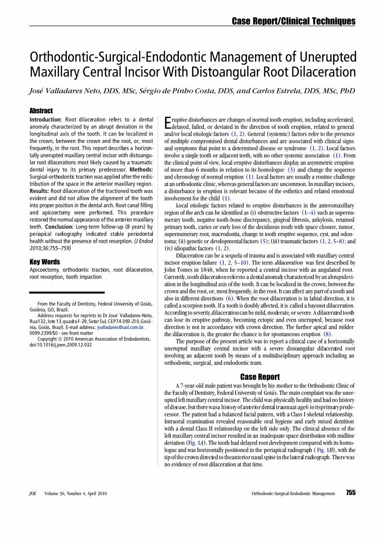
the Faculty of Dentistry, Federal University of Goia ´ s. The main complaint was the uner-upted left maxillary central incisor. The child was physically healthy and had no history of disease, but there wasa history of anterior dental traumaat age6 to itsprimary prede-cessor. The patient had a balanced facial pattern, with a Class I skeletal relationship.Intraoral examination revealed reasonable oral hygiene and early mixed dentitionwith a dental Class II relationship on the left side only. The clinical absence of theleft maxillary central incisor resulted in an inadequate space distribution with midlinedeviation (Fig. 1A). The tooth had delayed root development compared with its homo-logue and was horizontally positioned in the periapical radiograph (Fig. 1B ), with thetip of the crown directed to theanterior nasal spine in the lateral radiograph. There wasno evidence of root dilaceration at that time.
From the Faculty of Dentistry, Federal University of Goia s,Goia nia, GO, Brazil.
Address requests for reprints to Dr Jose Valladares-Neto,Rua132, lote 13,quadra F-29, Setor Sul, CEP74.093-210,Goia -nia, Goia s, Brazil. E-mail address: [email protected]/$0 - see front matter
Copyright ª 2010 American Association of Endodontists.doi:10.1016/j.joen.2009.12.032
Case Report/Clinical Techniques
JOE Volume 36, Number 4, April 2010 Orthodontic-Surgical-Endodontic Management 755
8/7/2019 Orthodontic-Surgical-Endodontic Management of Unerupted
http://slidepdf.com/reader/full/orthodontic-surgical-endodontic-management-of-unerupted 2/5
Four treatment alternatives were developed: (1) extraction of the unerupted central incisor and restoration with a bridge or animplant later when growth had ceased; (2) extraction of the unerup-ted central incisor and orthodontic closure of the space, substitutingthe lateral incisor for the central incisor with subsequent estheticrestoration improvement; (3) nonextraction of the unerupted central incisor with autotransplantation after orthodontic space opening; and(4) nonextraction of the unerupted central incisor with orthodonticspace opening, traction the involved tooth, and align in properposition.
The chosen option was to conduct the treatment without extrac-tion. The objective in the treatment was to open the space in the maxil-lary anterior region, surgical exposure, and traction the involved toothinto proper position.
The prognosis was considered doubtful because there were chan-ces of failure as a result of ankylosis, loss of attachment with root expo-sure, root anomaly, and external root resorption.
Initial treatment used an asymmetrical Klo ehn-type headgear tocorrect the left side Class II and gain some space. Concomitantly,brackets (Edgewise, 0.022 Â 0.025 inch slot; Dental Morelli, Sa ˜ oPaulo, Brazil) were placed on the 3 maxillary permanent incisors,and a 3Â 2 segmented alignment with open coil spring was performedto redistributespacein thearch,with special emphasisto theleft central incisor region (Fig. 1C ). Once adequate space was achieved, a surgical-orthodontic traction was programmed. Surgery was performed toexpose the unerupted incisor and attach an accessory (lingual buttontype) with a 0.010-inch ligature wire on it. Local anesthesia was given,and 2 mucoperiosteal incisions wereperformed to access the tooth, one
Figure 1. (A) Frontal view showing clinical absence of left maxillary central incisor with space closure and midline deviation; ( B ) pretreatment periapical radio-graph; (C ) compressed open-coil spring to open enough space for unerupted tooth; (D) surgical crown exposure showing its palatal surface; (E ) elastic attached toalignment wire and hook end for initial movement; (F ) left maxillary central incisor engaged in continuous arch wire showing improper tooth alignment due todivergent long axis.
Case Report/Clinical Techniques
756 Valladares Neto et al. JOE Volume 36, Number 4, April 2010
8/7/2019 Orthodontic-Surgical-Endodontic Management of Unerupted
http://slidepdf.com/reader/full/orthodontic-surgical-endodontic-management-of-unerupted 3/5
8/7/2019 Orthodontic-Surgical-Endodontic Management of Unerupted
http://slidepdf.com/reader/full/orthodontic-surgical-endodontic-management-of-unerupted 4/5
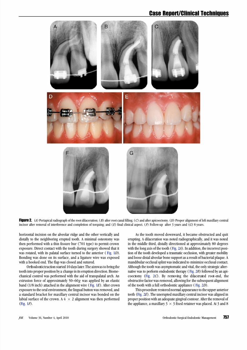
years after treatment, there were preserved bone, normal probingdepth, no apparent root resorption, and an acceptable gingival contour(Fig. 2F, G ).
DiscussionThe present case represents an orthodontic-surgical-endodontic
approach to an unerupted maxillary central incisor with normal pulp
vitality and a severely dilacerated root involving the neighboring tooth.Uematsu et al (11) described a similar case but with a labial-angulardilaceration, with the root apex projected just beneath the mobilemucosa of the labial sulcus. For both cases, endodontic treatment with an apicoectomy performed was the only feasible alternative that allowed the correct alignment of the tractioned tooth in the dental arch. The long-term follow-up supported this conduct, because therewas no evidence of unfavorable biologic response.
Root dilaceration is an uncommondental anomaly causedby trau-matic dental injury to the primary predecessors (2, 5–8, 12) and canresult in ectopic eruption (5, 12). It has also been proposed that it might be associated with some developmental syndromes (12).
Traumatic dental injury to a primary tooth and/or a bone fracturehas the potential to damage the underlying permanent tooth germ,
which might disturb its development. The effect of trauma to the Hert-wig’s epithelial root sheath can promote deflection or displacement of the permanent bud (6). Zilberman et al (6) reported that the severity of thetrauma to theprimary incisors is related to theeffect on their perma-nent successors. A prevalence of 4.7% of root dilacerationin theperma-nent incisors was noted after traumatic dental injury to their primary predecessors (6). In the present clinical case, the traumatic injury ismost likely the cause of dilaceration. After orthodontic traction androot formation, dilaceration became clearly evident.
The best time for treatment of eruptive disturbances is in the early stages (1–3). The objective of early treatment is to minimize emotional involvement of the child as a result of functional and esthetic problemscaused by the failure of maxillary anterior tooth eruption.
A severely dilacerated root of maxillary incisor is a clinical chal-lenge. Various therapeutic alternatives are described in the literature(1–3, 9–11, 13, 14). Even though surgical extraction is reported forseverely dilacerated roots (3, 10, 13, 14), a modified technique isalso cited, with the utilization of the crown of the extracted tooth asits own space maintainer or as an adhesive bridge construction whilefacial growth is active (13). The definitive restoration with a bridgeor an osseointegrated implant would be recommended laterwhen facial growth is complete. Another treatment alternative considering theextraction of the unerupted central incisor would be the orthodonticclosure of the space, substituting the lateral incisor for the central incisor with subsequent resin restoration. However, more recent casereports have shown that unerupted teeth could be properly positioned
with the aid of surgical-orthodontic traction (1, 2, 9–11, 15–18) andautotransplantation or intra-alveolar surgical uprighting (19–21). If the root dilaceration is severely labially directioned, endodontic treat-ment and apicoectomy have been suggested (11) instead of extraction.In the present case, the choice of orthodontic-surgical traction associ-ated with endodontic treatment and apicectomy was considereda feasible alternative.
The surgical-orthodontic approach to unerupted teeth iscommonly directed to surgical exposure of the crown and the bondingof a bracket for traction by light mechanical forces (1–2, 9, 10, 22).Other methods with a magnet system have been described with goodresults (23). The strategy adopted for the surgical exposure wasminimal bone removal and closed eruption after placing an attachment on the unerupted tooth. Kohavi et al (24) reported that the more bone
removed during surgical exposure of impacted canines, the greater theboneloss afterorthodontic treatment. The closed-eruption technique isconsidered a good surgical choice for unerupted teeth, considering thelong-term esthetic-periodontal status (25, 26). Other alternatives suchas the simple surgicalcrown exposurewithoutmechanical forces (4) orthe apically positioned flap technique have led to negative esthetics (25,26).
Removable appliances are considered better than fixed ones in
terms of anchorage control of the tooth extrusion, but they are limitedto patient compliance, proper root movement, and the application of continuous force. Thus, fixed appliances are preferable, becauseadequate bacterial plaque control and proper biomechanical control can be used.
Root dilaceration was not an impediment to movement, but ratherits spacial relationship with the neighboring tooth was. The decision forendodontic therapy with apicoectomy was doubtful because of its prog-nosis: first, because of the distal looseness of alveolar bone support;second, because of the unfavorable crown/root proportion; and third,because of the external root resorption after associated apicoectomy and orthodontic movement. In the present case, the radiograph control 3 and8 years after treatment showedno progression of root resorption,probably as a result of cementum repair (27).
In thecurrentcase report, theoutcome wasbased on radiographicaspects associated withclinical conditions. The 8-year recall showed theradiographic appearance of normal. With the introduction of conebeam computed tomography scans it is well-determined that theseimaging exams might change diagnostic hypotheses and treatmentplansand might affect the prognosis of certain clinical conditions. New methods to evaluate apical periodontitis and root resorption wererecently suggested by using cone beam computed tomography (28,29), and certainly this advanced imaging technology will be consideredin future studies.
The orthodontic treatment of a child with unerupted left maxillary central incisor with a severe root dilaceration and neighboring toothinvolvement was successfully performed by uncovering the crown,
closed eruption of the tooth, followed by root canal filling and apicoec-tomy. These procedures released the obstructive factor, favoring thesubsequent orthodontic alignment of the tooth. The long-term follow-up showed periodontal stabilitywithout the presenceof rootresorption.
References1. Valladares Neto J, Silva FA, Kaadi OB. Delayed eruption of permanent incisor asso-
ciated to prolonged retention of deciduous predecessor: obstructive, traumatic,developmental or idiopathic? Rev Odontol Brasil Central 1995;5:4–10.
2. Brin I, Zilberman Y, Azaz B. The unerupted maxillary central incisor: review of itsetiology and treatment. J Dent Child 1982;5:352–6.
3. Munns D. Unerupted incisors. Br J Orthod 1981;8:39–42.4. Di Biase DD. Mucous membrane and delayed eruption. Dental Pract 1971;21:
241–9.5. Stewart DJ. Dilacerated unerupted maxillary central incisors. Br Dent J 1978;145:
229–33.6. Zilberman Y, Ben Bassat Y, Lustmann J, Fuks A, Lustmann J. Effect of trauma to
primary incisors on root development of their permanent successors. PediatrDent 1986;8:289–93.
7. Ravn JJ. Sequelae of acute mechanical traumata in the primary dentition: a clinical study. J Dent Child 1968;35:281–9.
8. Mattison GD, Bernstein ML, Fischer JW. Lateral root dilaceration: a multi-disci-plinary approach to treatment. Endod Dent Traumatol 1987;3:135–40.
9. Kolokithas G, Karakasis D. Orthodontic movement of dilacerated maxillary central incisor. Am J Orthod Dentofac Orthop 1979;76:310–5.
10. Davis PH, Lewis DH. Dilaceration: a surgical/orthodontic solution. Br Dent J 1984;156:16–8.
11. Uematsu S, Uematsu T, Furusawa K, Deguchi T, Kurihara S. Orthodontic treatment of an impacted dilacerated maxillary central incisor combined with surgical exposureand apicoectomy. Angle Orthodont 2004;74:132–6.
Case Report/Clinical Techniques
758 Valladares Neto et al. JOE Volume 36, Number 4, April 2010
8/7/2019 Orthodontic-Surgical-Endodontic Management of Unerupted
http://slidepdf.com/reader/full/orthodontic-surgical-endodontic-management-of-unerupted 5/5
12. Jafarzadeh H, Abbott PV. Dilaceration: review of an endodontic challenge. J Endod2007;33:1025–30.
13. Becker A, Stern N, Zelcer Z. Utilization of a dilacerated incisor tooth as its own spacemantainer. J Dent 1976;4:263–4.
14. Smith DM, Winter GB. Root dilacerations of maxillary incisors. Br Dent J 1981;150:125–7.
15. Lin YTJ. Treatment of an impacted dilacerated maxillary central incisor. Am J OrthodDentofacial Orthop 1999;115:406–9.
16. Kolokithas G, Karakasis D. Orthodontic movement of dilacerated maxillary central incisor: report of a case. Am J Orthod 1979;76:310–5.
17. Tanaka E, Watanabe M, Nagaoka K, Yamaguchi K, Tanne K. Orthodontic traction of an impacted maxillary central incisor. J Clin Orthod 2001;35:375–8.18. Chew MT, Ong MM- A. Orthodontic-surgical management of an impacted dilacer-
ated maxillary central incisor: a clinical case report. Pediatr Dent 2004;26:341–4.19. Schatz J-P, Baets J, Joho J- P. Intra-alveolar surgical uprighting of impacted teeth:
a case report. Endod Dent Traumatol 1997;13:92–5.20. Maia RL, Vieira AP. Auto-transplantation of central incisor with root dilacerations:
technical note. Int J Oral Maxillofac Surg 2005;34:89–91.21. Filippi A, Pohl Y, Tekin U. Transplantation of displaced and dilacerated anterior
teeth. Endod Dent Traumatol 1998;14:93–8.
22. McNamara T, Woolfe SN, McNamara CM. Orthodontic management of a dilacer-ated maxillary central incisor with an unusual sequela. J Clin Orthod 1998;32:293–7.
23. Sandler JP. An attractive solution to unerupted teeth. Am J Orthod Dentofac Orthop1991;100:489–93.
24. Kohavi D, Becker A, Zilberman Y. Surgical exposure, orthodontic movement, andfinal tooth position as factors in periodontal breakdown of treated palatally impacted canines. Am J Orthod 1984;5:72–7.
25. Vermette ME, Kokich VD, Kennedy DB. Uncovering labially impacted teeth: apically positioned flap and closed eruption techniques. Angle Orthod 1995;65:211–21.
26. Chaushu S, Dykstein N, Ben-Bassat Y, Becker A. Periodontal status of impactedmaxillary incisors uncovered by 2 diferrent surgical techniques. J Oral MaxillofacSurg 2009;67:120–4.
27. Andreasen JO. Cementum repair after apicoetomy in humans. Acta Odontol Scand1973;31:211–21.
28. Estrela C, Bueno MR, Azevedo B, Azevedo JR, Pecora JD. A new periapical ı ndex based on cone beam computed tomography. J Endod 2008;34:1325–31.
29. Estrela C, Bueno MR, Alencar AHG, et al. Method to evaluate inflammatory root resorption by using cone beam computed tomography. J Endod 2009;35:1491–7.
Case Report/Clinical Techniques
JOE Volume 36, Number 4, April 2010 Orthodontic-Surgical-Endodontic Management 759