Embed Size (px)
Citation preview
223
IntroductionRehabilitative treatment in patients suffering from tooth loss is a challenge for dentists, and interactions among the dental specialties are required for optimal results. With the increasing demand for orthodontic treatment by adult patients, orthodontists must be aware of the various oral rehabilitation techniques that are currently available. Such knowledge will allow them, together with the general practitioner, to determine the best option for the patient. The orthodontist must be well-acquainted with and use a wide range of techniques that can improve masticatory function and smile aesthetics for their patients.
Although osseointegrated implants have shown consistent improvement in terms of their application in cases of tooth loss, nevertheless, the quality and quantity of the alveolar bone and gingival tissue are determining factors for the prognosis of the implant [1]. Rehabilitation success depends on the osseointegration of the implant, health of the hard and soft tissues, and harmony with the adjacent teeth [2]. In some cases of implant insertion soon after tooth extraction, numerous factors may influence the functional and aesthetic results. A conically shaped socket and socket expansion during tooth extraction may contribute to a poor interface between the implant and the bone (especially in the coronal third), which may interfere with the primary stabilization and rehabilitation time of the implant [3]. A vertical or horizontal bone deficiency may exist at the implant placement site, especially in the anterior maxillary and mandibular regions, requiring procedures to preserve or reconstruct the alveolar crest [4]. Some of these procedures, such as distraction osteogenesis [5], guided tissue regeneration [6], and bone graft application [7],
can be used during or after the extraction process.Allogeneic or autologous bone grafts of the intraoral
or extraoral regions are used to improve the bone contour before implant placement. The use of a graft removed from an extraoral area (e.g., the iliac region) is one of the only surgical methods for reconstructing the alveolar crest in cases of severe vertical bone loss. However, this approach is
expensive because it requires hospital surgery under general anesthesia, and it exposes the patient to the inherent risks of surgery. For gingival deficiencies, mucogingival surgical procedures are adopted, such as grafting of the gingival tissue and repositioning [4].
Forced orthodontic eruption is an alternative approach to surgery for increasing and improving the hard and soft tissue contours. Initially described by Heithersay [8] and Ingber [9], this technique has been used to correct isolated bone defects, reposition the gingival margin, and lengthen the crown [8-13]. The aims of this paper were to discuss the role of orthodontics in the oral rehabilitation of patients, with particular emphasis on the procedures used to treat bone defects, and to present a clinical case in which the orthodontic extrusive remodeling approach was used for bone gain in the posterior maxillary region, with subsequent implant placement.
Case ReportA 53-year-old woman presented with severe bone loss in the region of tooth 24, the upper left first premolar (Figure 1), due to periodontal disease associated with occlusal trauma. Prosthetic rehabilitation was not possible due to a low bone level at the apical third of the root. Extraction of this element was contraindicated because of later implant placement. Therefore, to complete the restoration of the alveolar architecture, a block bone graft was needed to increase the height of the alveolar ridge. To achieve appropriate function and aesthetics, tooth extraction for further rehabilitation with an implant would require a preliminary vertical reconstruction of the alveolar architecture. Depending on the extent of the vertical bone loss, this reconstruction would require a bone graft of autogenous origin (e.g., from the iliac crest or mandible) or even of allogenic, heterogenous, or alloplastic origin, for the rehabilitation of a single tooth.
In addition to bone loss, the patient had a Class I malocclusion with lower incisor crowding, lower premolar rotation, spaces between the upper premolars, and a poor relationship of the canines with no function; thus, orthodontic
Orthodontic Extrusion as an Aid in Oral Rehabilitation
Pedro Marcelo Tondelli1, Fabiana Akemy Kay2, Marcos Rikio Kuabara3
1DDS, MS, PhD, Collaborator Professor of Orthodontics, Department of Oral Medicine and Pediatric Dentistry, UEL – Londrina State University, Rua Pernambuco, 540, CEP 86020-070, Londrina, PR, Brazil. 2DDS, Department of Implantodontology, IMPPAR. Av. Arthur Tomaz, 100, CEP 86065-000, Londrina, PR, Brazil. 3DDS, MS, Department of Implantodontology, IMPPAR. Av. Arthur Tomaz, 100, CEP 86065-000, Londrina, PR, Brazil.
AbstractThe aims of this study were to discuss the use of orthodontics for oral rehabilitation in patients, particularly for dealing with bone defects, and to present a clinical case in which the orthodontic extrusive remodeling approach was used for bone gain in the posterior maxillary region, with subsequent implant placement. After 18 months of treatment, the dimensions of the alveolar process were restored and an implant was fixed in the maxillary region, with appropriate bone and gingival levels. Based on the literature and clinical evidence, orthodontic extrusion may be considered as an alternative to surgery to obtain a suitable implant site.
Key words: Orthodontic extrusion, Implant site, Periodontal regeneration
Corresponding author: Pedro Marcelo Tondelli, DDS, MS, PhD, Collaborator Professor of Orthodontics, Department of Oral Medicine and Pediatric Dentistry, UEL – Londrina State University, Rua Pernambuco, 540, CEP 86020-070, Londrina, PR, Brazil, Tel: +55(43)33574008; e-mail: [email protected]
224
OHDM - Vol. 13 - No. 2 - June, 2014
treatment was indicated. Proper hygiene instruction was provided and basic periodontal procedures were performed to establish a healthy periodontal environment that was free of tartar and plaque.
After 2 months, Roth braces were fixed in both arches, slot 0.022” × 0.028”, with the bracket in tooth 24 being positioned adjacent to the cervical region (Figure 2). Alignment and leveling of the teeth were performed with coaxial wires of 0.015” and 0.018”, followed by stainless steel wires of 0.016”, 0.018”, 0.020”, and 0.19” × 0.025”. Premolar extrusion was performed slowly to obtain bone gain. Bends were placed in the orthodontic wire to finish the premolar extrusion. Through this process, the bone and gingival architectures of the site were reconstructed.
During extrusion, the tooth was worn in its occlusal surface to prevent premature contact with the antagonist teeth and to allow movement. Because the tooth had undergone root canal treatment and had a ceramic crown, dental cutting could be made in large quantities. Torque was applied on the rectangular section wire, to move the premolar root apex in the buccal direction. This step enabled bone gain in the same direction for a better aesthetic restoration (Figure 3). A sequence of radiographs was obtained by the paralleling technique.
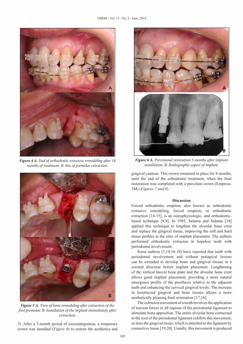
After 18 months of treatment, the dimensions of the alveolar process were restored, with appropriate bone and gingival levels. Afterwards, the tooth was extracted, and a 3.75 x 15 mm implant (Pi-Branemark) was fixed (Figures 4 and
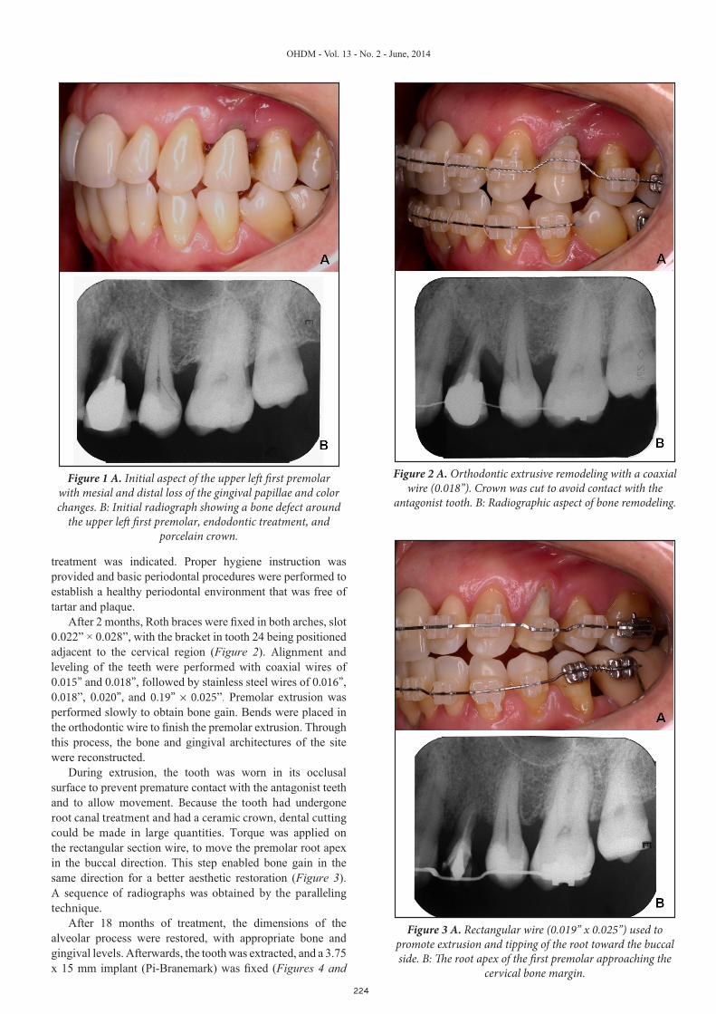
Figure 1 A. Initial aspect of the upper left first premolar with mesial and distal loss of the gingival papillae and color changes. B: Initial radiograph showing a bone defect around
the upper left first premolar, endodontic treatment, and porcelain crown.
Figure 2 A. Orthodontic extrusive remodeling with a coaxial wire (0.018”). Crown was cut to avoid contact with the
antagonist tooth. B: Radiographic aspect of bone remodeling.
Figure 3 A. Rectangular wire (0.019” x 0.025”) used to promote extrusion and tipping of the root toward the buccal side. B: The root apex of the first premolar approaching the
cervical bone margin.
225
OHDM - Vol. 13 - No. 2 - June, 2014
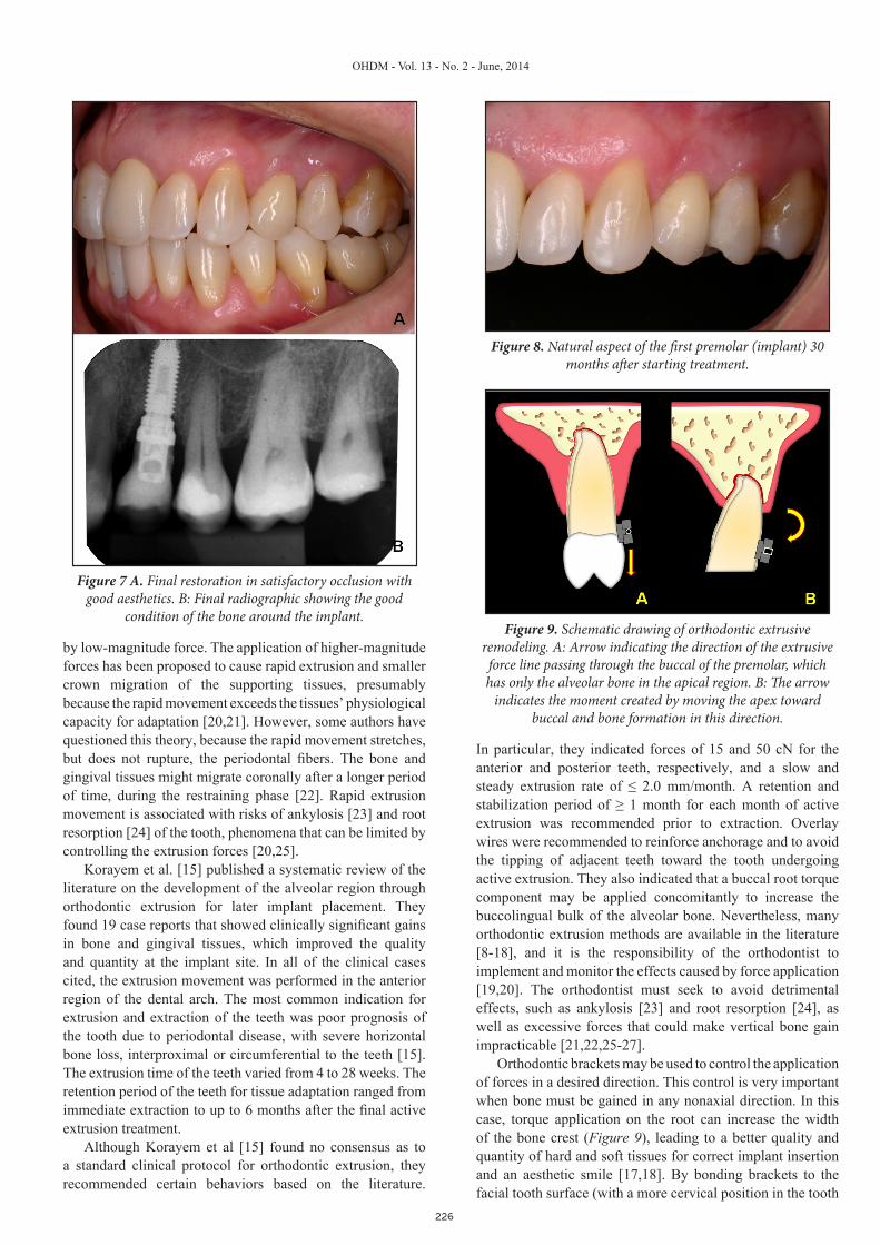
gingival contour. This crown remained in place for 8 months, until the end of the orthodontic treatment, when the final restoration was completed with a porcelain crown (Empress-3M) (Figures 7 and 8).
DiscussionForced orthodontic eruption, also known as orthodontic extrusive remodeling, forced eruption, or orthodontic extraction [14-15], is an osteophysiologic- and orthodontic-based technique [8,9]. In 1993, Salama and Salama [14] applied this technique to lengthen the alveolar bone crest and replace the gingival tissue, improving the soft and hard tissue profiles at the sites of implant placement. The authors performed orthodontic extrusion in hopeless teeth with periodontal involvement.
Some authors [1,14,16-18] have reported that teeth with periodontal involvement and without periapical lesions can be extruded to develop bone and gingival tissues in a coronal direction before implant placement. Lengthening of the vertical buccal bone plate and the alveolar bone crest allows good implant placement, providing a more natural emergence profile of the prosthesis relative to the adjacent teeth and enhancing the cervical gingival levels. The increase in keratinized gingival and bone tissues allows a more aesthetically pleasing final restoration [17,18].
The extrusion movement of a tooth involves the application of tension forces in all regions of the periodontal ligament to stimulate bone apposition. The entire alveolar bone connected to the root of the periodontal ligament exhibits this movement, as does the gingival tissue, which is attached to the ligament by connective tissue [19,20]. Usually, this movement is produced
Figure 4 A. End of orthodontic extrusive remodeling after 18 months of treatment. B: Site of premolar extraction.
Figure 5 A. View of bone remodeling after extraction of the first premolar. B: Installation of the implant immediately after
extraction.
Figure 6 A. Provisional restoration 5 months after implant installation. B: Radiographic aspect of implant.
5). After a 5-month period of osseointegration, a temporary crown was installed (Figure 6) to restore the aesthetics and
226
OHDM - Vol. 13 - No. 2 - June, 2014
by low-magnitude force. The application of higher-magnitude forces has been proposed to cause rapid extrusion and smaller crown migration of the supporting tissues, presumably because the rapid movement exceeds the tissues’ physiological capacity for adaptation [20,21]. However, some authors have questioned this theory, because the rapid movement stretches, but does not rupture, the periodontal fibers. The bone and gingival tissues might migrate coronally after a longer period of time, during the restraining phase [22]. Rapid extrusion movement is associated with risks of ankylosis [23] and root resorption [24] of the tooth, phenomena that can be limited by controlling the extrusion forces [20,25].
Korayem et al. [15] published a systematic review of the literature on the development of the alveolar region through orthodontic extrusion for later implant placement. They found 19 case reports that showed clinically significant gains in bone and gingival tissues, which improved the quality and quantity at the implant site. In all of the clinical cases cited, the extrusion movement was performed in the anterior region of the dental arch. The most common indication for extrusion and extraction of the teeth was poor prognosis of the tooth due to periodontal disease, with severe horizontal bone loss, interproximal or circumferential to the teeth [15]. The extrusion time of the teeth varied from 4 to 28 weeks. The retention period of the teeth for tissue adaptation ranged from immediate extraction to up to 6 months after the final active extrusion treatment.
Although Korayem et al [15] found no consensus as to a standard clinical protocol for orthodontic extrusion, they recommended certain behaviors based on the literature.
In particular, they indicated forces of 15 and 50 cN for the anterior and posterior teeth, respectively, and a slow and steady extrusion rate of ≤ 2.0 mm/month. A retention and stabilization period of ≥ 1 month for each month of active extrusion was recommended prior to extraction. Overlay wires were recommended to reinforce anchorage and to avoid the tipping of adjacent teeth toward the tooth undergoing active extrusion. They also indicated that a buccal root torque component may be applied concomitantly to increase the buccolingual bulk of the alveolar bone. Nevertheless, many orthodontic extrusion methods are available in the literature [8-18], and it is the responsibility of the orthodontist to implement and monitor the effects caused by force application [19,20]. The orthodontist must seek to avoid detrimental effects, such as ankylosis [23] and root resorption [24], as well as excessive forces that could make vertical bone gain impracticable [21,22,25-27].
Orthodontic brackets may be used to control the application of forces in a desired direction. This control is very important when bone must be gained in any nonaxial direction. In this case, torque application on the root can increase the width of the bone crest (Figure 9), leading to a better quality and quantity of hard and soft tissues for correct implant insertion and an aesthetic smile [17,18]. By bonding brackets to the facial tooth surface (with a more cervical position in the tooth
Figure 7 A. Final restoration in satisfactory occlusion with good aesthetics. B: Final radiographic showing the good
condition of the bone around the implant.
Figure 8. Natural aspect of the first premolar (implant) 30 months after starting treatment.
Figure 9. Schematic drawing of orthodontic extrusive remodeling. A: Arrow indicating the direction of the extrusive force line passing through the buccal of the premolar, which has only the alveolar bone in the apical region. B: The arrow
indicates the moment created by moving the apex toward buccal and bone formation in this direction.
227
OHDM - Vol. 13 - No. 2 - June, 2014
to be extruded), extrusion can be performed at low-magnitude forces, allowing new tissues to form [14]. The action line of the extrusion force passes buccally to the resistant center of the tooth and creates a moment (i.e., tendency to incline) on the tooth, which moves the root apex and leads to bone formation in the buccal direction [15]. Inclination and torque control must be performed later in the extrusion process and with a rectangular wire. The orthodontist must avoid loss of mechanical control, which can lead to excessive root apex tipping.
In the clinical case presented, the sequence of alignment and leveling of the upper teeth and the torque applied by the rectangular wire promoted vertical bone gain. Subsequent reconstruction of the alveolar architecture allowed the implant to be installed and a good aesthetic result to be achieved. In addition to bone loss, the radiographic results revealed decalcified bone around the first right upper premolar (24), with more evidence on the mesial side. This decalcification was improved after the basic periodontal procedures and “orthodontic extrusive remodeling” had been performed. Although the technique of parallelism was used, variations in the exposure parameters may account for this radiographic evidence.
Undoubtedly, the use of three-dimensional (3D) computed tomography would have been more efficient in providing results than 2D periapical radiography. However, although the lack of 3D radiography is a limitation in this clinical case, periapical radiography is a routine dental procedure. Moreover, because the amount of bone around the implant cannot be measured by the 2D method, the stability of the results may be questioned. However, clinically, the gingival aspect without papillae at the start of treatment (Figure 1), the improved bone quantity and quality after implant surgery (Figures 4 and 5), and the final restoration, which showed periodontal health and gain of papillae (Figure 8), indicated that the treatment resulted in many improvements, mainly in the papillae, which are very difficult to obtain in bone defects or around implants through periodontal surgery or any other procedure.
The use of orthodontic techniques to aid in the rehabilitation of teeth can avoid the need for more invasive procedures, such as mucogingival surgeries [4], distraction osteogenesis [5], guided tissue repair [6], or bone graft application [7]. The discomfort caused by the use of devices such as wires, braces, or other appliances for orthodontic extrusion can be a disadvantage from the aesthetic and hygienic perspectives. Similarly, there may be a need for conservative periodontal surgery, such as gingivoplasty, for correction of the cervical levels between adjacent teeth [8,19,20].
Orthodontic extrusion allows a tooth that has suffered bone loss to be restored, by repairing the proportion between the crown and the root. It avoids the need for implant placement or the mutilation of adjacent teeth through the installation of a fixed prosthesis. In the case of a hopeless tooth, the technique enables the implant to be fixed at a better angle and position, resulting in better function and aesthetics [14]. Orthodontic extrusion is indicated for the treatment of subgingival lesions (e.g., caries, fractures, and perforations), restorations invading the biologic width, vertical or horizontal bone loss, reduction of angular bone defects or isolated periodontal pockets [1,15,19,20], treatment of trauma and impacted teeth [19], as well as gains in the bone and gingival regions for implant insertions [1-4,8,10-12,15-17,19]. Orthodontic extrusion for hard and soft tissue gains is contraindicated in cases of ankylosis, hypercementosis, and periapical or vertical fracture of the root to be moved [19]. When rehabilitation of the root is proposed, the final crown-to-root ratio should not be less than 1:1, there must be sufficient prosthetic space, and there should not be furcation exposition in the multiradicular teeth; otherwise, the restorative procedure is unfeasible.
During orthodontic extrusive remodeling, inflammation can interfere directly with the bone tissue gain and stimulate gingival hyperplasia. The periodontal status, the quality and quantity of the attached gingiva, the presence and depth of periodontal pockets, the aesthetics of the site (e.g., gingival contour, occlusion, overjet, or overbite), the mandible movement, the prosthetic space after extrusion, and the general condition of the dentition must be evaluated. The patient should be informed about the risks of ankylosis, root resorption, recurrence, adjacent tooth movement, and treatment failure, which would necessitate changes in the treatment plan [19].
Orthodontic extrusion promotes the development of a better area for implant installation at sites of moderate or severe periodontal destruction, thereby increasing the amount and quality of bone, which directly influences implant stability [3]. Other treatment modalities, such as allogeneic or autologous bone grafts, guided tissue regeneration, distraction osteogenesis, and procedures to increase gingival aesthetics, may contribute to the same goal [4-7]. We cannot conclude whether any orthodontic procedure is superior to the others; to our knowledge, no study has compared these methods [15].
ConclusionBased on the literature and clinical evidence, orthodontic extrusion of teeth affected by severe periodontal disease can be considered as an alternative to surgery to obtain a suitable site for implants and to restore health and quality to the tissues.
References1. Mantzikos T, Shamus I. Case report: forced eruption and
implant site development. Angle Orthodontist. 1998; 68: 179–186.2. Phillips K, Kois JC. Aesthetic peri-implant site development.
The restorative connection. Dental Clinics of North America. 1998; 42: 57–70.
3. Buskin R, Castellon P, Hochstedler JL. Orthodontic extrusion and orthodontic extraction in preprosthetic treatment using implant therapy. Practical Periodontics & Aesthetic Dentistry. 2000; 12: 213–219.
4. Seibert JS, Salama H. Alveolar ridge preservation and reconstruction. Periodontology 2000. 1996; 11: 69–84.
5. Chin M, Toth B. Distraction osteogenesis in maxillofacial surgery using internal devices: report of five cases. Journal of Oral and Maxillofacial Surgery. 1996; 54: 45–53.
6. Hämmerle CH, Jung RE. Bone augmentation by means of barrier membranes. Periodontology 2000. 2003; 33: 36–53.
7. van Seenberghe D, Naert I, Bossuyt M, De Mars G, Calberson L, et al. The rehabilitation of the severely resorbed maxilla by simultaneous placement of autogenous bone grafts and implants: a
228
OHDM - Vol. 13 - No. 2 - June, 2014
10-year evaluation. Clinical Oral Investigations. 1997; 1: 102–108.8. Heithersay GS. Combined endodontic-orthodontic treatment
of transverse root fractures in the region of the alveolar crest. Oral Surgery, Oral Medicine, Oral Pathology. 1973; 36: 404–415.
9. Ingber JS. Forced eruption. I. A method of treating isolated one and two wall infrabony osseous defects - rationale and case report. Journal of Periodontology. 1974; 45: 199–206.
10. Potashnick SR, Rosenberg ES. Forced eruption: principles in periodontics and restorative dentistry. Journal of Prosthetic Dentistry. 1982; 48: 141–148.
11. Ingber JS. Forced eruption: part II. A method of treating nonrestorable teeth - periodontal and restorative considerations. Journal of Periodontology. 1976; 47: 203–216.
12. Ingber JS. Forced eruption: alteration of soft tissue cosmetic deformities. International Journal of Periodontics and Restorative Dentistry. 1989; 9: 416–425.
13. Johnson GK, Sivers JE. Forced eruption in crown-lengthening procedures. Journal of Prosthetic Dentistry. 1986; 56: 424–427.
14. Salama H, Salama M. The role of orthodontic extrusive remodeling in the enhancement of soft and hard tissue profiles prior to implant placement: a systematic approach to the management of extraction site defects. International Journal of Periodontics and Restorative Dentistry. 1993; 13: 312–333.
15. Korayem M, Flores-Mir C, Nassar U, Olfert K. Implant site development by orthodontic extrusion. Angle Orthodontist. 2008; 78: 752-760.
16. Mantzikos T, Shamus I. Forced eruption and implant site development: soft tissue response. American Journal of Orthodontics and Dentofacial Orthopedics. 1997; 112: 596–606.
17. Mantzikos T, Shamus I. Forced eruption and implant site development: an osteophysiologic response. American Journal of Orthodontics and Dentofacial Orthopedics. 1999; 115: 583–591.
18. O’Neal RB, Butler BL. Restoration or implant placement: a growing treatment planning quandary. Periodontology 2000. 2002; 30: 111–122.
19. Bach N, Baylard JF, Voyer R. Orthodontic extrusion: periodontal considerations and applications. Journal of the Canadian Dental Association. 2004; 70: 775–780.
20. Cuoghi OA, Bosco AF, Mendonça MR, Tondelli PM, Miranda-Zamalloa YM. Multidisciplinary treatment of a fractured root: a case report. Australian Orthodontics Journal. 2010; 26: 90-96.
21. Sabri R. L’allongement coronaire par l’égression orthodontique. Principes et techniques. Journal of Periodontology. 1989; 8: 197–204.
22. Reitan K. Principles of retention and avoidance of post-treatment relapse. American Journal of Orthodontics. 1991; 55: 776-790.
23. Oesterle LJ, Wood LW. Raising the root. A look at orthodontic extrusion. Journal of the American Dental Association. 1991; 122: 193–198.
24. Minsk L. Orthodontic tooth extrusion as an adjunct to periodontal therapy. Compendium of Continuing Education in Dentistry. 2000; 21: 768–770.
25. Malmgren O, Malmgren B, Frykholm A. Rapid orthodontic extrusion of crown root and cervical root fractured teeth. Endodontics & Dental Traumatology. 1991; 7: 49–54.
26. Erkut S, Arman A, Gulsahi A, Uckan S, Gulsahi K. Forced eruption and implant treatment in posterior maxilla: a clinical report. Journal of Prosthetic Dentistry. 2007; 97: 70-74.
27. Holst S, Hegenbarth EA, Schlegel KA, Holst AI. Restoration of a nonrestorable central incisor using forced orthodontic eruption, immediate implant placement, and an all-ceramic restoration: a clinical report. Journal of Prosthetic Dentistry. 2007; 98: 251-255.


![Dental Extrusion with Orthodontic Miniscrew Anchorage: A ... · Miniscrews for orthodontic treatments are available in severallengths(5–12mm)anddiameters(1.2–2.0mm)[17]. E. Mizrahi](https://img.pdfslide.us/doc/110x75/5ed55049eb5803601c17fbed/dental-extrusion-with-orthodontic-miniscrew-anchorage-a-miniscrews-for-orthodontic.jpg)