Embed Size (px)
Citation preview
RESEARCH ARTICLE Open Access
Orthodenticle is necessary for survival of a clusterof clonally related dopaminergic neurons in theDrosophila larval and adult brainJorge Blanco1,2*, Rahul Pandey1, Martin Wasser3 and Gerald Udolph1*
Abstract
Background: The dopaminergic (DA) neurons present in the central brain of the Drosophila larva are spatiallyarranged in stereotyped groups that define clusters of bilaterally symmetrical neurons. These clusters have beenclassified according to anatomical criteria (position of the cell bodies within the cortex and/or projection pattern ofthe axonal tracts). However, information pertaining to the developmental biology, such as lineage relationship ofclustered DA neurons and differential cell subtype-specific molecular markers and mechanisms of differentiationand/or survival, is currently not available.
Results: Using MARCM and twin-spot MARCM techniques together with anti-tyrosine hydroxylaseimmunoreactivity, we have analyzed the larval central brain DA neurons from a developmental point of view anddetermined their time of birth, their maturation into a DA neurotransmitter phenotype as well as their lineagerelationships. In addition, we have found that the homeodomain containing transcription factor Orthodenticle(Otd) is present in a cluster of clonally related DA neurons in both the larval and adult brain. Taking advantage ofthe otd hypomorphic mutation ocelliless (oc) and the oc2-Gal4 reporter line, we have studied the involvement oforthodenticle (otd) in the survival and/or cell fate specification of these post-mitotic neurons.
Conclusions: Our findings provide evidence of the presence of seven neuroblast lineages responsible for thegeneration of the larval central brain DA neurons during embryogenesis. otd is expressed in a defined group ofclonally related DA neurons from first instar larvae to adulthood, making it possible to establish an identityrelationship between the larval DL2a and the adult PPL2 DA clusters. This poses otd as a lineage-specific anddifferential marker of a subset of clonally related DA neurons. Finally, we show that otd is required in those DAneurons for their survival.
BackgroundThe Drosophila adult brain is a highly organized and com-plex structure that contains thousands of neurons (in theorder of 105) [1] exhibiting multiple cell-type identities, ascharacterized by various morphological, electrophysiologi-cal and molecular features. All these neurons arise fromthe mitotic activity of a small number of progenitor cells(neuroblasts (NBs)), which generate lineages of clonallyrelated neurons via two proliferative phases of neurogen-esis [2,3]. The first phase of neurogenesis takes place dur-ing embryogenesis and starts with the specification anddelamination of the NBs from the procephalic neurogenic
region. Between embryonic stages 8 and late 11, around100 NBs delaminate from this region on either side of theembryo in a reproducible spatiotemporal pattern [4]. EachNB assumes a unique identity, as revealed by the expres-sion of a specific set of marker genes such as the proneuralgenes, gap genes and segment polarity genes [5-7], andgives rise to an invariant cell lineage through multiplerounds of asymmetric cell divisions. In each cell division,the NB self-renews and generates a smaller daughter cell(named the ganglion mother cell), which divides only onceto give rise to two post-mitotic neurons or glial cells(reviewed in [8-10]). The first neurogenic process termi-nates at the end of embryogenesis, when most NBs stopdividing and enter a dormant phase called quiescence [11].The cells so far generated (primary neurons) wire the
* Correspondence: [email protected]; [email protected] of Medical Biology, 8A Biomedical Grove, Singapore 138648Full list of author information is available at the end of the article
Blanco et al. Neural Development 2011, 6:34http://www.neuraldevelopment.com/content/6/1/34
© 2011 Blanco et al; licensee BioMed Central Ltd. This is an Open Access article distributed under the terms of the Creative CommonsAttribution License (http://creativecommons.org/licenses/by/2.0), which permits unrestricted use, distribution, and reproduction inany medium, provided the original work is properly cited.

larval nervous system and eventually may remodel duringmetamorphosis to contribute to the adult brain [12,13].The second phase of neurogenesis starts during the latefirst (L1) and second (L2) instar larval stage, when theinactive NBs resume mitotic activity and, through roundsof asymmetric cell divisions, generate the population ofsecondary neurons and glial cells that accounts for morethan 90% of the adult brain [11,14]. Hence, a complete NBlineage can be divided into two discrete cell populations,each containing the cells generated by the NB during dis-tinct developmental phases (primary neurons duringembryogenesis and secondary neurons during larval devel-opment) and each harboring multiple neuronal cell types.It has been proposed that neuron identity within a NBlineage depends on a combination of spatial and temporalcues provided, firstly, by the unique identity the NBacquires during its specification/delamination time [7,15]and, secondly, as a result of a birth time/order-dependentmechanism [16], whereby cell-type specification of thenascent post-mitotic neurons depends on the identity ofthe progenitor temporal transcription factor expressed bythe NB at each particular time during lineage progression[17].The homeobox gene orthodenticle (otd), as a cephalic
gap gene, is expressed in broad domains of the procephalicectoderm during early neurogenesis, covering most of theprotocerebral anlage and the anterior part of the deutocer-ebral anlage [18]. Subsequently, its expression is alsodetected in NBs delaminating from these domains, whereit plays instructive roles important for cell viability andspatial identity of the nascent NBs [5]. It has also beenproposed that otd might control brain NB formation bytriggering proneural gene expression [19]. Inactivation ofotd at this early embryonic phase impairs NB formationand leads to a gap-like phenotype in the anterior head thatincludes the deletion of the protocerebral anlage and partof the deutocerebral anlage [20]. Later in development, otdexpression is also detected in post-mitotic neurons of thedeveloping brain and ventral nerve cord, not only in theembryo, but also in the larval and pupal brain and even inmature neurons of the adult brain. In this regard, flieshomozygous for an otd viable hypomorphic mutationcalled ocelliless (oc) show developmental defects that affectthe protocerebral bridge, an important neuropile structurein the adult brain that is part of the central complex [20].However, whether this phenotype is due to the alteredexpression of otd in progenitor cells, post-mitotic cells orboth is not known.A comprehensive study of gene function during neuro-
nal cell fate specification requires a previous and thor-ough cell lineage analysis and demands cell type-specificmolecular markers to trace the cells under study. In thispaper, we have focused our attention on the array ofdopaminergic (DA) neurons that populate the Drosophila
central brain during larval development. We have usedcell lineage tracing genetic techniques together withimmunoreactivity against the enzyme tyrosine hydroxy-lase (TH), the rate-limiting enzyme in dopamine bio-synthesis [21,22], to study their development, phenotypicmaturation and lineage relationships. Interestingly, onecluster of clonally related DA neurons expresses otd fromearly L1 to adulthood, allowing us to examine the post-mitotic role of otd in controlling identity and/or survivalof DA neurons in the Drosophila larval and adult brain.
ResultsBirth, clustering and phenotypic maturation ofdopaminergic neurons in the larval central brainUsing immunoreactivity against the enzyme TH as a DAneuron-specific molecular marker, an array of 21 DAneurons could be visualized in the central part of eachbrain hemisphere during the third instar larval stage (L3;Figure 1A). The stereotypical arrangement of their cellbodies in various groups has been used previously todefine four clusters of DA neurons that occupy distinctanatomical positions in the central brain [23-25]: dorsomedial 1 cluster (DM1: four cells), dorso medial 2 cluster(DM2: four cells), dorso lateral 1 cluster (DL1: sevencells) and dorso lateral 2 cluster (DL2: six cells). Asjudged by their projection and innervation patterns, DM2and DL1 cell clusters contained apparently homogeneouspopulations of DA neurons. On the contrary, both DM1and DL2 clusters could be further subdivided into twosubclusters according to their differential projection pat-terns. A single neuron in the DM1 cluster (namedDM1a) projected ventrally into the lower part of the ipsi-lateral brain lobe (Figure 1B), whereas the remainingthree DM1 DA neurons (named the DM1b cell cluster)innervated more ventrally localized regions of the brainlobe (subesophagial ganglion) and further extended intothe thoracic segments of the ventral ganglion (Figure1C). DM2 neurons projected ipsilaterally into the ante-rior part of the protocerebrum (Figure 1D), whereas DL1neurons were characterized by dorsally projecting neur-ites that bifurcated into dorsal and ventral branchesbefore crossing the midline to reach the contralateralbrain lobe (Figure 1E). Although described as homoge-neous, DM2 and DL1 cell clusters are possibly heteroge-neous at the single-cell level. Indeed, six different celltypes with slightly different innervation patterns havebeen described within the DL1 cell cluster using single-cell labeling techniques [25]. Similarly to the DM1cellcluster, the six DL2 DA neurons displayed two distinctprojection patterns: four neurons (named the DL2a cellcluster) projected their neurites dorsally into the anteriorpart of the ipsilateral brain lobe (Figure 1F). The remain-ing two cells (named the DL2b cell cluster) projected lat-erally and arborized in the ventral part of the brain lobe
Blanco et al. Neural Development 2011, 6:34http://www.neuraldevelopment.com/content/6/1/34
Page 2 of 14

Figure 1 Birth, clustering and differentiation of larval central brain dopaminergic neurons. (A) Third instar larval stage (L3) brain showingbilaterally symmetrical groups of TH-positive DA neurons clustered according to the position of the cell bodies within the cortex (the number ofneurons per cluster is given in parentheses) and the neurite projection patterns. Scale bar: 50 μm. (B-G’) Neurite projection patterns. Wild-typecell clones were induced in progenitor cells during early embryogenesis (3 to 7 h after egg laying (AEL)) and analyzed during L3. The cloneswere labeled with membrane tethered green and red flourescent proteins (both in grey) using the Twin-spot MARCM technique together withthe TH-gal4 driver. When possible, neurite projection patterns of the entire cell cluster are shown (B,E,F); otherwise, just one DA neuron as arepresentative for the cell cluster is produced (C,D). (G,G’) The neurite projection pattern (red arrowheads) of DL2b DA neurons (yellow arrows) isshown in two consecutive confocal optical sections. (H) Early L1 brain (24 to 28 h AEL) showing clusters of TH-positive cells (the number of DAneurons per cluster is given in parentheses) Scale bar: 10 μm. (I) Most larval DA neurons in the central brain are born during early neurogenesisbetween 3 to 13 h AEL. Wild-type cell clones, labeled with the MARCM technique in combination with the TH-gal4 driver, were induced inprogenitor cells at different times during development as indicated and analyzed in L3 brains. Images, except for (G,G’), represent Z projectionsof individual confocal optical sections. AEL, after egg laying; DA, dopaminergic; DL, dorso lateral; DM, dorso medial; L, instar larval stage; MARCM,mosaic analysis with a repressible cell marker; ML, midline; TH, tyrosine hydroxylase.
Blanco et al. Neural Development 2011, 6:34http://www.neuraldevelopment.com/content/6/1/34
Page 3 of 14

before crossing the midline to innervate the contralateralbrain lobe (Figure 1G,G’).We next determined the time of birth of the larval
central brain DA neurons by using the MARCM(mosaic analysis with a repressible cell marker) techni-que in combination with the TH-Gal4 driver. Flippaserecognition target (FRT)-mediated mitotic recombina-tion was randomly induced, exposing progenitor cells toa one hour heat-shock treatment (37°C) at differentdevelopmental stages and L3 brains were assayed for thepresence of green fluorescent protein (GFP)-labeled DAneurons. When mitotic recombination was induced dur-ing early L1, a time point commonly used to label neu-rons born during larval development, GFP-positive DAneurons were not detected in the resulting wild-type cellclones. This result agreed with the observation that THexpression was already detectable in early L1 brains(Figure 1H). On the contrary, a heat-shock treatmentapplied during early embryogenesis efficiently labeledDA neurons in the L3 central brain, with the highestlabeling efficiencies achieved when cell clones wereinduced at early embryonic stages (between 3 and 7 and7 and 11 hours after egg laying (AEL); Figure 1I). Theseresults demonstrate that DA neurons present in the L3central brain are primary neurons that arise during earlyembryogenesis.Despite their early embryonic origin, the larval central
brain DA neurons did not express the cell type-specificmarker gene TH during embryogenesis, and even at lateembryonic stages (stage 17) anti-TH immunoreactivity inthe central nervous system was restricted to the ventralnerve cord (data not shown and [24]). However, duringearly L1 (24 to 28 h AEL) most of the central brain DAneurons already displayed TH expression (Figure 1H),with two exceptions: the DM1a DA neuron started toshow anti-TH labeling during mid-late L1, whereas aDL2a DA neuron only showed anti-TH immunoreactivityat mid-late L3 (data not shown).In summary, DA neurons present in the central brain
of the Drosophila larva at L3 are generated during earlyembryogenesis and most of them acquire a mature neu-rotransmitter phenotype during early L1.
Dopaminergic neurons present in the central brain duringlarval development are generated by seven neuroblastlineagesIn Drosophila, clonally related neurons typically remainclustered in the mature brain and project their neuritesinto specific neuropile compartments [26,27]. To addresswhether the anatomical clustering of DA neurons in theL3 central brain is due to a clonal origin, we analyzedlineage relationships among DA neurons in each cellcluster. For this purpose, we utilized the twin-spotMARCM technique in combination with the tubulin-
Gal4 driver. Wild-type cell clones were induced duringearly embryogenesis (3 to 7 h AEL) and assayed in earlyL1 brains (24 to 28 h AEL) for the presence of GFP- andred fluorescent protein (RFP)-labeled NB clones contain-ing TH-positive neurons. Although at this developmentalstage two DA neurons still did not express the cell type-specific marker gene TH and thus could not be consid-ered in our analysis, two reasons justified our decision.Firstly, the reduced number of total cells present in thelarval brain during L1 facilitated an easier lineage analy-sis. Secondly, we observed that the tubulin promoterunderwent partial down-regulation in primary neuronsduring L3 (data not shown), impairing a reliable analysisof the lineage relationships among DA neurons.Our analysis revealed that all the DA neurons assigned
to the DM1b (Figure 2A), DM2 (Figure 2B), DL2a (Figure2D) and DL2b (Figure 2E) clusters were contained withinindividual NB clones and thus were clonally related. Bycontrast, we only found NB clones containing at most sixout of the seven DL1 DA neurons (Figure 2C,C’), indicat-ing that two NBs generate the seven DL1 DA neurons.As mentioned above, due to their delayed TH expres-
sion, two DA neurons were initially not included in thelineage analysis. In order to validate their allocation to therespective DA cell clusters, we induced MARCM-labeledwild-type NB clones during early embryogenesis (3 to 7 hAEL) and analyzed L3 brains. We tried to circumvent thedown-regulation of the tubulin promoter by including anadditional copy of the UAS-CD8::GFP transgene in thegenotype of the analyzed larvae. Although this strategy didnot always work reliably (for example, we did not observesingle NB clones containing the group of six DL1 DA neu-rons), we were still able to assign the missing DM1 DAneuron to an independent NB lineage (named DM1a), aswell as to confirm the allocation of the missing DL2 DAneuron to the DL2a NB lineage (Additional file 1).In summary, we found that seven NB lineages generate
the DA neurons present in each hemisphere of the Droso-phila larval central brain and that their clustered appear-ance is, at least in part, a consequence of a clonalrelationship. Moreover, the assignment of DA neurons toparticular NB lineages provides access to study geneticmechanisms of DA neuron cell fate specification.
orthodenticle is expressed in DL2a dopaminergic neuronsThe homeobox containing gene otd is broadly expressedin the anterior ectoderm during the blastoderm stage andis necessary for the specification of most of the NBs thatpopulate the protocerebrum and part of the deutocereb-rum [5,19]. Using Otd immunostainings, we found thatotd was expressed in DL2a DA neurons already at earlyL1 (Figure 3A). At mid-late L3, when the entire comple-ment of DA neurons in the larval central brain was
Blanco et al. Neural Development 2011, 6:34http://www.neuraldevelopment.com/content/6/1/34
Page 4 of 14

DL2a
B
A
C
D
C´
E
CD8-GFPCD2-RFP TH
CD8-GFP
CD8-GFPCD2-RFP
CD8-GFPCD2-RFP
CD8-GFPCD2-RFP
CD8-GFP
TH
TH
TH
TH
DM1b
DM2
DL1
DL1
DL2b
A11 A21
B11
C11
C1́1
D11
E 11
B21
C21
C2́1
D21
E 21E
*
*
*
TH
Figure 2 Twin-spot MARCM lineage analysis of larval central brain dopaminergic neurons at L1 reveals clonal relationship betweendopaminergic neurons. (A-E) Membrane-tethered GFP- and RFP-labeled wild-type NB clones containing TH-positive (blue) DA neurons fromdifferent DA cell clusters: DM1b cluster (A), DM2 cluster (B), DL1 cluster (C,C’), DL2a cluster (D) and DL2b cluster (E). All panels representindividual confocal optical sections (0.5 μm thick). The red asterisk in C2 identifies the DL1 DA neuron that belongs to a different NB lineage(DL1b). The yellow asterisk in C2 and C’2 identifies the same neuron in different optical sections. Scale bars: 10 μm. DA, dopaminergic; DL, dorsolateral; DM, dorso medial; GFP, green fluorescent protein; L, instar larval stage; NB, neuroblast; RFP, red fluorescent protein; TH, tyrosinehydroxylase.
Blanco et al. Neural Development 2011, 6:34http://www.neuraldevelopment.com/content/6/1/34
Page 5 of 14

Figure 3 otd is expressed in DL2a dopaminergic neurons in the larval brain. (A,C) otd (blue) is expressed in a specific cluster of TH-positive(red) DA neurons of the larval central brain during L1 (A) and L3 (C). (B,D) In ocga1 hemizygous larvae, two out of three DL2a DA neurons at L1(B) and three out of four DL2a DA neurons at L3 (D) are not detected using anti-TH immunoreactivity. Scale bars: 10 μm (A,B); 20 μm (C,D). (E)The oc2 enhancer is active in three out of the four DL2a DA neurons at L3. Scale bar: 10 μm. (E1-E3) otd expression and (E’1-E’3) oc2 enhanceractivity in DL2a DA neurons are analyzed in individual optical sections. (E4,E’4) DL2b DA neurons display neither otd expression nor oc2enhancer activity. Scale bars: 5 μm (E1-E4). (F,F1) In L3 ocga1 hemizygous larvae, oc2 enhancer activity in DL2a DA neurons is lost. Scale bar: 10μm. (G) otdYH13 mutant cell clones induced during early embryogenesis impair the development of DL2a DA neurons (white arrow). Yellowarrows point to the DL2b DA neurons in the left hemisphere Scale bar: 50 μm. All the panels, except for (E1-E4) and (E’1-E’4), represent Zprojections of individual confocal optical sections. DA, dopaminergic; DL, dorso lateral; GFP, green fluorescent protein; L, instar larval stage; oc,ocelliless; otd, orthodenticle; TH, tyrosine hydroxylase.
Blanco et al. Neural Development 2011, 6:34http://www.neuraldevelopment.com/content/6/1/34
Page 6 of 14

already visible with anti-TH labeling, Otd was specificallydetected in the four DL2a DA neurons (Figure 3C,E1-E3).To analyze the significance of otd expression in the spe-
cification and/or survival of post-mitotic DL2a DA neu-rons, we made use of the hypomorphic otd allele oc. ochemizygous flies are viable, but they show a lack of ocelliand associated bristles in the head vertex. These flies alsolack the protocerebral bridge, a neuropile structurepresent in the fly adult brain that is part of the centralcomplex [20]. These phenotypes arise as a consequence ofchromosomal rearrangements that remove cis-acting regu-latory sequences (oc enhancer) important for otd expres-sion during ocelli and protocerebral bridge development[18,28]. During early L1, otd expression in oc hemizygouslarvae was downregulated and the number of TH-positiveDL2a DA neurons was reduced to one or two neurons, ascompared to the three DL2a DA neurons present in wild-type brains (Figure 3B). In L3 brains, the lack of anti-THimmunoreactivity affected three out of the four DL2a DAneurons (Figure 3D). To investigate whether this pheno-type is due to the loss of DL2a DA neurons per se or toTH downregulation in these neurons, we made use of theoc enhancer and the oc2-Gal4 driver [28] as an alternativeway of labeling DL2a DA neurons. Detection of oc enhan-cer activity in DL2a DA neurons in oc mutant L3 brainswould imply that otd is involved in the activation and/ormaintenance of TH expression in these neurons. By con-trast, the absence of oc enhancer activity in DL2a DA neu-rons in oc mutant L3 brains would indicate otd isprimarily necessary for survival of DL2a DA neurons.Reporter gene expression under the control of the oc2-Gal4 driver was detected in three out of the four DL2aDA neurons in wild-type L3 brains (Figure 3E’1-E’3). Inter-estingly, when oc2-gal4 transcriptional activity was ana-lyzed in oc mutant L3 brains, reporter gene expression wasnot detected in DL2a DA neurons (Figure 3F,F1).We also analyzed the relevance of otd expression during
embryogenesis for the generation of DL2a DA neurons.We induced MARCM-labeled cell clones mutant for anull otd allele during early embryogenesis and assayed L3brains for the presence of TH-positive cells within theseotd mutant clones. Very few otd- NB clones were recov-ered in the central brain and none of them affected theDL2a DA neurons. However, L3 brains lacking some ofthe DL2a DA neurons were observed (white arrow inFigure 3G), though the corresponding NB clone was notdetected. The simplest interpretation for this result is thatOtd depletion during early neurogenesis induces NB celldeath and, as a result, loss of DL2a DA neurons.Taken together, the lack of anti-TH labeling observed
in three DL2a DA neurons in oc mutant L3 brains isnot due to TH downregulation, but likely reflects theabsence of these neurons. Thus, we conclude that otd isnecessary for survival of the larval DL2a DA neurons.
Adult PPL2 dopaminergic neurons derive from the larvalDL2a clusterSimilarly to the larval brain, DA neurons also cluster inthe adult Drosophila brain and these clusters have beenannotated according to their anatomical position[23,24,29]. In order to investigate whether adult DAneurons express otd, we assayed wild-type young adultbrains (3 to 7 days old after eclosion) for the co-expres-sion of otd and TH. We observed that seven DA neu-rons, assigned to the protocerebral posterior lateral 2(PPL2) cluster, expressed otd (Figure 4A,A1-A4) and fiveof them also showed oc2 enhancer transcriptional activ-ity (Figure 4A,A’1-A’4). Interestingly, the entire PPL2cluster was not detected in oc mutant hemizygous fliesusing anti-TH immunoreactivity (Figure 4B). Also, tar-geted knockdown of otd in DA neurons by RNA inter-ference (RNAi) resulted in a partial phenocopy of thismutant phenotype (TH expression was detected in threeout of the seven PPL2 DA neurons; Figure 4C,C1-C3)and this effect could be rescued by the simultaneousexpression of the anti-apoptotic gene P35 (Figure 4D1-D4). Otd protein levels in the rescued DA neurons (cells1, 2, 5 and 7) were indistinguishable from backgroundlevels or drastically reduced (Figure 4D’1-D’4); yet, cellviability and TH expression were recovered by P35 co-expression. These results indicate that otd is dispensablefor TH expression in PPL2 DA neurons and it is mainlyrequired for their survival in the adult brain.otd expression and oc2 enhancer activity in DA neu-
rons of the larval and adult brain might establish an iden-tity connection between the four larval DL2a DA neuronsand the seven PPL2 DA neurons present in the adultbrain. However, where did the additional three DA neu-rons present in the adult PPL2 cluster come from? A clo-ser examination of L3 of the NB lineage that generatesthe DL2a DA neurons (Figure 5A-C) revealed that otdwas expressed not only in the four TH-positive neurons,but also in three adjacent TH-negative cells (arrows inFigure 5A,B). These three cells might represent neuronsborn during larval development that acquire anti-THimmunoreactivity during pupal stages or, alternatively,they might be undifferentiated embryonic neurons thatundergo phenotypic maturation only during pupal devel-opment. To distinguish between these two possibilities,twin-spot MARCM-labeled wild-type cell clones wereinduced in combination with the TH-gal4 driver duringearly L1 and adult brains were assayed for the presenceof labeled DA neurons. In 13% of the analyzed adultbrains (n = 30), one or two PPL2 DA neurons werelabeled (Figure 5D-E), yet marker gene expression wasnot detected in three PPL2 DA neurons simultaneously.This result indicates that at least two PPL2 DA neuronsin the adult brain are born during larval development.Conversely, the remaining five PPL2 DA neurons are
Blanco et al. Neural Development 2011, 6:34http://www.neuraldevelopment.com/content/6/1/34
Page 7 of 14

A A1 A’1
A2 A’2
A3 A’3
A4 A’4
B
C C1
C2
C3
oc2>CD8-GFP
oc a1
TH OTD CD8-GFP
TH OTD
TH
TH
OTD
MERGE
CD8-GFP OTD
CD8-GFP OTD
CD8-GFP OTD
CD8-GFP OTD
THOTD
THOTD
THOTD
THOTD
PPL2
PPL2
PPL2
2 2
1 1
2 2
3 3
4 4
4 4
5 5
6 6
7 7
D1
D’1
D2 D3 D4
D’2 D’3 D’4
THOTD
otdYH13/+;TH>otdRNAi,P35
THOTD
THOTD
THOTD
OTD OTD OTD OTD
2
1 3
4
5
6
7 2
6
7
2
1 3
4
2
5
6
7
6
7
*
*
otdYH13/+;TH>otdRNAi
Figure 4 otd is expressed in PPL2 dopaminergic neurons in the adult brain. (A) otd (blue) expression in the PPL2 cell cluster of TH-positive(red) DA neurons in the adult brain. The PPL2 cell cluster labeled with a yellow asterisk is magnified and otd expression (A1-A4) and oc2enhancer activity (A’1-A’4) are analyzed in single optical sections. Scale bars: 10 μm. (B) In ocga1 hemizygous flies, the seven PPL2 DA neuronsare not detected using anti-TH immunoreactivity. (C) Targeted expression of an otd-specific RNA interference construct in DA neurons (using theTH-gal4 driver) impairs the viability of four PPL2 DA neurons in the adult brain of otdYH13 heterozygous flies (7 days old). (C,C1-C3) TH and otdexpression in the resulting PPL2 cluster (labeled with a yellow asterisk in (C)) is analyzed at higher magnification. Scale bars: 10 μm. (D1-D4)Targeted expression of P35 in Otd-depleted DA neurons (using the TH-gal4 driver) rescues cell viability and TH expression in PPL2 DA neurons inthe adult brain of otdYH13 heterozygous flies (7 days old). Pictures represent single optical sections. Scale bars: 10 μm. (D’1-D’4) Otd protein levelsin four PPL2 DA neurons (1, 2, 5 and 7) are not recovered. Panels (A-C,C1-C3) represent Z projections of individual confocal optical sections. Scalebars in (A-C): 50 μm. DA, dopaminergic; GFP, green fluorescent protein; oc, ocelliless; otd, orthodenticle; PPL, protocerebral posterior lateral; RNAi,RNA interference; TH, tyrosine hydroxylase.
Blanco et al. Neural Development 2011, 6:34http://www.neuraldevelopment.com/content/6/1/34
Page 8 of 14

Figure 5 PPL2 dopaminergic neurons in the adult brain are lineage related to the larval DL2a dopaminergic cell cluster. (A-C’) DL2a NBlineage (labeled with membrane tethered GFP) was analyzed during L3 using individual optical sections. Within the NB clone, seven cells areOtd-positive (blue). Three of them (arrows) only express otd, whereas the other four express both otd and TH (red). Scale bars: 10 μm. (D) Twin-spot MARCM-labeled wild-type cell clones were induced during early L1 and analyzed in the adult brain. Scale bar: 50 μm. (E-E’’) Magnified viewof the PPL2 cell cluster showing two PPL2 DA neurons (arrows) labeled with membrane tethered RFP. Two additional RFP-labeled neurons(arrowheads) do not express TH and correspond to neurons in which the TH-gal4 driver is ectopically activated. Scale bars: 10 μm. DA,dopaminergic; GFP, green fluorescent protein; L, instar larval stage; MARCM, mosaic analysis with a repressible cell marker; NB, neuroblast; otd,orthodenticle; PPL, protocerebral posterior lateral; RFP, red fluorescent protein; TH, tyrosine hydroxylase.
Blanco et al. Neural Development 2011, 6:34http://www.neuraldevelopment.com/content/6/1/34
Page 9 of 14

likely to be of embryonic origin. Together, our data sup-port the notion that the adult PPL2 cluster is derivedfrom the larval DL2a cluster.
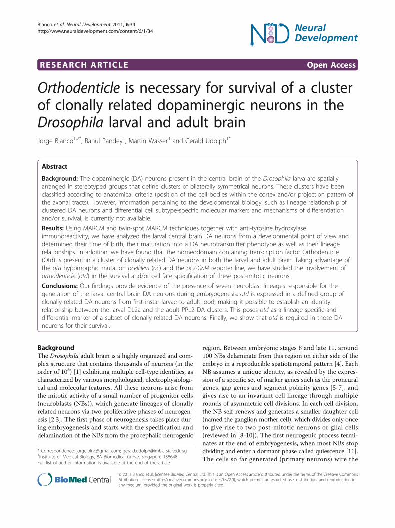
DiscussionSeven neuroblasts generate the larval central braindopaminergic neuronsThe Drosophila larval central brain contains 21 DA neu-rons per hemisphere during L3, which express the celltype-specific marker gene TH [21,22]. Different methodshave been proposed to classify and annotate these neu-rons according to anatomical criteria (position of the cellbodies within the cortex and/or projection pattern of theaxonal tracts) [23-25]. In this paper, we have analyzedthese neurons from a developmental point of view andclassified them according to their lineage relationship(Figure 6). The MARCM technique is a powerful tool tostudy lineage progression and cellular pedigrees duringDrosophila brain development [30]. It allows the labelingof progenitor cells and their offspring at different timesduring development, depending on the timing of a heat-shock-induced flippase-mediated mitotic recombinationevent [31]. Using this technique, we have shown that thelarval central brain DA neurons are primary neuronsborn during early embryogenesis. However, when analyz-ing the lineage relationship among these neurons, twomajor problems were encountered. Firstly, implicit in thetechnique is the fact that a labeled NB clone is accompa-nied by a non-labeled twin clone (two post-mitotic cellsderive from the first ganglion mother cell born just afterthe mitotic recombination event). The exclusion of twocells from the lineage analysis is negligible when larvallineages are analyzed (the average size of a standard larvallineage at L3 is 120 cells [32]). However, embryoniclineages are small (on average between 10 and 20 cells atthe end of embryogenesis [3]) and the exclusion of twocells can be significant. Secondly, MARCM-labeled NBclones induced during early embryogenesis can only bevisualized with a considerable delay after their generation(from L2 onwards) due to the persistence of the Gal80repressor protein [30]. These two problems have recentlybeen circumvented by the development of the twin-spotMARCM technique [33]. This technique not only allowsthe visualization of cell clones earlier in development butalso differentially labels the NB clone and the twin clone;thus, the study of the entire NB lineage is now possible.Using this technique, we have analyzed the lineage rela-tionship among the DA neurons present in the Droso-phila central brain during larval development. We foundthat seven NB lineages generate the 21 DA neurons pre-sent in the larval central brain (Figures 2 and 6B; Addi-tional file 1): DM1a (one DA neuron), DM1b (three DAneurons), DM2 (four DA neurons), DL1a (six DA neu-rons), DL1b (one DA neuron), DL2a (four DA neurons)
and DL2b (two neurons). At large, the lineage analysesagree with the clustering of DA neurons according toanatomical criteria, supporting the general assumptionthat cell bodies arrangement and axonal projection pat-terns are reliable ways to classify neurons in Drosophila.Just in the case of the DL1 cell cluster was a discrepancyfound. The cell bodies of the seven DL1 DA neurons arecompactly arranged in a cell cluster that occupies medial-lateral positions in the L3 central brain and their neuritesdisplay similar projection patterns (Figure 1E). Yet, sixDL1 DA neurons are clonally related (DL1a NB lineage;Figure 2C,C’) and the remaining DL1 DA neuron (redasterisk in Figure 2C2) is generated by an additional NB(DL1b NB lineage). For future functional studies, it wouldbe interesting to find molecular markers differentiallylabeling these two populations of DL1 DA neurons.
Otd acts as a survival factor in DL2a dopaminergicneuronsMost studies involving the homeodomain transcriptionfactor Otd in central nervous system development in Dro-sophila have dealt with its role in the specification andproliferation of progenitor cells during early neurogenesis[5,19], whereas a possible function in post-mitotic neuronshas been largely overlooked. Our observation that otd isexpressed in the DL2a DA neurons during larval develop-ment prompted us to investigate its role in the specifica-tion and/or survival of this DA cell cluster. According toanti-TH labeling, DL2a DA neurons mature mainly duringearly L1. Thus, null otd alleles, which are embryonic lethal,could not be used in our analysis. Therefore, we investi-gated the hypomorphic otd allele oc. We found that in ocmutant hemizygous larvae, otd expression in dorsolateralregions of the central brain was reduced and, as a conse-quence, only one of the four DL2a DA neurons showedanti-TH labeling during L3. The failure to detect three ofthe four DA neurons can be due to a defect in the regula-tion of TH expression or to the loss of DA neurons per se.Several lines of evidence support the latter hypothesis.Firstly, a general regulator of TH expression would beexpected to be present in all or most of the central brainDA neurons; yet, otd expression during larval developmentis restricted to the DL2a DA cell cluster. Secondly, misex-pression of otd in randomly induced cell clones in the cen-tral brain during larval development does not result inectopic TH-expressing DA neurons (data not shown).Thirdly, labeling of DL2a DA neurons with the oc2-gal4driver shows that reporter gene expression is also abol-ished in oc mutant hemizygous larvae during L3. The oc2enhancer has been shown to be positively regulated by otdduring ocelli development [28] and might not, therefore,be suitable to label DL2a DA neurons in an otd-indepen-dent way. However, a minimal version of this enhancerharboring the characterized Otd binding site (oc7) was
Blanco et al. Neural Development 2011, 6:34http://www.neuraldevelopment.com/content/6/1/34
Page 10 of 14
DM1a
DM1b
DM2
DL1
DL2a (Otd+)
DL2b
Early L1 Late L3 Adult
DM1b
DM2
DL1a
DL1b
DL2a
DM1a 0
DL2b
Lineage
1
A
B
?
3 3 ?
4 4 ?
6 6 ?
1 1 ?
3 4 7 (PPL2)
2 2 ?
Figure 6 Summary of the classification of dopaminergic neurons present in the larval central brain at L3, according to anatomical andlineage relationship criteria. (A) A cartoon depicting a dorsal view of a L3 larva central brain showing cell body locations and neuriteprojection patterns of the different types of DA neurons. For simplicity, just one neuron per cluster and per brain hemisphere is shown. Thecartoon does not pretend to precisely reproduce the innervation pattern of every type of DA neuron, but just gives a general description. (B)Table showing the distribution of DA neurons in different NB lineages at different times during development. The lineage harboring Otd+ DAneurons is highlighted in blue. DA, dopaminergic; DL, dorso lateral; DM, dorso medial; L, instar larval stage; NB, neuroblast; PPL, protocerebralposterior lateral.
Blanco et al. Neural Development 2011, 6:34http://www.neuraldevelopment.com/content/6/1/34
Page 11 of 14

active in the ocelli primordium [28], but did not showenhancer activity in DL2a DA neurons during larval devel-opment (data not shown). This indicates that the oc2enhancer is differentially regulated in the ocelli primor-dium and in DA neurons during development and, hence,the oc2-gal4 driver may be used to label DL2a DA neuronsin an otd-independent fashion.Taken together, our observations support the hypoth-
esis that otd expression is required for survival of DL2aDA neurons during larval development.
DL2a dopaminergic neurons survive into adulthood andparticipate in the PPL2 dopaminergic cell clusterThe wild-type Drosophila adult brain is populated byabout 200 DA neurons distributed in several bilaterallysymmetric clusters [23,24,29]. The PPL2 cluster containsseven cells that express otd and five of them also show oc2enhancer activity in young adult brains (Figure 4A1-A4).Similarly to the larval brain, otd expression in PPL2 DAneurons seems to be necessary for their survival, sinceneither anti-TH immunoreactivity nor transcriptionalactivity of the oc2 enhancer is detected in oc mutant adultbrains (Figure 4B and data not shown). Moreover, theeffects of targeted depletion of Otd in PPL2 DA neurons(loss of cell viability and/or TH expression; Figure 4C) canbe rescued by the simultaneous expression of the anti-apoptotic gene P35, pointing out a role in cell survival asthe main function of otd in PPL2 DA neurons. Altogether,the simplest interpretation for these results would be thatotd expression labels homologous DA neuron populationsin both the larval (DL2a cell cluster) and adult (PPL2 cellcluster) brains and, hence, both clusters contain the sameDA neurons. The discrepancy in cell number betweenboth clusters of DA neurons can be interpreted by analyz-ing the NB lineage responsible for the generation of theDL2a DA neurons. At L3, this lineage contains seven otdexpressing cells, four of them are primary neurons thathave already undergone maturation and express TH. Theother three cells might represent immature secondaryneurons that differentiate during pupal stages to give riseto the additional three DA neurons present in the adultPPL2 cluster. The distinction between early-differentiating(four cells) and late-differentiating (three cells) PPL2 DAneurons finds support in the targeted depletion of Otd inDA neurons by RNAi. Expression of an otd-specific RNAiconstruct in DA neurons (using the TH-Gal4 driver) hasno effect on the larval brain (data not shown), but impairsthe viability of four PPL2 DA neurons in the adult brain.Since these four cells differentiate during larval develop-ment, the RNAi machinery would have more time tocompletely deplete Otd than in the case of the late differ-entiating DA neurons. Further support for this interpreta-tion also comes from the analysis in the adult brain of
wild-type twin-spot MARCM cell clones induced duringearly L1. According to this analysis, at least two PPL2 DAneurons in the adult brain are secondary neurons, whereasthe third DA neuron might represent an undifferentiatedprimary neuron that only matures during pupaldevelopment.Recently, the expression of Otx2, an otd ortholog, in
DA neurons in the mouse adult brain has also beenreported [34]. It is selectively expressed in the centralDA neurons of the ventral tegmental area, where it iscell autonomously required to antagonize identity fea-tures of the dorsal-lateral ventral tegmental area DAneurons [35]. Thus, contrary to Drosophila, depletion ofOtx2 in these DA neurons does not induce cell death,but it changes neuron subtype identity. Interestingly,otx2 expression in these DA neurons has been asso-ciated with their reduced vulnerability to Parkinsonianneurodegeneration [35].Finally, in oc mutant adult flies most of the protocereb-
ral bridge, a neuropile structure that is part of the centralcomplex, is also missing [20]. In several behavioral para-digms, these mutant flies walk slowly and show alteredorientation behavior toward visual objects [36,37]. It hasbeen recently proposed that the protocerebral bridge isan essential part of a visual targeting network that trans-mits directional clues to the motor output [37]. Thus,with regards to the data presented here, it would beinteresting to analyze whether the lack of PPL2 DA neu-rons in oc mutant adult flies contributes to the behavioralphenotypes observed in these mutant flies.
ConclusionsUsing MARCM and twin-spot MARCM techniquestogether with anti-TH immunoreactivity, we have classi-fied the 21 DA neurons present in the Drosophila larvalcentral brain into seven clusters of clonally related DAneurons. The homeobox gene otd is specificallyexpressed in DA neurons belonging to one of theseclusters (DL2a cluster); thus, otd expression differentiallylabels a subset of DA neurons. Furthermore, by takingadvantage of an otd hypomorphic mutation and the oc2-Gal4 reporter line, we have established a cell lineagerelationship between the larval DL2a and the adult PPL2DA cell clusters. We also studied the role of otd in thesurvival and/or cell fate specification of these post-mito-tic neurons. Contrary to mice, where Otx2 expression inDA neurons of the adult brain is necessary for neuronsubtype identity, otd is required in the Drosophila larvaland adult brain for survival of DL2a and PPL2 DA neu-rons. These findings suggest that otd acts as a post-mitotic selector gene whose differential expressionamong DA neurons might help to establish functionaldifferences.
Blanco et al. Neural Development 2011, 6:34http://www.neuraldevelopment.com/content/6/1/34
Page 12 of 14

Materials and methodsFly strains, clonal analysis and RNAi experimentsFlies were reared on standard medium at 25°C. The fol-lowing transgene and reporter lines were used: UAS-P35(Bloomington Drosophila Stock Center, Bloomington,Indiana, USA), UAS-otd (J Blanco, unpublished), oc2-gal4[28], TH-gal4 [24]. Mutant alleles used in this study: ocga1,otdYH13 [38].Mitotic clones were generated and positively labeled
(with membrane tethered CD8::GFP and CD2::RFP)according to the MARCM [30] and twin-spot MARCM[33] techniques. Unless indicated, recombination wasinduced 3 to 7 hours AEL by a one hour heat shock at37°C and the larvae were dissected 21 hours later (earlyL1) or 110 hours later (L3). Genotypes of the analyzed lar-vae were as follows: otdYH13 MARCM clones, w otdYH13
FRT19A/w hs-FLP tubP-GAL80LL1 FRT19A; tubP-GAL4UAS-mCD8::GFPLL5/+;wild-type MARCM clones, y w hs-FLP/+; FRT82/
FRT82 tubP-GAL80LL10; tubP-GAL4LL7 (or TH-GAL4)UAS-mCD8::GFPLL6/UAS-mCD8::GFPLL6; wild-typetwin-spot MARCM clones, y w hs-FLP/+; FRT40A UAS-mCD8::GFP UAS-CD2-Mir/FRT40A UAS-rCD2::RFPUAS-GFP-Mir; tubP-GAL4LL7 (or TH-GAL4)/+.Depletion of Otd by RNAi was carried out by targeted
expression of an otd-specific RNAi construct (VDRC-105764) in DA neurons using the TH-gal4 driver. Toincrease knockdown efficiency, the experiment was doneat 29°C in otdYH13 heterozygous flies.
ImmunohistochemistryAntibody staining on brains was performed as previouslydescribed [39]. Primary antibodies were as follows: rabbitanti-Otd (1:250) [40], mouse anti-TH (1:100; Chemicon,Millipore AG, Temecula, California, USA), rabbit anti-TH(1:250) [41], rabbit anti-RFP (1:100; Abcam, Cambridge,UK). Secondary antibodies were Alexa488-, Alexa568- andAlexa647-conjugated antibodies generated in goat (1:200;Molecular Probes, Invitrogen, Paisley, Renfrewshire, UK).Fluorescent images were captured with an OlympusFV1000 confocal laser scanning microscope and analyzedin ImageJ [42]. Unless otherwise indicated, pictures corre-spond to single optical sections (1 μm thick). Figures wereassembled using Adobe Illustrator and Photoshop.
Additional material
Additional file 1: Supplementary Figure 1 - MARCM lineage analysisof late differentiating dopaminergic neurons in the larval centralbrain at L3. MARCM-labeled wild-type NB clones were induced duringearly embryogenesis (3 to 7 h AEL) and analyzed at L3. To circumventthe down-regulation of the tubulin promoter, an additional copy of theUAS-CD8::GFP transgene was included in the genotype of the analyzedlarvae. (A-C) The DM1a DA neuron belongs to a NB lineage independentof the DM1b DA cell lineage. (D,E) The late differentiating DL2 DA
neuron belongs to the DL2a DA cell lineage. Scale bars: 10 μm. Allpanels correspond to Z projections of individual confocal opticalsections. AEL, after egg laying; DA, dopaminergic; DL, dorso lateral; DM,dorso medial; GFP, green fluorescent protein; MARCM, mosaic analysiswith a repressible cell marker; NB, neuroblast.
AbbreviationsAEL: after egg laying; DA, dopaminergic; DL: dorso lateral; DM: dorso medial;FRT: flippase recognition target; GFP: green fluorescent protein; L: instarlarval stage; MARCM: mosaic analysis with a repressible cell marker; NB:neuroblast; oc: ocelliless; otd: orthodenticle; PPL: protocerebral posterior lateral;RFP: red fluorescent protein; RNAi: RNA interference; TH: tyrosinehydroxylase.
AcknowledgementsWe acknowledge B Bello, C Desplan, T Lee, B Lu, U Walldorf and theBloomington Stock Center for kindly providing fly strains and reagents. Thiswork was supported by the Joint Singapore Bioimaging Consortium (SBIC)-Singapore Stem Cell Consortium (SSCC).
Author details1Institute of Medical Biology, 8A Biomedical Grove, Singapore 138648.2Institute of Biotechnology, University of Helsinki, Viikinkaari 1, PO Box 65,Fin-00014 Finland. 3Bioinformatics Institute, 30 Biopolis Street, #07-01 Matrix,Singapore 138671.
Authors’ contributionsJB and RP carried out all the experiments. JB, MW and GU conceptualizedthe project. JB and GU wrote the manuscript. All authors read and approvedthe final manuscript.
Competing interestsThe authors declare that they have no competing interests.
Received: 1 June 2011 Accepted: 14 October 2011Published: 14 October 2011
References1. Ito K, Awasaki T: Clonal unit architecture of the adult fly brain. Adv Exp
Med Biol 2008, 628:137-158.2. Urbach R, Technau GM: Neuroblast formation and patterning during early
brain development in Drosophila. Bioessays 2004, 26:739-751.3. Hartenstein V, Spindler S, Pereanu W, Fung S: The development of the
Drosophila larval brain. Adv Exp Med Biol 2008, 628:1-31.4. Urbach R, Schnabel R, Technau GM: The pattern of neuroblast formation,
mitotic domains and proneural gene expression during early braindevelopment in Drosophila. Development 2003, 130:3589-3606.
5. Urbach R, Technau GM: Molecular markers for identified neuroblasts inthe developing brain of Drosophila. Development 2003, 130:3621-3637.
6. Urbach R, Technau GM: Segment polarity and D/V patterning geneexpression reveals segmental organization of the Drosophila brain.Development 2003, 130:3607-3620.
7. Technau GM, Berger C, Urbach R: Generation of cell diversity andsegmental pattern in the embryonic central nervous system ofDrosophila. Dev Dyn 2006, 235:861-869.
8. Egger B, Chell JM, Brand AH: Insights into neural stem cell biology fromflies. Philos Trans R Soc Lond B Biol Sci 2008, 363:39-56.
9. Knoblich JA: Mechanisms of asymmetric stem cell division. Cell 2008,132:583-597.
10. Doe CQ: Neural stem cells: balancing self-renewal with differentiation.Development 2008, 135:1575-1587.
11. Truman JW, Bate M: Spatial and temporal patterns of neurogenesis in thecentral nervous system of Drosophila melanogaster. Dev Biol 1988,125:145-157.
12. Truman JW: Metamorphosis of the central nervous system of Drosophila.J Neurobiol 1990, 21:1072-1084.
13. Truman JW: Steroid receptors and nervous system metamorphosis ininsects. Dev Neurosci 1996, 18:87-101.
Blanco et al. Neural Development 2011, 6:34http://www.neuraldevelopment.com/content/6/1/34
Page 13 of 14

14. Prokop A, Technau GM: The origin of postembryonic neuroblasts in theventral nerve cord of Drosophila melanogaster. Development 1991,111:79-88.
15. Skeath JB, Thor S: Genetic control of Drosophila nerve cord development.Curr Opin Neurobiol 2003, 13:8-15.
16. Kao CF, Lee T: Birth time/order-dependent neuron type specification. CurrOpin Neurobiol 2010, 20:14-21.
17. Jacob J, Maurange C, Gould AP: Temporal control of neuronal diversity:common regulatory principles in insects and vertebrates? Development2008, 135:3481-3489.
18. Finkelstein R, Smouse D, Capaci TM, Spradling AC, Perrimon N: Theorthodenticle gene encodes a novel homeo domain protein involved inthe development of the Drosophila nervous system and ocellar visualstructures. Genes Dev 1990, 4:1516-1527.
19. Younossi-Hartenstein A, Green P, Liaw GJ, Rudolph K, Lengyel J,Hartenstein V: Control of early neurogenesis of the Drosophila brain bythe head gap genes tll, otd, ems, and btd. Dev Biol 1997, 182:270-83.
20. Hirth F, Therianos S, Loop T, Gehring WJ, Reichert H, Furukubo-Tokunaga K:Developmental defects in brain segmentation caused by mutations ofthe homeobox genes orthodenticle and empty spiracles in Drosophila.Neuron 1995, 15:769-778.
21. Budnick V, White K: Catecholamine-containing neurons in Drosophilamelanogaster: distribution and development. J Comp Neurol 1988,268:400-413.
22. Nässel DR, Elekes K: Aminergic neurons in the brain of blowflies andDrosophila dopamine- and tyrosine hydroxylase-immunoreactiveneurons and their relationship with putative histaminergic neurons. CellTissue Res 1992, 267:147-167.
23. Monastirioti M: Biogenic amine systems in the fruit fly Drosophilamelanogaster. Microsc Res Tech 1999, 45:106-121.
24. Friggi-Grelin F, Coulom H, Meller M, Gomez D, Hirsh J, Birman S: Targetedgene expression in Drosophila dopaminergic cells using regulatorysequences from tyrosine hydroxylase. J Neurobiol 2003, 54:618-627.
25. Selcho M, Pauls D, Han KA, Stocker RF, Thum AS: The role of dopamine inDrosophila larval classical olfactory conditioning. PLoS ONE 2009, 4:e5897.
26. Truman JW, Schuppe H, Sheperd D, Williams DW: Developmentalarchitecture of adult-specific lineages in the ventral CNS of Drosophila.Development 2004, 131:5167-5184.
27. Pereanu W, Hartenstein V: Neural lineages of the Drosophila brain: athree-dimensional digital atlas of the pattern of lineage location andprojection at the late larval stage. J Neurosci 2006, 26:5534-5553.
28. Blanco J, Seimiya M, Pauli T, Reichert H, Gehring WJ: Wingless andHedgehog signaling pathways regulate orthodenticle and eyes absentduring ocelli development in Drosophila. Dev Biol 2009, 329:104-115.
29. Mao Y, Davis RL: Eight different types of dopaminergic neurons innervatethe Drosophila mushroom body neuropil: anatomical and physiologicalheterogeneity. Front Neural Circuits 2009, 3:5.
30. Lee T, Luo L: Mosaic analysis with a repressible cell marker for studies ofgene function in neuronal morphogenesis. Neuron 1999, 22:451-461.
31. Wu JS, Luo L: A protocol for mosaic analysis with a repressible cellmarker (MARCM) in Drosophila. Nat Protoc 2006, 1:2583-2589.
32. Bello BC, Izergina N, Caussinus E, Reichert H: Amplification of neural stemcell proliferation by intermediate progenitor cells in Drosophila braindevelopment. Neural Dev 2008, 3:5.
33. Yu HH, Chen CH, Shi L, Huang Y, Lee T: Twin-spot MARCM to reveal thedevelopmental origin and identity of neurons. Nat Neurosci 2009,12:947-953.
34. Di Salvio M, Di Giovannantonio LG, Omodei D, Acampora D, Simeone A:Otx2 expression is restricted to dopaminergic neurons of the ventraltegmental area in the adult brain. Int J Dev Biol 2010, 54:939-945.
35. Di Salvio M, Di Giovannantonio LG, Acampora D, Prosperi R, Omodei D,Prakash N, Wurst W, Simeone A: Otx2 controls neuron subtype identity inventral tegmental area and antagonizes vulnerability to MPTP. NatNeurosci 2010, 13:1481-1488.
36. Strauss R: The central complex and the genetic dissection of locomotorbehaviour. Curr Opin Neurobiol 2002, 12:633-638.
37. Triphan T, Poeck B, Neuser K, Strauss R: Visual targeting of motor actionsin climbing Drosophila. Curr Biol 2010, 20:663-668.
38. FlyBase. [http://flybase.bio.indiana.edu].
39. Bello B, Holbro N, Reichert H: Polycomb group genes are required forneural stem cell survival in postembryonic neurogenesis of Drosophila.Development 2007, 134:1091-1099.
40. Hirth F, Kammermeier L, Frei E, Walldorf U, Noll M, Reichert H: Anurbilaterian origin of the tripartite brain: developmental genetic insightsfrom Drosophila. Development 2003, 130:2365-2373.
41. Yang Y, Gehrke S, Imai Y, Huang Z, Ouyang Y, Wang JW, Yang L, Beal MF,Vogel H, Lu B: Mitochondrial pathology and muscle and dopaminergicneuron degeneration caused by inactivation of Drosophila Pink1 isrescued by Parkin. Proc Natl Acad Sci USA 2006, 103:10793-10798.
42. Image Processing and Analysis in Java. [http://rsbweb.nih.gov/ij/].
doi:10.1186/1749-8104-6-34Cite this article as: Blanco et al.: Orthodenticle is necessary for survivalof a cluster of clonally related dopaminergic neurons in the Drosophilalarval and adult brain. Neural Development 2011 6:34.
Submit your next manuscript to BioMed Centraland take full advantage of:
• Convenient online submission
• Thorough peer review
• No space constraints or color figure charges
• Immediate publication on acceptance
• Inclusion in PubMed, CAS, Scopus and Google Scholar
• Research which is freely available for redistribution
Submit your manuscript at www.biomedcentral.com/submit
Blanco et al. Neural Development 2011, 6:34http://www.neuraldevelopment.com/content/6/1/34
Page 14 of 14


![[18F]Fluorodopa PETshows striatal dopaminergic dysfunction](https://img.pdfslide.us/doc/110x75/628e71a806be7c7a267428b6/18ffluorodopa-petshows-striatal-dopaminergic-dysfunction-.jpg)