Embed Size (px)
Citation preview
Orofacial trauma in child abuse: types, prevalence, management, and the dental profession’s involvement
Howard L. Needleman, DMD
Abstract This article: (1) reviews the available data describing
and documenting the types and prevalence of orofacial injuries in physically abused children; (2) reviews surveys indicating the dental profession’s awareness of, experience with, and reporting of child abuse; and (3) recommends treatment for those injuries.
Studies demonstrate that: (1) trauma to the head and associated areas occurs in approximately 50% of the cases of physical abuse to children; (2) soft tissue injuries - most frequently bruises - are the most common injury sustained to the head and face and are the single most common inju y sustained in child abuse; and (3) injuries to the upper lip and maxilla y labial frenum may be a characteristic lesion in the severely abused young child.
Surveys of the dental profession clearly demonstrate that dentists: (1) do see suspicious cases of child abuse; (2) often fail to report their suspicions as is legally required; (3) do not have adequate training or knowledge of child abuse and neglect; and (4) report more cases if made aware of child abuse and neglect and their responsibility to report.
T h e physical abuse of children is a problem not limited to the medical or social service professions. Our profession has become increasingly aware of its
role in the detection, reporting, and treatment of the abused child. Numerous editorials have appeared in the dental literature alerting us to our moral and legal responsibility as health professionals in the recogni- tion of child abuse.l-I3 in addition, many articles have appeared reviewing child abuse in general and dis- cussing the dentist’s role.1650 Several of these articles have appeared in state dental journals listing their particular state laws, reporting agencies, and hotline numbers.
The purposes of this paper are to: (1) review the available data describing and documenting the types and prevalence of orofacial injuries in physically abused children; (2) review surveys indicating the dental profession’s awareness of, experience with, and reporting of child abuse; and (3) recommend treat- ment for those injuries.
Types and Prevalence of Orofacial Injuries in Child Abuse
It is a common finding that when an individual is attacked for whatever reason, the head and/or facial areas often are involved. This is because these areas are exposed and accessible and because the head often is considered representative of the whole being or
PEDIATRIC DENTISTRY: May 1986Nol. 8 Special Issue 1 71
elf.''^^,^^ It is therefore not suprising that physical abuse of children often involves the head and facial areas.
In 1946 C a f f e ~ ~ ~ described 6 infants suffering from chronic subdural hematomas who presented with multiple fractures in their long bones. It was in this classic article that the abused child and some of the common characteristics of abuse first were described. In 3 of the 6 cases, orofacial injuries were noted. One child presented with swollen and hemorrhagic gums, petechiae in the oral mucosa and ecchymosis of the face. The other 2 children both exhibited bruises of the face.
The term ”battered child syndrome” was coined by Dr. Henry Kempe in his milestone article in 1962.53 According to Kempe, this syndrome should be con- sidered in any child exhibiting evidence of any bone fracture, subdural hematoma, failure to thrive, soft tissue swelling, or skin bruising.
The first study to examine the types of injuries sus- tained in the physically abused child was published in 1966 by Cameron et al.54 This study examined the autopsy findings of 29 fatal cases of abuse seen over a 2-year period in the Department of Forensic Med- icine at The London Hospital Medical College. Half of the children studied (mean age 14.3 months), had obvious bruises of the head, face, and neck and all exhibited soft tissue injuries. The prevalence and lo- cation of these injuries were as follows: 79%, scalp; 59%, neck; 52%, forehead; 49%, cheek; 48%, lower jaw and right leg; and 45%, upper lip region. Of the 13 areas described as sustaining soft tissue trauma, the head and neck areas were among the most fre- quently described. Lesions to the jaw and neck were well circumscribed and of a “finger-tip” character suggestive of gripping.
Lacerations of the mucosa of the inner aspect of the upper lip near the frenum and/or the occasional tearing of the lip from the alveolar margin of the gums occurred in 45% of Cameron’s cases. The age of the child is significant in this type of injury. A frenum tear is not uncommon in the young child who acci- dentally falls while learning to walk (generally be- tween 6 months and l 1/2 years). However, a frenum tear in a very young, nonambulatory patient (< 1 year), or an older, more stable child (> 2 years) should arouse one’s suspicion as to the possibility of this injury being nonaccidental. This type of injury may be the result of a blow to the mouth, an effort to silence a screaming child, or having forced a spoon or bottle into a baby’s mouth by an angry adult who is frustrated with a slow eater. It is based on this report alone that the torn frenum injury has been purported to be pathognomonic of child abuse in the dental literature. In no other study is such a high frequency reported.
Cameron et al. also state that bruises of the cheeks and sides of the head suggest blows or slaps with a fist or open hand. If the lesions are more localized and have underlying severe injuries, they may rep- resent a severe blow or impact with a hard object.
The following year a British study by Skinner and Castle (1967) was published documenting the injuries to 78 abused children requiring medical attention.55 Of these children, at least 34 (43.5%) sustained trauma to the face and mouth. This may in fact be an un- derestimate since some of the bruises were reported without location noted. The majority of the injuries were bruises, but also included lacerations, bites, and abrasions. Soft tissue injuries occurred in 77% of the cases, head injuries (skull fractures, subdural hema- tomas) in 28%, and bony injuries (fractures and dis- locations) in 46%. Bums were the single most common injury in this study, occurring in 56 (72%) of the cases.
O’Neill et al. (1973) studied 110 cases of child abuse brought to the hospital over a 5-year period.51 Their study population included a large number of infants and children with head injuries. Thirty-two (29%) of the children had some form of intracranial injury, 15 (14%) had skull fractures and 23 (21%) were coma- tose. Almost all of the children had some degree of soft tissue injury and 55 (50%) were admitted pri- marily because of the soft tissue trauma.
Baetz et al. (1977) examined the records of 58 cases of battered children with 87 injuries occurring over a 4-year period.56 Twenty-five (43%) of these children had head injuries of the following types (decreasing prevalence): hematomas, lacerations and swelling, and skull fractures. Of the injuries, bruises were the most common (33%) followed by fractures and joint inju- ries (25%).
The largest and most detailed study examining the types of injuries that children sustain when being abused was reported by Becker et al. (1978).57 The medical records of 260 cases of child abuse admitted to The Children’s Hospital in Boston between 1970 and 1975 were reviewed. One hundred and twenty- eight (49%) of the patients had facial and/or intraoral trauma. An additional 16% of the children had inju- ries to the head, such as skull fractures, subdural hematomas, contusions, and lacerations of the scalp. This brought the total of head/face/intraoral injuries to 65% of the abused children in the study. Of the 386 injuries sustained by the 260 children (Table l), 33% were to the head, 61% to the face (contusions, ecchymoses, abrasions, lacerations, fractures, burns, and bites) and 6% to intraoral structures. The single most common type of injury was a facial contusion which occurred twice as often as the second most common injury, contusions to the body or organs. In 45% of the cases, the head injuries were severe enough to be the reason for admission to the hospital.
72 OROFACIAL TRAUMA IN CHILD ABUSE: Needleman

TABLE 1. Types and Locations of Injuries in 260 Abused Children
Abrasions Contusions Subdural and and Dental
Location Fractures Hematoma Lacerations Ecchymoses Burns Bites Trauma Miscellaneous Total
Head (scalp) 33 23 6 14 0 0 0 1 77 Face 3 0 40 96 4 2 0 0 145 Intraoral 0 0 4 6 0 0 4 0 14 Body (organ) 38 0 20 52 25 6 0 9 150
Reprinted from Becker DB, Needleman HL, Kotelchuck M: Child abuse and dentistry; orofacial trauma and its recognition by dentists. J Am Dent Assoc 97:24-28, 447, 1978.
Malcez (1979) cited 25 cases of suspected abuse re- ported by pediatric dentists.58 The principal dental injuries reported in these cases were: fractured teeth, 32%; oral lacerations, 14%; fractures of the maxilla or mandible, 11%; and oral burns, 5%.
The literature contains numerous reports and ar- ticles documenting bite marks in child abuse and the role the dental profession plays in using this infor- mation. This topic is dealt with in detail by Dr. Wag- ner in this issue. Bite marks can help to identify the abuser.
In summary, studies demonstrate that: (1) trauma to the head and associated areas occurs in approxi- mately 50% of the cases of physically abused chil- dren; (2) soft tissue injuries - most frequently bruises - are the most common injury sustained to the head and face and are the single most common injury sus- tained in child abuse; and (3) injuries to the upper lip and maxillary labial frenum may be a characteristic lesion in the severely abused young child.
These findings make it obvious that dentists are in a position to detect child abuse. National figures in- dicate that as many as 1 million (625,000 substanti- ated) children are abused and/or neglected annually and of these about 1000 die each year.59 If we assume that half of these cases involve trauma to the head, as is indicated in the literature, our profession is def- initely in a position to detect substantial numbers of abused children. In this way we can help to prevent further trauma to the children by bringing help to these troubled families.
The Dentist’s Involvement in Reporting Child Abuse
There are numerous reports of abused children in the dental literature in which the dentist was the professional who initially suspected that injuries in- volving the orofacial structure were the result of physical Such children first were treated and subsequently reported to appropriate agencies. Most of the cases cited were instances of severe child abuse resulting in hospital admission or death and involving the head and orofacial areas. It can be as-
sumed, then, that less severe abuse cases may be appearing in medical and/or dental office settings. One may hypothesize that these cases of abuse re- sulting in less serious injuries may go undetected by the dentist or physician due to lack of suspicion and/ or knowledge of child abuse and neglect. Several studies have been published which address the is- sues of how frequently dentists actually are involved in child abuse reporting and the extent of their knowledge of child abuse and
The first evidence of a lack of reporting of child abuse by dentists appeared in the Journal of the American Dental Association in 1967.69 In this article, reports of child abuse in New York and Illinois were documented. During 1966 in New York, 416 cases of suspected child abuse were reported: 85% of these reports came from hospitals, 12% from physicians, and no reports came from dentists. Illinois records indicate that 934 reports of child abuse were received between 1965 and 1967, only 1 of which was from a dentist.
It was not until 1978 that a large-scale study inves- tigating the dentist’s involvement in child abuse re- porting was completed. Becker et al. (1978) sent questionnaires to all pediatric dentists, all oral sur- geons, and one-third of all general dentists in the Commonwealth of Massach~set ts .~~ Based on 537 re- sponses, the following observations were made (Ta- ble 2).
1.
2.
3.
4.
5.
Eight per cent of all dentists responding saw sus- pected cases of child abuse (22%, oral surgeons; 18%, pediatric dentists). Of the 22 suspected cases of child abuse seen, only 4 cases actually were reported. The main reason cited for nonreporting was that it was difficult to confirm these suspicions. Only 45% of dentists were aware of their legal responsibility to report instances of suspected chdd abuse (77%, pediatric dentists; 62%, oral sur- geons). Only 28% of dentists knew the name of the agency to which to report these cases. Although oral surgeons and pediatric dentists rep-
PEDIATRIC DENTISTRY: May 1986Nol. 8 Special Issue 1 73
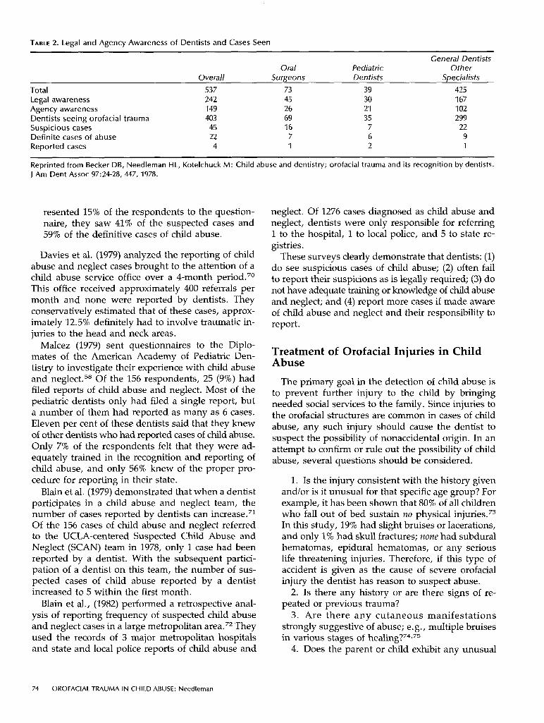
TABLE 2. Legal and Agency Awareness of Dentists and Cases Seen
Total Legal awareness Agency awareness Dentists seeing orofacial trauma Suspicious cases Definite cases of abuse Reported cases
Oral Overall Surgeons
537 73 242 45 149 26 403 69 45 16 22 7 4 1
Pediatric Dentists
39 30 21 35 7 6 2
General Dentists Other
Specialists
425 167 1 02 299
22 9 1
Reprinted from Becker DB, Needleman HL, Kotelchuck M: Child abuse and dentistry; orofacial trauma and i ts recognition by dentists. J Am Dent Assoc 97:24-28, 447, 1978.
resented 15% of the respondents to the question- naire, they saw 41% of the suspected cases and 59% of the definitive cases of child abuse.
Davies et al. (1979) analyzed the reporting of child abuse and neglect cases brought to the attention of a child abuse service office over a 4-month period.70 This office received approximately 400 referrals per month and none were reported by dentists. They conservatively estimated that of these cases, approx- imately 12.5% definitely had to involve traumatic in- juries to the head and neck areas.
Malcez (1979) sent questionnaires to the Diplo- mates of the American Academy of Pediatric Den- tistry to investigate their experience with child abuse and neglect.58 Of the 156 respondents, 25 (9%) had filed reports of child abuse and neglect. Most of the pediatric dentists only had filed a single report, but a number of them had reported as many as 6 cases. Eleven per cent of these dentists said that they knew of other dentists who had reported cases of child abuse. Only 7% of the respondents felt that they were ad- equately trained in the recognition and reporting of child abuse, and only 56% knew of the proper pro- cedure for reporting in their state.
Blain et al. (1979) demonstrated that when a dentist participates in a child abuse and neglect team, the number of cases reported by dentists can increase.71 Of the 156 cases of child abuse and neglect referred to the UCLA-centered Suspected Child Abuse and Neglect (SCAN) team in 1978, only 1 case had been reported by a dentist. With the subsequent partici- pation of a dentist on this team, the number of sus- pected cases of child abuse reported by a dentist increased to 5 within the first month.
Blain et al., (1982) performed a retrospective anal- ysis of reporting frequency of suspected child abuse and neglect cases in a large metropolitan area.72 They used the records of 3 major metropolitan hospitals and state and local police reports of child abuse and
neglect. Of 1276 cases diagnosed as child abuse and neglect, dentists were only responsible for referring 1 to the hospital, 1 to local police, and 5 to state re- gistries.
These surveys clearly demonstrate that dentists: (1) do see suspicious cases of child abuse; (2) often fail to report their suspicions as is legally required; (3) do not have adequate training or knowledge of child abuse and neglect; and (4) report more cases if made aware of child abuse and neglect and their responsibility to report.
Treatment of Orofacial Injuries in Child Abuse
The primary goal in the detection of child abuse is to prevent further injury to the child by bringing needed social services to the family. Since injuries to the orofacial structures are common in cases of child abuse, any such injury should cause the dentist to suspect the possibility of nonaccidental origin. In an attempt to confirm or rule out the possibility of child abuse, several questions should be considered.
1. Is the injury consistent with the history given and/or is it unusual for that specific age group? For example, it has been shown that 80% of all children who fall out of bed sustain no physical injuries.73 In this study, 19% had slight bruises or lacerations, and only 1% had skull fractures; none had subdural hematomas, epidural hematomas, or any serious life threatening injuries. Therefore, if this type of accident is given as the cause of severe orofacial injury the dentist has reason to suspect abuse.
2. Is there any history or are there signs of re- peated or previous trauma?
3 . Are there any cutaneous manifestations strongly suggestive of abuse; e.g., multiple bruises in various stages of healing?74,75
4. Does the parent or child exhibit any unusual
74 OROFACIAL TRAUMA IN CHILD ABUSE: Needleman
behavior which might indicate abuse; e.g., an ex- aggerated or detached response to questioning?
5. Is there any evidence of neglect or poor su- pervision of the child?
As with any orofacial injury and especially in cases of suspected child abuse, a neurological assessment should be made initially. Croll et al. (1980) described a rapid, systematic, and meaningful neurological as- sessment for dentists which is essential in these cases.76 This assessment includes: observing the child’s com- munication and motor skills; patency of airway; ob- taining a history of any loss of consciousness, cyanosis or seizure activity; obtaining vital signs; observation for signs of rhinorrhea or otorrhea; rapid testing of the cranial nerves; and alerting the parents to the possible signs of neurological damage.
Needleman (1984) described the approaches for management of orofacial injuries in suspected cases of abuse.77 If the initial examination reveals any pos- itive signs of neurological damage or other injuries beyond the scope of the attending dentist, an appro- priate referral should be made. In instances of severe trauma to the jaws, alveoli, or intraoral soft tissues, an oral and maxillofacial surgeon is best qualified to provide treatment. Facial lacerations requiring exten- sive suturing might best be treated by a plastic sur- geon. Unfamiliar oral lesions can be referred to an oral pathologist or oral surgeon. Trauma to the body possibly involving internal organs, to the head in- volving the CNS, or to the extremities always must be evaluated further by a physician. These profes- sionals should be made aware of your suspicions so they also can be sensitive to and helpful in confirming the possibility of abuse. The initial orofacial injuries accompanying these signs and symptoms must be treated, the referrals must be made and carried out before discussing the issue of child abuse with the parents or guardian, since referral recommendations and follow up can be jeopardized if the parents or guardian feel threatened.
If the initial examination reveals trauma limited to the oral cavity and treatment is within the scope of the attending dentist, definitive treatment should commence. When treatment is completed, the dentist should discuss with the accompanying adult the treatment rendered, prognosis, necessary follow-up care and the symptoms of more serious head injuries (Appendix). Once this has been accomplished, the issue of suspected child abuse should be discussed.
Prior to rendering specific treatment modalities, some general treatment considerations should be re- viewed. Is the child old enough and/or mature enough to cooperate? Ideal treatment may have to be com- promised or modified with an unmanageable child.
Use of sedative premedication, nitrous oxide/oxygen analgesia, and/or the use of physical restraints such as a Papoose Board” may be essential for managing the very young or very apprehensive patient. Parents or guardians should not be present in the room dur- ing treatment as their presence may hinder commu- nication with the distraught child.
The attitudes of parents or caretakers toward den- tal care are sometimes a factor in treatment decisions. In cases in which child abuse is suspected, parental input might not be sought. Treatment approaches with the best prognoses should be selected since risky and/ or complex procedures can increase the chance of fail- ure and are often dependent on faithful parental fol-
Treatment considerations also should include: (1) space maintenance of primary units; (2) root devel- opment of involved teeth; (3) coronal development of the succedaneous teeth; (4) dental occlusion; and (5) medical status. Each of the variables can affect the treatment modalities selected.78
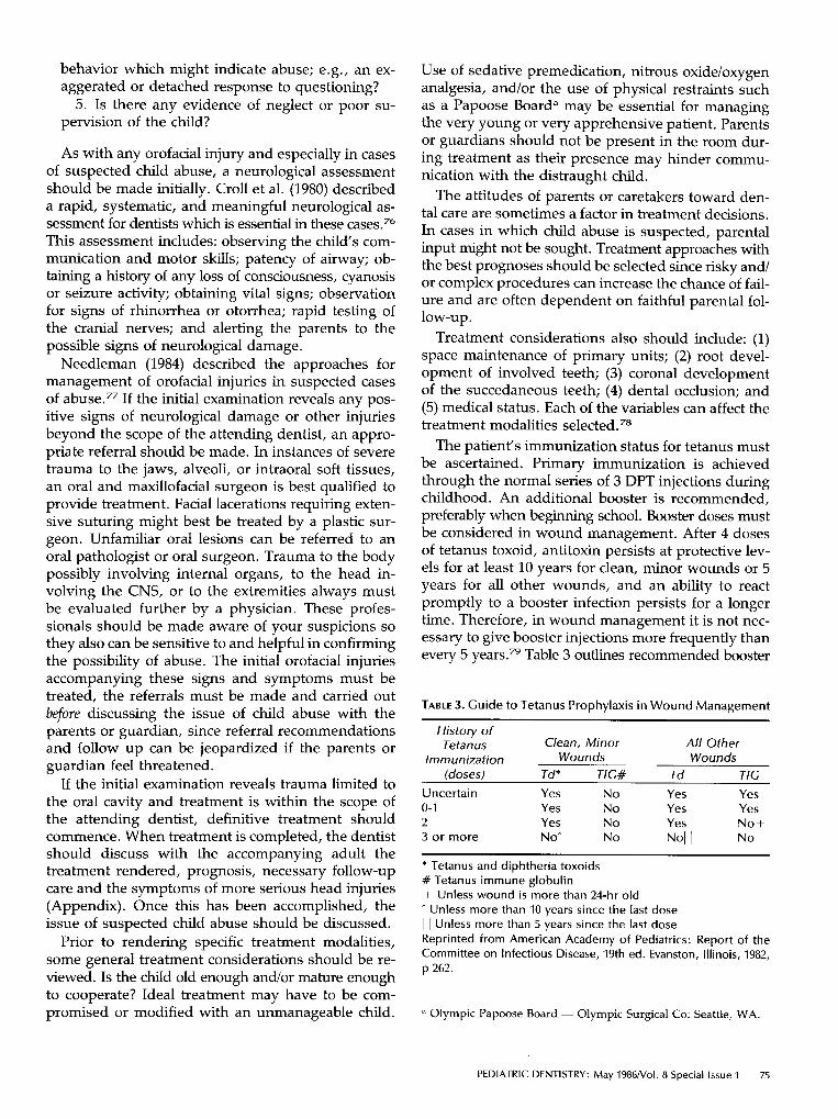
The patient’s immunization status for tetanus must be ascertained. Primary immunization is achieved through the normal series of 3 DPT injections during childhood. An additional booster is recommended, preferably when beginning school. Booster doses must be considered in wound management. After 4 doses of tetanus toxoid, antitoxin persists at protective lev- els for at least 10 years for clean, minor wounds or 5 years for all other wounds, and an ability to react promptly to a booster infection persists for a longer time. Therefore, in wound management it is not nec- essary to give booster injections more frequently than every 5 years.79 Table 3 outlines recommended booster
low-up.
TABLE 3. Guide to Tetanus Prophylaxis in Wound Management
History of A l l Other Tetanus Clean, Minor
Immunization Wounds Wounds (doses) Td* TIC# Td TIC
Uncertain Yes No Yes Yes 0-1 Yes No Yes Yes 2 Yes No Yes No + 3 o r more NoA No No1 I No
* Tetanus and diphtheria toxoids # Tetanus immune globulin + Unless wound i s more than 24-hr old
A Unless more than 10 years since the last dose I I Unless more than 5 years since the last dose Reprinted from American Academy of Pediatrics: Report of the Committee on Infectious Disease, 19th ed. Evanston, Illinois, 1982, p 262.
a Olympic Papoose Board - Olympic Surgical Co: Seattle, WA.
PEDIATRIC DENTISTRY: May 1986Nol. 8 Special Issue 1 75
doses according to immunization status and wound risk level.
Soft Tissue Injuries Contusions and ecchymoses are best treated with
ice packs to accessible areas for the first 24 hr. This results in local vasoconstriction and minimizes the flow of blood to the traumatized area. In instances of extensive swelling, pressure dressings may be help- ful. Later in the course of treatment, applications of warm packs help resolve extravasated blood in the tissues. It is important to note that these lesions are often a diagnostic sign of possible underlying or re- lated bony fractures; e.g., a contusion in the floor of the mouth often signals a fracture of the mandible.
Treatment of abrasions primarily consists of careful cleansing of the wound. Gentle irrigation of the wound with warm water and soap helps to remove dirt and foreign material. The wound then is irrigated with copious amounts of normal saline and the surround- ing skin is prepared with an antiseptic solution. If foreign material still remains, cleansing with a brush and surgical soap may be required along with the removal of individual debris with a sterile cotton tip, tissue forcep, or the tip of a #11 scalpel. Placement of a sterile dressing or gauze over the wound helps to protect the wound from further irritation and pro- motes healing. The dressing should be changed daily until the wound is healed.
In small and medium frenum tears, suturing is usually not necessary since healing will be satisfac- tory with secondary intention. However, if the wound is large, the alveolar bone is exposed, and/or the wound separates when the lip is pulled upwards, suturing is required.
It is essential to use local anesthesia in managing any lacerations in children. When possible, regional block anesthesia should be used to avoid distortion of wounds. As with abrasions, one should be alert for foreign bodies within a wound. It is not uncom- mon for tooth fragments to be present. Appropriate soft tissue radiographs should be taken to rule out this possibility when the child sustains fractured teeth with lacerations. Following debridement, the wound should be explored to delineate its anatomy and to assess any damage to nearby structures such as a parotid duct or gland, Wharton’s duct, or a facial nerve. Only obviously nonviable tissue should be excised. It is best to use 5-0 or 6-0 nylon or silk sutures for skin suturing, 3-0 or 4-0 chromic gut sutures for re- pairing the musculature such as the lip or tongue, and 4-0 or 5-0 plain gut sutures for mucosal closures. Suturing is done in layers, starting from the inside and moving to the outside. Interrupted suturing should be used, placing the sutures no farther than 2.5 mm apart. Nonresorbable sutures should be re-
moved 4 days after placement. Dry wounds can be dressed with steri-strips and draining wounds can be covered with gauze. The use of prophylactic anti- biotics is recommended and penicillin is the drug of choice.
Puncture wounds are treated similarly to lacera- tions. The wound must be cleansed, the damage as- sessed, and, if large enough, the puncture must be sutured in layers. Small punctures can be left to heal by secondary intention, especially in areas such as the palate.
Human bites must be regarded as serious injuries since devastating complications can result if proper treatment is not rendered. Frequently reported com- plications include recurrent infections, permanent joint stiffness, osteomyelitis, and digital amputations. Less frequent are extremity amputations, systemic sepsis, and death.80 The wound first should be cleansed, debrided if necessary, and then left open. Adminis- tration of tetanus toxoid is recommended according to the previously described protocol. Staphylococcus aureus is the organism most frequently encountered in human bite wound infections, although other strains have been implicated.80,81 Antibiotics recommended include cephalosporin, penicillin, or clindamycin.
Minor bite wounds can be treated as traumatic ul- cers with topical protectants. A corticosteroid such as triamcinolene, when added to the protectant can help control resultant inflammation.
Burns to the orofacial structure are not uncommon. They may be classified as electrical, thermal, or chem- ical.
The etiologic, histologic, and clinical features of electrical burns have been well reviewed.82 Minor electrical burns can be treated conservatively by re- peated applications of topical antibiotics. They heal with little deformity. Tetanus toxoid should be ad- ministered if the patient is in need of a booster.
Major electrical burns require hospitalization be- cause severe complications often occur. These in- clude fluid loss, poor nutrition, and shock, as well as secondary hemorrhage from the labial arteries that may occur 3-4 days after injury. Careful daily de- bridement is necessary to remove necrotic tissue and stimulate the formation of healthy granulation tissue. Adhesive strips can be used for good approximation of wound edges and manipulation of muscle pull. Home care should include frequent saline rinses and massaging burned tissue to increase blood supply. Many electrical burns require surgical repair, but the exact timing of the repair has been controversial. A delayed or conservative approach to the management of the acquired oral deformity now is recom- mended. 83-90 Several recently published articles rec- ommended that intraoral acrylic splints with extraoral extensions be inserted soon after the t r a ~ m a . ~ l - ~ ~ This
76 OROFACIAL TRAUMA I N CHILD ABUSE: Needleman
appliance has proven successful in minimizing the contraction of the commissure, a long-term sequela of electrical burns to the mouth.
Thermal burns involving small portions of the face should be referred to a physician. The wound usually is covered with sterile dressings and needs to be in- spected every 3-4 days for healing progress and signs of infection. Antibiotics should be applied during the healing process, but greasy ointments should be avoided. Extensive second- or third-degree burns of the face are best referred to a hospital burn center.
Ulcerations of the oral mucosa that occur as the result of thermal injuries usually heal ~neven t fu l ly .~~ The ulcerative area should be kept covered with a thick paste. Topical steroids, such as triamcinolene in an adhering paste can be helpful for healing and pain control. Complete healing of an ulcerative area caused by a thermal burn usually takes 2-3 weeks. Systemic analgesics may be helpful for 1-2 days fol- lowing the injury.
Chemical agents accidentally or intentionally placed in the mouth result in severe and acute trauma to the oral mucosa.'oo A white slough forms representing necrotic epithelium. As the slough is rubbed off, a bleeding, raw, and painful area is exposed. Caustic or corrosive burns of the mouth can result from in- gesting liquid or solid (granules or powder) forms of alkalines or acids.
Symptoms from burns of the oral cavity occur im- mediately and range from minor discomfort to severe pain. Extensive burns, however, may destroy mu- cosal nerve endings and produce anesthesia. Exces- sive salivation, drooling, and dysphagia occur due to irritation of oral and esophageal mucosa. Examina- tion of the mouth may show edema, inflammation, and/or whitish areas. The presence of oral burns only documents exposure to a caustic agent and does not predict accurately the presence or absence of esoph- ageal burns.
Immediate treatment consists of flushing the caus- tic substance off the skin or mucosa with copious amounts of water. Neutralizing the caustic agent may produce heat and is no longer recommended.
Injuries to the Dentition The pediatric dentist is well acquainted with the
management of injuries to both the primary and per- manent dentitions. In cases of suspected child abuse, follow-up dental care may not be possible due to the lack of familial compliance and/or delays in the dis- position of the case by the investigating agency. Thus, treatment of dental injuries needs to be as definitive as possible. For example, teeth with Class I1 fractures should be restored at the emergency visit to their original size and shape using the acid-etch technique and the appropriate resin material. The same ration-
ale would apply to Class 111 fractures. To decrease chances for failure and the need for careful follow up, one should consider definitive 1-stage pulp ther- apy (i.e., pulpectomy and gutta-percha obturation) for these pulpal exposures. Obviously, many other types of injuries require repeated appointments for treatment and/or observation and this must be made known to the individual who will be investigating the case.
Injuries to the Facial Bones Facial fractures are relatively uncommon in chil-
dren. They can, however, occur during physical as- sault with nasal fractures occurring most frequently (45%), followed by mandibular fractures (32%), and zygomatic maxillary complex and orbit fractures (20.5%).'01 Initial management of facial fractures re- quires attention to basic life-support means, such as airway maintenance, control of bleeding, and fluid management. Sedation of the young patient may be necessary to fully evaluate the fracture both clinically and radiographically. Temporary stabilization of the fracture is often helpful in controlling pain during acute stages. Proper bandaging (i.e., using Barton's bandage) of the facial bones can provide support and help to sedate the patient before he/she is transferred into the care of an oral and maxillofacial surgeon.
Diagnosis of mandibular fractures usually can be made by clinical examination. The fracture appears as an irregularity in the mandibular arch, with loss of proper dental occlusion, and with occasional tears in the oral mucosa. Mandibular fractures most com- monly occur in the bicuspid area. Active treatment is not necessary if there are no signs of displacement. Often there is little discomfort with these injuries. A soft or liquid diet should be prescribed for 4 weeks, allowing time for sufficient union so that the patient can eat normally. If displacement is evident and teeth are adjacent to the fracture site, interdental wire fix- ation can be undertaken. If no teeth are present, sta- bilization of mandibular fractures may be accomplished by open reduction.
Mandibular fractures also can occur in the subcon- dylar region. These often cause pain and tenderness in the TMJ region as well as considerable trismus and displacement of the lower dental arch. Active treat- ment of such fractures is seldom necessary, but re- ferral to an oral and maxillofacial surgeon is mandatory for further evaluation and follow up. A comprehen- sive discussion of the definitive management of facial fractures is presented elsewhere in the literature.lo2
Conclusions This article has shown that dentists need to be alert
to the possibility that orofacial trauma may be the
PEDIATRIC DENTISTRY: May 1986NoI. 8 Special Issue 1 77
result of child abuse. By heightening the dental profession’s awareness of this issue, child abuse de- tection will increase. This will help to insure that these troubled families will receive the appropriate social services, thus preventing further physical and psy- chological trauma to the child.
Dr. Needleman is an assistant professor, pediatric dentistry, Har- vard University, and associate dentist in chief, Children’s Hospital, Boston. Reprint requests should be sent to: Dr. Howard L. Needle- man, Children’s Hospital - Dental Department, 300 Longwood Ave., Boston, MA 02115.
1. 2.
3.
4.
5.
6.
7.
8.
9.
10. 11.
12.
13.
14. 15.
16.
17.
18.
19.
20.
21.
22.
23.
24,
25.
26
78
Child abuse reporting laws. J Am Dent Assoc 75:1070, 1967. Danielsen K Scandinavian Society of Forensic Odontology Newsletter. 6:93-98, 1972. Dentist required to report cases of abused and maltreated children. NY State Dent J 39:629, 1973. Laskin DM. The battered-child syndrome. J Oral Surg 31:903, 1973. Teuscher GW. The battered child: a social enigma. J Dent
Simley DO: Abused and neglected. J Wisc Dent Assoc 51:377, 1975. Woods W: Expanding our responsibility. J Dent Child 4286, 1975. Laskin DM The recognition of child abuse. J Oral Surg 36349, 1978. Scholle RH: Sobs in silence? J Am Dent Assoc 97153-54, 1978. Child abuse and the dentist. J Can Dent Assoc 45:581, 1979. Dentists should be alert for child abuse evidence. J Am Dent
Robinson HB: Detecting abused children: dentistry’s several roles. Dent Surv 55:4,59, 1979. Teuscher GW: It is our problem, too. J Dent Child 46:184, 1979. Child abuse. Bull Cleve Dent SOC 25:18-19, 1962. Wald M. Child abuse in Wisconsin. The dentist’s responsi- bility in reporting. Great Milwaukee Dent Bull 34:113-16, 1968. Lux 8: A dentist’s-eye view of delinquency. Dent News 6:7, 1969. Tapp NE: Child abuse. Great Milwaukee Dent Bull 35:318- 19, 1969. Hazelwood AI: Child abuse: the dentist‘s role. NY State Dent
Dentists required to report cases of abused and maltreated children. NY State Dent J 39:629, 1973. MacGregor SA: A day in court could be an unpleasant and costly experience. Ont Dent 52:lO-13, 1975. ten Bensel RW, King KJ: Neglect and abuse of children: his- torical aspects, identification and management. J Dent Child
Field FO: Battered child syndrome. Orange City Dent SOC
Jackson AD. Nonaccidental injuries to children.Proc Brit Paedod SOC 6:ll-13, 1976. Benusis K: Child abuse: what dentists shouid know. North- west Dent 56260-63, 1977. Kershon H, Marsh E: Child abuse and the dentist. Ont Dent
Schwartz S, Woolridge E, Stege D: Oral manifestations and
Child 41~335-36, 1974.
ASSOC 99:116-17, 1979.
J 36:289-91, 1970.
42:348-58, 1975.
Bull p 15-17, 1976.
54:11-12, 1977.
OROFACIAL TRAUMA IN CHILD ABUSE: Needleman
~
27.
28.
29.
30.
31.
32.
33.
34.
35.
36.
37.
38.
39. 40.
41.
42.
43.
44.
45.
46.
47.
48.
49.
50.
51.
52.
53.
54.
55,
legal aspects of child abuse. J Am Dent Assoc 95586-91, 1977. Sopher I M The dentist and the battered child syndrome. Dent Clin North Am 21:113-22, 1977. ten Bensel RW, King KJ, Bastein SA: Child abuse and ne- glect history, identification, and reporting. Dent Hyg 51:119- 25, 1977. Smith J, Frehvell LD, Zucker SB: The dentist’s role in com- bating child abuse. Va Dent J 55:18-21, 1978. Tymiak R Dentists - face to face with a battered child. I11 Dent J 47484-87, 1978. Beaver HA, McClendon EJ: The dentist’s responsibility in child abuse and neglect. J Mich Dent Assoc 61:131-35, 1979. Sonis AL: Child abuse and neglect: the dentist’s role. J La Dent Assoc 3733-35, 1979. Avery KT: The responsibility of the dental profession in the prevention of child abuse. J Okla Dent Assoc 7:9-11, 1980. Axelband AA, Travin MS: Child abuse and what you should do about it. NY State Dent J 46:76-77, 1980. Bowen PL: Child neglect identification: the hygienist and child advocacy. Dent Hyg 5471-73, 1980. Sperber N The dual responsibility of dentistry in child abuse. J Calif Dent Assoc 8:31-38, 1980. KelIer DL: The dentist’s responsibility in child abuse and neglect. Milit Med 146:190-93, 1981. Kenney JP: Child abuse and neglect. A continuing concern of the dental profession. Ill Dent J 50:181-82, 1981. Nye S: Child abuse - an overview. I11 Dent J 50:183-84,1981. Sperber N The dual responsibility of dentistry in child abuse. Int J Orthod 19:21-28, 1981. Stanley RT: Child abuse - What’s a dentist to do? Ohio Dent J 55:16-27, 1981. Badger G R Dental neglect: a solution. J Dent Child 49:285- 87, 1982. Case JH, Phillips GP, Zatopek DL: Be aware! Child abuse or neglect? Tex Dent J 99:6-10, 1982. Doline SL: Child abuse: the role and responsibility of the dentist. J NJ Dent Assoc 53:21-22, 46-47, 1982. Knudson KG, Richardson DS: Child abuse: how you can help when your patients are victims. Dent Stud 61:32-37, 1982. Sanger RG, Bross DC: Implications of child abuse and ne- glect for the dental profession. J Am Dent Assoc 10455-56, 1982. Gallo LG: Child abuse: who is involved? NY State Dent J
Johnson CF: Sudden infant death syndrome versus child abuse: the teenage connection. J Pedod 7196-208, 1983. Gooch BF: An introduction to the dentist’s role in the pro- tection of abused and neglected children and youth. J Colo Dent Assoc 625-6, 1984. Simson G: Battered child syndrome. Tex Dent J 101:lO-13, 1984. ONeill JA, Meacham WF, Griffin JP, Sawyers JL: Patterns of injury in the battered child syndrome. J Trauma 13:332- 39, 1973. Caffey J: Multiple fractures in the long bones of infants suf- fering from chronic subdural hematoma. Am J Roentgen01
Kempe CH, Silverman FN, Steele BF, Droegemueller W, Sil- ver HK The battered child syndrome. J Am Med Assoc 181:17- 24, 1962. Cameron JM, Johnson HR, Camps FE: The battered child syndrome. Med Sci Law 6:2-21, 1966. Skinner AE, Castle RL: 78 battered children: a retrospective
49~77-78, 1983.
56163-73, 1946.
56.
57.
58.
59.
60.
61.
62.
63.
64.
65.
66.
67.
68.
69.
70.
71.
72.
73.
74.
75.
76.
77.
study. London; National Society for the Prevention of Cru- elty to Children, 1969 pp 1-21. Baetz K, Sledziewski W, Margetts D: Recognition and man- agement of the battered child syndrome. J Dent Assoc South Africa 32:13-18, 1977. Becker DB, Needleman HL, Kotelchuck M: Child abuse and dentistry: orofacial trauma and its recognition by dentists. J Am Dent Assoc 9724-28, 447, 1978. Malcez RE: Child abuse, its relationship to pedodontics: a survey. J Dent Child 46:25-26, 1979. National study of the incidence and severity of child abuse and neglect: Executive summary. Washington; U.S. Depart- ment of Health and Human Services, DHHS Publication, no
Tate RJ: Facial injuries associated with the battered child syndrome. Br J Oral Surg 9:41-45, 1971. Maidwell-Smith MA: The role of the dental surgeon in a case of suspected nonaccidental injury occurring in a child. Its aetiology, recognition and management. Apex 12:ll-12, 1980. Primosch RE, Young SK Pseudobattering of Vietnamese children (cao gio) J Am Dent Assoc 101:47-48, 1980. Blumberg ML, Kunken FR The dentist’s involvement with child abuse. NY State Dent J 4765-69, 1981. Croll TP, Menna VJ, Evans C A Primary identification of an abused child in a dental office: a case report. Pediatr Dent
Creason K: A case of child abuse. J Mich Dent Assoc 64235- 86, 1982. Schuman NJ, Hamilton RL: Discovery of child abuse with associated dental fracture in a hospital-affiliated clinic: report of a case with a four-year follow up. Spec Care Dent 2250- 51, 1982. Wright JT, Thornton JB: Osteogenesis imperfecta with den- tinogenesis imperfecta: a mistaken case of child abuse. Pe- diatr. Dent 5:207-9, 1983. Sobel RS, Kerns DL: A case of child abuse, in Clinical Man- agement of Child Abuse, Sanger RG, Bross DC, eds., Chi- cago; Quintessence Pub Co, 1984 pp 159-64. From the States: Legislation and Litigation. J Am Dent Assoc
Davis GR, Domoto PK, Levy RL: The dentist’s role in child abuse and neglect. Issues, identification, and management. J Dent Child 46:185-92, 1979. Blain SM, Winegarden T, Barber TK, Sognnaes RF: Child abuse and neglect. 11. Dentistry’s role. IADR Program and Abstracts, no 1105, 1979. Blain SM: Child abuse, in Pediatric Dentistry: Scientific foun- dations and clinical practice, Stewart RE, Barber TK, Trout- man KC, Wei S H Y , eds., St Louis; CV Mosby CO, 1982, p 962. Helfer RE, Slovis TL, Black M: Injuries resulting when small children fall out of bed. Pediatrics 60:533-35, 1977. Sussman SJ: Skin manifestations of the battered-child syn- drome. J Pediatr 72:99-101, 1968. Ellerstein NS: The cutaneous manifestations of child abuse and neglect. Am J Dis Child 133:906-9, 1979. Croll Tl’, Brooks EB, Schut L, Laurent JP: Rapid neurologic assessment and initial management for the patient with traumatic dental injuries. J Am Dent Assoc 100:530-34,1980. Needleman HL: Treatment of oral facial injuries and lesions in child abuse. in Clinical Management of Child Abuse and Neglect, Sanger RG, Bross DC, eds. Chicago; Quintessence
(OHDS) 81-30329, 1981.
3:339-41, 1981.
75:1081-82, 1967.
Pub CO, 1984 pp 83-96.
78.
79.
80.
81.
82.
83.
84.
85.
86.
87.
88.
89.
90.
91.
92.
93.
94.
95.
96.
97.
98.
99.
100. 101.
102.
Craig JW, Hargreaves JA. Assessment of injury with aims and principles of treatment, in The Management of Trau- matized Anterior Teeth of Children, 2nd ed. Hargreaves JA, Craig JW, Needleman HL, eds. Edinburgh; Churchill Liv- ingstone, 1981 pp 10-20. American Academy of Pediatrics: Report of the Committee on Infectious Disease, 19th ed. Evanston, IL; American Academy of Pediatrics, 1982 p 262-63. Malinowski RW, Strate RG, Perry JF, Fischer RP: Manage- ment of human bite injuries of the hand. J Trauma 19:655- 59, 1979. Goldstein EJ, Citron DM, Wield B, Blackman U, Sutter VL, Miller TA, Finegold SM: Bacteriology of human and animal bite wounds. J Clin Microbiol 8:667-72, 1978. Needleman HL, Berkowitz RJ: Electric burns to the oral tis- sues in children. J Dent Child 41:19-22, 1974. Oeconomopoulos CT: Electrical burns in infancy and early childhood. Am J Dis Child 10367-70, 1962. Thomson GH, Juckes AM, Famer AM: Electrical burns to the mouth of children. Plast Reconstr Surg 35:466-77, 1965. Fogh-Anderson P, Sorensen B: Electric mouth bums in chil- dren, treatment and prevention. Acta Chir Scand 131:214- 18, 1966. Pitts W, Pickrell K, Quinn G, Massengill R Electrical burns of lips and mouths in infants and children. Plast Reconstr Surg 44:471-79, 1969. Goldberg MH: Electrical trauma to the oral cavity. J Oral Surg 27190-93, 1969. Gifford GH, Marty AT, MacCollum DW: The management of electrical mouth burns in children. Pediatrics 47:113-19, 1971. Ackerman AB, Goldfaden GL: Electrical burns of the mouth in children. Arch Dermatol 104:308-11, 1971. Gormley MB, Marshall J, Jarrett W, Bromberg B: Thermal trauma: a review of 22 electrical burns of the lip. Oral Surg
Colcleugh RG, Ryan JE: Splinting electrical bums of the mouth in children. Plast Reconstr Surg 58:239-41, 1976. Larson TH. Splinting oral electrical bums in children: report of two cases. J Dent Child 44:382-87, 1977. Wright GZ, Colcleugh RG, Davidge L K Electrical bums to the commisures of the lips. J Dent Child 44:377-81, 1977. Ryan JE: Prosthetic treatment of electrical burns to the oral cavity. J Prosthet Dent 42:434-36, 1979. Lecompte EJ, Goldman BM: Oral burns in children - early treatment and fabrication. Pediatr Dent 4:333-37, 1982. Silverglade D: Splinting electrical bums utilizing a fixed splint technique: a report of 48 cases. J Dent Child 50:455-58, 1983. Gay W: Prostheses for oral burn patients. J Prosthet Dent
Jose11 SD, Owen D, Kreutzer LW, Goldberg NH: Extraoral management for electrical bums of the mouth. J Dent Child
McCarthy PL, Shklar G: Traumatic lesions of oral mucosa, in Diseases of the Oral Mucosa, 2nd ed. McCarthy PL, Shklar G, eds. Philadelphia; Lea and Febiger, 1980 p 334. McGuigan MA: Caustics. Clin Toxic Rev 1:12, 1979. Kaban LB, Mulliken JB, Murray JE: Facial fractures in chil- dren. Plast Reconstr Surg 59:15-20, 1977. Sanders B, Brady FA, Johnson R Injuries, in Pediatric Oral and Maxillofacial Surgery, Sanders B, ed. St Louis; CV Mosby Co, 1979 p 330.
30:531-33, 1972.
521564-66, 1984.
51147-52, 1984.
PEDIATRIC DENTISTRY: May 1986Nol. 8 Special Issue 1 79
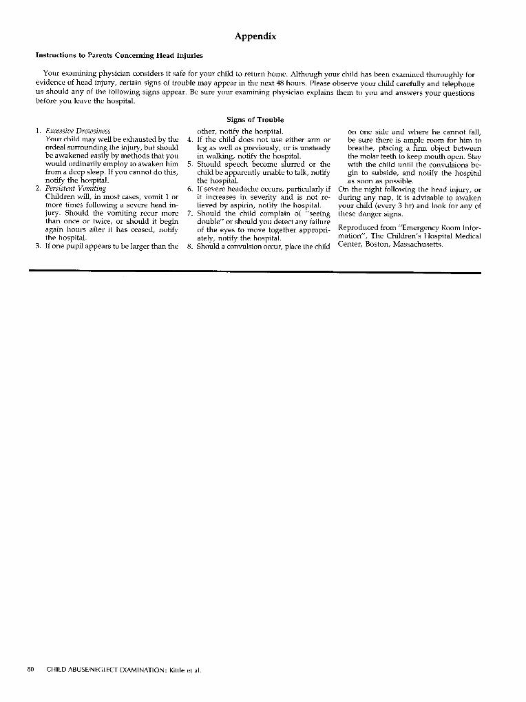
Appendix
Instructions to Parents Concerning Head Injuries
Your examining physician considers it safe for your child to return home. Although your child has been examined thoroughly for evidence of head injury, certain signs of trouble may appear in the next 48 hours. Please observe your child carefully and telephone us should any of the following signs appear. Be sure your examining physician explains them to you and answers your questions before you leave the hospital.
Signs of Trouble
1. Excessive Drowsiness Your child may well be exhausted by the ordeal surrounding the injury, but should be awakened easily by methods that you would ordinarily employ to awaken him from a deep sleep. If you cannot do this, notify the hospital.
Children will, in most cases, vomit 1 or more times following a severe head in- jury. Should the vomiting recur more than once or twice, or should it begin again hours after it has ceased, notify the hospital.
3. If one pupil appears to be larger than the
2. Persistent Vomiting
other, notify the hospital. 4. If the child does not use either arm or
leg as well as previously, or is unsteady in walking, notify the hospital.
5. Should speech become slurred or the child be apparently unable to talk, n o t e the hospital.
6. If severe headache occurs, particularly if it increases in severity and is not re- lieved by aspirin, notify the hospital.
7. Should the child complain of “seeing double” or should you detect any failure of the eyes to move together appropri- ately, notify the hospital.
8. Should a convulsion occur, place the child
on one side and where he cannot fall, be sure there is ample room for him to breathe, placing a firm object between the molar teeth to keep mouth open. Stay with the child until the convulsions be- gin to subside, and notify the hospital as soon as possible.
On the night following the head injury, or during any nap, it is advisable to awaken your child (every 3 hr) and look for any of these danger signs.
Reproduced from ”Emergency Room Infor- mation”, The Children’s Hospital Medical Center, Boston, Massachusetts.
80 CHILD ABUSE/NECLECT EXAMINATION: Kittle et al.