Embed Size (px)
Citation preview
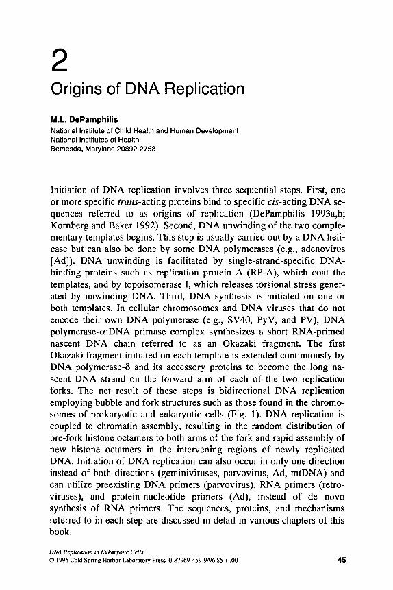
2 Origins of DNA Replication
M.L. DePamphilis National Institute of Child Health and Human Development National Institutes of Health Bethesda, Maryland 20892-2753
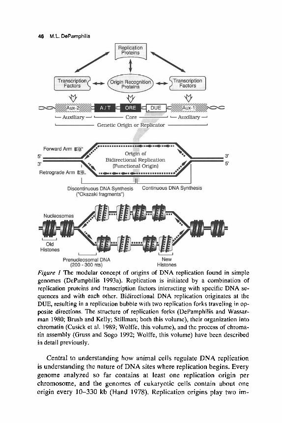
Initiation of DNA replication involves three sequential steps. First, one or more specific trans-acting proteins bind to specific cis-acting DNA se- quences referred to as origins of replication (DePamphilis 1993a,b; Kornberg and Baker 1992). Second, DNA unwinding of the two comple- mentary templates begins. This step is usually carried out by a DNA heli- case but can also be done by some DNA polymerases (e.g., adenovirus [Ad]). DNA unwinding is facilitated by single-strand-specific DNA- binding proteins such as replication protein A (RP-A), which coat the templates, and by topoisomerase I, which releases torsional stress gener- ated by unwinding DNA. Third, DNA synthesis is initiated on one or both templates. In cellular chromosomes and DNA viruses that do not encode their own DNA polymerase (e.g., SV40, PyV, and PV), DNA polymerase-a:DNA primase complex synthesizes a short RNA-primed nascent DNA chain referred to as an Okazaki fragment. The first Okazaki fragment initiated on each template is extended continuously by DNA polymerase-& and its accessory proteins to become the long na- scent DNA strand on the forward arm of each of the two replication forks. The net result of these steps is bidirectional DNA replication employing bubble and fork structures such as those found in the chromo- somes of prokaryotic and eukaryotic cells (Fig. 1). DNA replication is coupled to chromatin assembly, resulting in the random distribution of pre-fork histone octamers to both arms of the fork and rapid assembly of new histone octamers in the intervening regions of newly replicated DNA. Initiation of DNA replication can also occur in only one direction instead of both directions (geminiviruses, parvovirus, Ad, mtDNA) and can utilize preexisting DNA primers (parvovirus), RNA primers (retro- viruses), and protein-nucleotide primers (Ad), instead of de novo synthesis of RNA primers. The sequences, proteins, and mechanisms referred to in each step are discussed in detail in various chapters of this book.
DNA Replicalion in Eukaryolic Cells 0 1996 Cold Spring Harbor Laboratory Press 0-87969-459-9/96 $5 + .OO 4s
46 M.L. DePamphilis
.............. .*r)mmm..
origin of 3 (Functional Origin) 5’
Bidirectional Replication
.t.t.t.h* .............. Discontinuous DNA Synthesis
(“Okazaki fragments”) Continuous DNA Synthesis
Nucleosomes
Prenucleosornal DNA (200 - 300 nts)
U
New Histones
Figure 1 The modular concept of origins of DNA replication found in simple genomes (DePamphilis 1993a). Replication is initiated by a combination of replication proteins and transcription factors interacting with specific DNA se- quences and with each other. Bidirectional DNA replication originates at the DUE, resulting in a replication bubble with two replication forks traveling in op- posite directions. The structure of replication forks (DePamphilis and Wassar- man 1980; Brush and Kelly; Stillman; both this volume), their organization into chromatin (Cusick et al. 1989; Wolffe, this volume), and the process of chroma- tin assembly (Gruss and Sogo 1992; Wolffe, this volume) have been described in detail previously.
Central to understanding how animal cells regulate DNA replication is understanding the nature of DNA sites where replication begins. Every genome analyzed so far contains at least one replication origin per chromosome, and the genomes of eukaryotic cells contain about one origin every 10-330 kb (Hand 1978). Replication origins play two im-
Eukaryotic Replication Origins 47
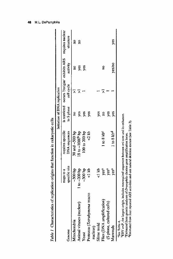
portant roles in DNA replication. First, they ensure that each time a cell divides, the entire genome is replicated efficiently. For example, a single mammalian cell contains about 1.8 meters of DNA that must be repli- cated in 6-8 hours, and early embryos of frogs, flies, and sea urchins replicate comparable amounts of DNA in 10-40 minutes. Second, replication origins provide a way in which to regulate when and where initiation events occur (Table 1). Replication of cellular chromosomes is restricted to one phase of the cell proliferation cycle (S phase), and initia- tion at each of several thousand replication origins is restricted to once per S phase (Blumenthal et al. 1974). Multiple initiation events at the same locus (gene amplification) can occur in tumors and transformed cell lines, but only rarely are genes amplified during normal animal develop- ment (Tlsty 1990). In contrast, replication of mtDNA and large viral DNA genomes such as herpes and vaccinia is not dependent on the cell entering S phase. mtDNA replicates randomly throughout the cell divi- sion cycle, and large viral genomes usurp the cell's machinery to provide their own replication components. Replication of small viral DNA genomes such as papovaviruses is restricted to S phase, but each genome copy may undergo two or more rounds of replication during a single S phase. Thus, it appears that mitochondria1 and viral genomes in mam- malian cells were designed to escape the very controls required for cel- lular DNA replication. This may not be true for simpler organisms such as flagellated protozoa, where replication of mtDNA (kinetoplast DNA) appears to follow the same rules as nuclear DNA.
Origins of DNA replication that function in eukaryotic cells can be divided into two groups: those found in "simple genomesl' such as animal viruses (SV40, pol yomavirus [PyV], PV, Ad, herpes simplex virus [HSV], Epstein-Barr virus [EBV]), mitochondria (human, mouse), protozoa (Tetrahymena), yeast (Saccharomyces cerevisiae, Schizosac- charomyces pombe), and slime mold (Physarum); and those found in 'lcomplex genomes" of metazoa such as flies (Drosophila, Sciara), frogs (Xenopus), and mammals (rodents, human). Simple origins that function in eukaryotes are similar to those that function in prokaryotes (Kornberg and Baker 1992). They have a modular anatomy composed of unique DNA sequence motifs and interactions with soluble proteins (DePam- philis 1993a,b). Whether or not metazoan chromosomes contain sequence-specific replication origins analogous to those found in simpler genomes is controversial, and therefore is discussed in more detail. What is clear is that replication begins at specific sites in metazoan chromo- somes, that initiation of DNA replication requires nuclear structure, and that nuclear structure imposes site specificity.
Tabl
e I
Cha
ract
eris
tics o
f rep
licat
ion
orig
ins t
hat f
unct
ion
in e
ukar
yotic
cells
3 -
-. In
itiat
ion
of D
NA
repl
icat
ion
v)
map
s to
a re
quire
s spe
cific
is
restr
icte
d oc
curs
?/or
igin
/ ex
hibi
ts A
RS
requ
ires n
ucle
ar
Gen
ome
spec
ific s
ite
DN
A se
quen
ces
to S
pha
se
cell
cycl
e ac
tivity
str
uctu
re
Mito
chon
dria
-3
00 b
p 30
and
-500
bp
no
>1
no
no
Ani
mal
vir
uses
(nuc
lear
) 1
to -2
00 b
p 18
to -l
OOOb
bp
Yes
>1
Yes
no
Yea
st
-300
bp
100
to 2
00 b
p Ye
s 1
Yes
Prot
ozoa
(Tet
rahy
men
a m
acro
c1
kb
c2 k
b Ye
s
Slim
e mol
d c1
kb
Yes
1
(S p
hase
, cul
ture
d ce
lls)
yesa
Ye
s 1
Mam
mal
s ye
sa
1 to
8 k
bd
Yes
1
yesl
no
Yes
nucl
eus)
Flie
s (D
NA
am
plifi
catio
n)
yesa
1 to
8 k
bc
no
>1
no
'See
Tab
le 4
. bE
BV
oriP
, the
larg
est o
rigin
, incl
udes
nonr
equi
red
sequ
ence
s bet
wee
n or
i-cor
e and
its
enha
ncer.
'E
stim
ated
fro
m g
enet
ic an
alys
es o
f Dro
soph
ila c
horio
n ge
ne a
mpl
ifica
tion
locu
s. dE
stim
ated
from
four
repo
rted
AR
S ac
tiviti
es a
nd o
ne n
atur
al d
elet
ion
mut
ant (
see
Tab
le 3
).
Eukaryotic Replication Origins 49
CHARACTERISTICS OF SIMPLE ORIGINS
Replication origins in simple genomes are composed of modular units acting in concert to determine where and when DNA replication will oc- cur (Table 1). In this sense, replication origins are equivalent to tran- scription promoters, but whereas each promoter is uniquely responsible for transcription of its associated gene, large chromosomes like yeast often contain more origins than are needed for their replication (Newlon et al. 1993). This flexibility in origin number is allowed because DNA regions that lack a replication origin can be replicated by replication forks that originate from origins located many kilobases away.
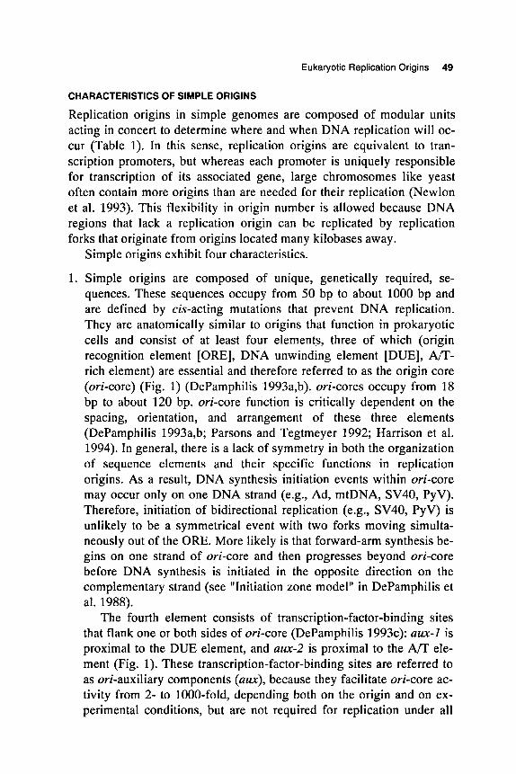
1. Simple origins are composed of unique, genetically required, se- quences. These sequences occupy from 50 bp to about 1000 bp and are defined by cis-acting mutations that prevent DNA replication. They are anatomically similar to origins that function in prokaryotic cells and consist of at least four elements, three of which (origin recognition element [ORE], DNA unwinding element [DUE], A n - rich element) are essential and therefore referred to as the origin core (ori-core) (Fig. 1) (DePamphilis 1993a,b). ori-cores occupy from 18 bp to about 120 bp. ori-core function is critically dependent on the spacing, orientation, and arrangement of these three elements (DePamphilis 1993a,b; Parsons and Tegtmeyer 1992; Harrison et al. 1994). In general, there is a lack of symmetry in both the organization of sequence elements and their specific functions in replication origins. As a result, DNA synthesis initiation events within ori-core may occur only on one DNA strand (e.g., Ad, mtDNA, SV40, PyV). Therefore, initiation of bidirectional replication (e.g., SV40, PyV) is unlikely to be a symmetrical event with two forks moving simulta- neously out of the ORE. More likely is that forward-arm synthesis be- gins on one strand of ori-core and then progresses beyond ori-core before DNA synthesis is initiated in the opposite direction on the complementary strand (see "Initiation zone model" in DePamphilis et al. 1988).
The fourth element consists of transcription-factor-binding sites that flank one or both sides of ori-core (DePamphilis 1993~): aux-I is proximal to the DUE element, and aux-2 is proximal to the A/T ele- ment (Fig. 1). These transcription-factor-binding sites are referred to as ori-auxiliary components (am), because they facilitate ori-core ac- tivity from 2- to 1000-fold, depending both on the origin and on ex- perimental conditions, but are not required for replication under all
Simple origins exhibit four characteristics.
50 M.L. DePamphilis
conditions and do not affect the mechanism by which replication oc- curs (DePamphilis 1993a,b). Some yeast origins, for example, contain a binding site (element B3) for transcription factor ABF1, whereas others do not. In this sense, auxiliary components are analogous to transcription enhancers. However, although enhancers that stimulate transcription are generally independent of their distance and orienta- tion relative to the promoter, orientation and spacing between auxil- iary sequences and ori-core are critical in some origins (Ad, SV40, PyV), whereas in other origins (EBV, yeast), these parameters are flexible. These differences presumably reflect differences in the specificity and strength of interactions between transcription factors and origin recognition proteins, as well as differences in the mecha- nism by which ori-auxiliary components function.
2. The genetic origin is coincident with the functional origin. The func- tional origin is the site where DNA synthesis actually begins. It has been mapped to within +300 bp of the genetic origin in yeast chromosomes and plasmids, and the transition from discontinuous to continuous DNA synthesis on each template (Fig. 1) mapped with nucleotide resolution to the genetic origins in SV40, PyV, Ad, mtDNA, Escherichia coli (Kohara et al. 1985; Rokeach and Zyskind 1986; Seufert and Messer 1987), and bacteriophage h (Yoda et al. 1988). However, these transitions can lie outside ori when SV40 DNA replication is initiated in vitro (Bullock et al. 1991), suggesting that extensive DNA unwinding can precede initiation of DNA synthe- sis, thereby providing DNA polymerase-a:DNA primase with the op- portunity to begin synthesis outside of ori (see "Initiation zone model" in DePamphilis et al. 1988). In vivo, the rate of DNA unwind- ing may be retarded by chromatin structure.
3. Simple origins can act as autonomously replicating sequences (ARSs). ARS elements confer on other DNA molecules the ability to replicate when transferred to either cells or cell extracts containing the required replication proteins. So far, ARS elements have been demonstrated only with viral and yeast origins. Functional origins in the chromosomes of S. cerevisiae have been shown to correspond to individual ARS elements that are genetically required for origin ac- tivity (Newlon, this volume). The same appears true for S. pombe, al- though sequence requirements for origins in S. pombe appear more diffuse and origin function less efficient than in S. cerevisiae (Caddle and Calos 1994; Dubey et al. 1994; Wohlgemuth et al. 1994).
4. Simple origins can function in a soluble cell-free DNA replication system. So far, this has been demonstrated only with viral origins, a
Eukaryotic Replication Origins 51
problem that may be overcome when large amounts of purified replication proteins become available for other systems.
ANATOMY OF SIMPLE REPLICATION ORIGINS
Origin Recognition Element
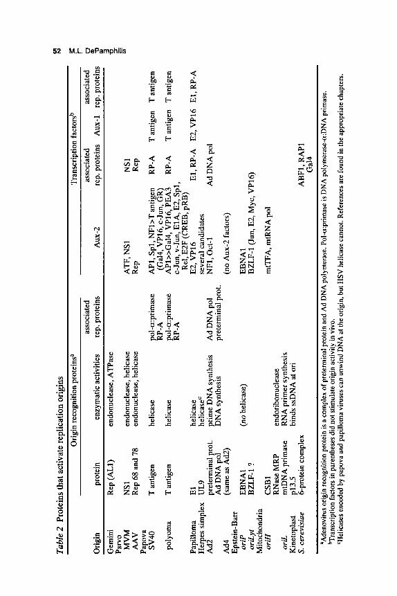
An ORE is the DNA-binding site for one or more origin recognition proteins (Table 2) that are required for initiation of DNA replication. This DNA-protein interaction can be regulated by posttranslational modi- fications, such as phosphorylation of specific amino acid residues in SV40 T antigen (see Weisshart and Fanning, this volume). Sequences within or flanking the ORE often exhibit the characteristics of bent DNA (Zahn and Blattner 1985; Deb et al. 1986; Williams et al. 1988), which may facilitate protein binding (Ryder et al. 1986). Moreover, although an ORE may exhibit a twofold symmetry, interaction with its recognition protein can be asymmetrical, leading to an asymmetrical opening of ori- core (SenGupta and Borowiec 1994).
Origin recognition proteins serve at least two functions. The first is to initiate DNA unwinding using either their own helicase activity (e.g., parvovirus, SV40, PyV, PV, HSV) or one that associates with it (EBV may be an example). The second function is to guide other replication proteins to the origin. For example, SV40 and PyV T antigen associate with DNA polymerase-a:DNA primase, the enzyme responsible for ini- tiation of the first RNA-primed DNA chain (Okazaki fragment) at their respective origins, and with RP-A, a single-stranded DNA-binding protein that stimulates both helicase and polymerase action (Melendy and Stillman 1993; Schneider et al. 1994 and references therein). Similarly, Ad preterminal protein, the protein-dCTP primer that initiates Ad DNA synthesis, is complexed with Ad DNA polymerase, the enzyme responsible for Ad DNA synthesis (see Hay, this volume). In yeast, a six- protein origin recognition complex binds to an approximately 50-bp se- quence that includes elements A and B1. All yeast origins require ele- ment A (containing the 11-bp conserved ARS consequence sequence) and at least two of the three B elements that lie to one side of element A (see Newlon, this volume). Origin recognition proteins such as EBV EBNAl that do not have their own helicase activity (Frappier and O’Donnell 1991) presumably associate with a cellular helicase. These in- teractions between origin recognition proteins and replication proteins can determine the host-cell specificity of a replication origin (Melendy and Stillman 1993; Schneider et al. 1994).
UI
N
Tabl
e 2 P
rote
ins t
hat a
ctiv
ate
repl
icat
ion
orig
ins
Orig
in re
cogn
ition
pro
tein
sa
Tran
scrip
tion
fact
orsb
n r-
asso
ciat
ed
asso
ciat
ed
asso
ciat
ed
0
Orig
in
prot
ein
enzy
mat
ic a
ctiv
ities
re
p. p
rote
ins
AU
X-2
re
p. p
rote
ins
Aux
-1
rep.
pro
tein
s 2 3
Gem
ini
Rep
w-1
) en
donu
clea
se, A
TPas
e 'p zr
Parv
o -.
Papo
va
(D - UI
MV
M
NS1
en
donu
clea
se, h
elic
ase
ATF
, NSl
N
S1
AA
V
Rep
68
and
78
endo
nucl
ease
, hel
icas
e R
ep
Rep
SV40
T
ant
igen
he
licas
e po
l-a:p
rimas
e A
P1, S
pl, N
Fb
T an
tigen
R
P-A
T
antig
en
T an
tigen
pol y
oma
T an
tigen
he
licas
e po
l-a:p
rimas
e A
Pl>>
Gal
4, V
P16,
PEA
3 R
P-A
T
antig
en
T a
ntig
en
RP-
A
(Ga1
4, V
P16,
c-J
un, G
R)
RP-
A
c-Ju
n, v
-Jun
, ElA
, E2,
Spl
, R
el, E
2F (C
REB
, pR
B)
Papi
llom
a E
l he
licas
e E2
, VP1
6 E
l, R
P-A
E2
, VP1
6 E
l, R
P-A
H
erpe
s sim
plex
U
L9
helic
aseC
se
vera
l can
dida
tes
Ad2
pr
eter
min
al p
rot.
prim
e D
NA
synt
hesi
s A
d D
NA
pol
N
F1,O
ct-1
A
d D
NA
pol
Ad4
(s
ame
as A
d2)
(no
Aux
-2 fa
ctor
s)
Epst
ein-
Bar
r
Mito
chon
dria
Ad
DN
A p
ol
DN
A sy
nthe
sis
pret
erm
inal
pro
t.
oriP
E
BN
Al
(no h
elic
ase)
E
BN
Al
oriL
yt
BZL
F-1
? B
ZLF-
1 (J
un, E
2, M
yc, V
P16)
oriH
C
SBl
mtT
FA, m
tRN
A p
ol
oriL
m
tDN
A p
rimas
e R
NA
prim
er s
ynth
esis
R
Nas
e M
RP
endo
ribon
ucle
ase
Kin
etop
last
p1
3.5
bind
s ss
DN
A a
t ori
S. c
erev
isiu
e 6-
prot
ein
com
plex
A
BF1
, RA
P1
Gal
4 ~
~~~~
~~
~~
~~~~
~ ~~
~ ~~
~~~~
~~
~~
__
__
__
_
aAde
novi
rus o
rigi
n re
cogn
ition
pro
tein
is a
com
plex
of p
rete
rmin
al p
rote
in a
nd A
d D
NA
pol
ymer
ase.
Pol
-a:p
rim
ase
is D
NA
pol
ymer
ase-
a:D
NA
pri
mas
e.
bTra
nscr
iptio
n fac
tors
in p
aren
thes
es d
id n
ot st
imul
ate o
rigi
n ac
tivity
in v
ivo.
'H
elic
ases
enc
oded
by
papo
va a
nd p
apill
oma v
irus
es c
an u
nwin
d D
NA
at t
he o
rigi
n, b
ut H
SV h
elic
ase
cann
ot. R
efer
ence
s are
foun
d in
the
appr
opri
ate c
hapt
ers.
Eukaryotic Replication Origins 53
DNA Unwinding Element
DNA unwinding appears to begin at an easily unwound DNA sequence referred to as the DNA unwinding element (DUE, Fig. 1). A DUE is identified by cis-acting mutations that both increase the stability of the double helix (i.e., make DNA unwinding more difficult) and reduce DNA replication (Umek and Kowalski 1990b). Computer programs are available that determine DNA helical stability in known sequences (Rychlik and Rhoads 1989; Natale et al. 1992), and the effects of muta- tions on helical stability can be assessed within the context of a super- coiled plasmid using a single-strand-specific endonuclease, or two- dimensional gel electrophoresis of plasmid topoisomers. These ap- proaches have identified DUEs in E. coli oriC (Kowalski and Eddy 1989), yeast ARS elements in plasmids (Natale et al. 1992; Huang and Kowalski 1993; Miller and Kowalski 1993), and replication origins in yeast chromosomes (Huang and Kowalski 1993) and SV40 (Lin and Kowalski 1994). In yeast replication origins the genetic and physical properties of element B2 are consistent with those of a DUE (Rao et al. 1994; Theis and Newlon 1994).
Although primary sequence is an important determinant of the energy required for DNA unwinding (because of the importance of base- stacking interactions), there is no unique consensus sequence for a DUE. DUE sequences in yeast are not conserved, and easily unwound DNA se- quences that are not components of origins can substitute for the DUEs in both yeast and E. coli (Umek and Kowalski 1988; Kowalski and Eddy 1989). Therefore, DUEs are unlikely to be binding sites for specific replication proteins. Instead, it appears that one of the proteins binding to the ORE must interact with the DUE because the spatial relationship be- tween these two core elements is critical. In yeast, the DUE always is lo- cated 3 ' to the T-rich strand of the ARS consensus sequence (Natale et al. 1993), and reversal of the orientation of the ARS consensus sequence with respect to the DUE abolishes DNA replication (Holmes and Smith 1989). Binding of proteins to ORE results in DNA unwinding in the DUEs of E. coli oriC (Kowalski and Eddy 1989), SV40, and PyV. In SV40 and PyV, the DUE, the site where T antigen begins unwinding DNA, and the origin of bidirectional replication (OBR; defined by the transition between continuous and discontinuous DNA synthesis on each template [Fig. 11) are all coincident (DePamphilis 1993a; Lin and Kowalski 1994). Similarly, in yeast ARS307, a majority of leading strands emanate from the proposed DUE (unpublished data cited in Theis and Newlon 1994). Therefore, the DUE appears to be the entry site for the replication machinery.
54 M.L. DePamphilis
Not all easily unwound DNA sequences are part of replication origins, but the fact that such sequences can substitute for known DUEs qualifies them as "potential DUEs." Since the energetic, length, and spacing requirements of true DUEs remain to be defined, it is difficult to estimate the frequency at which potential DUEs occur in natural DNA. For purposes of comparison, a potential DUE can be defined as a 100-bp sequence whose helical stability is 20 or more kcal/mole below the aver- age for the sequence analyzed. This definition is stringent enough to ex- clude some real DUEs. Such a potential DUE would be expected once every 3.2 kb in a random sequence of 60% A+T content (D. Natale, pers. comm.), suggesting that potential DUEs occur much more frequently than replication origins in yeast (1 origin/36 kb) and mammalian (1 origin/100 kb) DNA. Thus, the ability of an easily unwound sequence to function as a DUE likely depends on conditions such as its proximity to other origin elements (Fig. l), the concentration of initiation factors, the influence of chromatin structure, and the amount of negative superhelical energy available (Umek and Kowalski 1990a).
An-rich Element
Most, but not all (e.g., EBV), replication origins contain an Am-rich se- quence consisting of a T-rich and an A-rich strand. The length of the A/T-rich element is critical for origin function (Gerard and Gluzman 1986; Koff et al. 1991), a fact that may be related to its bent character (Deb et al. 1986). Bent DNA can interact more easily with proteins, which may account for the fact that binding of origin recognition proteins to ORES frequently distorts the Am-rich element (Koff et al. 1991; Gillette et al. 1994; SenGupta and Borowiec 1994). Distortion of the A/T-rich element may facilitate either binding to the ORE or melting of the DUE.
Auxiliary Components
ori-auxiliary components stimulate replication only when they bind one or more transcription factors, and only when the transcription factor con- tains an activation domain that specifically interacts with the replication machinery (Table 2) (DePamphilis 1993c; van der Wet , this volume). In some genomes (SV40, PyV, PV, EBV), the same sequence elements that function as promoters or enhancers in transcription also function as aux components in replication; cis-acting mutations that affect one process also affect the other. Auxiliary components could be used to regulate
Eukaryotic Replication Origins 55
origin activity in two ways. First, the ability of a particular transcription factor to stimulate a particular origin may be limited to specific members of a transcription factor family, and to the availability of specific coac- tivator proteins (Guo and DePamphilis 1992). Thus, auxiliary com- ponents can stimulate the same origin to different extents in different cell types as the composition of available transcription factors changes during animal development (Rochford and Villarreal 1991). Second, just as transcription factors can initiate transcription of different genes at dif- ferent times during the cell division cycle, they could regulate the temporal order of DNA replication during S phase, accounting for the observation that active genes are replicated early and inactive genes are replicated late (see Simon and Cedar, this volume).
There are four basic mechanisms by which transcription factors can stimulate ori-core (DePamphilis 1993~):
1. An upstream promoter can direct transcription through ori-core. The resulting mRNA is then cut by an endonuclease at specific sites to generate RNA primers for initiation of DNA synthesis. This mecha- nism occurs at mtDNA oriH, E. coli filamentous and T-odd bacterio- phage, and E. coli plasmid ColEl (Kornberg and Baker 1992).
2. Transcription factors can facilitate binding of origin recognition proteins to ori-core. For example, NFI binding to am-2 facilitates binding of subsaturating concentrations of the Ad2 preterminal protein/Ad DNA polymerase complex (pTP-pol) (see Hay, this volume). Binding of PV-encoded enhancer-specific activation protein, E2, to am-l and possibly am-2 facilitates binding of E l to ori-core (see Stenlund, this volume). Binding of EBV-encoded EBNAl protein to the EBV enhancer (am-2) stabilizes interaction of EBNAl to the EBV ori-core (Frappier et al. 1994).
3. Transcription factors can facilitate the activity of an initiation com- plex after it has formed. For example, SV40 ori-auxiliary components stimulate SV40 ori-core by facilitating T-antigen-dependent DNA un- winding at ori-core (Gutierrez et al. 1990). This may occur by stabi- lizing the interaction of T antigen with ssDNA as it disrupts its own binding site by unwinding it (Gutierrez et al. 1990), and by recruiting RP-A, a single-strand DNA-binding protein, to stabilize ssDNA (He et al. 1993; Li and Botchan 1993). The T-antigen dimer that binds to am-2 appears to interact with the T-antigen hexamer bound to ORE (Guo et al. 1991), thus accounting for the observation that the need for am-l to stimulate papovavirus origin activity is inversely related to the ability of T antigen to activate ori-core (Sock et al. 1993). In the chromosomes of E. coli, bacteriophage h, and plasmid R6K, tran-
56 M.L. DePamphilis
scription or association with RNA polymerase near ori can stimulate ori activity by removing torsional stress in DNA and thus facilitating DNA unwinding (Kornberg and Baker 1992).
4. Transcription factors can prevent chromatin structure from interfering with binding of replication factors to origins. Nucleosomes can repress replication origins in yeast (Simpson 1990), Drosophilu (Kar- pen and Spradling 1990), and mammalian chromosomes (Forrester et al. 1990). Therefore, since transcriptionally active DNA sequences are not incorporated into nucleosomes (Morse et al. 1992), transcrip- tion through a replication origin may provide access to replication factors. Alternatively, binding of transcription factors to enhancers may allow interactions between the enhancer and ori-core, analogous to those between enhancer and promoter (Majumder and DePamphilis 1994), that prevent nucleosomes from repressing origins in the same way that it prevents nucleosomes from repressing promoters (Paran- jape et al. 1994). This mechanism may apply to PyV, where the PyV enhancer (am-2) is dispensable under conditions where a repressive chromatin structure appears to be absent (Prives et al. 1987; Martinez- Salas et al. 1988; Majumder et al. 1993), and to SV40 am-2 where prebinding some transcription factors (e.g., NFI, Ga14:VP16) can pre- vent chromatin assembly from interfering with SV40 DNA replica- tion in vitro (see Hassell and Brinton, this volume). However, the facts that Ga14:VP16 does not stimulate the SV40 origin in vivo, and that prebinding T antigen alone can also prevent nucleosome repres- sion, suggest that other mechanisms should be considered (DePam- philis 1993~).
VIRAL ORIGINS AS MODELS FOR CELLULAR ORIGINS
PV and EBV have been considered models for cellular DNA replication because their genomes replicate in the nucleus, their DNA replication is restricted to S phase, and they maintain a low number of genome copies per cell. Moreover, early studies on PV DNA replication concluded that a complex interaction between positive and negative controls restricted initiation of replication to once per origin per S phase (Roberts and Weintraub 1988). However, later studies (see Stenlund, this volume) revealed that PV origins are remarkably similar to those in papova- viruses, and that although cis-acting PV sequences can suppress the ac- tivity of lytic origins such as SV40 and PyV, they do not restrict them to one initiation event per S phase (Nallaseth and DePamphilis 1994). In
Eukaryotic Replication Origins 57
fact, initiation of PV DNA replication is not restricted to once per S phase (Gilbert and Cohen 1987; Ravnan et a]. 1992). EBV remains a can- didate because its DNA replicates at the same rate as cellular chromo- somes (Haase and Calos 1991; Yates and Guan 1991), but i t remains to be determined whether or not EBV DNA replication, like cellular DNA replication, does not reinitiate when cells are limited to a single S phase in the presence of a mitotic inhibitor such as nocodazoie (Nallaseth and DePamphilis 1994). This test should be applied to all putative mam- malian ARS elements (Table 3) (Krysan et al. 1993; Masukata et al. 1993).
COMPLEX (METAZOAN) ORIGINS
In comparison with simple genomes, origins of replication in multi- cellular animals (the metazoa) often appear paradoxical. Early attempts to identify ori sequences in mammalian chromosomes by their ability to function as ARS elements were difficult to reproduce and therefore con- troversial (Gutierrez et al. 1988; Burhans and Huberman 1994), although some recent reports (Table 3) look promising. Nevertheless, most large (>lo kb) DNA fragments from mammalian chromosomes can provide some ARS activity in mammalian cells (Krysan et al. 1993; Masukata et al. 1993), suggesting that DNA length is more critical than DNA se- quence. The same conclusion is reached when DNA is injected into the eggs of frogs, sea urchins, or fish, or when DNA is added to extracts of Xenopus eggs or Drosophila embryos (Coverley and Laskey 1994; see Laskey and Madine; Blow and Chong; both this volume). DNA replica- tion is initiated at a single randomly chosen site within virtually any DNA molecule (Hines and Benbow 1982; MCchali and Kearsey 1984; Hyrien and MCchali 1992; Mahbubani et al. 1992). This lack of site- specific initiation also appears during chromosome replication in Xenopus (Hyrien and MCchali 1993) and Drosophila (Shinomiya and Ina 1991) embryos, suggesting that the lack of sequence requirements and site specificity observed when DNA is introduced into cultured cells, eggs, or egg extracts accurately reflects chromosome replication in situ.
Another approach to identify replication origins in metazoan chromo- somes is in situ mapping of initiation sites for DNA replication in the hope that subsequent physical and genetic analysis of those loci will reveal the nature of a metazoan origin of DNA replication. This approach has revealed that DNA replication in metazoan chromosomes occurs at specific sites.

5 k?
Sam
e or
igin
5
r
W
Iu
TI
Tabl
e 3 M
etaz
oan
orig
ins o
f rep
licat
ion
are
gene
tical
ly d
eter
min
ed
3
in s
ingl
e co
py
Tra
nslo
cate
d D
elet
ion
AR
S A
PE
Org
anis
m
Loc
atio
n an
d m
ulti
copy
gen
omes
a to
oth
er s
ites
in
OBR
ac
tivi
ty
acti
vity
Ham
ster
D
HFR
gen
e (o
r$)
chro
mos
omal
(1)
activ
e (2
) M
ouse
rR
NA
gen
e D
roso
phila
H
uman
M
ouse
M
ouse
H
uman
H
uman
H
amst
er
Hum
an
Mou
se
chor
ion
gene
hs
p70
gene
Ig
H g
ene
enha
ncer
A
DA
gen
e (la
te S
pha
se)
c-m
yc g
ene
cDN
A 3
43
CA
D g
ene
grou
p P-
glob
in g
ene
AD
A g
ene
(ear
ly S
pha
se)
activ
e (5
) Ye
s (6)
Ye
s (7)
ep
isom
al(8
) Ye
s (8)
Ye
s (9)
Ye
s (10
) ep
isom
al(l
1)
epis
omal
(l3)
in
activ
e (1
2)
aAm
plifi
ed g
ene
copi
es ar
e fo
und
in e
ither
chro
mos
omal
or e
piso
mal
loca
tions
. R
efer
ence
s (in
par
enth
eses
): (1
) H
ande
li et
al.
1989
; Bur
hans
et
al.
1990
, 19
91; V
assi
lev
et a
l. 19
90; D
ijkw
el a
nd H
amlin
199
2, 1
995;
DeP
amph
ilis
1993
d;
Tash
eva
and
Rou
fa 1
994a
; Gilb
ert e
t al
. 19
95; M.
Gia
cca,
unp
ubl.;
(2)
Han
deli
et a
l. 19
89; (
3) S
tolz
enbu
rg et
al.
1994
; (4)
Her
man
n et
al.
1994
; (5)
Orr
-Wea
ver
1991
; (6)
Tai
ra e
t al.
1994
; (7)
Arii
zum
i et a
l. 19
93; I
guch
i-Arig
a et
al.
1993
; (8)
Virt
a-Pe
arlm
an e
t al.
1993
; (9)
Ber
beric
h et
al.
1995
; (10
) Wu
et a
l. 19
95; (
11) K
el-
ly e
t al.
1995
; (12
) Kits
berg
et a
l. 19
93; (
13) P
ears
on e
t al.
1994
.
Eukaryotic Replication Origins 59
MAPPING ORIGINS OF REPLICATION IN METAZOAN CHROMOSOMES
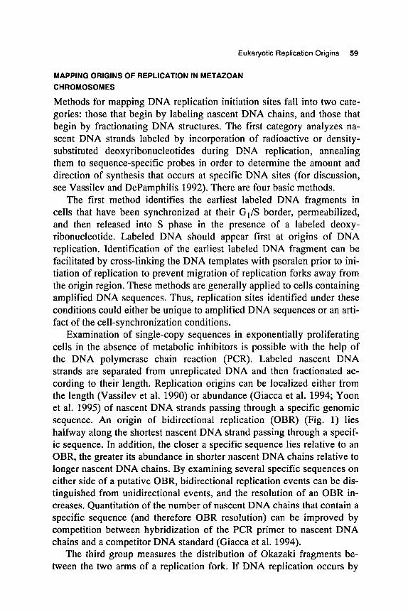
Methods for mapping DNA replication initiation sites fall into two cate- gories: those that begin by labeling nascent DNA chains, and those that begin by fractionating DNA structures. The first category analyzes na- scent DNA strands labeled by incorporation of radioactive or density- substituted deoxyribonucleotides during DNA replication, annealing them to sequence-specific probes in order to determine the amount and direction of synthesis that occurs at specific DNA sites (for discussion, see Vassilev and DePamphilis 1992). There are four basic methods.
The first method identifies the earliest labeled DNA fragments in cells that have been synchronized at their G1/S border, permeabilized, and then released into S phase in the presence of a labeled deoxy- ribonucleotide. Labeled DNA should appear first at origins of DNA replication. Identification of the earliest labeled DNA fragment can be facilitated by cross-linking the DNA templates with psoralen prior to ini- tiation of replication to prevent migration of replication forks away from the origin region. These methods are generally applied to cells containing amplified DNA sequences. Thus, replication sites identified under these conditions could either be unique to amplified DNA sequences or an arti- fact of the cell-synchronization conditions.
Examination of single-copy sequences in exponentially proliferating cells in the absence of metabolic inhibitors is possible with the help of the DNA polymerase chain reaction (PCR). Labeled nascent DNA strands are separated from unreplicated DNA and then fractionated ac- cording to their length. Replication origins can be localized either from the length (Vassilev et al. 1990) or abundance (Giacca et al. 1994; Yoon et al. 1995) of nascent DNA strands passing through a specific genomic sequence. An origin of bidirectional replication (OBR) (Fig. 1) lies halfway along the shortest nascent DNA strand passing through a specif- ic sequence. In addition, the closer a specific sequence lies relative to an OBR, the greater its abundance in shorter nascent DNA chains relative to longer nascent DNA chains. By examining several specific sequences on either side of a putative OBR, bidirectional replication events can be dis- tinguished from unidirectional events, and the resolution of an OBR in- creases. Quantitation of the number of nascent DNA chains that contain a specific sequence (and therefore OBR resolution) can be improved by competition between hybridization of the PCR primer to nascent DNA chains and a competitor DNA standard (Giacca et al. 1994).
The third group measures the distribution of Okazaki fragments be- tween the two arms of a replication fork. If DNA replication occurs by
60 M.L. DePamphilis
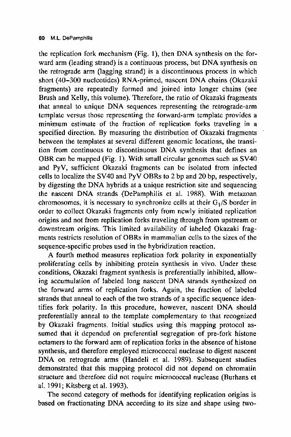
the replication fork mechanism (Fig. l), then DNA synthesis on the for- ward arm (leading strand) is a continuous process, but DNA synthesis on the retrograde arm (lagging strand) is a discontinuous process in which short (40-300 nucleotides) RNA-primed, nascent DNA chains (Okazaki fragments) are repeatedly formed and joined into longer chains (see Brush and Kelly, this volume). Therefore, the ratio of Okazaki fragments that anneal to unique DNA sequences representing the retrograde-arm template versus those representing the fonvard-arm template provides a minimum estimate of the fraction of replication forks traveling in a specified direction. By measuring the distribution of Okazaki fragments between the templates at several different genomic locations, the transi- tion from continuous to discontinuous DNA synthesis that defines an OBR can be mapped (Fig. 1). With small circular genomes such as SV40 and PyV, sufficient Okazaki fragments can be isolated from infected cells to localize the SV40 and PyV OBRs to 2 bp and 20 bp, respectively, by digesting the DNA hybrids at a unique restriction site and sequencing the nascent DNA strands (DePamphilis et al. 1988). With metazoan chromosomes, it is necessary to synchronize cells at their GI/S border in order to collect Okazaki fragments only from newly initiated replication origins and not from replication forks traveling through from upstream or downstream origins. This limited availability of labeled Okazaki frag- ments restricts resolution of OBRs in mammalian cells to the sizes of the sequence-specific probes used in the hybridization reaction.
A fourth method measures replication fork polarity in exponentially proliferating cells by inhibiting protein synthesis in vivo. Under these conditions, Okazaki fragment synthesis is preferentially inhibited, allow- ing accumulation of labeled long nascent DNA strands synthesized on the forward arms of replication forks. Again, the fraction of labeled strands that anneal to each of the two strands of a specific sequence iden- tifies fork polarity. In this procedure, however, nascent DNA should preferentially anneal to the template complementary to that recognized by Okazaki fragments. Initial studies using this mapping protocol as- sumed that it depended on preferential segregation of pre-fork histone octamers to the forward arm of replication forks in the absence of histone synthesis, and therefore employed micrococcal nuclease to digest nascent DNA on retrograde arms (Handeli et al. 1989). Subsequent studies demonstrated that this mapping protocol did not depend on chromatin structure and therefore did not require micrococcal nuclease (Burhans et al. 1991; Kitsberg et al. 1993).
The second category of methods for identifying replication origins is based on fractionating DNA according to its size and shape using two-
Eukaryotic Replication Origins 61
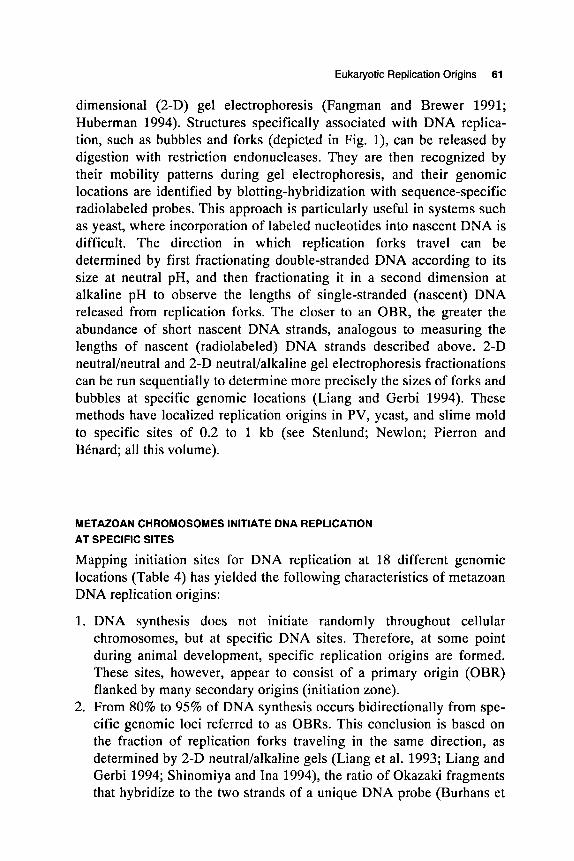
dimensional (2-D) gel electrophoresis (Fangman and Brewer 1991; Huberman 1994). Structures specifically associated with DNA replica- tion, such as bubbles and forks (depicted in Fig. l), can be released by digestion with restriction endonucleases. They are then recognized by their mobility patterns during gel electrophoresis, and their genomic locations are identified by blotting-hybridization with sequence-specific radiolabeled probes. This approach is particularly useful in systems such as yeast, where incorporation of labeled nucleotides into nascent DNA is difficult. The direction in which replication forks travel can be determined by first fractionating double-stranded DNA according to its size at neutral pH, and then fractionating it in a second dimension at alkaline pH to observe the lengths of single-stranded (nascent) DNA released from replication forks. The closer to an OBR, the greater the abundance of short nascent DNA strands, analogous to measuring the lengths of nascent (radiolabeled) DNA strands described above. 2-D neutralheutral and 2-D neutral/alkaline gel electrophoresis fractionations can be run sequentially to determine more precisely the sizes of forks and bubbles at specific genomic locations (Liang and Gerbi 1994). These methods have localized replication origins in PV, yeast, and slime mold to specific sites of 0.2 to 1 kb (see Stenlund; Newlon; Pierron and BCnard; all this volume).
METAZOAN CHROMOSOMES INITIATE DNA REPLICATION AT SPECIFIC SITES
Mapping initiation sites for DNA replication at 18 different genomic locations (Table 4) has yielded the following characteristics of metazoan DNA replication origins:
1. DNA synthesis does not initiate randomly throughout cellular chromosomes, but at specific DNA sites. Therefore, at some point during animal development, specific replication origins are formed. These sites, however, appear to consist of a primary origin (OBR) flanked by many secondary origins (initiation zone).
2. From 80% to 95% of DNA synthesis occurs bidirectionally from spe- cific genomic loci referred to as OBRs. This conclusion is based on the fraction of replication forks traveling in the same direction, as determined by 2-D neutral/alkaline gels (Liang et al. 1993; Liang and Gerbi 1994; Shinomiya and Ina 1994), the ratio of Okazaki fragments that hybridize to the two strands of a unique DNA probe (Burhans et
62 M.L. DePamphilis
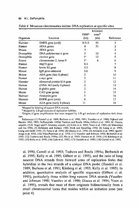
Table 4 Metazoan chromosomes initiate DNA replication at specific sites Initiation
OBRa zoneb Organism Location (kb) (kb) Reference
Hamster DHFR gene (or$) 0.5-3 55 1 Human rRNA genes 8 31 2 Mouse rRNA genes 3 3 Drosophilu DNA polymerase a gene 5 10 4 Drosophila chorion gene 1 C 12 5
Human Hsp70 gene 0.4 7 Human lamin B2 gene 0.5 8 Mouse IgH gene enhancer 0.6 9 Mouse ADA gene (late S phase) 2 10
Sciara chromosome-2, locus 9 1 C 6 6
Human c-myc gene 2 Hamster ribosomal protein S14 gene 2 Human cDNA 343 (early S phase) 2 Human P-globin gene Mouse CAD gene group Hamster rhodopsin gene Hamster DHFR gene (oriy)
11 12 13 14 15 16 17
Mouse ADA gene (early S phase) 11 18
aMapped by labeling of nascent DNA strands.
‘Origins of gene amplification that were mapped by 2-D gel analyses of replication fork direc- tion.
References: (1) Handeli et al. 1989; Burhans et al. 1990, 1991; Vassilev et al. 1990; Dijkwel and Hamlin 1992, 1995; DePamphilis 1993d; Tasheva and Roufa 1994a; Gilbert et al. 1995; M. Giacca, unpubl.; (2) E. Gogel and F. Grummt, unpubl.; (3) Little et al. 1993; Yoon et al. 1995; (4) Shinomiya and Ina 1994; (5) Delidakis and Kafatos 1989; Heck and Spradling 1990; (6) Liang et al. 1993; Liang and Gerbi 1994; (7) Taira et al. 1994; (8) Giacca et al. 1994; (9) Ariizumi et al. 1993; Iguchi- Ariga et al. 1993; (10) Virta-Pearlman et al. 1993; (11) Vassilev and Johnson 1990; Berberich et al. 1995; (12) Tasheva and Roufa 1994a; (13) Wu et al. 1993; Pearson et al. 1994; (14) Kitsberg et al. 1993; (15) Kelly et al. 1995; (16) Gale et al. 1992; (17) Handeli et al. 1989; (18) Carroll et al. 1993.
Mapped by 2-D gel analyses of replication bubbles.
al. 1990; Carroll et al. 1993; Tasheva and Roufa 1994a; Berberich et al. 1995; Kelly et al. 1995; Gilbert et al. 1995), and the ratio of long nascent DNA strands from forward arms of replication forks that hybridize to the two strands of a unique DNA probe (Handeli et al. 1989; Burhans et al. 1991; Kitsberg et al. 1993; Kelly et al. 1995). In addition, quantitative analysis of specific sequences (Gilbert et al. 1995), particularly those within long nascent DNA strands (Vassilev and Johnson 1990; Vassilev et al. 1990; Giacca et al. 1994; Yoon et al. 1995), reveals that most of them originate bidirectionally from a small chromosomal locus that resides within an initiation zone (see point 4).
Eukaryotic Replication Origins 63
3. An OBR is contained within 0.5 kb to 2 kb. This conclusion is based on 11 different OBRs that appear to lie within a 2-kb region and on the DHFR orif3 locus where five different nascent DNA strand meth- ods have been applied with remarkable agreement (Table 4). Some OBRs lie within larger regions of 5-11 kb. All of these estimates are for the maximum size of an OBR; resolution is limited by the dif- ficulty in preparing probes large enough to give a strong hybridization signal with radiolabeled nascent DNA chains, and devoid of repetitive sequences. Future refinements in origin-mapping techniques will like- ly resolve these OBRs to a smaller locus. Thus, metazoan replication origins appear to be 3-10 times larger than replication origins in simple genomes (0.05-1 kb). The fact that 18 OBRs have been identi- fied by independent investigators using a variety of different methods gives confidence that site-specific initiation is not an artifact of the experimental conditions used to map them. Similar results were ob- tained with synchronized and unsynchronized cells, with cells con- taining single-copy sequences and with cells containing amplified multicopy sequences, with untreated cells and with cells treated with metabolic inhibitors, and with different methods for detecting specific DNA sequences.
Neutral/alkaline 2-D gel electrophoresis has been used to map an OBR to 1 kb at an amplification locus in Sciuru (Liang et al. 1993; Liang and Gerbi 1994), giving credence to an earlier interpretation of 2-D gel electrophoresis mapping data that 80% of replication forks at the chorion gene amplification locus in Drosophilu originate from a specific 1-kb site (Heck and Spradling 1990). Neutral/alkaline 2-D gel electrophoresis also identified an S-phase OBR 15-20 kb downstream from the Drosophilu DNA polymerase a gene (Shinomiya and Ina 1994).
4. Replication bubbles are detected throughout a larger "initiation zone" of 6-55 kb that includes the OBR. This conclusion is based on analyses of DNA structures by neutralheutral 2-D gel electrophoresis at five different genomic loci (Table 3) (Delidakis and Kafatos 1989; Heck and Spradling 1990; Dijkwel and Hamlin 1992, 1995; Liang et al. 1993; Little et al. 1993; Liang and Gerbi 1994), two of which (Drosophilu chorion gene, Sciuru locus 9) are developmentally pro- grammed amplification origins. Four of these "initiation zones" en- compass an OBR that was detected either by nascent strand analyses (rRNA genes, DHFR orif3) or by measuring the direction of fork movement using neutral/alkaline 2-D gel electrophoresis (Sciuru locus 9, Drosophilu pol a gene). One study on the Drosophilu
64 M.L. DePamphilis
chorion gene locus (Heck and Spradling 1990) concluded that al- though multiple initiation sites may exist within a 12-kb locus, a model in which a single origin is preferred 70-80% of the time could explain their neutralheutral 2-D gel electrophoresis data.
Results of neutralheutral 2-D gel analyses are consistent with newly synthesized DNA analyses and most neutral/alkaline gel analyses if one assumes that the frequency of initiation events at the OBR is much greater than the frequency of initiation events outside the OBR. In fact, replication bubbles detected by neutralheutral 2-D gel analyses appear more abundant in the 12-kb region containing the DHFR orip OBR (Dijkwel and Hamlin 1992), and in the 8-kb region at the 5 '-end of the rRNA transcription unit (Little et al. 1993) where nascent DNA strand analyses revealed a >lO-fold excess of newly synthesized DNA relative to other sites within the initiation zone (DePamphilis 1993d; Gilbert et al. 1995; Yoon et al. 1995). In prac- tice, the relative number of initiation events in different DNA seg- ments is difficult to quantify by 2-D gel analysis because of concerns over variable loss of replication bubbles and other technical problems (Dijkwel and Hamlin 1992; Krysan et al. 1993; Little et al. 1993), whereas analysis of labeled nascent DNA chains lends itself readily to quantification and thus reveals the preference for one site relative to another. For example, the ratio of DNA synthesis between the two templates of a specific DNA fragment automatically provides the minimum fraction of replication forks moving in the same direction through this region. Initiation events distributed randomly outside the OBR simply contribute to the background level in these mapping protocols.
METAZOAN ORIGINS OF REPLICATION ARE GENETICALLY DETERMINED
The simple fact that metazoan origins map to specific sites that replicate at specific times during S phase (see Simon and Cedar, this volume) demonstrates that origins of replication are inherited from one cell divi- sion to the next. This conclusion is reinforced by reports that the same OBR identified in cells containing two copies per diploid genome is also identified in cells containing 1,000 (hamster DHFR gene) to 30,000 (mouse ADA gene) tandem copies of either chromosomal or extrachro- mosomal (episomal) sequences (Table 3). Therefore, each copy of the amplified region that initiates replication must use the same OBR; other- wise, initiation would appear to occur at many different sites within the same DNA locus.
Eukaryotic Replication Origins 65
Direct evidence that metazoan replication origins are genetically determined comes from reports that DHFR orip (Handeli et al. 1989) arid the chorion gene amplification origin (Orr-Weaver 1991) retain their ac- tivity when translocated to other chromosomal sites. Conversely, an 8-kb deletion between the human &globin and (3-globin genes that includes the only OBR found within a 135-kb region eliminates bidirectional replication from this site; all replication forks now move in one direction through this 135-kb region (Kitsberg et al. 1993). These data demonstrate that metazoan origins of replication are determined by as yet undefined DNA sequences. Nevertheless, identification of genetically required DNA sequences that function as ARS elements has been difficult.
To date, five reports of ARS elements that function in mammalian cells and cell extracts have been documented in detail and shown to cor- respond to sites where replication occurs in mammalian chromosomes (Table 3). In other plasmid assays, replication appears to depend on the distribution of as yet undefined sequence signals over a large area (>lo kb), signals that are more prevalent in human DNA than in bacterial DNA (Krysan et al. 1993). Sequences have been identified in human DNA that stimulate plasmid replication about 3-fold and are present at a 2-kb OBR mapped in chromosomal DNA (Masukata et al. 1993). These results suggest that replication is stimulated by simple sequence features that occur frequently in mammalian DNA and therefore may promote initiation events throughout the initiation zone.
ARS activity in mammalian cells may depend on several variables. For example, some OBR regions may exhibit stronger ARS activity than others. Incubation of negatively supercoiled plasmid DNA with DNA polymerase-a:DNA primase, RP-A, T-antigen helicase, and DNA gyrase resulted in site-specific initiation of DNA replication at the strong yeast origin, ARS1, and at the c-myc OBR (Ishimi et al. 1994). These condi- tions employ the energy derived from negative superhelical turns to ini- tiate DNA replication at DUES that can be unwound by T antigen in the presence of RP-A. However, in the DHFR ori(3 region where ARS ac- tivity has not been detected (Burhans et al. 1990), preference for the OBR region was observed, but it was less pronounced than with the other two origins.
Other studies on plasmid DNA replication in human cells (Caddle and Calos 1992) or in Xenopus eggs and egg extracts (Gilbert et al. 1995) failed to observe either preferential replication of plasmids containing the DHFR or$ region or site-specific initiation within or@ in those plasmids that contained this sequence. However, when nuclei from G1-phase ham- ster cells were incubated in Xenopus egg extract, DNA replication was
66 M.L. DePamphilis
initiated specifically at or near the same or$ OBR utilized by hamster cells (Gilbert et al. 1995). Therefore, site-specific initiation of DNA replication in metazoan chromosomes involves nuclear structure, a re- quirement that may be difficult to fulfill with plasmid DNA. For exam- ple, matrix (scaffold) attachment regions (Schlake et al. 1994) or locus control regions (Bonifer et al. 1994) can increase transcription rates for integrated but not episomal templates, demonstrating that some potential components of replication origins function only in the context of cell chromosomes. Conversely, many sequences that can function as ARS elements in plasmids do not function as replication origins in chromosomes (Kipling and Kearsey 1990; Newlon et al. 1993). There- fore, plasmid DNA replication may not be an appropriate model for metazoan cellular DNA replication, because metazoan origins may func- tion efficiently only within the context of a real chromosome.
Finally, the sequence context of an origin can strongly affect its ac- tivity. When two or more yeast ARS elements are in close proximity (-6 kb), the efficiency of each is reduced, and only one is activated in each cell cycle (Brewer and Fangman 1993; Marahrens and Stillman 1994). This phenomenon has been demonstrated in S. pombe chromosomes, where initiation zones have been shown to be composed of two or three independent origins (Dubey et al. 1994; Wohlgemuth et al. 1994). Other sequences in the neighborhood also can affect origin activity, making one yeast ARS element preferred over its neighbor (Newlon et al. 1993; Brewer and Fangman 1994). Thus, one could imagine that a metazoan initiation zone is composed of many "simple origins" of the type found in yeast, for example, and that the resulting interference patterns from neighboring origins and extraneous sequences would impose an OBR at one particular site. Moreover, the anatomical complexity observed for metazoan initiation zones could vary considerably as a function of the number and arrangement of the simple origins that comprise them.
The ability to detect ARS activity in mammalian cells thus may depend on a number of factors, among which are negative superhelical density in the extrachromosomal DNA, sequence context of the cellular OBR, number and proximity of initiation signals that comprise a replica- tion origin, size of the extrachromosomal DNA, and the relative strengths of various OBRs. In addition, detection of ARS activity may require stringent selection conditions (Virta-Pearlman et al. 1993). Detection of ARS in S. pombe, for example, is more difficult than in S. cerevisiae, be- cause virtually every DNA sequence, even vector DNA, is capable of replicating to a limited extent and therefore requires stringent selection conditions to identify "true" ARS elements (Caddle and Calos 1994;
Eukaryotic Replication Origins 67
Dubey et al. 1994; Wohlgemuth et al. 1994). Furthermore, if replication sites in nuclei are limited, only a small number of extrachromosomal origins will be accommodated, and detection may require a sensitive PCR-based assay (Taira et al. 1994).
An alternative assay for cis-acting sequences that initiate DNA replication is to look for an amplification promoting element (APE) that promotes formation of large numbers of integrated copies of a DNA se- quence, rather than replication of extrachromosomal DNA sequences. A 370-bp APE has been identified in the nontranscribed spacer region of mouse rRNA gene (Hermann et al. 1994) and mediates a 40- to 800-fold amplification of the vector DNA in transformed cells. This DNA seg- ment also contains an OBR that maps from 0.5 to -3.5 kb upstream of the transcription initiation site for mouse rRNA gene (E. Gogel and F. Grummt, unpubl.), in agreement with the OBR at the 5'end of human rRNA genes (Little et al. 1993; Yoon et al. 1995). The 4.5-kb OBR region in the hamster DHFR orip also acts as an APE and contains homologies with the APE found in rRNA genes (Stolzenburg et al. 1994). APE activity may provide a more reproducible assay for metazoan replication origin sequences, if these sequences function effi- ciently only in the context of a large chromosome.
DNA FEATURES OF A METAZOAN REPLICATION ORIGIN
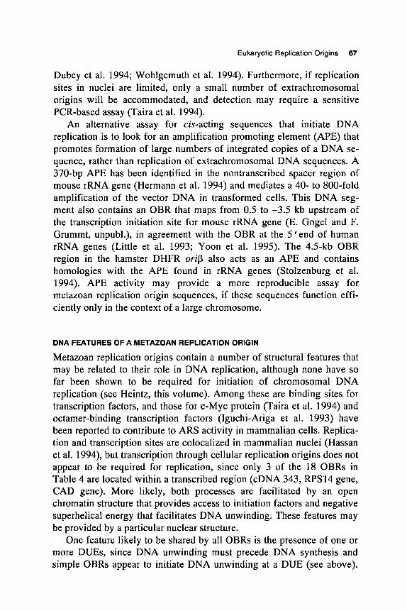
Metazoan replication origins contain a number of structural features that may be related to their role in DNA replication, although none have so far been shown to be required for initiation of chromosomal DNA replication (see Heintz, this volume). Among these are binding sites for transcription factors, and those for c-Myc protein (Taira et al. 1994) and octamer-binding transcription factors (Iguchi-Ariga et al. 1993) have been reported to contribute to ARS activity in mammalian cells. Replica- tion and transcription sites are colocalized in mammalian nuclei (Hassan et al. 1994), but transcription through cellular replication origins does not appear to be required for replication, since only 3 of the 18 OBRs in Table 4 are located within a transcribed region (cDNA 343, RPS14 gene, CAD gene). More likely, both processes are facilitated by an open chromatin structure that provides access to initiation factors and negative superhelical energy that facilitates DNA unwinding. These features may be provided by a particular nuclear structure.
One feature likely to be shared by all OBRs is the presence of one or more DUES, since DNA unwinding must precede DNA synthesis and simple OBRs appear to initiate DNA unwinding at a DUE (see above).
68 M.L. DePamphilis
Potential DUEs exist at or close to several OBRs, including DHFR orip (Dobbs et al. 1994).
A second feature is a densely methylated island (DMI) that consists of 127 bp (RPS14 gene OBR) to 512 bp (DHFR orip) of DNA in which all dC residues are methylated on both strands, regardless of the adjacent nucleotide (Tasheva and Roufa 1994b). This unusual methylation pattern has been observed only in association with replication origins, and then only in proliferating cells. Intriguingly, DNA methyltransferase, the en- zyme responsible for converting hemimethylated sites to methylated sites in nascent DNA, becomes associated with replication foci during S phase (Leonhardt et al. 1992). DMIs might act in a positive way by providing a binding site for a replication-specific factor, by altering DNA structure to promote unwinding at a neighboring DUE, or by altering chromatin structure to increase accessibility to initiation factors. In fact, the DMI overlapping the DHFR orip OBR is flanked by binding sites for RIP60, a protein that can link the two sites to form a 736-bp DNA loop that en- compasses the DMI and flanks a potential DUE (Mastrangelo et al. 1993). By analogy to E. coli DnaA, bacteriophage h 0 protein (Kornberg and Baker 1992), and SV40 T-antigen (see Borowiec, this volume), bind- ing of origin recognition proteins can impose superhelical tension that causes untwisting of DNA in nearby DUEs. Alternatively, by analogy to E. coli oriC (Herrick et al. 1994), the DMI may help to limit initiation to once per S phase. oriC is methylated on both strands at 11 sites. When these sites become hemimethylated as a result of replication, oriC associ- ates with an outer membrane component, delaying rebinding of its origin recognition protein, DnaA, and thus delaying reinitiation.
A third feature is attachment sites for nuclear matrix or nuclear scaf- fold (Nakayasu and Berezny 1989; Hozik et al. 1993), components of nuclear structure that are commonly associated with newly replicated DNA and, in some cases, origins of cellular replication (see Laskey and Madine, this volume).
A fourth feature is palindromic sequences that can collapse into cruciform structures when sufficient negative superhelical energy is pro- vided. These structures may promote initiation of DNA replication at specific sites. Cruciform extrusion at the origin of E. coli plasmid pT181 is promoted by the plasmid-encoded initiator protein RepC (Noirot et al. 1990), and antibodies directed against DNA cruciforms can stimulate overall DNA synthesis and copy number of specific genes in permeabil- ized mammalian cells (Zannis-Hadjopoulos et al. 1988). Moreover, stain- ing mammalian cells with anti-cruciform antibodies suggests that cruciform structures accumulate as cells prepare to enter S phase (Ward
Eukaryotic Replication Origins 69
et al. 1991). A role for cruciforms in replication origins must be consid- ered cautiously, however, since cruciform structures can be a direct result of aberrant DNA replication involved in gene amplification (Cohen et al. 1994).
THE ROLE OF NUCLEAR STRUCTURE IN METAZOAN DNA REPLICATION
Prokaryotic genomes and animal virus genomes can all replicate in the presence of purified soluble proteins and cofactors; no requirement for a cellular structure has been observed, although there exists a transient in- teraction between E. coli oriC and the outer membrane that regulates the rate at which reinitiation can occur at oriC (Herrick et al. 1994). Whether or not the same is true for replication origins in simple eukaryotic organisms remains to be seen. However, one of the most striking require- ments for initiation of DNA replication in metazoan chromosomes is that of nuclear structure (see Laskey and Madine, this volume).
DNA replication in metazoan chromosomes occurs at discrete nuclear foci. Clusters of replication origins initiate replication synchronously (Hand 1978), giving rise to discrete "replication complexes" that contain from 100 to 300 replication forks (see Laskey and Madine, this volume). Formation of these replication complexes accounts for the many observa- tions that newly synthesized DNA is preferentially bound to components of nuclear structure generally referred to as nuclear matrix or nuclear scaffold (Nakayasu and Berezney 1989; Hozik et al. 1993). These replication complexes appear to be assembled in an energy-dependent process prior to S phase at the sites where replication begins (Fig. 2). RP- A, a heterotrimeric single-strand DNA-binding protein that is required for replication of metazoan chromosomes (Fang and Newport 1993), is bound at discrete foci in nuclei prior to DNA unwinding and DNA synthesis (Adachi and Laemmli 1994). High levels of cyclin B/ cdc2 protein kinase, an enzyme that is required for entrance into mitosis, pre- vents the appearance of these RP-A foci, consistent with their absence in mitotic chromosomes (Adachi and Laemmli 1994). Cyclin-A-dependent cdk2 protein kinase, an enzyme that is required for entrance into S phase (Fang and Newport 1991), colocalizes with RP-A (Cardoso et al. 1993). Proliferating cell nuclear antigen (PCNA), a cofactor for DNA poly- merase-6, and DNA polymerase-a, an enzyme required for synthesis of Okazaki fragments, are also found at replication foci in S-phase nuclei (Kill et al. 1991). Whether or not they are prebound to these foci before replication begins is not clear. Presumably, licensing factor (see Laskey
70 M.L. DePamphilis
and Madine; Blow and Chong; both this volume), a cytoplasmic initia- tion factor that gains access to replication origins only when the nuclei become permeable during mitosis, also binds to preinitiation complexes.
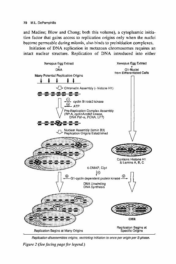
Initiation of DNA replication in metazoan chromosomes requires an intact nuclear structure. Replication of DNA introduced into either
Xenopus Egg Extract
DNA
Many Potential Replication Origins
+ Xenopus Egg Extract
G1-Nuclei from Differentiated Cells
+ - 0 Chromatin Assembly (- histone H1)
- cyclin Bl/cdc2 kinase
Pre-Replication Complex Assembly (RP-A, cyclinNcdk2 kinase,
DNA Pol-a, PCNA, LF?)
Nuclear Assembly (lamin 83) Replication Origins Established
10 s G 1 - c y c l i n dependent protein k i n a s e x fl
V DNA Unwinding DNA Synthesis
Replication disassembles origins, restricting initiation to once per origin per S-phase.
Figure 2 (See facing page for legend.)
Eukaryotic Replication Origins 71
Xenopus eggs or egg extracts does not occur unless DNA is first as- sembled into chromatin and then organized into a nuclear structure that includes lamin B3 and functional nuclear pores (see Laskey and Madine; Blow; both this volume). In addition, the nuclear envelope is instrumen- tal in regulating the onset of S phase, apparently by regulating access of chromosomal DNA to one or more initiation factors (licensing factor) present in the cytoplasm (Coverley and Laskey 1994).
Site-specific initiation requires an intact nuclear structure. Xenopus egg extract can initiate DNA replication in purified DNA molecules only after the DNA is organized into a pseudo-nucleus, but under these condi- tions, DNA replication is independent of DNA sequence and begins at many sites distributed throughout the molecules. However, Xenopus egg extract can initiate DNA replication at specific sites in mammalian chromosomes, but only when the DNA is presented in the form of an in- tact nucleus from differentiated cells (Gilbert et al. 1995). Initiation of DNA synthesis in nuclei isolated from G1-phase hamster cells is dis- tinguished from continuation of DNA synthesis at preformed replication forks in S-phase nuclei by a delay that precedes DNA synthesis, a depen- dence on soluble Xenopus egg factors, sensitivity to the protein kinase in- hibitor 6-dimethylaminopurine (DMAP), and complete labeling of na- scent DNA chains. Initiation sites for DNA replication were mapped downstream from the amplified DHFR gene region by (1) identification of the earliest labeled DNA fragments (Gilbert et al. 1993), (2) quantita- tive hybridization of newly synthesized DNA to double-stranded DNA probes to reveal genomic loci where DNA synthesis began, and (3)
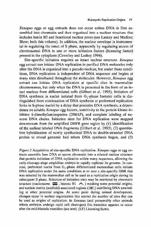
Figure 2 Acquisition of site-specific DNA replication. Xenopus eggs or egg ex- tracts assemble bare DNA or sperm chromatin into a relaxed nuclear structure that permits initiation of DNA replication within many sequences, allowing the early-cleavage-stage amphibian embryo to rapidly replicate its genome. In con- trast, preformed nuclei from G1-phase differentiated mammalian cells initiate DNA replication under the same conditions at or near a site-specific OBR that was selected by the mammalian cell to be used as a replication origin during its subsequent S phase. Selection of initiation sites may be restricted by chromatin structure (nucleosome , histone H1 A ) masking some potential origins, and nuclear matrix (scaffold) associated regions (X) stabilizing DNA unwind- ing at other potential origins. At some point during animal development, changes occur in nuclear organization that restrict the number of sites that can be used as origins of replication. In Xenopus (and presumably other animals whose embryos undergo rapid cell cleavages) this transition appears to occur after the mid-blastula transition (see text). (LF) Licensing factor.
72 M.L. DePamphilis
quantitative hybridization of Okazaki fragments to single-stranded DNA probes to reveal the transition between continuous and discontinuous DNA synthesis on each template within this initiation locus. When bare DNA substrates are used, then Xenopus eggs or egg extracts do not dis- tinguish between prokaryotic DNA, hamster DNA that does not contain a replication origin, and hamster DNA that does contain a replication origin. Moreover, initiation events were distributed equally throughout a 30-kb cosmid containing the DHFR orip region. When nuclei are used, Xenopus egg extract continues DNA synthesis in S-phase nuclei at sites that had been initiated in hamster cells (e.g, DHFR orip). When the in- tegrity of the nuclear membrane is preserved, Xenopus egg extract ini- tiates DNA replication in G1-phase nuclei specifically at or near the OBR (orip) utilized by hamster cells. When nuclear integrity is damaged, pref- erence for initiation at orip is significantly reduced or eliminated. There- fore, initiation sites for DNA replication in mammalian cells are estab- lished prior to S phase by some component of differentiated nuclear structure, and this replication origin can be recognized by soluble initia- tion factors present in Xenopus eggs.
Subsequent studies (Wu and Gilbert 1996) have revealed that Xenopus egg extract initiates replication at many sites throughout the DHFR gene region in nuclei isolated from early G1-phase hamster cells, whereas the same extract initiates specifically at orip in nuclei isolated from late G1-phase hamster cells. Therefore, specific origins of replica- tion in mammalian chromosomes are reestablished during each cell divi- sion cycle several hours after nuclear assembly occurs.
MODEL FOR METAZOAN ORIGINS OF DNA REPLICATION
There are three basic models for understanding the cumulative data on metazoan replication origins. The first is the "strand separation model'' in which extensive DNA unwinding precedes initiation of RNA-primed DNA synthesis at many sites concurrently on both templates (Benbow et al. 1992). This model was based largely on reports of single-stranded DNA in Xenopus and Drosophilu embryos, but the single-stranded DNA bubbles that one would expect as replication intermediates have not been detected by 2-D gel analyses of DNA replication in Xenopus eggs (Hyrien and MCchali 1992; Mahbubani et al. 1992) or Drosophilu em- bryos (Liang and Gerbi 1994), or by electron microscopy of DNA replicating in mammalian cells (Hamlin et al. 1992; Gruss et al. 1994). On the contrary, the fact that replication forks and bubbles are detected by these methods, and the fact that replication fork polarity is observed
Eukaryotic Replication Origins 73
by hybridization of either Okazaki fragments or long nascent DNA chains to complementary DNA templates (see above, Mapping Origins of Replication in Metazoan Chromosomes), provide compelling evidence that DNA synthesis occurs at replication forks in both simple and com- plex genomes.
A second model is one in which a replication complex assembles at the OBR, but then migrates to other positions on either side of the OBR before initiating DNA unwinding and DNA synthesis. This would gener- ate a Gaussian distribution of initiation events with the OBR at its apex. However, one would expect the OBR sequence to strongly stimulate DNA replication under all conditions, a prediction that is difficult to reconcile with the lack of DNA sequence preference observed in the eggs and cleavage-stage embryos of frogs, flies, fish, and sea urchins.
A third model is suggested by the Jesuit dictum that "many are called, but few are chosen" and perhaps offers the simplest way of interpreting all the data (Fig. 3) (DePamphilis 1993a,b,d; Burhans and Huberman 1994). Whereas naked DNA contains many possible sites where replica- tion can begin, assembly of DNA into chromatin can suppress initiation at some of these sites, and organization of chromatin into a nuclear struc- ture can activate DNA replication at other sites by promoting DNA un- winding. Thus, initiation sites for DNA replication in metazoan chromosomes would consist of three parts:
1. OBR. Most initiation events occur bidirectionally within a 0.5- to 2- kb locus. In differentiated cells, where most potential replication origins are suppressed, favored sites may consist of an easily un- wound DNA sequence in combination with other origin components such as DMIs, ORES, and transcription-factor-binding sites. These features, in addition to sequences that may attach to nuclear matrix (scaffold) in order to stabilize unwound DNA at the OBR (Bode et a]. 1992), would comprise the site-specific, heritable replication origins that have been mapped in metazoan chromosomes (Table 1).
2. Initiation zone. Some initiation events are detected randomly distrib- uted throughout a 6-kb to 55-kb region that encompasses the OBR. These sequences may remain accessible to replication factors so that the same cell can occasionally initiate replication at a nearby DUE. Alternatively, some cells within a population simply may have selected a different OBR in the same region, or are capable of utiliz- ing more than one OBR in the same region, the choice of which can change during successive S phases. Secondary initiation sites also oc- cur in simple genomes, although usually at a lower frequency. For ex- ample, papovavirus replication origins are at least 100-fold more effi-
74 M.L. DePamphilis
1001 loo]
Simple
A
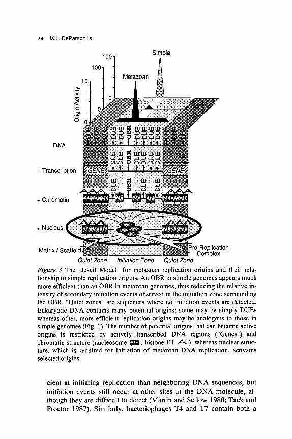
Quiet Zone Initiation Zone Quiet Zone Figure 3 The "Jesuit Model" for metazoan replication origins and their rela- tionship to simple replication origins. An OBR in simple genomes appears much more efficient than an OBR in metazoan genomes, thus reducing the relative in- tensity of secondary initiation events observed in the initiation zone surrounding the OBR. "Quiet zones" are sequences where no initiation events are detected. Eukaryotic DNA contains many potential origins; some may be simply DUES whereas other, more efficient replication origins may be analogous to those in simple genomes (Fig. 1). The number of potential origins that can become active origins is restricted by actively transcribed DNA regions ("Genes") and chromatin structure (nucleosome m, histone H1 A), whereas nuclear struc- ture, which is required for initiation of metazoan DNA replication, activates selected origins.
cient at initiating replication than neighboring DNA sequences, but initiation events still occur at other sites in the DNA molecule, al- though they are difficult to detect (Martin and Setlow 1980; Tack and Proctor 1987). Similarly, bacteriophages T4 and T7 contain both a
Eukaryotic Replication Origins 75
primary replication origin and several secondary origins (Kornberg and Baker 1992). If we compare site specificity in a metazoan ge- nome with that in a simple genome, initiation events outside the meta- zoan OBR would go undetected unless the scale is expanded to ac- commodate the lower activity of a metazoan replication origin (Fig.
3. Quiet zone. In contrast to DNA replication in rapidly cleaving em- bryos where initiation events are detected throughout the genome, ini- tiation events in differentiated cells are restricted to specific sites (OBR + initiation zone). Initiation events outside the initiation zone may be further suppressed by active transcription (Haase et al. 1994) and higher-order chromatin structure (Fig. 3) (Hand 1978; Forrester et al. 1990; Simpson 1990; Ferguson and Fangman 1992; Karpen and Spradling 1992), two features that are generally absent in rapidly cleaving embryos (Fig. 2) (Wolffe 1994).
DNA replication in Xenopus egg extracts provides a simple paradigm for the selection and assembly of initiation sites for DNA replication (Fig. 2). Naked DNA contains many potential sites where DNA replica- tion can begin. These sites most likely correspond to easily unwound DNA sequences that are components of replication origins in simple (Natale et al. 1993) as well as metazoan (Dobbs et al. 1994) genomes, and that can promote site-specific replication in plasmid DNA (Ishimi et al. 1994). When naked DNA is introduced into Xenopus eggs or egg ex- tract, chromatin is assembled in the absence of histone H1, which is re- quired for compaction of DNA into 30-nm fibers (Wolffe 1994). In addi- tion, nuclei assembled in Xenopus eggs are less compact than nuclei in differentiated cells. This may be due partially to the fact that the com- position of nuclear lamin proteins, a component of nuclear structure re- quired for DNA replication (Newport et al. 1990; Jenkins et al. 1993), is simpler in Xenopus embryos than in Xenopus differentiated cells (Bena- vante et al. 1985; Stick and Hausen 1985). This relaxed environment may allow assembly of replication complexes at any one of the many potential origins located within a large DNA region, thus facilitating rapid replication of the Xenopus embryonic genome. The fact that repli- cation does not initiate at more than one site within a single plasmid molecule (Hines and Benbow 1982; MCchali and Kearsey 1984; Mahbu- bani et al. 1992; Hyrien and MCchali 1992) suggests that replication in embryos undergoing rapid nuclear division cycles (e.g., frogs, flies, fish, sea urchins) occurs at only one of many potential sites within a large DNA region. Formation of replication complexes does not require nuclear membrane formation (Adachi and Laemmli 1994), but initiation
3).
76 M.L. DePamphilis
of DNA replication does, suggesting that some component of nuclear structure interacts directly with each replication origin. Moreover, activa- tion of replication complexes appears to require the action of G1-cyclin- dependent protein kinase cdk2. General inhibitors of protein kinases (e.g., DMAP) prevent initiation of DNA replication in either sperm chromatin or GI nuclei, but do not inhibit active replication forks (Blow 1993; Kubota and Takisawa 1993; Gilbert et al 1995). Cipl protein, a specific inhibitor of cdk2 kinase, exhibits a similar effect, and this inhibi- tion can be overcome with cyclin E (Strausfeld et al. 1994). Cipl inhibi- tion occurs after formation of replication complexes but before DNA un- winding has begun (Adachi and Laemmli 1994).
It has long been observed that the density of initiation sites (origin- to-origin distance) in metazoan chromosomes varies in different cell types and under different experimental conditions, leading to the conclu- sion that many more replication sites are available than are used during a normal S phase, and that the number of active replication origins depends on the ratio of initiation factors to DNA and accessibility of origins as determined by higher-order chromatin structure (Hand 1978). As animal development progresses and cells begin to undergo differentiation, a 5- to 10-fold reduction can occur in the frequency of initiation sites (Blumenthal et al. 1974; McKnight and Miller 1977; Buongiorno- Nardelli et al. 1982; Shinomiya and Ina 1991). In fact, although initiation events in Xenopus rRNA gene region are distributed randomly through- out these sequences during early development (prior to gastrulation), ini- tiation events become confined to the intergenic regions as development progresses (Hyrien et al. 1995). Changes in chromatin and nuclear struc- ture that occur at the mid-blastula transition in Xenopus, for example, may repress initiation of DNA replication at some loci while facilitating it at others (Fig. 3) (Gilbert et al. 1995). In yeast, for example, at least half of the ARS elements on chromosome I11 are not active as chromo- somal replication origins (Newlon et al. 1993). Factors that likely con- tribute to these changes in metazoa include the onset of transcription and cell differentiation, and the appearance of histone H1 and lamins A and C. Thus, some of the initiation sites chosen in fertilized Xenopus eggs may remain as origins throughout development, whereas other sites may be eliminated.
SUMMARY AND PERSPECTIVE
DNA replication in differentiated eukaryotic cells is initiated at specific DNA sites, regardless of whether the genome is viral or mitochondrial,
Eukaryotic Replication Origins 77
or whether the organism consists of one cell type or many cell types. Per- haps the most basic difference among origins is whether they are designed to initiate once per cell division cycle or more than once. How- ever, whereas. the anatomy of initiation sites in most simple genomes is fairly clear and their mechanism of action is understood in principle, if not in detail, initiation sites in metazoan chromosomes remain elusive: Their requirement for nuclear structure makes them more complex than those in simple genomes, and the sequences that determine them have not yet been identified. Metazoan replication origins may follow the same principles revealed by replication origins in simple genomes but solve the problems of determining where, when, and how to initiate DNA synthesis in different ways. Why such complexity appears in the chromosomes of metazoa but not single-cell organisms may involve dif- ferences in chromatin structure and nuclear organization that accompany the appearance of histone H1, nuclear lamins, and nuclear envelope breakdown during mitosis in metazoans.
The concept of a metazoan origin as a specific nucleoprotein complex that interacts with soluble replication factors, rather than simply a specific sequence that is recognized by soluble factors, can account for three important features of metazoan DNA replication. First, it provides a way to change the number of origins per chromosomal locus at different stages in animal development, thus accommodating the need for shorter S phases in the early developmental stages of some animals. Second, it provides a way to limit the number of origins per genome without simul- taneously limiting the cell’s capacity to sustain genetic alterations. A large number of less efficient origins that demand less sequence specificity allow more flexibility for rearranging genetic information than a small number of highly efficient origins that require a great deal of sequence specificity. Finally, it provides a way to restrict initiation at each origin to once per S phase. Initiation at each replication origin would be restricted to once per cell cycle if the nucleoprotein complex that defines a replication origin in GI nuclei was disrupted by the act of replication. In that event, reinitiation at the same site would be prevented, even in the presence of excess soluble initiation factors.
Metazoan replication origins in G , nuclei of differentiated mam- malian cells may be analogous to the six-protein origin replication com- plex assembled at yeast ARS sites (see Newlon, this volume) or the EBNAl protein/origin core DNA complex in EBV episomes (see Yates, this volume), both of which appear to be stable DNA-protein complexes that exist throughout the cell cycle. Thus, a licensing factor (see Laskey and Madine, this volume) may recognize this DNNprotein platform,
78 M.L. DePamphilis
permitting it to be activated by a G1-cyclin-dependent protein kinase. Licensing factor is inactivated by the replication process and then re- placed when the nuclear membrane becomes permeable to cytoplasmic factors during mitosis.
ACKNOWLEDGMENTS
I am indebted to Darren Natale, David Gilbert, William Burhans, and many other friends and colleagues for their intellectual contributions to this paper.
REFERENCES
Adachi. Y. and U.K. Laemmli. 1994. Study of the cell cycle-dependent assembly of the DNA pre-replication centres in Xenopus egg extract. EMBO J. 13: 4153-4164.
Ariizumi, K., Z. Wang, and P.W. Tucker. 1993. Immunoglobulin heavy chain enhancer is located near or in an initiation zone of chromosomal DNA replication. Proc. Natl. Acad. Sci. 90: 3695-3699.
Benavante, R., G. Krohne, and W.W. Franke. 1985. Cell type-specific expression of nuclear lamina proteins during development of Xenopus laevis. Cell 41: 177-190.
Benbow, R.M., J. Zhao, and D.D. Larson. 1992. On the nature of origins of DNA replica- tion in eukaryotes. BioEssays 14: 661-670.
Berberich, S., A. Trivedi, D.C. Daniel, E.M. Johnson, and M. Leffak. 1995. In vitro replication of plasmids containing human c-myc DNA. J. Mol. Biol. 245: 92-109.
Blow, J.J. 1993. Preventing re-replication of DNA in a single cell cycle: Evidence for a replication licensing factor. J. Cell Biol. 122: 993-1002.
Blumenthal, A.B., H.J. Kriegstein, and D.S. Hogness. 1974. The units of DNA replica- tion in Drosophila melanogaster chromosomes. Cold Spring Harbor Symp. Quant. Biol. 38: 205-223.
Bode, J., Y. Kohwi, L. Dickinson, T. Joh, D. Klehr, C. Mielke, and T. Kohwi- Shigematsu. 1992. Biological significance of unwinding capability of nuclear matrix- associating DNAs. Science 55: 195-1 97.
Bonifer, C., N. Yannoutsos, G. Kriiger, F. Grosveld, and A.E. Sippel. 1994. Dissection of the locus control function located on the chicken lysozyme gene domain in transgenic mice. Nucleic Acids Res. 22: 4202-4210.
Brewer, B.J. and W.L. Fangman. 1993. Initiation at closely spaced replication origins in a yeast chromosome. Science 262: 1728-1731.
. 1994. Initiation preference at a yeast origin of replication. Proc. Natl. Acad. Sci.
Bullock, P.A., Y.S. Seo, and J. Hurwitz. 1991. Initiation of SV40 DNA synthesis in vitro. Mol. Cell. Biol. 11: 2350-2361.
Buongiorno-Nardelli, M., G. Micheli, M.T. Carri, and M. Marilley. 1982. A relationship between replicon size and supercoiled loop domains in the eukaryotic genome. Nature
Burhans, W.C. and J.A. Huberman. 1994. DNA replication origins in animal cells: A
91: 3418-3422.
298: 100-102.
question of context? Science 263: 639-640.
Eukaryotic Replication Origins 79
Burhans, W.C., L.T. Vassilev, M.S. Caddle, N.H. Heintz, and M.L. DePamphilis. 1990. Identification of an origin of bidirectional DNA replication in mammalian chromosomes. Cell 62: 955-965.
Burhans, W.C., L.T. Vassilev, J. Wu, J.M. Sogo, F.N. Nallaseth, and M.L. DePamphilis. 1991. Emetine allows identification of origins of mammalian DNA replication by im- balanced DNA synthesis, not through conservative nucleosome segregation. EMBO J.
Caddle, M.S. and M.P. Calos. 1992. Analysis of the autonomous replication behavior in human cells of the DHFR putative origin of replication. Nucleic Acids Res. 20:
-. 1994. Specific initiation at an origin of replication from Schizosaccharomyces pombe. Mol. Cell. Biol. 14: 1796-1805.
Cardoso, M.C., H. Leonhardt, and B. Nadal-Ginard. 1993. Reversal of terminal differen- tiation and control of DNA replication: Cyclin A and cdk2 specifically localize at sub- nuclear sites of DNA replication. Cell 74: 979-992.
Carroll, S.M., M.L. DeRose, J.L. Kolman, G.H. Nonet, R.E. Kelly, and G.M. Wahl. 1993. Localization of a bidirectional DNA replication origin in the wild type and in episomally amplified murine ADA loci. Mol. Cell. Biol. 13: 2971-2981.
Cohen, S., D. Hassin, S. Karby, and S. Lavi. 1994. Hairpin structures are the primary amplification products: A novel mechanism for generation of inverted repeats during gene amplification. Mol. Cell. Biol. 14: 7782-7791.
Coverley, D. and R.A. Laskey. 1994. Regulation of eukaryotic DNA replication. Annu. Rev. Biochem. 63: 745-776.
Cusick, M.E., P.M. Wassarman, and M.L. DePamphilis. 1989. Application of nucleases to visualizing chromatin organization at replication forks. Methods Enzymol. 170:
Deb, S.P., A.L. DeLucia, C. Bauer, A. Koff, and P. Tegtmeyer. 1986. Domain and struc-
Delidakis, C. and F.C. Kafatos. 1989. Amplification enhancers and replication origins in
DePamphilis, M.L. 1993a. Eukaryotic DNA replication: Anatomy of an origin. Annu.
-. 1993b. Origins of DNA replication that function in eukaryotic cells. Curr. Opin.
-. 1993c. How transcription factors regulate origins of DNA replication in
-. 1993d. Origins of DNA replication in metazoan chromosomes. J. Biol. Chem.
DePamphilis, M.L. and P.M. Wassarman. 1980. Replication of eukaryotic chromosomes: A close-up of the replication fork. Annu. Rev. Biochem. 49: 627-666.
DePamphilis, M.L., E. Martinez-Salas, D.Y. Cupo, E.A. Hendrickson, C.E. Fritze, W.R. Folk, and U. Heine. 1988. Initiation of polyomavirus and SV40 DNA replication, and the requirements for DNA replication during mammalian development. Cancer Cells 6:
Dijkwel, P.A. and J.L. Hamlin. 1992. Initiation of DNA replication in the DHFR locus is confined to the early S-period in CHO cells synchronized with the plant amino acid mimosine. Mol. Cell. Biol. 12: 3715-3722.
-. 1995. The Chinese hamster DHFR origin consists of multiple potential nascent
10: 4351-4360.
5971-5978.
290-316.
ture of the SV40 core origin of replication. Mol Cell. Biol. 6: 1663-1670.
the autosomal chorion gene cluster of Drosophila. EMBO J. 8: 891-901.
Rev. Biochem. 62: 29-63.
Cell Biol. 5: 434-441.
eukaryotic cells. Trends Cell Biol. 3: 161-167.
268: 1-4.
1 65- 175.
80 M.L. DePamphilis
strand start sites. Mol. Cell. Biol. 15: 3023-3031. Dobbs, D.L., W.-L. Shaiu, and R.M. Benbow. 1994. Modular sequence elements associ-
ated with origin regions in eukaryotic chromosomal DNA. Nucleic Acids Res. 22:
Dubey, D.D., J. Zhu, D.L. Carlson, K. Sharma, and J.A. Huberman. 1994. Three ARS elements contribute to the ura4 replication origin region in the fission yeast, Schizosac- charomyces pombe. EMBO J. 13: 3638-3647.
Fang, F. and J. Newport. 1991. Evidence that the G1-S and G2-M transitions are con- trolled by different cdc2 proteins in higher eukaryotes. Cell 66: 731-742.
-. 1993. Distinct roles of cdk2 and cdc2 in RP-A phosphorylation during the cell cycle. J. Cell Sci. 106: 983-994.
Fangman, W.L. and B.J. Brewer. 1991. Activation of replication origins within yeast chromosomes. Annu. Rev. Cell Biol. 7 : 375-402.
Ferguson, B.M. and W.L. Fangman. 1992. A position effect on the time of replication origin activation in yeast. Cell 68: 333-339.
Forrester, W.C., E. Epner, M.C. Driscoll, T. Enver, M. Brice, T. Papayannopoulou, and M. Groudine. 1990. A deletion of the human 0-globin locus activation region causes a major alteration in chromatin structure and replication across the entire 0-globin region. Genes Dev. 4: 1637- 1649.
Frappier, L. and M. O’Donnell. 1991. Overproduction, purification and characterization of EBNAl, the origin binding protein of Epstein-Barr virus. J. Biol. Chem. 266:
Frappier, L., K. Goldsmith, and L. Bendell. 1994. Stabilization of the EBNAl protein on the EBV latent origin of DNA replication by a DNA looping mechanism. J. Biol. Chem. 269: 1057-1062.
Gale, J.M., R.A. Tobey, and J.A. D’Anna. 1992. Localization and DNA sequence of a replication origin in the rhodopsin gene locus of Chinese hamster cells. J. Mol. Biol.
Gerard, R. and Y. Gluzman. 1986. Functional analysis of the role of the A m rich region and upstream flanking sequences in SV40 DNA replication. Mol. Cell. Biol. 6:
Giacca, M., L. Zentilin, P. Norio, S. Diviacco, D. Dimitrova, G. Contreas, G. Biamonti, G. Perini, F. Weighardt, S. Riva, and A. Falaschi. 1994. Fine mapping of a replication origin of human DNA. Proc. Natl. Acad. Sci. 91: 7119-7124.
Gilbert, D.M. and S.N. Cohen. 1987. BPV plasmids replicate randomly in mouse fibroblasts throughout S-phase of the cell cycle. Cell 50: 59-68.
Gilbert, D.M., H. Miyazawa, and M.L. DePamphilis. 1995. Site-specific initiation of DNA replication in Xenopus egg extract requires nuclear structure. Mol. Cell. Biol.. 15:
Gilbert, D.M., H. Miyazawa, F.S. Nallaseth, J.M. Ortega, J.J. Blow, and M.L. DePamphilis. 1993. Site-specific initiation of DNA replication in metazoan chromo- somes and the role of nuclear organization. Cold Spring Harbor Symp. Quant. Biol. 58: 475-485.
Gillette, T.G., M. Lusky, and J. Borowiec. 1994. Induction of structural changes in the BPV-1 origin of replication by the viral E l and E2 proteins. Proc. Natl. Acad. Sci. 91:
2479-2489.
7819-7826.
224: 343-352.
4570-4577.
2942-2954.
8846-8850. Gruss, C. and J.M. Sogo. 1992. Chromatin replication. BioEssays 14: 1-8. Gruss, C., J. Wu, and JM. Sogo. 1994. Disruption of nucleosomes at the replication fork.
Eukaryotic Replication Origins 81
E M 6 0 J . 12: 4533-4545. Guo, Z.4. and M.L. DePamphilis. 1992. Specific transcription factors stimulate both
simian virus 40 and polyomavirus origins of DNA replication. Mol. Cell. Biol. 12:
Guo, Z.-S., U. Heine, and M.L. DePamphilis. 1991. T-antigen binding to site 1 facilitates initiation of SV40 DNA replication but does not affect bidirectionality. Nucleic Acids Res. 19: 7081-7088.
Gutieirez, C., Z-S. Guo, J. Roberts, M.L. DePamphilis. 1990. Simian virus origin auxil- iary sequences weakly facilitate binding of T-antigen, but strongly facilitate initiation of DNA unwinding. Mol. Cell. Biol. 10: 1719-1728.
Gutierrez, C., Z.-S. Guo, W. Burhans, M.L. DePamphilis, J. Farrell-Towt, and G. Ju. 1988. Is c-myc protein directly involved in DNA replication? Science 240: 1202-1203.
Haase, S.B. and M.P. Calos. 1991. Replication control of autonomously replicating hu- man sequences. Nucleic Acids Res. 19: 5053-5058.
Haase, S.B., S.S. Heinzel, and M.P. Calos. 1994. Transcription inhibits the replication of autonomously replicating plasmids in human cells. Mol. Cell. Biol. 14: 2516-2526.
Hamlin, J.L., P.A. Dijkwel, and J.P. Vaughn. 1992. Initiation of replication in the Chinese hamster DHFR domain. Chromosomu (suppl. 1) 102: 17-23.
Hand, R. 1978. Eucaryotic DNA: Organization of the genome for replication. Cell 15:
Handeli, S., A. Klar, M. Meuth, and H. Cedar. 1989. Mapping replication units in animal cells. Cell 57: 909-920.
Harrison, S., K. Fisenne, and J. Hearing. 1994. Sequence requirements of the EBV latent origin of DNA replication. J. Virol. 68: 1913-1925.
Hassan, A.B., R.J. Errington, N.S. White, D.A. Jackson, and P.R. Cook. 1994. Replica- tion and transcription sites are colocalized in human cells. J. Cell Sci. 107: 425-434.
He, Z., B.T. Brinton, J. Greenblatt, J.A. Hassell, and C.J. Ingles. 1993. Transactivator proteins VP16 and GAL4 bind replication factor A. Cell 73: 1223-1232.
Heck, M.M.S. and A.C. Spradling. 1990. Multiple replication origins are used during Drosophilu chorion gene amplification. J. Cell Biol. 4: 903-914.
Hermann, C., E. Gartner, U.H. Weidle, and F. Grummt. 1994. High copy expression vec- tor based on amplification promoting sequences. DNA Cell Biol. 13: 437-445.
Herrick, J., R. Kern, S. Guha, A. Landulsi, 0. Fayet, A. Malki, and M. Kohiyama. 1994. Parental strand recognition of the DNA replication origin by the outer membrane in E. coli. EMBO J. 13: 4695-4703.
Hines, P.J. and R.M. Benbow. 1982. Initiation of replication at specific origins in DNA molecules microinjected into unfertilized of the frog Xenopus Iuevis. Cell 30: 459-468.
Holmes, S.G. and M.M. Smith. 1989. Interaction of the H4 autonomously replicating se- quence core consensus sequence and its 3'-flanking domain. Mol. Cell. Biol. 9:
Hozhk P., A.B. Hassan, D.A. Jackson, and P.R. Cook. 1993. Visualization of replication factories attached to nucleoskeleton. Cell 73: 361-373.
Huang, R.-Y. and D. Kowalski. 1993. A DNA unwinding element and an ARS consensus comprise a replication origin within a yeast chromosome. E M 6 0 J. 12: 4521-4531.
Huberman, J.A. 1994. Analysis of DNA replication origins and directions by two- dimensional gel electrophoresis. In The cell cycle: A practical approach (ed. P. Fantes and R.F. Brooks), pp. 213-234. Oxford University Press, United Kingdom.
Hyrien, 0. and M. Mechali. 1992. Plasmid replication in Xenopus eggs and egg extracts:
25 14-2524.
31 7-325.
5464-5472.
82 M.L. DePamphilis
A 2D gel electrophoretic analysis. Nucleic Acids Res. 20: 1463-1469. -. 1993. Chromosomal replication initiates and terminates at random sequences but
at regular intervals in the ribosomal DNA of Xenopus early embryos. EMBO J. 12:
Hyrien, O., C. Maric, and M. Mtchali. 1995. Transition in specification of embryonic metazoan DNA replication origins. Science 270: 994-997.
Iguchi-Ariga, S.M.M., N. Ogawa, and H. Ariga. 1993. Identification of the initiation region of DNA replication in the murine immunoglobulin heavy chain gene and pos- sible function of the octamer motif as a putative DNA replication origin in mammalian cells. Biochim. Biophys. Acta 1172: 73-81.
Ishimi, Y., K. Matsumoto, and R. Ohba. 1994. DNA replication from initiation zones of mammalian cells in a model system. Mol. Cell. Biol. 14: 6489-6496.
Jenkins, H., T. Holman, C. Lyon, B. Lane, R. Stick, and C. Hutchison. 1993. Nuclei that lack a lamina accumulate karyophilic proteins and assemble a nuclear matrix. J. Cell
Karpen, G.H. and A.C. Spradling. 1992. Reduced DNA polytenization of a mini- chromosome region undergoing position effect variegation in Drosophila. Cell 63:
Kelly, R.E., M.L. DeRose, B.W. Draper, and G.M. Wahl. 1995. Identification of an origin of bidirectional replication within the coding region of the ubiquitously ex- pressed CAD gene. Mol. Cell. Biol. 15: 4136-4148.
Kill, I.R., J.M. Bridges, K.H.S. Campbell, G. Maldonado-Codina, and C.J. Hutchison. 1991. The timing of the formation and usage of replicase clusters in S-nuclei of human diploid fibroblasts. J. Cell Sci. 100: 869-876.
Kipling, D. and S.E. Kearsey. 1990. Reversion of autonomously replicating sequence mutations in 5’. cerevisiae: Creation of a eucaryotic replication origin within pro- caryotic vector DNA. Mol. Cell. Biol. 10: 265-272.
Kitsberg, D., S. Selig, 1. Keshet, and H. Cedar. 1993. Replication structure of the human p-globin gene domain. Nature 366: 588-590.
Koff, A., J.F. Schwedes, and P. Tegtmeyer. 1991. HSV origin-binding protein (UL9) loops and distorts the viral replication origin. J. Virol. 65: 3284-3292.
Kohara, Y., N. Tohdoh, X.-W. Jiang, and T. Okazaki. 1985. The distribution and properties of RNA primed initiation sites of DNA synthesis at the replication origin of Escherichia coli chromosome. Nucleic Acids Res. 13: 6847-6866.
451 1-4520.
Sci. 106: 275-285.
97-107.
Kornberg, A. and T. Baker. 1992. DNA replication. W.H. Freeman, New York. Kowalski, D. and M.J. Eddy. 1989. The DNA unwinding element: A novel, cis-acting
component that facilitates opening of the Escherichia coli replication origin. EMBO J. 8: 4335-4344.
Krysan, P.J., J.G. Smith, and M.P. Calos. 1993. Autonomous replication in human cells of multimers of specific human and bacterial DNA sequences. Mol. Cell. Biol. 13:
Kubota, Y. and H. Takisawa. 1993. Determination of initiation of DNA replication before and after nuclear formation in Xenopus egg cell free extracts. J. Cell Biol. 123:
Leonhardt, H, A.W. Page, H.U. Weier, and T.H. Bestor. 1992. A targeting sequence directs DNA methyltransferase to sites of DNA replication in mammalian nuclei. Cell
Li, R. and M.R. Botchan. 1993. Acidic transcription activation domains of VP16 and p53
2688-2696.
1321-1331.
71: 865-873.
Eukaryotic Replication Origins 83
bind cellular replication protein A and stimulate in vitro BPV-1 DNA replication. Cell
Liang, C. and S.A. Gerbi. 1994. Analysis of an origin of DNA amplification in Sciara coprophila by a novel three dimensional gel method. Mol. Cell. Biol. 14: 1520-1530.
Liang, C., J.D. Spitzer, H.S. Smith, and S.A. Gerbi. 1993. Replication initiates at a con- fined region during DNA amplification in Sciara DNA puff II/9A. Genes Dev. 7 :
Lin, S . and D. Kowalski. 1994. DNA helical instability facilitates initiation at the SV40 replication origin. J. Mol. Biol. 235: 496-507.
Little, R.D., T.H.K. Platt, and C.L. Schildkraut. 1993. Initiation and termination of DNA replication in human rRNA genes. Mol. Cell. Biol. 13: 6600-6610.
Mahbubani, H.M., T. Paull, J.K. Elder, and J.J. Blow. 1992. DNA replication initiates at multiple sites on plasmid DNA in Xenopus egg extracts. Nucleic Acids Res. 22:
Majumder, S. and M.L. DePamphilis. 1994. TATA-dependent enhancer stimulation of promoter activity in mice is developmentally acquired. Mol. Cell. Biol. 14: 4258-4268.
Majumder, S., M. Miranda, and M.L. DePamphilis. 1993. Analysis of gene expression in mouse preimplantation embryos demonstrates that the primary role of enhancers is to relieve repression of promoters. EMBO J. 12: 1131-1140.
Marahrens, Y. and B. Stillman. 1994. Replicator dominance in a eukaryotic chromosome.
Martin, R.G. and V.P. Setlow. 1980. Initiation of SV40 DNA synthesis is not unique to the replication origin. Cell 20: 381-391.
Martinez-Salas, E., D.Y. Cupo, and M.L. DePamphilis. 1988. The need for enhancers is acquired upon formation of a diploid nucleus during early mouse development. Genes Dev. 2: 1115-1126.
Mastrangelo, LA., P.G. Held, L. Dailey, J.S. Wall, P.V.C. Hough, N. Heintz, and N.H. Heintz. 1993. RIP60 dimers assemble link structures at an origin of bidirectional replication in the DHFR amplicon of CHO cells. J . Mol. Biol. 232: 766-778.
Masukata, H., H. Satoh, C. Obuse, and T. Okazaki. 1993. Autonomous replication of hu- man chromosomal DNA fragments in human cells. Mol. Biol. Cell 4: 1121-1132.
McKnight, S.L. and O.L. Miller, Jr. 1977. Electron microscopic analysis of chromatin replication in the cellular blastoderm Drosophila melanogaster embryo. Cell 12:
MCchali, M. and S. Kearsey. 1984. Lack of specific sequence requirement for DNA replication in Xenopus eggs compared with high sequence specificity in yeast. Cell 38:
Melendy, T. and B. Stillman. 1993. An interaction between replication protein A and T- antigen appears essential for primosome assembly during SV40 DNA replication. J. Biol. Chem. 268: 3389-3395.
Miller, C.A. and D. Kowalski. 1993. cis-Acting components in the replication origin from ribosomal DNA of Saccharomyces cerevisiae. Mol. Cell. Biol. 13: 5360-5369.
Morse, R.H., S.Y. Roth, and R.T. Simpson. 1992. A transcriptionally active tRNA gene interferes with nucleosome positioning in vivo. Mol. Cell. Biol. 12: 4015-4025.
Nakayasu, H. and R. Berezney. 1989. Mapping replication sites in the eukaryotic cell nucleus. J. CellBiol. 108: 1-11.
Nallaseth, F.S. and M.L. DePamphilis. 1994. Papillomavirus contains cis-acting se- quences that can suppress but not regulate origins of DNA replication. J. Virol. 68:
73: 1207-1221.
1072- 1084.
1457-1462.
EMBO J. 13: 3395-3400.
795-804.
55-64.
84 M.L. DePamphilis
305 1-3064. Natale, D.A., A.E. Schubert, and D. Kowalski. 1992. DNA helical stability accounts for
mutational defects in a yeast replication origin. Proc. Natl. Acad. Sci. 89: 2654-2658. Natale, D.A., R.M. Umek, and D. Kowalski. 1993. Ease of DNA unwinding is a con-
served property of yeast replication origins. Nucleic Acids Res. 21: 555-560. Newlon, C., I. Collins, A. Dershowitz, A.M. Deshpande, S.A. Greenfeder, L.Y. Ong, and
J.F. Theis. 1993. Analysis of replication origin function on chromosome 111 of Sac- charomyces cerevisiae. Cold Spring Harbor Symp. Quant. Biol. 58: 415-423.
Newport, J.W, K.L. Wilson, and W.G. Dunphy. 1990. A lamin-independent pathway for nuclear envelope assembly. J. Cell Biol. 111: 2247-2259.
Noirot, P., J. Bargonetti, and R.P. Novick. 1990. Initiation of rolling circle replication in pT181 plasmid: Initiator protein enhances cruciform extrusion at the origin. Proc. Natl. Acad. Sci. 87: 8560-8564.
Om-Weaver, T.L. 1991. Drosophila chorion genes: Cracking the eggshell’s secrets. BioEssuys 13: 97-105.
Paranjape, S.M., R.T. Kamakaka, and J.T. Kadonaga. 1994. Role of chromatin structure in the regulation of transcription by RNA polymerase 11. Annu Rev. Biochem. 63:
Parsons, R. and P. Tegtmeyer. 1992. Spacing is crucial for coordination of domain func- tions within the SV40 core 0rigin.J. Virol. 66: 1933-1942.
Pearson, C.E., A. Shihab-el-Deen, G.B. Price, and M. Zannis-Hadjopoulos. 1994. Elec- tron microscopic analysis of in vitro replication products of ors 8, a mammalian origin enriched sequence. Somatic Cell Mol. Genet. 20: 147-152.
Prives, C., Y. Murakami, F.G. Kern, W. Folk, C. Basilico, and J. Hukitz . 1987. DNA se- quence requirements for replication of polyomavirus DNA in vivo and in vitro. Mol. Cell. Biol. 7: 3694-3704.
Rao, H., Y. Marahrens, and B. Stillman. 1994. Functional conservation of multiple ele- ments in yeast chromosomal replicators. Mol. Cell. Biol. 14: 7643-7651.
Ravnan, J.-B., D.M. Gilbert, G. Kelly, T. Hagen, and S.N. Cohen. 1992. Random-choice replication of extrachromosomal BPV molecules in heterogeneous, clonally derived BPV-infected cell lines. J. Virol. 66: 6946-6952.
Roberts, J.M. and H. Weintraub. 1988. cis-Acting negative control of DNA replication in eukaryotic cells. Cell 52: 397-404.
Rochford, R. and L.P. Villarreal. 1991. Polyomavirus DNA replication in the pancreas and in a transformed pancreas cell line has distinct enhancer requirements. J. Virol. 65:
Rokeach, L.A. and J.W. Zyskind. 1986. RNA termination within the E. coli origin of replication: Stringent regulation and control by DnaA protein. Cell 46: 763-771.
Rychlik, W. and R.E. Rhoads. 1989. A computer program for choosing optimal oligo- nucleotides for filter hybridization, sequencing and in vitro amplification of DNA. Nucleic Acids Res. 17: 8543-8551.
Ryder, K., S. Silver, A.L. DeLucia, E. Fanning, and P. Tegtmeyer. 1986. An altered DNA conformation in origin region I is a determinant for the binding of SV40 large T- antigen. Cell 44: 719-725.
Schlake, T., D. Klehr-Wirth, M. Yoshida, T. Beppu, and J. Bode. 1994. Gene expression within a chromatin domain: The role of core histone hyperacetylation. Biochemistry 33:
Schneider, C., K. Weisshart, L.A. Guarino, I. Dornreiter, and E. Fanning. 1994. Species
265-297.
2108-21 12.
4197-4206.
Eukaryotic Replication Origins 85
specific functional interactions of DNA polymerase-a-primase with SV40 T-antigen require SV40 origin DNA. Mol. Cell. Biol. 14: 3176-3185.
SenGupta, D.J. and J.A. Borowiec. 1994. Strand and face: Topography of interactions be- tween the SV40 origin of replication and T-antigen during initiation of replication.
Seufert, W., and W. Messer. 1987. Start sites for bidirectional in vitro DNA replication inside the replication origin, oriC, of E. coli. EMBO J. 6: 2469-2472.
Shinomiya, T. and S. h a . 1991. Analysis of chromosomal replicons in early embryos of Drosophilu melunoguster by two-dimensional gel electrophoresis. Nucleic Acids Res.
-. 1994. Mapping an initiation region of DNA replication at a single-copy chromosomal locus in Drosophilu melunoguster cells by two-dimensional gel methods and PCR-mediated nascent-strand analysis: Multiple replication origins in a broad zone. Mol. Cell. Biol. 14: 7394-7403.
Simpson, R.T. 1990. Nucleosome positioning can affect the function of a cis-acting DNA element in vivo. Nature 343: 387-389.
Sock, E., M. Wegner, E.A. Fortunato, and F. Grummt. 1993. Large T-antigen and se- quences within the regulatory region of JC virus both contribute to the features of JC virus DNA replication. Virology 197: 537-548.
Stick, R. and P. Hausen. 1985. Changes in the nuclear lamina composition during early developmnet of Xenopus luevis. Cell 42: 191-200.
Stolzenburg, F., R. Gerwig., E. Dinkl, and F. Grummt. 1994. Structural homologies and functional similarities between mammalian origins of replication and amplification promoting sequences. Chromosomu 103: 209-214.
Strausfeld, U.P., M. Howell, R. Rempel, J.L. Maller, T. Hunt, and J.J. Blow. 1994. Cipl blocks the initiation of DNA replication in Xenopus extracts by inhibition of cyclin- dependent kinases. Curr. Biol. 4: 876-883.
Tack, L.C. and G.N. Proctor. 1987. Two major replicating SV40 chromosome classes: Synchronous replication fork movement is associated with bound large T antigen dur- ing elongation. J. Biol. Chem. 262: 6339-6349.
Taira, T., S.M.M. Iguchi-Ariga, and H. Ariga. 1994. A novel DNA replication origin identified in the human heat shock protein 70 gene promoter. Mol. Cell. Biol. 14:
Tasheva, E.S. and D.J. Roufa. 1994a. A mammalian origin of bidirectional DNA replica- tion within the Chinese hamster RPS14 locus. Mol. Cell. Biol. 14: 5628-5635.
-. 1994b. Densely methylated DNA islands in mammalian chromosomal replica- tion origins. Mol. Cell. Biol. 14: 5636-5644.
Theis, J.F. and C.S. Newlon. 1994. Domain B of ARS307 contains two functional ele- ments and contributes to chromosomal replication origin function. Mol. Cell. Biol. 14:
Tlsty, T.D. 1990. Normal diploid human and rodent cells lack a detectable frequency of gene amplification. Proc. Nutl. Acud. Sci. 87: 3132-3136.
Umek, R.M. and D. Kowalski. 1988. The ease of DNA unwinding as a determinant of in- itiation at yeast replication origins. Cell 52: 559-567.
-. 1990a. The DNA unwinding element in a yeast replication origin functions inde- pendently of easily unwound sequences present elsewhere on a plasmid. Nucleic Acids Rex 18: 6601-6605.
. 1990b. Thermal energy suppresses mutational defects in DNA unwinding at a
EMBO J. 13: 982-992.
19: 3935-3941.
6386-6397.
7652-7659.
86 M.L. DePamphilis
yeast replication origin. Proc. Natl. Acad. Sci. 87: 2486-2490. Vassilev, L.T. and M.L. DePamphilis. 1992. Guide to identification of origins of DNA
replication in eukaryotic cell chromosomes. Crit. Rev. Biochem. Mol. Biol. 27:
Vassilev, L.T. and E.M. Johnson. 1990. An initiation zone of chromosomal DNA replica- tion located upstream of the c-myc gene in proliferating HeLa cells. Mol. Cell Biol. 10:
Vassilev, L.T., W.C. Burhans, and M.L. DePamphilis. 1990. Mapping an origin of DNA replication at a single copy locus in exponentially proliferating mammalian cells. Mol. Cell. Biol. 10: 4685-4689.
Virta-Pearlman, V.J., P.H. Gunaratne, and A.C. Chinault. 1993. Analysis of a replication initiation sequence from the adenosine deaminase region of the mouse genome. Mol. Cell. Biol. 13: 5931-5942.
Ward, G.K., A. Shihab-el-Deen, and M. Zannis-Hadjopoulos. 1991. DNA cruciforms and the nuclear supporting stucture. Exp. Cell. Res. 195: 92-98.
Williams, J.S., T.T. Eckdahl, and J.N. Anderson. 1988. Bent DNA as a replication enhan- cer in S. cerevisiae. Mol. Cell. Biol. 8: 2763-2769.
Wohlgemuth, J.G., G.H. Bulboaca, M. Moghadam, M.S. Caddle, and M.P. Calos. 1994. Physical mapping of origins of replication in the fission yeast Schizosaccharomyces pombe. Mol. Biol. Cell. 5: 839-849.
Wolffe, A.P. 1994. The role of transcription factors, chromatin structure and DNA replication in 5 s RNA gene regulation. J. Cell Sci. 107: 2055-2063.
Wu, C., M. Zannis-Hadjopoulos, and G.B. Price. 1993. In vivo activity for initiation of DNA replication resides in a transcribed region of the human genome. Biochim. Biophys. Acta 1174: 258-268.
Wu, J.-R. and D.M. Gilbert. 1996. A distinct GI-phase step required to specify a mam- malian replication origin. Science (in press).
Yates, J.L. and N. Guan. 1991. Epstein-Barr virus-derived plasmids replicate only once per cell cycle and are not amplified after entry into cells. J. Virol. 65: 483-488.
Yoda, K.-Y., H. Yasuda, X.-W. Jiang, and T. Okazaki. 1988. Nucleic Acids Res. 16:
Yoon, Y., J.A. Sanchez, C. Brun, and J.A. Huberman. 1995. Mapping of replication in- itiation sites in human ribosomal DNA by nascent strand abundance analysis. Mol. Cell. Biol. 15: 2482-2489.
Zahn, K. and F.R. Blattner. 1987. Direct evidence for DNA bending at the h-replication origin. Science 236: 416-422.
Zannis-Hadjopoulos, M., L. Frappier, M. Khoury, and G.B.Price. 1988. Effect of anti- cruciform DNA monoclonal antibodies on DNA replication. EMBO J. 7 : 1837-1844.
445-472.
4899-4904.
6531-6546.
![NEU,docs.neu.edu.tr/library/6293831129.pdf · DNA [1], DNA hybridization [2], DNA biosensors [3,4], action mechanisms and determination of some DNA targeted drugs, origins of some](https://img.pdfslide.us/doc/110x75/5d5306ab88c993ff0e8b8b3c/neudocsneuedutrlibrary-dna-1-dna-hybridization-2-dna-biosensors.jpg)