Embed Size (px)
Citation preview
Borgers et al. EJNMMI Research 2013, 3:34http://www.ejnmmires.com/content/3/1/34
ORIGINAL RESEARCH Open Access
Imaging of serotonin transporters with [123I]FP-CITSPECT in the human hypothalamusAnke J Borgers1*, Anneke Alkemade1, Elsmarieke M Van de Giessen2, Madeleine L Drent3, Jan Booij2,Peter H Bisschop1 and Eric Fliers1
Abstract
Background: Serotonergic neurons in the rodent hypothalamus are implicated in key neuroendocrine andmetabolic functions, including circadian rhythmicity. However, the assessment of the serotonergic system in thehuman hypothalamus in vivo is difficult as delineation of the hypothalamus is cumbersome with conventionalregion-of-interest analysis. In the present study, we aimed to develop a method to visualize serotonin transporters(SERT) in the hypothalamus. Additionally, we tested the hypothesis that hypothalamic SERT binding ratios aredifferent between patients with hypothalamic impairment (HI), pituitary insufficiency (PI), and control subjects (C).
Methods: SERT availability was determined in 17 subjects (6 HI, 5 PI, and 6 healthy controls), 2 h after injection of123I-N-ω-fluoropropyl-2β-carboxymethoxy-3β-(4-iodophenyl) nortropane ([123I]FP-CIT), using single-photon emissioncomputed tomography (performed on a brain-dedicated system) fused with individual magnetic resonanceimaging (MRI) scans of the brain. The hypothalamus (representing specific SERT binding) and cerebellum(representing nonspecific binding) were manually delineated on each MRI to assess [123I]FP-CIT binding andspecific-to-nonspecific binding ratios.
Results: In each healthy subject, [123I]FP-CIT binding was higher in the hypothalamus than in the cerebellum, andthe mean hypothalamic binding ratio of SERT was 0.29 ± 0.23. We found no difference in hypothalamic bindingratios between HI, PI, and control subjects (HI 0.16 ± 0.24, PI 0.45 ± 0.39, C 0.29 ± 0.23, p value 0.281).
Conclusions: We were able to demonstrate SERT binding in the human hypothalamus in vivo. However, we didnot find altered hypothalamic SERT binding in patients with hypothalamic impairment.
Trial registration: Netherlands Trial Register: NTR2520
Keywords: Serotonin transporter imaging, [123I]FP-CIT, SPECT, Human, Pituitary insufficiency, Hypothalamus, SERT
BackgroundThe human hypothalamus is a small brain structure ofonly 4 ml in the diencephalon that directs a multitude ofimportant functions in the body, including pituitary hor-mone release, diurnal rhythmicity, energy homeostasis,and autonomic regulation [1]. The serotonergic systemis one of the key regulators of these functions [2-6]. Ani-mal studies showed that numerous hypothalamic areasreceive axon collaterals from serotonergic perikarya lo-cated in the midbrain [7,8]. Hypothalamic microinjection
* Correspondence: [email protected] of Endocrinology and Metabolism, Academic Medical Centre,University of Amsterdam, Meibergdreef 9, Room F5-168, Amsterdam 1105AZ, The NetherlandsFull list of author information is available at the end of the article
© 2013 Borgers et al.; licensee Springer. This isAttribution License (http://creativecommons.orin any medium, provided the original work is p
of serotonergic agents into brain-cannulated rats producespotent and selective effects on feeding patterns and foodchoice [9]. Moreover, serotonergic stimulation of selectedhypothalamic areas in rodents affects energy metabolism[10], circadian rhythmicity [11], and cardiovascular re-sponses [4]. By inference, dysfunction of the serotonergicsystem is likely to be one of the determinants of symptomsin patients with hypothalamic dysfunction such as obesity,disturbed sleep, and drowsiness [12-14].Imaging of serotonin transporters (SERT) with single-
photon emission computed tomography (SPECT) orpositron emission tomography (PET) provides an import-ant opportunity to study the serotonergic system in vivo.SERT are expressed exclusively in the membrane of seroto-nergic neurons and regulate intrasynaptic neurotransmitter
an Open Access article distributed under the terms of the Creative Commonsg/licenses/by/2.0), which permits unrestricted use, distribution, and reproductionroperly cited.
Borgers et al. EJNMMI Research 2013, 3:34 Page 2 of 7http://www.ejnmmires.com/content/3/1/34
levels. The concentration of transporters is assumed to re-flect the homeostatic tone of neurotransmitter systems[15]. Several studies have investigated SERT in vivo in thediencephalon in humans [16-19], providing strong evi-dence for expression of SERT in the human diencephalon.However, the expression of SERT in the hypothalamuswas poorly defined as spatial resolution of nuclear imagingtechniques is limited, and delineation of a structure assmall and heterogeneous as the hypothalamus is cumber-some with conventional region-of-interest (ROI) analysis[20]. To our knowledge, only one study demonstratedhypothalamic SERT binding using PET and [11C]DASB,although the delineation of the hypothalamus was notstrictly defined [21].The aim of this study was to evaluate whether SERT
binding can be demonstrated in the human hypothalamusin vivo using SPECT imaging and 123I-N-ω-fluoropropyl-2β-carboxymethoxy-3β-(4-iodophenyl)nortropane ([123I]FP-CIT). For this purpose, we combined conventionalmagnetic resonance imaging (MRI) for anatomical refer-ence with SPECT imaging of the SERT with [123I]FP-CITusing a brain-dedicated system [22,23]. This radiotracer isapproved to visualize and quantify dopamine transportersas early as 3 h after injection [24], but more recent studiesshowed its capacity to assess specific binding toextrastriatal SERT as well. For instance, in rats, [123I]FP-CIT binding in the hypothalamus could be blocked as wellas displaced by a selective serotonin reuptake inhibitor[25,26]. MDMA (a selective neurotoxic drug for serotoninneurons) was able to reduce hypothalamic binding of β-CIT (a radiotracer pharmacologically comparable to [123I]FP-CIT) in rats and monkeys [27]. In nonhuman primates,[11C]FP-CIT binding in the diencephalon was displaced byβ-CIT. In humans, [123I]FP-CIT binding in the midbrainand diencephalon could be blocked by a selective sero-tonin reuptake inhibitor [22]. In addition, in an autoradio-graphic study of the postmortem human brain, [125I]β-CITbinding in the thalamus, hypothalamus, and midbrain,with the exception of the substantia nigra, could be com-pletely displaced by addition of the selective serotonin re-uptake inhibitor citalopram, indicating that binding inthese areas is almost exclusive to SERT and not to dopa-mine transporters [28]. The capacity to assess specificbinding to extrastriatal SERT in humans is optimal be-tween 2 and 3 h after injection [23,29].As a next step, we investigated if hypothalamic specific-
to-nonspecific [123I]FP-CIT binding ratios are impaired inpatients treated for a large sellar tumor giving rise to vis-ual field defects. These tumors are highly suspect for giv-ing rise to hypothalamic impairment by various factorsincluding direct tumor invasion or involvement, traumarelated to surgery, and radiation [30]. As these patientssuffer from pituitary insufficiency, we included a thirdgroup with pituitary insufficiency without a history of
visual field defects, radiotherapy, and surgery to correctfor potential confounding by endocrine factors.
MethodsSubjectsSix healthy control subjects were included in the presentstudy. Exclusion criteria were age below 18 or above65 years; the use of medication interfering with serotoninor dopamine metabolism (e.g., psychotropic medicationlike SSRIs or other antidepressants); lifetime ecstasy, am-phetamine, or cocaine use; intravenous drug abuse asmeasured by self-report; participation in another study as-sociated with exposure to ionizing radiation during thelast 12 months; pregnancy; and the presence of any con-traindication for MRI. All subjects completed the BeckDepression Inventory, the Mini-Mental State Examination,the Symptoms Checklist, and Snaith-Hamilton PleasureScale before inclusion to exclude subjects with severeneuropsychiatric problems.Furthermore, eligible patients with clinical suspicion of
hypothalamic impairment and patients with pituitary in-sufficiency, i.e., at least one impaired anterior pituitaryhormonal axis, were recruited from the outpatient clinicof the Department of Endocrinology and Metabolism ofthe Academic Medical Centre and the Department ofEndocrinology of the VU Medical Centre. All patients wereseen on a regular basis by an internist-endocrinologistfor clinical and biochemical evaluation. They receivedconventional hormone replacement therapy consisting ofL-thyroxin, hydrocortisone, testosterone, recombinanthuman growth hormone, and/or vasopressin analogueswhen indicated. Exclusion criteria were identical to thosefor healthy control subjects.We selected three groups that were carefully matched
for age and gender: (1) six healthy control subjects (C); (2)six subjects with probable hypothalamic impairment (HI),defined as having a history of surgery in the sellar region,cranial radiotherapy, as well as compression of the opticchiasm; and (3) five subjects with pituitary insufficiency(PI), without a history of cranial surgery, radiotherapy, orcompression of the optic chiasm. The HI group consistedof subjects treated for non-functioning macroadenoma(n = 3), craniopharyngioma (n = 1), growth hormone(GH)-producing macroadenoma (n = 1), or dysgerminoma(n = 1). All subjects with HI were adrenocorticotropic hor-mone (ACTH)-, thyroid-stimulating hormone (TSH)-,and luteinizing hormone/follicle-stimulating hormone(LH/FSH)-deficient; n = 5 had GH deficiency, and n = 2had antidiuretic hormone (ADH) deficiency. In the PIgroup, n = 3 subjects had Sheehan syndrome, and n = 2subjects had pituitary apoplexy. All subjects with PIwere ACTH-, GH-, and LH/FSH-deficient, and n = 4were TSH-deficient. As expected, the three groups werecomparable with respect to age, sex, and body mass index
Borgers et al. EJNMMI Research 2013, 3:34 Page 3 of 7http://www.ejnmmires.com/content/3/1/34
(Table 1). Written informed consent was obtained fromall subjects, and the study was approved by the MedicalEthical Committee of the Academic Medical Centre fromthe University of Amsterdam and performed in accord-ance with the Declaration of Helsinki.
[123I]FP-CIT brain SPECT imagingSubjects were examined using SPECT with the ligand[123I]FP-CIT, which has a high affinity for the dopaminetransporter and somewhat lower affinity for the SERT.Radiosynthesis of [123I]FP-CIT was performed as de-scribed earlier [31]. To block the uptake of free radioactiveiodide in the thyroid, each subject received 300 mg of po-tassium iodide in the 24 h before the SPECT imaging.Acquisition of the SPECT images took place at 2 h afteran intravenous bolus injection of approximately 115 MBq[123I]FP-CIT (range, 110 to 120 MBq). They were per-formed using a 12-detector single-slice brain-dedicatedscanner (Neurofocus 810, which is an upgrade of theStrichmann Medical Equipment, Cleveland, OH, USA)with a full-width at half-maximum resolution of approxi-mately 6.5 mm throughout the 20-cm field of view. Sub-jects were positioned with their head parallel to theorbitomeatal line to acquire axial slices parallel and up-ward from this line to the vertex in 5-mm steps. The en-ergy window was set at 135 to 190 keV. Attenuationcorrection of all images was performed as described earl-ier [32], and all images were reconstructed in three-dimensional (3-D) mode.
MRIFor anatomical reference, a T1-weighted 3-D MRI scanwas acquired from each individual using a 3-T PhilipsIntera scanner (Philips Healthcare, Best, The Netherlands)with a standard head coil.
Image analysisTo analyze the brain SPECT images, we defined ROIs forthe hypothalamus and cerebellar cortex (excluding the
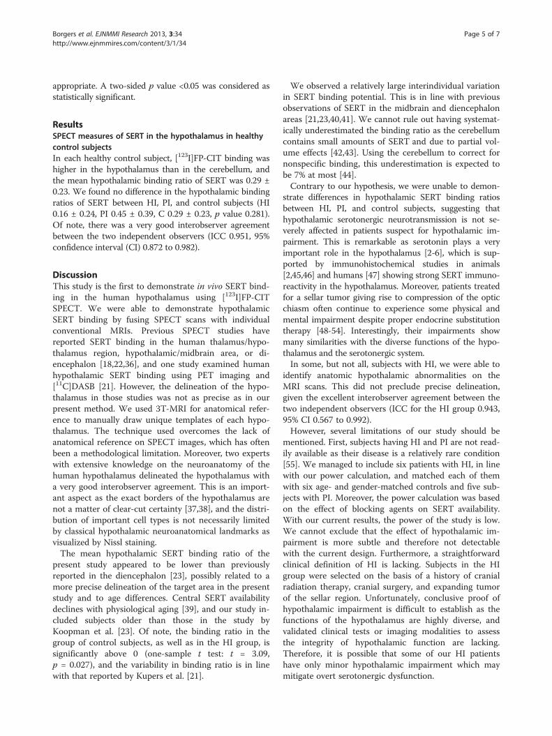
Table 1 Clinical characteristics
HI
n = 6
Age (year) 51.0 ± 6.0
Male/female (n) 2/4
Body mass index, kg/(height)2 32.7 ± 10.1
ACTH deficiency, n (%) 6 (100)
GH deficiency, n (%) 5 (83.3)
TSH deficiency, n (%) 6 (100)
LH/FSH deficiency, n (%) 6 (100)
ADH deficiency, n (%) 2 (33.3)
HI, patients with probable hypothalamic impairment, defined as having a history ofchiasm; PI, subjects with pituitary insufficiency but without a history of cranial surge
vermis) in each participant. These unique ROIs weremanually drawn by experienced researchers in the field ofthe hypothalamus (AA and EF) on each individual T1-weighted 3-D MRI scan using in-house-developed soft-ware [33]. AA and EF were blinded to the clinical data.Using the same software, SPECT scans of the subjectswere manually matched with their individual T1-weighted3-D MRI scan. In the first step, the individual MRI scanwas reoriented towards the anterior-posterior commissureline. Second, the individual SPECT data were overlaidonto the individual MRI and manually matched in allthree (x, y, z) planes. Finally, the mean amounts of radio-activity/voxel were determined for each ROI. Activityin the cerebellar cortex (excluding the vermis) was as-sumed to represent nonspecific binding. The specific-to-nonspecific binding ratios were calculated as follows:(Binding in hypothalamus − Nonspecific binding in thecerebellar cortex) / Nonspecific binding in the cerebellarcortex [34,35].
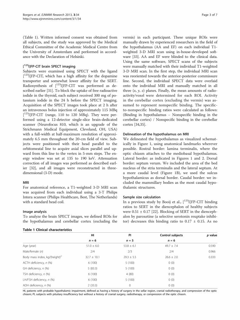
Delineation of the hypothalamus on MRIWe delineated the hypothalamus as visualized schemat-ically in Figure 1, using anatomical landmarks whereverpossible. Rostral border: lamina terminalis, where theoptic chiasm attaches to the mediobasal hypothalamus.Lateral border: as indicated in Figures 1 and 2. Dorsalborder: septum verum. We included the area of the bednucleus of the stria terminalis and the lateral septum. Ata more caudal level (Figure 1B), we used the sulcushypothalamicus as dorsal border. Caudal border: we in-cluded the mammillary bodies as the most caudal hypo-thalamic structures.
Sample size calculationIn a previous study by Booij et al., [123I]FP-CIT bindingratios to SERT in the diencephalon of healthy subjectswere 0.51 ± 0.17 [22]. Blocking of SERT in the dienceph-alon by paroxetine (a selective serotonin reuptake inhibi-tor) decreases this binding ratio to 0.17 ± 0.15. As no
PI Control subjects p value
n = 5 n = 6
53.8 ± 6.1 49.7 ± 7.4 0.590
2/3 2/4 0.966
29.3 ± 5.5 26.6 ± 2.0 0.333
5 (100) 0 (0)
5 (100) 0 (0)
4 (80) 0 (0)
5 (100) 0 (0)
0 0 (0)
surgery in the sellar region, cranial radiotherapy, and compression of the opticry, radiotherapy, or compression of the optic chiasm.
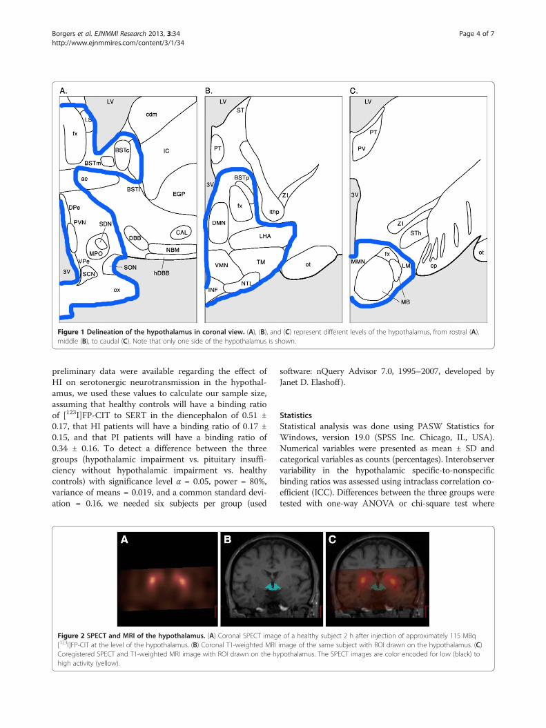
Figure 1 Delineation of the hypothalamus in coronal view. (A), (B), and (C) represent different levels of the hypothalamus, from rostral (A),middle (B), to caudal (C). Note that only one side of the hypothalamus is shown.
Borgers et al. EJNMMI Research 2013, 3:34 Page 4 of 7http://www.ejnmmires.com/content/3/1/34
preliminary data were available regarding the effect ofHI on serotonergic neurotransmission in the hypothal-amus, we used these values to calculate our sample size,assuming that healthy controls will have a binding ratioof [123I]FP-CIT to SERT in the diencephalon of 0.51 ±0.17, that HI patients will have a binding ratio of 0.17 ±0.15, and that PI patients will have a binding ratio of0.34 ± 0.16. To detect a difference between the threegroups (hypothalamic impairment vs. pituitary insuffi-ciency without hypothalamic impairment vs. healthycontrols) with significance level α = 0.05, power = 80%,variance of means = 0.019, and a common standard devi-ation = 0.16, we needed six subjects per group (used
Figure 2 SPECT and MRI of the hypothalamus. (A) Coronal SPECT imag[123I]FP-CIT at the level of the hypothalamus. (B) Coronal T1-weighted MRICoregistered SPECT and T1-weighted MRI image with ROI drawn on the hyhigh activity (yellow).
software: nQuery Advisor 7.0, 1995–2007, developed byJanet D. Elashoff).
StatisticsStatistical analysis was done using PASW Statistics forWindows, version 19.0 (SPSS Inc. Chicago, IL, USA).Numerical variables were presented as mean ± SD andcategorical variables as counts (percentages). Interobservervariability in the hypothalamic specific-to-nonspecificbinding ratios was assessed using intraclass correlation co-efficient (ICC). Differences between the three groups weretested with one-way ANOVA or chi-square test where
e of a healthy subject 2 h after injection of approximately 115 MBqimage of the same subject with ROI drawn on the hypothalamus. (C)pothalamus. The SPECT images are color encoded for low (black) to
Borgers et al. EJNMMI Research 2013, 3:34 Page 5 of 7http://www.ejnmmires.com/content/3/1/34
appropriate. A two-sided p value <0.05 was considered asstatistically significant.
ResultsSPECT measures of SERT in the hypothalamus in healthycontrol subjectsIn each healthy control subject, [123I]FP-CIT binding washigher in the hypothalamus than in the cerebellum, andthe mean hypothalamic binding ratio of SERT was 0.29 ±0.23. We found no difference in the hypothalamic bindingratios of SERT between HI, PI, and control subjects (HI0.16 ± 0.24, PI 0.45 ± 0.39, C 0.29 ± 0.23, p value 0.281).Of note, there was a very good interobserver agreementbetween the two independent observers (ICC 0.951, 95%confidence interval (CI) 0.872 to 0.982).
DiscussionThis study is the first to demonstrate in vivo SERT bind-ing in the human hypothalamus using [123I]FP-CITSPECT. We were able to demonstrate hypothalamicSERT binding by fusing SPECT scans with individualconventional MRIs. Previous SPECT studies havereported SERT binding in the human thalamus/hypo-thalamus region, hypothalamic/midbrain area, or di-encephalon [18,22,36], and one study examined humanhypothalamic SERT binding using PET imaging and[11C]DASB [21]. However, the delineation of the hypo-thalamus in those studies was not as precise as in ourpresent method. We used 3T-MRI for anatomical refer-ence to manually draw unique templates of each hypo-thalamus. The technique used overcomes the lack ofanatomical reference on SPECT images, which has oftenbeen a methodological limitation. Moreover, two expertswith extensive knowledge on the neuroanatomy of thehuman hypothalamus delineated the hypothalamus witha very good interobserver agreement. This is an import-ant aspect as the exact borders of the hypothalamus arenot a matter of clear-cut certainty [37,38], and the distri-bution of important cell types is not necessarily limitedby classical hypothalamic neuroanatomical landmarks asvisualized by Nissl staining.The mean hypothalamic SERT binding ratio of the
present study appeared to be lower than previouslyreported in the diencephalon [23], possibly related to amore precise delineation of the target area in the presentstudy and to age differences. Central SERT availabilitydeclines with physiological aging [39], and our study in-cluded subjects older than those in the study byKoopman et al. [23]. Of note, the binding ratio in thegroup of control subjects, as well as in the HI group, issignificantly above 0 (one-sample t test: t = 3.09,p = 0.027), and the variability in binding ratio is in linewith that reported by Kupers et al. [21].
We observed a relatively large interindividual variationin SERT binding potential. This is in line with previousobservations of SERT in the midbrain and diencephalonareas [21,23,40,41]. We cannot rule out having systemat-ically underestimated the binding ratio as the cerebellumcontains small amounts of SERT and due to partial vol-ume effects [42,43]. Using the cerebellum to correct fornonspecific binding, this underestimation is expected tobe 7% at most [44].Contrary to our hypothesis, we were unable to demon-
strate differences in hypothalamic SERT binding ratiosbetween HI, PI, and control subjects, suggesting thathypothalamic serotonergic neurotransmission is not se-verely affected in patients suspect for hypothalamic im-pairment. This is remarkable as serotonin plays a veryimportant role in the hypothalamus [2-6], which is sup-ported by immunohistochemical studies in animals[2,45,46] and humans [47] showing strong SERT immuno-reactivity in the hypothalamus. Moreover, patients treatedfor a sellar tumor giving rise to compression of the opticchiasm often continue to experience some physical andmental impairment despite proper endocrine substitutiontherapy [48-54]. Interestingly, their impairments showmany similarities with the diverse functions of the hypo-thalamus and the serotonergic system.In some, but not all, subjects with HI, we were able to
identify anatomic hypothalamic abnormalities on theMRI scans. This did not preclude precise delineation,given the excellent interobserver agreement between thetwo independent observers (ICC for the HI group 0.943,95% CI 0.567 to 0.992).However, several limitations of our study should be
mentioned. First, subjects having HI and PI are not read-ily available as their disease is a relatively rare condition[55]. We managed to include six patients with HI, in linewith our power calculation, and matched each of themwith six age- and gender-matched controls and five sub-jects with PI. Moreover, the power calculation was basedon the effect of blocking agents on SERT availability.With our current results, the power of the study is low.We cannot exclude that the effect of hypothalamic im-pairment is more subtle and therefore not detectablewith the current design. Furthermore, a straightforwardclinical definition of HI is lacking. Subjects in the HIgroup were selected on the basis of a history of cranialradiation therapy, cranial surgery, and expanding tumorof the sellar region. Unfortunately, conclusive proof ofhypothalamic impairment is difficult to establish as thefunctions of the hypothalamus are highly diverse, andvalidated clinical tests or imaging modalities to assessthe integrity of hypothalamic function are lacking.Therefore, it is possible that some of our HI patientshave only minor hypothalamic impairment which maymitigate overt serotonergic dysfunction.
Borgers et al. EJNMMI Research 2013, 3:34 Page 6 of 7http://www.ejnmmires.com/content/3/1/34
ConclusionsWe were able to demonstrate SERT binding in the hu-man hypothalamus in vivo. This technique will allowfunctional studies on hypothalamic SERT in variouspathologies. In particular, our technique might be ofinterest for future studies on mood disorders and foodintake regulation, given the importance of the serotoner-gic system and hypothalamus in these processes. We didnot find altered specific-to-nonspecific [123I]FP-CITbinding ratios in patients treated for a large sellar tumorgiving rise to visual field defects, although a number ofmethodological issues preclude a definitive conclusion.
Competing interestsJB is a consultant at GE Healthcare. The other authors declare that they haveno competing interests.
Authors' contributionsAll authors contributed substantially to the scientific process leading to thismanuscript. Authors AA, AJB, EF, JB, and PHB contributed to the concept anddesign of the study. AJB and EMG acquired data on the subjects. AA, AJB,and EF analyzed the data. AJB drafted the manuscript, which was revised byEF and PHB. AA, EMG, JB, and MLD critically contributed to the manuscript.All authors read and approved the final manuscript.
AcknowledgmentsWe wish to acknowledge Martine van Vessem-Timmermans, Anne Klomp,and Marieke Schouw for their excellent technical assistance. AA received aVENI grant of the Netherlands Organization for Health Research andDevelopment (grant no. 916.86.020).
Author details1Department of Endocrinology and Metabolism, Academic Medical Centre,University of Amsterdam, Meibergdreef 9, Room F5-168, Amsterdam 1105AZ, The Netherlands. 2Department of Nuclear Medicine, Academic MedicalCentre, University of Amsterdam, Amsterdam, The Netherlands. 3Departmentof Internal Medicine, Section of Endocrinology, Neuroscience CampusAmsterdam, VU University Medical Centre, Amsterdam, The Netherlands.
Received: 8 January 2013 Accepted: 2 April 2013Published: 25 April 2013
References1. Swaab DF: Human Hypothalamus: Basic and Clinical Aspects, Part II.
Amsterdam: Elsevier; 2004.2. Amir S, Robinson B, Ratovitski T, Rea MA, Stewart J, Simantov R: A role for
serotonin in the circadian system revealed by the distribution ofserotonin transporter and light-induced Fos immunoreactivity in thesuprachiasmatic nucleus and intergeniculate leaflet. Neuroscience 1998,84:1059–1073.
3. Curzon G: Serotonin and appetite. Ann N Y Acad Sci 1990, 600:521–530.4. Horiuchi J, McDowall LM, Dampney RA: Differential control of cardiac and
sympathetic vasomotor activity from the dorsomedial hypothalamus.Clin Exp Pharmacol Physiol 2006, 33:1265–1268.
5. Jorgensen HS: Studies on the neuroendocrine role of serotonin. Dan MedBull 2007, 54:266–288.
6. Oberndorfer S, Saletu-Zyhlarz G, Saletu B: Effects of selective serotoninreuptake inhibitors on objective and subjective sleep quality.Neuropsychobiology 2000, 42:69–81.
7. Ljubic-Thibal V, Morin A, Diksic M, Hamel E: Origin of the serotonergicinnervation to the rat dorsolateral hypothalamus: retrograde transport ofcholera toxin and upregulation of tryptophan hydroxylase mRNAexpression following selective nerve terminals lesion. Synapse 1999,32:177–186.
8. Petrov T, Krukoff TL, Jhamandas JH: Chemically defined collateralprojections from the pons to the central nucleus of the amygdala andhypothalamic paraventricular nucleus in the rat. Cell Tissue Res 1994,277:289–295.
9. Leibowitz SF, Alexander JT: Hypothalamic serotonin in control of eatingbehavior, meal size, and body weight. Biol Psychiatry 1998, 44:851–864.
10. Sakaguchi T, Arase K, Fisler JS, Bray GA: Effect of a high-fat diet on firingrate of sympathetic nerves innervating brown adipose tissue inanesthetized rats. Physiol Behav 1989, 45:1177–1182.
11. Rosenwasser AM: Functional neuroanatomy of sleep and circadianrhythms. Brain Res Rev 2009, 61:281–306.
12. Borgers AJ, Romeijn N, Van SE, Fliers E, Alkemade A, Bisschop PH:Compression of the optic chiasm is associated with permanent shortersleep duration in patients with pituitary insufficiency. Clin Endocrinol (Oxf )2011, 75:347–353.
13. Bray GA, Gallagher TF Jr: Manifestations of hypothalamic obesity in man:a comprehensive investigation of eight patients and a review of theliterature. Medicine (Baltimore) 1975, 54:301–330.
14. Martin JB, Riskind PN: Neurologic manifestations of hypothalamic disease.Prog Brain Res 1992, 93:31–40.
15. Stahl: Essential Psychopharmacology: Neuroscientific Basis and PracticalApplications. New York: Cambridge University Press; 2000.
16. Koch W, Schaaff N, Popperl G, Mulert C, Juckel G, Reicherzer M, Ehmer-vonGC, Moller HJ, Hegerl U, Tatsch K, Pogarell O: [I-123] ADAM and SPECT inpatients with borderline personality disorder and healthy controlsubjects. J Psychiatry Neurosci 2007, 32:234–240.
17. Willeit M, Stastny J, Pirker W, Praschak-Rieder N, Neumeister A, Asenbaum S,Tauscher J, Fuchs K, Sieghart W, Hornik K, Aschauer HN, Brücke T, Kasper S:No evidence for in vivo regulation of midbrain serotonin transporteravailability by serotonin transporter promoter gene polymorphism. BiolPsychiatry 2001, 50:8–12.
18. Zitterl W, Aigner M, Stompe T, Zitterl-Eglseer K, Gutierrez-Lobos K, Schmidl-Mohl B, Wenzel T, Demal U, Zettinig G, Hornik K, Thau K: [123I]-beta-CITSPECT imaging shows reduced thalamus-hypothalamus serotonintransporter availability in 24 drug-free obsessive-compulsive checkers.Neuropsychopharmacology 2007, 32:1661–1668.
19. Zitterl W, Aigner M, Stompe T, Zitterl-Eglseer K, Gutierrez-Lobos K, Wenzel T,Zettinig G, Hornik K, Pirker W, Thau K: Changes in thalamus-hypothalamusserotonin transporter availability during clomipramine administration inpatients with obsessive-compulsive disorder. Neuropsychopharmacology2008, 33:3126–3134.
20. Peterson TE, Shokouhi S: Advances in preclinical SPECT instrumentation.J Nucl Med 2012, 53:841–844.
21. Kupers R, Frokjaer VG, Erritzoe D, Naert A, Budtz-Joergensen E, Nielsen FA,Kehlet H, Knudsen GM: Serotonin transporter binding in thehypothalamus correlates negatively with tonic heat pain ratings inhealthy subjects: a [11C]DASB PET study. NeuroImage 2011, 54:1336–1343.
22. Booij J, De JJ, De BK, Knol R, De Win MM, Van Eck-Smit BL: Quantificationof striatal dopamine transporters with 123I-FP-CIT SPECT is influenced bythe selective serotonin reuptake inhibitor paroxetine: a double-blind,placebo-controlled, crossover study in healthy control subjects. J NuclMed 2007, 48:359–366.
23. Koopman KE, la Fleur SE, Fliers E, Serlie MJ, Booij J: Assessing the optimaltime point for the measurement of extrastriatal serotonin transporterbinding with 123I-FP-CIT SPECT in healthy, male subjects. J Nucl Med2012, 53:1087–1090.
24. Booij J, Knol RJ, Reneman L, De BK, Janssen AG, Van Royen EA: Iodine-123labelled nor-beta-CIT binds to the serotonin transporter in vivo as assessedby biodistribution studies in rats. Eur J Nucl Med 1998, 25:1666–1669.
25. Booij J, Andringa G, Rijks LJ, Vermeulen RJ, De BK, Boer GJ, Janssen AG, VanRoyen EA: [123I]FP-CIT binds to the dopamine transporter as assessed bybiodistribution studies in rats and SPECT studies in MPTP-lesionedmonkeys. Synapse 1997, 27:183–190.
26. Lavalaye J, Knol RJ, De BK, Reneman L, Janssen AG, Booij J: [123I]FP-CITbinding in rat brain after acute and sub-chronic administration ofdopaminergic medication. Eur J Nucl Med 2000, 27:346–349.
27. Reneman L, Booij J, Habraken JB, De BK, Hatzidimitriou G, Den Heeten GJ,Ricaurte GA: Validity of [123I]beta-CIT SPECT in detecting MDMA-inducedserotonergic neurotoxicity. Synapse 2002, 46:199–205.
28. Staley JK, Basile M, Flynn DD, Mash DC: Visualizing dopamine andserotonin transporters in the human brain with the potent cocaineanalogue [125I]RTI-55: in vitro binding and autoradiographiccharacterization. J Neurochem 1994, 62:549–556.
29. Hesse B, Lindhardt TB, Acampa W, Anagnostopoulos C, Ballinger J, Bax JJ,Edenbrandt L, Flotats A, Germano G, Stopar TG, Franken P, Kelion A, Kjaer A,
Borgers et al. EJNMMI Research 2013, 3:34 Page 7 of 7http://www.ejnmmires.com/content/3/1/34
Le Guludec D, Ljungberg M, Maenhout AF, Marcassa C, Marving J, McKiddieF, Schaefer WM, Stegger L, Underwood R: EANM/ESC guidelines forradionuclide imaging of cardiac function. Eur J Nucl Med Mol Imaging2008, 35:851–885.
30. Manley PE, McKendrick K, McGillicudy M, Chi SN, Kieran MW, Cohen LE,Kothare S, Michael SR, Goumnerova LC, Sun P, London W, Marcus KJ,Pomeroy SL, Ullrich NJ: Sleep dysfunction in long term survivors ofcraniopharyngioma. J Neurooncol 2012, 108:543–549.
31. Booij J, Busemann SE, Stabin MG, Janssen AG, De BK, Van Royen EA: Humanbiodistribution and dosimetry of [123I]FP-CIT: a potent radioligand forimaging of dopamine transporters. Eur J Nucl Med 1998, 25:24–30.
32. Booij J, Tissingh G, Boer GJ, Speelman JD, Stoof JC, Janssen AG, Wolters EC,van Royen EA: [123I]FP-CIT SPECT shows a pronounced decline of striataldopamine transporter labelling in early and advanced Parkinson'sdisease. J Neurol Neurosurg Psychiatry 1997, 62:133–140.
33. van Herk M, de Jaeger K, de Munck J, Hoogeman M, Meinders J, Ploeger L: Adelineation system for N modalities-software aspects [extended abstract]. In13th ICCR: May 22–25 2000. Heidelberg. New York: Springer; 2000:73.
34. Innis RB, Cunningham VJ, Delforge J, Fujita M, Gjedde A, Gunn RN, Holden J,Houle S, Huang SC, Ichise M, Iida H, Ito H, Kimura Y, Koeppe RA, KnudsenGM, Knuuti J, Lammertsma AA, Laruelle M, Logan J, Maguire RP, Mintun MA,Morris ED, Parsey R, Price JC, Slifstein M, Sossi V, Suhara T, Votaw JR, WongDF, Carson RE: Consensus nomenclature for in vivo imaging of reversiblybinding radioligands. J Cereb Blood Flow Metab 2007, 27:1533–1539.
35. Laruelle M, Wallace E, Seibyl JP, Baldwin RM, Zea-Ponce Y, Zoghbi SS,Neumeyer JL, Charney DS, Hoffer PB, Innis RB: Graphical, kinetic, andequilibrium analyses of in vivo [123I] beta-CIT binding to dopaminetransporters in healthy human subjects. J Cereb Blood Flow Metab 1994,14:982–994.
36. Dahlstrom M, Ahonen A, Ebeling H, Torniainen P, Heikkila J, Moilanen I:Elevated hypothalamic/midbrain serotonin (monoamine) transporteravailability in depressive drug-naive children and adolescents. Mol Psychiatry2000, 5:514–522.
37. Anderson E, Haymaker W: Breakthroughs in hypothalamic and pituitaryresearch. Prog Brain Res 1974, 41:1–60.
38. Braak H, Braak E: Anatomy of the human hypothalamus (chiasmatic andtuberal region). Prog Brain Res 1992, 93:3–14.
39. van Dyck CH, Malison RT, Seibyl JP, Laruelle M, Klumpp H, Zoghbi SS,Baldwin RM, Innis RB: Age-related decline in central serotonin transporteravailability with [(123)I]beta-CIT SPECT. Neurobiol Aging 2000, 21:497–501.
40. Chou YH, Yang BH, Chung MY, Chen SP, Su TP, Chen CC, Wang SJ: Imagingthe serotonin transporter using (123)I-ADAM in the human brain.Psychiatry Res 2009, 172:38–43.
41. van de Giessen E, Booij J: The SPECT tracer [123I]ADAM binds selectivelyto serotonin transporters: a double-blind, placebo-controlled study inhealthy young men. Eur J Nucl Med Mol Imaging 2010, 37:1507–1511.
42. Kent JM, Coplan JD, Lombardo I, Hwang DR, Huang Y, Mawlawi O, VanHeertum RL, Slifstein M, Abi-Dargham A, Gorman JM, Laruelle M:Occupancy of brain serotonin transporters during treatment withparoxetine in patients with social phobia: a positron emissiontomography study with 11C McN 5652. Psychopharmacology (Berl) 2002,164:341–348.
43. Parsey RV, Kent JM, Oquendo MA, Richards MC, Pratap M, Cooper TB,Arango V, Mann JJ: Acute occupancy of brain serotonin transporter bysertraline as measured by [11C]DASB and positron emission tomography.Biol Psychiatry 2006, 59:821–828.
44. Kish SJ, Furukawa Y, Chang LJ, Tong J, Ginovart N, Wilson A, Houle S, MeyerJH: Regional distribution of serotonin transporter protein in postmortemhuman brain: is the cerebellum a SERT-free brain region? Nucl Med Biol2005, 32:123–128.
45. Emiliano AB, Cruz T, Pannoni V, Fudge JL: The interface of oxytocin-labeled cells and serotonin transporter-containing fibers in the primatehypothalamus: a substrate for SSRIs therapeutic effects?Neuropsychopharmacology 2007, 32:977–988.
46. Legutko R, Gannon RL: Serotonin transporter localization in the hamstersuprachiasmatic nucleus. Brain Res 2001, 893:77–83.
47. Moore RY, Speh JC: Serotonin innervation of the primate suprachiasmaticnucleus. Brain Res 2004, 1010:169–173.
48. Biermasz NR, Joustra SD, Donga E, Pereira AM, Van DN, Van DM, Van derKlaauw AA, Corssmit EP, Lammers GJ, van Kralingen KW, van Dijk JG, RomijnJA: Patients previously treated for nonfunctioning pituitary
macroadenomas have disturbed sleep characteristics, circadianmovement rhythm, and subjective sleep quality. J Clin Endocrinol Metab2011, 96:1524–1532.
49. Dekkers OM, van der Klaauw AA, Pereira AM, Biermasz NR, Honkoop PJ,Roelfsema F, Smit JW, Romijn JA: Quality of life is decreased aftertreatment for nonfunctioning pituitary macroadenoma. J Clin EndocrinolMetab 2006, 91:3364–3369.
50. Kendall-Taylor P, Jonsson PJ, Abs R, Erfurth EM, Koltowska-Haggstrom M,Price DA, Verhelst J: The clinical, metabolic and endocrine features andthe quality of life in adults with childhood-onset craniopharyngiomacompared with adult-onset craniopharyngioma. Eur J Endocrinol 2005,152:557–567.
51. Rosen T, Eden S, Larson G, Wilhelmsen L, Bengtsson BA: Cardiovascular riskfactors in adult patients with growth hormone deficiency. Acta Endocrinol(Copenh) 1993, 129:195–200.
52. Tiemensma J, Kaptein AA, Pereira AM, Smit JW, Romijn JA, Biermasz NR: Copingstrategies in patients after treatment for functioning or nonfunctioningpituitary adenomas. J Clin Endocrinol Metab 2011, 96:964–971.
53. van der Klaauw AA, Dekkers OM, Pereira AM, van Kralingen KW, Romijn JA:Increased daytime somnolence despite normal sleep patterns in patientstreated for nonfunctioning pituitary macroadenoma. J Clin EndocrinolMetab 2007, 92:3898–3903.
54. Van Someren EJ, Swart-Heikens J, Endert E, Bisschop PH, Swaab DF, BakkerPJ, Romijn JA, Fliers E: Long-term effects of cranial irradiation forchildhood malignancy on sleep in adulthood. Eur J Endocrinol 2004,150:503–510.
55. Regal M, Paramo C, Sierra SM, Garcia-Mayor RV: Prevalence and incidenceof hypopituitarism in an adult Caucasian population in northwesternSpain. Clin Endocrinol (Oxf ) 2001, 55:735–740.
doi:10.1186/2191-219X-3-34Cite this article as: Borgers et al.: Imaging of serotonin transporters with[123I]FP-CIT SPECT in the human hypothalamus. EJNMMI Research 20133:34.
Submit your manuscript to a journal and benefi t from:
7 Convenient online submission
7 Rigorous peer review
7 Immediate publication on acceptance
7 Open access: articles freely available online
7 High visibility within the fi eld
7 Retaining the copyright to your article
Submit your next manuscript at 7 springeropen.com


![Selective serotonin reuptake inhibitors [SSRIs] for stroke recoveryclok.uclan.ac.uk/6814/19/17551 - Selective serotonin reuptake... · Hackett, Maree (2012) Selective serotonin reuptake](https://img.pdfslide.us/doc/110x75/5f9c1bce9667ca02083a93ee/selective-serotonin-reuptake-inhibitors-ssris-for-stroke-selective-serotonin.jpg)