Embed Size (px)
Citation preview
Olivecrona et al. Scandinavian Journal of Trauma, Resuscitation and Emergency Medicine 2013, 21:33http://www.sjtrem.com/content/21/1/33
ORIGINAL RESEARCH Open Access
Acute neuro-endocrine profile and predictionof outcome after severe brain injuryZandra Olivecrona1*, Per Dahlqvist2 and Lars-Owe D Koskinen1
Abstract
Object: The aim of the study was to evaluate the early changes in pituitary hormone levels after severe traumaticbrain injury (sTBI) and compare hormone levels to basic neuro-intensive care data, a systematic scoring of theCT-findings and to evaluate whether hormone changes are related to outcome.
Methods: Prospective study, including consecutive patients, 15–70 years, with sTBI, Glasgow Coma Scale (GCS)score ≤ 8, initial cerebral perfusion pressure > 10 mm Hg, and arrival to our level one trauma university hospitalwithin 24 hours after head trauma (n = 48). Serum samples were collected in the morning (08–10 am) day 1 andday 4 after sTBI for analysis of cortisol, growth hormone (GH), prolactin, insulin-like growth factor 1 (IGF-1),thyroid-stimulating hormone (TSH), free triiodothyronine (fT3), free thyroxine (fT4), follicular stimulating hormone(FSH), luteinizing hormone (LH), testosterone and sex hormone-binding globulin (SHBG) (men). Serum for cortisoland GH was also obtained in the evening (17–19 pm) at day 1 and day 4. The first CT of the brain was classifiedaccording to Marshall. Independent staff evaluated outcome at 3 months using GOS-E.
Results: Profound changes were found for most pituitary-dependent hormones in the acute phase after sTBI,i.e. low levels of thyroid hormones, strong suppression of the pituitary-gonadal axis and increased levels ofprolactin. The main findings of this study were: 1) A large proportion (54% day 1 and 70% day 4) of the patientsshowed morning s-cortisol levels below the proposed cut-off levels for critical illness related corticosteroidinsufficiency (CIRCI), i.e. <276 nmol/L (=10 ug/dL), 2) Low s-cortisol was not associated with higher mortality orworse outcome at 3 months, 3) There was a significant association between early (day 1) and strong suppression ofthe pituitary-gonadal axis and improved survival and favorable functional outcome 3 months after sTBI, 4)Significantly lower levels of fT3 and TSH at day 4 in patients with a poor outcome at 3 months. 5) A higher MarshallCT score was associated with higher day 1 LH/FSH- and lower day 4 TSH levels 6) In general no significantcorrelation between GCS, ICP or CPP and hormone levels were detected. Only ICPmax and LH day 1 in men wassignificantly correlated.
Conclusion: Profound dynamic changes in hormone levels are found in the acute phase of sTBI. This is consistentwith previous findings in different groups of critically ill patients, most of which are likely to be attributed tophysiological adaptation to acute illness. Low cortisol levels were a common finding, and not associated withunfavorable outcome. A retained ability to a dynamic hormonal response, i.e. fast and strong suppression of thepituitary-gonadal axis (day 1) and ability to restore activity in the pituitary-thyroid axis (day 4) was associated withless severe injury according to CT-findings and favorable outcome.
Keywords: Severe traumatic brain injury, Hypopituitarism, Outcome, ICP targeted therapy, Hypothalamic-pituitarydysfunction, Prostacyclin
* Correspondence: [email protected] of Pharmacology and Clinical Neuroscience,Division of Neurosurgery, Umeå University, SE 90185, Umeå, SwedenFull list of author information is available at the end of the article
© 2013 Olivecrona et al.; licensee BioMed Central Ltd. This is an Open Access article distributed under the terms of theCreative Commons Attribution License (http://creativecommons.org/licenses/by/2.0), which permits unrestricted use,distribution, and reproduction in any medium, provided the original work is properly cited.
Olivecrona et al. Scandinavian Journal of Trauma, Resuscitation and Emergency Medicine 2013, 21:33 Page 2 of 13http://www.sjtrem.com/content/21/1/33
IntroductionTraumatic brain injury (TBI) remains one of the majorcauses of death and disability worldwide. The pituitaryis particularly vulnerable to head trauma due to theanatomical location of the gland within the sella turcicaas well as its fragile infundibular hypothalamic structureand its vascular supply. Pituitary insufficiency after traumawas first reported in 1914 [1] and post-mortem evidencedating back several decades show pituitary gland infarctionsin up to one-third of patients deceased shortly after TBI[2]. Injury mechanisms of hypothalamic-pituitary damagedue to TBI include direct mechanical/shearing injuryto the pituitary stalk and the vulnerable long hypophysealvessels, which may result in anterior lobe infarction andsecondary injuries due to increased intracranial pressure,hypotension, hypoxia and vasospasm. The posteriorpituitary is less susceptible to injury due to less fragilevascular supply [3]. Nevertheless, pituitary insufficiencyafter TBI has until recently been considered a rare event,with sparse data derived from case reports and caseseries [3,4]. However, reports from recent years havesuggested permanent pituitary insufficiency after trau-matic head injury to be far more common than previouslythought [5,6].In the acute phase after TBI, depending on TBI severity
and location an acute post-traumatic hypothalamic andpituitary tissue damage is likely to occur in many patientsearly after trauma, with concomitant disturbances inhormone secretion. Most important in the acute phase isnot to overlook an acute insufficiency in the hypothalamus-pituitary-adrenal axis (HPA-axis) with inadequate cortisolsecretion, which is a life-threatening condition and mustbe correctly diagnosed and promptly treated. However,solid diagnostic criteria of cortisol insufficiency in criticalillness are still lacking and under debate [7,8]. Furthermore,the known roles of GH, IGF-1, estrogen and testosteroneupon brain function and plasticity propose that inadequatelevels after sTBI may have both acute and long-termsignificance upon the recovering brain [9-13]. GH andIGF-1 receptors are abundant in the brain, GH isinvolved in vascular reactivity, vascular tone and CNSrepair processes, while IGF-1 seems to be important inre-myelination and avoidance of demyelination [11,12,14].There is proof of that estrogen and progesterone areneuro-protective, whereas androgens have been reportedboth to exacerbate and protect against neuronal damage,probably in a time and dose-dependent manner [15].Previous reports on the neuro-endocrine changes in the
acute phase of moderate to severe TBI show evidenceof central hypogonadism in 25–80%, thyroid hormonedeficiency in 2–15%, hyperprolactinemia in more than50%, GH deficiency in 18% and cortisol deficiency in 13%.However, most previous reports are on mixed materials,i.e. mild, moderate and severe traumatic brain injury,
acute phase definition varies and the treatment givenfor the sTBI is not always clearly described.At the neurosurgical department at Umeå University
Hospital, all patients suffering from sTBI are treatedaccording to an intracranial pressure (ICP) targetedprotocol based on aggressive neurosurgery and the Lundconcept resulting in a mortality of less than 15% [16].The aims of this prospective study were to investigate
the prevalence and dynamics of very early pituitary-related hormonal dysfunction and the relation to basicneuro-intensive care data, CT-findings and possibleprognostic implications in a strictly defined group ofpatients with sTBI (Glasgow coma scale, GCS < 9),treated according to the Lund concept.
Material and methodsIn the northern part of Sweden, the department ofneurosurgery in Umeå has a regional responsibility ofabout 900 000 inhabitants. The area corresponds approxi-mately to the area of Great Britain. All referring hospitalsin the catchment area of the department refer patientswith sTBI. All patients treated for sTBI in the period fromJanuary 1st 2002 to December 31st 2005 were included inthe study if inclusion criteria were met. The inclusioncriteria were: age 15–70 years, arrival in the departmentwithin 24 hours of trauma, verified traumatic brain injury,GCS at intubation and sedation of GCS 8 or less and afirst measured CPP of 10 mmHg or more. TBI severitywas defined by the GCS, and based on the first scoreregistered after resuscitation. If there were doubts aboutthe GCS when the patients arrived from the referringhospitals a re-evaluation was done. Exclusion criteriawere: pregnant or breastfeeding woman, penetrating headinjury and medication with glucocorticoids.The patients were part of a prospective randomized
blinded placebo controlled study on the effect of prosta-cyclin in severe traumatic head injury [16].All patients were initially sedated with midazolam and
fentanyl. No patient received Etomidate. Multimodalmonitoring was applied. Invasive arterial blood pressureand ICP, using an intra-parenchymal pressure-measuringdevice (Codman MicroSensor, Johnson & Johnson Pro-fessional Inc., Raynham, MA, USA), were continuouslymeasured. CPP was automatically calculated. Data weredigitally stored using the Picis system (Picis, Inc., Wakefield,MA, USA) and the LabPilot (CMA Microdialysis, Solna,Sweden).Patients were treated with no head elevation and the
arterial baseline level was set at the heart level. Thus, nocorrection for CPP was needed. The patients weremechanically ventilated (PaO2 ≥12 kPa and PaCO2
4.5-5.5 kPa). The goal of the treatment was to maintainICP <20 mmHg, not allowing CPP <50 mmHg. HourlyICP and CPP were calculated by using all the minute-
Olivecrona et al. Scandinavian Journal of Trauma, Resuscitation and Emergency Medicine 2013, 21:33 Page 3 of 13http://www.sjtrem.com/content/21/1/33
to-minute ICP and CPP values during the first 5 days. Thehour with the highest ICP and lowest CPP was identifiedas ICPmax and CPPmin.The patients were kept normo-volemic. The crystalloid
and colloid osmotic pressures were kept normal by infusionof red blood cells, albumin, glucose solutions and Ringer’sacetate and sodium (S-Hb >110 g/L, S-alb >40 g/L,Na+ ≥ 140 mmol/L). The fluid balance was keptneutral and furosemide was used when indicated. Afterhemodynamic stabilization, clonidine and metoprololwere given as continuous intravenous infusions, inorder to normalize the blood pressure and to reduce thetranscapillary hydrostatic pressure. These drugs alsoreduce the stress level mediated by the sympatheticnervous system. Muscle relaxants and steroids are not apart of the treatment regime and thus not used. Further,mass lesions were surgically removed. If ICP, despite ofthe above measures, was not brought under control,additional sedation with low-dose thiopental, placementof a ventriculostomy and uni- or bilateral hemicraniectomywith duraplasty were used.The severity of the trauma was assessed by ISS [17] and
the brain tissue damage by the Marshall classification [18].Serum samples were collected in the morning (08–
10 am) day 1 and day 4 after sTBI for analysis of cortisol,growth hormone (GH), prolactin, insulin-like growthfactor 1 (IGF-1), thyroid-stimulating hormone (TSH),free triiodothyronine (fT3), free thyroxine (fT4), follicularstimulating hormone (FSH), luteinizing hormone (LH),testosterone and sex hormone-binding globulin (SHBG)(men). Serum for cortisol and GH was also obtained inthe evening (17–19 pm) at day 1 and day 4. The sampleswere immediately centrifuged and stored at -70°C untilanalysis. All hormones were analyzed at the accreditedclinical chemistry laboratory at Umeå university hospital.Serum cortisol, TSH, fT3, fT4, FSH, LH, prolactinand SHBG were analyzed by electrochemiluminescenceimmunoassay (ECLIA; Modular Analytics E170, Roche,GmbH, Hannheim, Germany). Serum testosterone wasanalyzed by Coat-a-count RIA (Siemens). Serum GH andIGF-1 was measured by DPC Immulite 2000 (Siemens).Calculated free testosterone (fc-testosterone) levels werecalculated using total testosterone, SHBG and albuminlevels [19]. Clinical outcome was assessed at 3 monthsafter trauma by independent staff and performed withstructured interviews according to the extended GlasgowOutcome Scale GOS-E. The clinical outcome is reportedas GOS score at 3 months. GOS was also dichotomizedinto unfavorable (GOS 1–3)/favorable (GOS 4–5) and intodeceased/alive for further outcome analysis.Values are reported as mean ± standard error of the mean
(SEM) for continuous data and for non-parametric and or-dinal variables as median and range. A two-tailed Student’st-test was applied for continuous data. Comparison of
cortisol levels between groups and comparison of allhormone levels between deceased and alive was madeusing the non-parametric Wilcoxon sign-rank test, due tolarge variations in the cortisol levels and very few patientdeceased at 3 months. Correlation analyses were madeusing Pearson test for continuous data and Spearman’srho test when at least one parameter was ordinal. Factorsinfluencing outcome was explored using logistic re-gression. Prediction of outcome was analyzed usingreceiver operated characteristics (ROC) curve method.The JMP (9.0.0) statistical package was used (SASInstitute Inc. USA). A p ≤ 0.05 was considered statisticallysignificant.The regional ethical board at Umeå University approved
the study (00–175, 05-007M). The study was also approvedby the Swedish Medical Products Agency (151:633/01) andthe study is registered as a clinical trial (ClinicalTrial.govidentifier NCT01363583).
ResultsOut of 89 patients, 48 patients fulfilled the inclusioncriteria [16]. One patient died soon after admission, thushormone samples could not be obtained. Due to cervicalspine injury two patients were treated with high-dosemethylprednisolone before the transfer to the neurosur-gical trauma level one unit in Umeå. This treatment waswithdrawn at our department and these patients wereexcluded from the study. Thus our results are based on45 patients, 15 women and 30 men, none of thesereceived glucocorticoid treatment. Due to technicalreasons one or two hormone analysis are sometimeslacking and this is denoted in the text. Mean age was35.7 ± 2.2, (range 15–64), median ISS was 29 [9-43] andmedian GCS at intubation and sedation was 6 [3-8]. Thetrauma was caused by road accidents (car, motorbike,bicycle and pedestrian) 28/45, snow mobile accidents 4/45,falls 10/45, assaults 2/45 and sport 1/45. Patients werehospitalized at the neuro-intensive care unit for a mean of12.5 ± 0.6 days, median 12.3 days (3.5-23.7). Two patientsdied during the neuro-intensive care treatment, due totherapy refractive high ICP. At 3 months mortalitywas 8.9% (4/45), median GOS was 4 [1-5] and favorableoutcome (GOS 4–5) was found in 53.3% of the patients.There was no significant difference regarding age, sex
distribution, initial GCS, ISS or clinical outcome at 3months between the prostacyclin and placebo treatedgroups [16]. There were also no significant differences inany of the measured hormone levels at day 1 or 4 aftersTBI between patients treated with prostacyclin andplacebo (data not shown). Therefore, the results representthe whole patient group (prostacyclin-and placebo treatedgroups together).Substantial effects of sTBI on hormone levels were
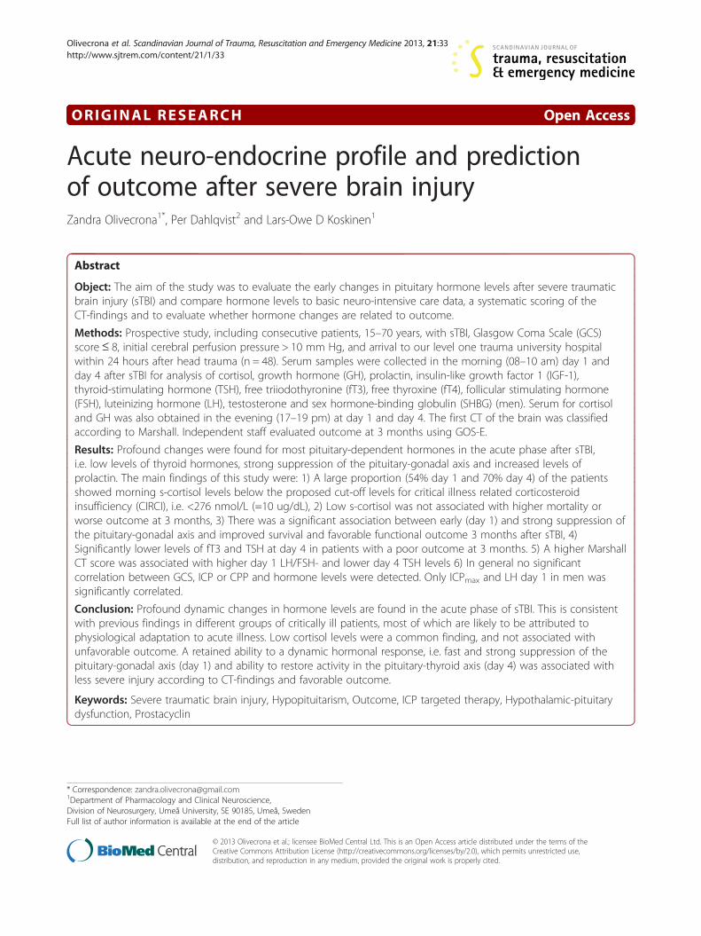
observed. Figure 1 depicts the proportion of patients

Figure 1 Proportions of patients (n = 45) presenting hormone values above or below laboratory reference interval day 1 and day 4after sTBI. Reference intervals are given in Table 1.
Olivecrona et al. Scandinavian Journal of Trauma, Resuscitation and Emergency Medicine 2013, 21:33 Page 4 of 13http://www.sjtrem.com/content/21/1/33
with hormone levels above or below normal referenceranges for our accredited laboratory. Large proportionsof the patients showed elevated levels of prolactin andlow cortisol, fT3, testosterone, LH and FSH levels.
Hypothalamic-pituitary-adrenal axisMean serum cortisol day 1 after sTBI was within referenceranges with a non-significant increase from day 1 to day 4(Table 1). However, there was a substantial individualvariation. Thus, using the proposed limit for criticalillness related corticosteroid insufficiency (CIRCI) [20]of total serum cortisol <276 nmol/L (10 ug/dL) day 1cortisol was low in 24/44 (54.5%) patients in the morningand 23/44 (52.3%) in the evening. Day 4 the correspondingfigures were 31/44 (70.5%) in the morning and 26/44(59.1%) in the evening. The number of patients withvery low serum cortisol (<100 nmol/L) day 1 after sTBIwas 8/43 (18.6%) in the morning and 7/44 (15.9%) in theevening. Day 4, the number of patients with morningcortisol below 100 nmol/L was 10/44 (22.7%) and inthe evening 11/45 (24.4%). Abnormally high morning cor-tisol levels were considered as >800 nmol/L and in theevening >600 nmol/L in accordance with laboratory refer-ence range. Cortisol levels exceeding reference range day1 after sTBI was found in 0/43 (morning) and 8/44(18.2%, evening). The corresponding figures day 4 was 3/
44 (6.8%) and 7/45 (15.6%). There was a trend towardshigher morning cortisol levels day 1 in subjects deceasedat 3 months (497 ± 143 nmol/L) as compared with survi-vors (282 ± 31 nmol/L) (p = 0.13). However, there were nostatistically significant differences in cortisol levels day 1or day 4 between deceased vs. alive subjects at 3 months orbetween patients with unfavorable vs. favorable outcome.No correlation was seen between cortisol and GCS, ISS,Marshall grade, GOS, ICPmax or CPPmin.
Thyroid axisMean serum fT4 levels decreased significantly (−20.4%)from day 1 to day 4 after TBI (p < 0.0001), (Table 1). Thelevel of fT4 was below reference range (12–22 pmol/L)in 4/44 (5.5%) of the patients at day 1 and in 12/44(27.3%) at day 4, whereas fT4 above normal was foundin 4/44 (9.1%) of the patients day 1 but none at day 4.There was no statistically significant difference in fT4levels between the deceased and alive patients or in theunfavorable/favorable outcome groups at 3 months. Nosignificant correlations were found between fT4 andGCS, ISS, Marshall grade, ICPmax, CPPmin or GOS.Mean fT3 levels followed fT4 and decreased signifi-
cantly (−24.3%) from day 1 to day 4 (p < 0.0001),(Table 1). Serum fT3 was below reference range (3.1-6.8 pmol/L) in 11/44 (25.0%) of the patients at day 1 and
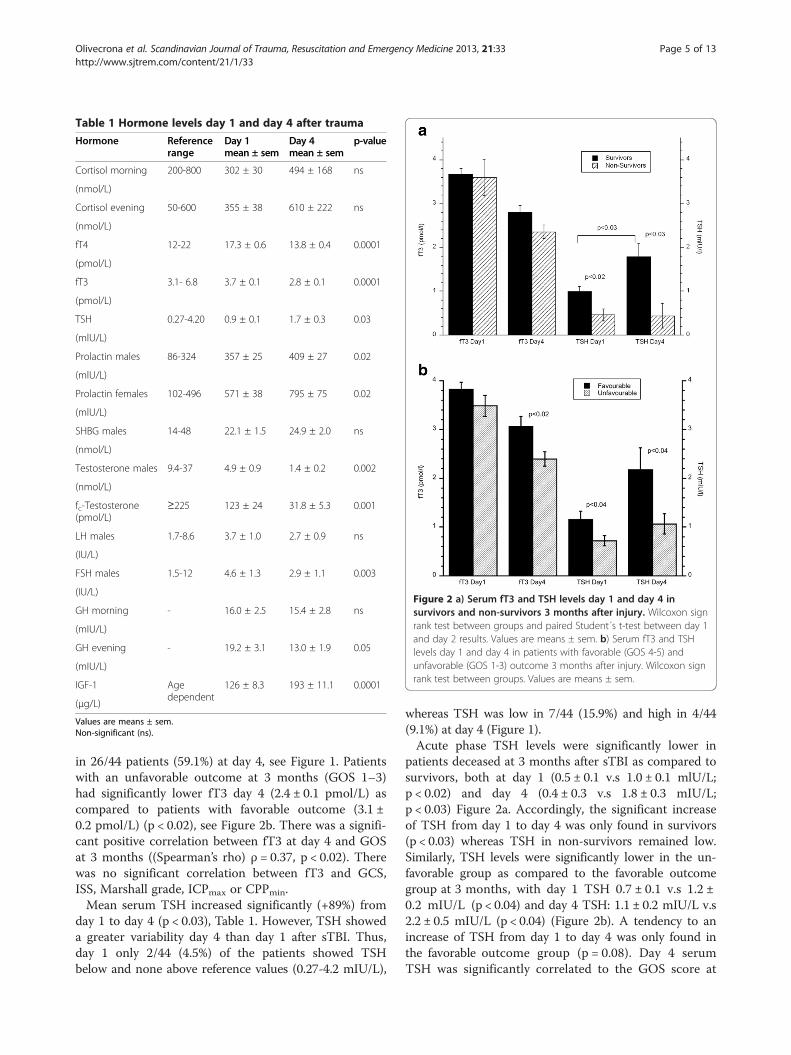
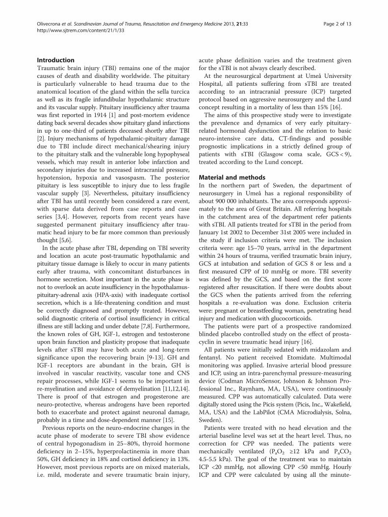
Figure 2 a) Serum fT3 and TSH levels day 1 and day 4 insurvivors and non-survivors 3 months after injury. Wilcoxon signrank test between groups and paired Student´s t-test between day 1and day 2 results. Values are means ± sem. b) Serum fT3 and TSHlevels day 1 and day 4 in patients with favorable (GOS 4-5) andunfavorable (GOS 1-3) outcome 3 months after injury. Wilcoxon signrank test between groups. Values are means ± sem.
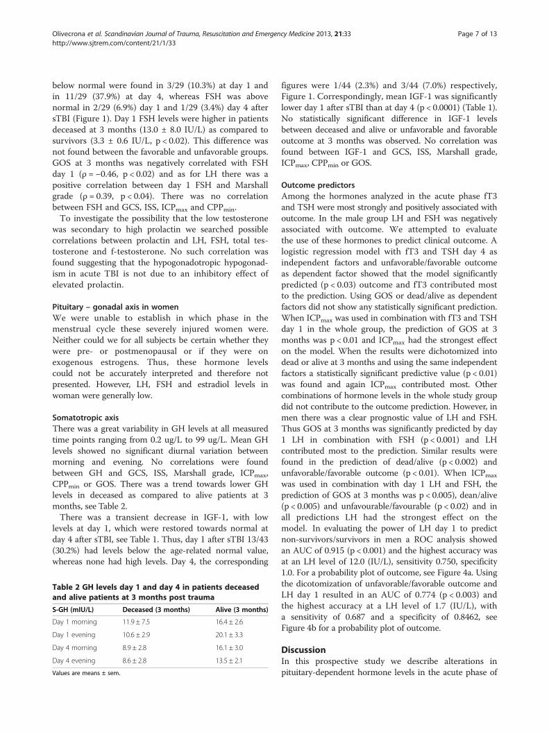
Table 1 Hormone levels day 1 and day 4 after trauma
Hormone Referencerange
Day 1mean ± sem
Day 4mean ± sem
p-value
Cortisol morning 200-800 302 ± 30 494 ± 168 ns
(nmol/L)
Cortisol evening 50-600 355 ± 38 610 ± 222 ns
(nmol/L)
fT4 12-22 17.3 ± 0.6 13.8 ± 0.4 0.0001
(pmol/L)
fT3 3.1- 6.8 3.7 ± 0.1 2.8 ± 0.1 0.0001
(pmol/L)
TSH 0.27-4.20 0.9 ± 0.1 1.7 ± 0.3 0.03
(mlU/L)
Prolactin males 86-324 357 ± 25 409 ± 27 0.02
(mlU/L)
Prolactin females 102-496 571 ± 38 795 ± 75 0.02
(mlU/L)
SHBG males 14-48 22.1 ± 1.5 24.9 ± 2.0 ns
(nmol/L)
Testosterone males 9.4-37 4.9 ± 0.9 1.4 ± 0.2 0.002
(nmol/L)
fc-Testosterone(pmol/L)
≥225 123 ± 24 31.8 ± 5.3 0.001
LH males 1.7-8.6 3.7 ± 1.0 2.7 ± 0.9 ns
(IU/L)
FSH males 1.5-12 4.6 ± 1.3 2.9 ± 1.1 0.003
(IU/L)
GH morning - 16.0 ± 2.5 15.4 ± 2.8 ns
(mIU/L)
GH evening - 19.2 ± 3.1 13.0 ± 1.9 0.05
(mIU/L)
IGF-1 Agedependent
126 ± 8.3 193 ± 11.1 0.0001
(μg/L)
Values are means ± sem.Non-significant (ns).
Olivecrona et al. Scandinavian Journal of Trauma, Resuscitation and Emergency Medicine 2013, 21:33 Page 5 of 13http://www.sjtrem.com/content/21/1/33
in 26/44 patients (59.1%) at day 4, see Figure 1. Patientswith an unfavorable outcome at 3 months (GOS 1–3)had significantly lower fT3 day 4 (2.4 ± 0.1 pmol/L) ascompared to patients with favorable outcome (3.1 ±0.2 pmol/L) (p < 0.02), see Figure 2b. There was a signifi-cant positive correlation between fT3 at day 4 and GOSat 3 months ((Spearman’s rho) ρ = 0.37, p < 0.02). Therewas no significant correlation between fT3 and GCS,ISS, Marshall grade, ICPmax or CPPmin.Mean serum TSH increased significantly (+89%) from
day 1 to day 4 (p < 0.03), Table 1. However, TSH showeda greater variability day 4 than day 1 after sTBI. Thus,day 1 only 2/44 (4.5%) of the patients showed TSHbelow and none above reference values (0.27-4.2 mIU/L),
whereas TSH was low in 7/44 (15.9%) and high in 4/44(9.1%) at day 4 (Figure 1).Acute phase TSH levels were significantly lower in
patients deceased at 3 months after sTBI as compared tosurvivors, both at day 1 (0.5 ± 0.1 v.s 1.0 ± 0.1 mlU/L;p < 0.02) and day 4 (0.4 ± 0.3 v.s 1.8 ± 0.3 mIU/L;p < 0.03) Figure 2a. Accordingly, the significant increaseof TSH from day 1 to day 4 was only found in survivors(p < 0.03) whereas TSH in non-survivors remained low.Similarly, TSH levels were significantly lower in the un-favorable group as compared to the favorable outcomegroup at 3 months, with day 1 TSH 0.7 ± 0.1 v.s 1.2 ±0.2 mIU/L (p < 0.04) and day 4 TSH: 1.1 ± 0.2 mIU/L v.s2.2 ± 0.5 mIU/L (p < 0.04) (Figure 2b). A tendency to anincrease of TSH from day 1 to day 4 was only found inthe favorable outcome group (p = 0.08). Day 4 serumTSH was significantly correlated to the GOS score at
Olivecrona et al. Scandinavian Journal of Trauma, Resuscitation and Emergency Medicine 2013, 21:33 Page 6 of 13http://www.sjtrem.com/content/21/1/33
3 months (ρ = 0.3, p < 0.05). Day 1 TSH was negativelycorrelated to Marshall grade (ρ = −0.48, p < 0.001), but notto GCS, ISS, CPPmin, ICPmax.
ProlactinElevated levels of serum prolactin were observed in 14/29(48.3%) of the men and in 10/15 (66.7%) of the women atday 1 after sTBI (Figure 1). At day 4 the number of malepatients with supra-normal prolactin levels had increasedto 21/29 (72.4%), whereas one male showed low prolactin.The corresponding results in women were 13/15 (86.7%)and 0/15. Mean prolactin levels increased from day 1 today 4 in both men and women (p < 0.02) (Table 1).There was no statistically significant difference in theprolactin levels between deceased/alive subjects or inthe unfavorable/favorable groups at 3 month. Prolactinlevels were not correlated to GCS, ISS, Marshall grade,GOS, CPPmin and ICPmax.
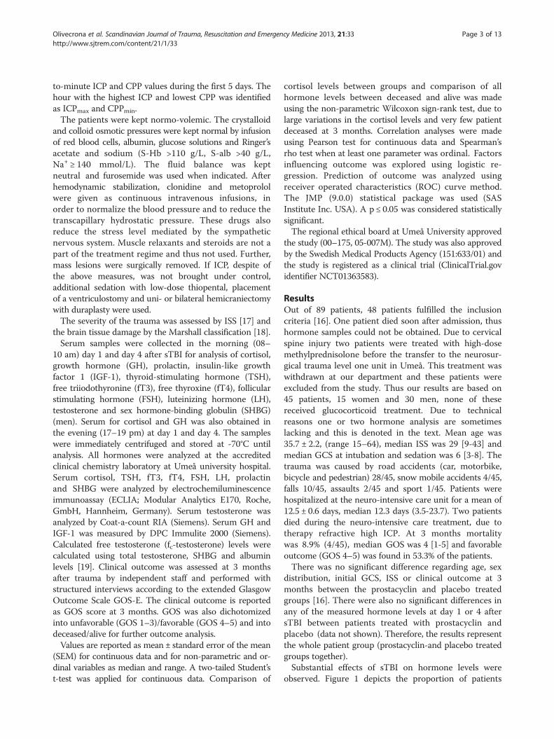
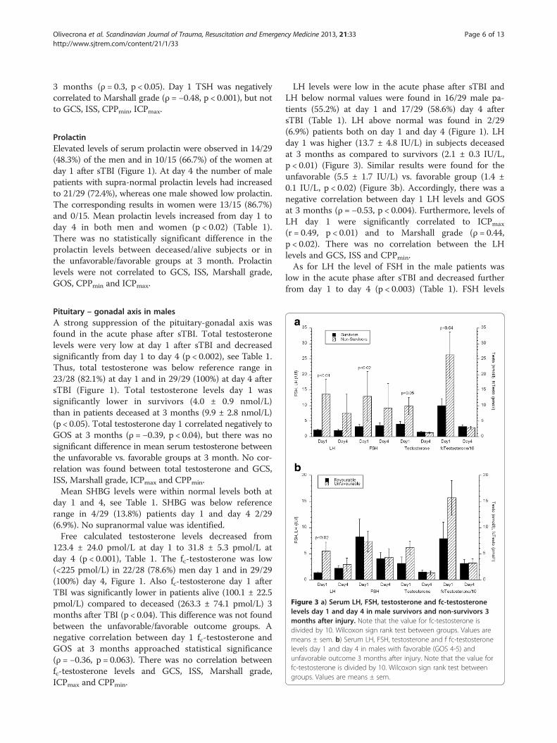
Figure 3 a) Serum LH, FSH, testosterone and fc-testosteronelevels day 1 and day 4 in male survivors and non-survivors 3months after injury. Note that the value for fc-testosterone isdivided by 10. Wilcoxon sign rank test between groups. Values aremeans ± sem. b) Serum LH, FSH, testosterone and f fc-testosteronelevels day 1 and day 4 in males with favorable (GOS 4-5) andunfavorable outcome 3 months after injury. Note that the value forfc-testosterone is divided by 10. Wilcoxon sign rank test betweengroups. Values are means ± sem.
Pituitary – gonadal axis in malesA strong suppression of the pituitary-gonadal axis wasfound in the acute phase after sTBI. Total testosteronelevels were very low at day 1 after sTBI and decreasedsignificantly from day 1 to day 4 (p < 0.002), see Table 1.Thus, total testosterone was below reference range in23/28 (82.1%) at day 1 and in 29/29 (100%) at day 4 aftersTBI (Figure 1). Total testosterone levels day 1 wassignificantly lower in survivors (4.0 ± 0.9 nmol/L)than in patients deceased at 3 months (9.9 ± 2.8 nmol/L)(p < 0.05). Total testosterone day 1 correlated negatively toGOS at 3 months (ρ = −0.39, p < 0.04), but there was nosignificant difference in mean serum testosterone betweenthe unfavorable vs. favorable groups at 3 month. No cor-relation was found between total testosterone and GCS,ISS, Marshall grade, ICPmax and CPPmin.Mean SHBG levels were within normal levels both at
day 1 and 4, see Table 1. SHBG was below referencerange in 4/29 (13.8%) patients day 1 and day 4 2/29(6.9%). No supranormal value was identified.Free calculated testosterone levels decreased from
123.4 ± 24.0 pmol/L at day 1 to 31.8 ± 5.3 pmol/L atday 4 (p < 0.001), Table 1. The fc-testosterone was low(<225 pmol/L) in 22/28 (78.6%) men day 1 and in 29/29(100%) day 4, Figure 1. Also fc-testosterone day 1 afterTBI was significantly lower in patients alive (100.1 ± 22.5pmol/L) compared to deceased (263.3 ± 74.1 pmol/L) 3months after TBI (p < 0.04). This difference was not foundbetween the unfavorable/favorable outcome groups. Anegative correlation between day 1 fc-testosterone andGOS at 3 months approached statistical significance(ρ = −0.36, p = 0.063). There was no correlation betweenfc-testosterone levels and GCS, ISS, Marshall grade,ICPmax and CPPmin.
LH levels were low in the acute phase after sTBI andLH below normal values were found in 16/29 male pa-tients (55.2%) at day 1 and 17/29 (58.6%) day 4 aftersTBI (Table 1). LH above normal was found in 2/29(6.9%) patients both on day 1 and day 4 (Figure 1). LHday 1 was higher (13.7 ± 4.8 IU/L) in subjects deceasedat 3 months as compared to survivors (2.1 ± 0.3 IU/L,p < 0.01) (Figure 3). Similar results were found for theunfavorable (5.5 ± 1.7 IU/L) vs. favorable group (1.4 ±0.1 IU/L, p < 0.02) (Figure 3b). Accordingly, there was anegative correlation between day 1 LH levels and GOSat 3 months (ρ = −0.53, p < 0.004). Furthermore, levels ofLH day 1 were significantly correlated to ICPmax
(r = 0.49, p < 0.01) and to Marshall grade (ρ = 0.44,p < 0.02). There was no correlation between the LHlevels and GCS, ISS and CPPmin.As for LH the level of FSH in the male patients was
low in the acute phase after sTBI and decreased furtherfrom day 1 to day 4 (p < 0.003) (Table 1). FSH levels
Olivecrona et al. Scandinavian Journal of Trauma, Resuscitation and Emergency Medicine 2013, 21:33 Page 7 of 13http://www.sjtrem.com/content/21/1/33
below normal were found in 3/29 (10.3%) at day 1 andin 11/29 (37.9%) at day 4, whereas FSH was abovenormal in 2/29 (6.9%) day 1 and 1/29 (3.4%) day 4 aftersTBI (Figure 1). Day 1 FSH levels were higher in patientsdeceased at 3 months (13.0 ± 8.0 IU/L) as compared tosurvivors (3.3 ± 0.6 IU/L, p < 0.02). This difference wasnot found between the favorable and unfavorable groups.GOS at 3 months was negatively correlated with FSHday 1 (ρ = −0.46, p < 0.02) and as for LH there was apositive correlation between day 1 FSH and Marshallgrade (ρ = 0.39, p < 0.04). There was no correlationbetween FSH and GCS, ISS, ICPmax and CPPmin.To investigate the possibility that the low testosterone
was secondary to high prolactin we searched possiblecorrelations between prolactin and LH, FSH, total tes-tosterone and f-testosterone. No such correlation wasfound suggesting that the hypogonadotropic hypogonad-ism in acute TBI is not due to an inhibitory effect ofelevated prolactin.
Pituitary – gonadal axis in womenWe were unable to establish in which phase in themenstrual cycle these severely injured women were.Neither could we for all subjects be certain whether theywere pre- or postmenopausal or if they were onexogenous estrogens. Thus, these hormone levelscould not be accurately interpreted and therefore notpresented. However, LH, FSH and estradiol levels inwoman were generally low.
Somatotropic axisThere was a great variability in GH levels at all measuredtime points ranging from 0.2 ug/L to 99 ug/L. Mean GHlevels showed no significant diurnal variation betweenmorning and evening. No correlations were foundbetween GH and GCS, ISS, Marshall grade, ICPmax,CPPmin or GOS. There was a trend towards lower GHlevels in deceased as compared to alive patients at 3months, see Table 2.There was a transient decrease in IGF-1, with low
levels at day 1, which were restored towards normal atday 4 after sTBI, see Table 1. Thus, day 1 after sTBI 13/43(30.2%) had levels below the age-related normal value,whereas none had high levels. Day 4, the corresponding
Table 2 GH levels day 1 and day 4 in patients deceasedand alive patients at 3 months post trauma
S-GH (mIU/L) Deceased (3 months) Alive (3 months)
Day 1 morning 11.9 ± 7.5 16.4 ± 2.6
Day 1 evening 10.6 ± 2.9 20.1 ± 3.3
Day 4 morning 8.9 ± 2.8 16.1 ± 3.0
Day 4 evening 8.6 ± 2.8 13.5 ± 2.1
Values are means ± sem.
figures were 1/44 (2.3%) and 3/44 (7.0%) respectively,Figure 1. Correspondingly, mean IGF-1 was significantlylower day 1 after sTBI than at day 4 (p < 0.0001) (Table 1).No statistically significant difference in IGF-1 levelsbetween deceased and alive or unfavorable and favorableoutcome at 3 months was observed. No correlation wasfound between IGF-1 and GCS, ISS, Marshall grade,ICPmax, CPPmin or GOS.
Outcome predictorsAmong the hormones analyzed in the acute phase fT3and TSH were most strongly and positively associated withoutcome. In the male group LH and FSH was negativelyassociated with outcome. We attempted to evaluatethe use of these hormones to predict clinical outcome. Alogistic regression model with fT3 and TSH day 4 asindependent factors and unfavorable/favorable outcomeas dependent factor showed that the model significantlypredicted (p < 0.03) outcome and fT3 contributed mostto the prediction. Using GOS or dead/alive as dependentfactors did not show any statistically significant prediction.When ICPmax was used in combination with fT3 and TSHday 1 in the whole group, the prediction of GOS at 3months was p < 0.01 and ICPmax had the strongest effecton the model. When the results were dichotomized intodead or alive at 3 months and using the same independentfactors a statistically significant predictive value (p < 0.01)was found and again ICPmax contributed most. Othercombinations of hormone levels in the whole study groupdid not contribute to the outcome prediction. However, inmen there was a clear prognostic value of LH and FSH.Thus GOS at 3 months was significantly predicted by day1 LH in combination with FSH (p < 0.001) and LHcontributed most to the prediction. Similar results werefound in the prediction of dead/alive (p < 0.002) andunfavorable/favorable outcome (p < 0.01). When ICPmax
was used in combination with day 1 LH and FSH, theprediction of GOS at 3 months was p < 0.005), dean/alive(p < 0.005) and unfavourable/favourable (p < 0.02) and inall predictions LH had the strongest effect on themodel. In evaluating the power of LH day 1 to predictnon-survivors/survivors in men a ROC analysis showedan AUC of 0.915 (p < 0.001) and the highest accuracy wasat an LH level of 12.0 (IU/L), sensitivity 0.750, specificity1.0. For a probability plot of outcome, see Figure 4a. Usingthe dicotomization of unfavorable/favorable outcome andLH day 1 resulted in an AUC of 0.774 (p < 0.003) andthe highest accuracy at a LH level of 1.7 (IU/L), witha sensitivity of 0.687 and a specificity of 0.8462, seeFigure 4b for a probability plot of outcome.
DiscussionIn this prospective study we describe alterations inpituitary-dependent hormone levels in the acute phase of

Figure 4 a) Probability of death at 3 months related to serumLH levels day 1 after sTBI. Probability results are from ROC analysis.b) Probability of unfavorable outcome at 3 months related to serumLH levels day 1 after sTBI. Probability results are from ROC analysis.
Olivecrona et al. Scandinavian Journal of Trauma, Resuscitation and Emergency Medicine 2013, 21:33 Page 8 of 13http://www.sjtrem.com/content/21/1/33
sTBI, in a well-defined patient group treated according toan ICP-targeted therapy in a protocol-guided manner.The main findings of this study were: 1) A large propor-tion of the patients had low s-cortisol levels, belowproposed cut-off levels for critical illness relatedcorticosteroid insufficiency (CIRCI). Low s-cortisol levels(<276 nmol/L = 10 ug/dL) and even <100 nmol/L wasnot associated with higher mortality or unfavorable outcomeat 3 months; 2) There was a significant association between
early (day 1) strong suppression of the pituitary-gonadalaxis and better survival and favorable outcome 3 monthsafter sTBI in men. 3) Survival and favorable outcome(GOS 4–5) was associated with significantly higher levelsof fT3 and TSH day 4 after sTBI. 4) In general there wasno correlation between GCS, ISS, ICPmax, CPPmin andhormone levels. Only in men day 1 LH was correlated toICPmax.In the last decade several studies have shown permanent
pituitary insufficiency after TBI to be considerably morecommon than previously recognized [3,8]. Therefore,screening to find patients and replace persistent hormonedeficiency has been proposed and implemented in clinicalpractice at many centers [21]. The value of measurehormone levels in the acute phase after TBI is less clear,except when suspecting an acute cortisol insufficiency [8].It is well known that the levels of most hormones aredramatically altered in acute critical illness as comparedwith resting baseline levels. Thus, interpreting hormonelevels in the acute phase after TBI is complex and has tobe related to the hormonal changes in any acute illness[22,23]. The potential benefits of replacement therapy ofendocrine dysfunction in acute TBI are not known, exceptfor the life-saving replacement of an acute ACTH-cortisoldeficiency [8]. The few intervention studies aiming toreplace (or over-replace) hormone deficiencies in acutecritical illness have generally failed to show benefits ofhormone treatment [23]. In contrast, there is evidencethat pharmacological doses of glucocorticoids in TBIpatients without cortisol deficiency increase mortality [24]and this has also been shown in a study using high dosesof recombinant growth hormone in acute critical illness[25]. Thus, it is likely that most of the hormonal changesin acute critical illness can be attributed to physiologicaladaptation to severe physiological stress [8].Most previous studies on hormone levels after TBI are
on mixed materials, including mild, moderate and severeTBI and report varying rates of acute hormonal dysfunction[5,26,27]. The present study is the first study whichprospectively studies hormone levels in the acutephase of severe TBI in a homogenous group of patients,strictly protocol-treated according to an ICP-targetedtherapy based on the Lund concept [28]. We foundprofound changes for most pituitary dependent hormonesin the acute phase after sTBI, i.e. low levels of thyroidhormones, strong suppression of the pituitary-gonadalaxis and increased levels of prolactin, much in linewith previous studies on acute TBI and other acutecritical illness.We have previously shown that our protocol-guided
ICP targeted treatment seems to protect patients withsevere TBI from clinical and subclinical seizures andthus reduces the risk of secondary brain injury and araise in prolactin due to seizures [29].
Olivecrona et al. Scandinavian Journal of Trauma, Resuscitation and Emergency Medicine 2013, 21:33 Page 9 of 13http://www.sjtrem.com/content/21/1/33
Prostacyclin has been proposed to have beneficialeffects in traumatic injury [30]. However, in a larger setof TBI patients these results were not confirmed [16].Interestingly, it has been shown that prostacyclin insome situations influences pituitary hormone release[31]. Both plasma cortisol and prolactin increased afterprostacyclin infusion. This effect has been suggested tobe due to stress during prostacyclin infusion. However,the mechanisms are not fully understood [32]. We wereunable to show any significant effect of prostacyclinupon hormone levels after acute sTBI. However, it islikely that the effect of the TBI upon hormone levels wereso pronounced that the potential effect of prostacyclinwas obscured.
Hypothalamic-pituitary-adrenal axisElevated cortisol levels is a physiological response to crit-ical illness to modulate metabolism to ensure energy sub-strates for vital organs, exert supporting effects on thecirculatory system and suppress excessive immune systemactivation [22,33,34]. The primary endocrine task in acuteTBI is not to overlook a clinically relevant deficiency ofthe hypothalamic-pituitary-adrenal (HPA) axis with acutecortisol insufficiency, a condition with life-threateninghyponatremia and hypotension, in need for promptdiagnosis and treatment with stress-doses of glucocorti-coids. It should however be recognized that correctdiagnosis of cortisol insufficiency in a critically illpatient is difficult and strict diagnostic criteria are stillmissing. Recent recommendations are to consider morningserum cortisol levels <300 nmol/L as highly suggestive ofacute adrenal insufficiency and treat with stress-dosesof i.v. hydrocortisone (i.e. 200–300 mg/d) [8]. We foundlow s-cortisol levels in more than 50% of the sTBI patientsin our study. No patients in the current study were givenglucocorticoids. In spite of this we found no associationbetween low cortisol levels and increased mortality orunfavorable outcome. This is opposed to the Dublin groupwho found increased mortality in the patients with thelowest s-cortisol [8]. Contrary, in this study patientsdeceased at 3 months tended to have higher serumcortisol in the acute phase compared with survivors. Thisis in line with previous studies of cortisol levels in differenttypes of critical illness [35] and may be attributed to amore severe critical illness and correspondingly higherstress response. Abnormalities in cortisol dynamicsafter severe traumatic brain injury are inconsistent,and correlations between serum cortisol levels and clinicaloutcomes have been conflicting since both high and lowserum cortisol levels have been associated with pooreroutcomes [36]. The CRASH-study reported increasedmortality in TBI-patients treated with pharmacologicaldoses of methylprednisolone [37]. Furthermore, experi-mental evidence suggests that suppressing elevated
cortisol levels reduce neuronal damage after differentinsults [38]. On the other hand, in a recent study treatmentwith stress doses of hydrocortisone (200 mg/d) decreasedthe risk of hospital-acquired pneumonia and shortenedmechanical ventilation in patients with sTBI and in-adequate adrenal function, defined as basal s-cortisol <15μg/dL (414 nmol/L) or rise in s-cortisol <9 μg/dL (248nmol/L) after cosyntropin stimulation [39]. In that study alarge proportion of the patients were treated withEtomidate and it has been criticized for difficulty toobjective outcome parameters, since glucocorticoidtherapy may blunt fever response, which was a criteriafor pneumonia. Thus, more studies are needed to answerthe question when a TBI-patient will benefit or not fromreplacement therapy with stress doses of glucocorticoids.In studies like this it is important to acknowledge the vastrange of factors potentially influencing hormone levels incritically ill patients under full neuro-intensive care. Inthis study the patients have been treated in a strictlyprotocol-guided manner and most of the patientsreceived the same drugs during the neuro-intensive care.Etomidate, which is a strong inhibitor of adrenal steroidhormone synthesis [40] was not used in any of thepatients. Even so, some degree of confounding effects ofe.g. anesthetics upon hormone levels cannot be excluded.Little is known about the effects of anesthetic drugs uponhormone levels in patients with sTBI. The effect ofpropofol, which was used in some of the patients is lessclear. Some studies demonstrate a drop in cortisol levelsduring propofol infusion [41,42], whereas others show noeffect on hormone levels [43]. More studies on theisolated effect of anesthetic drugs upon hormonelevels are needed, since these cannot be avoided dur-ing the early treatment of patients with severe trau-matic head injury.
Thyroid axisLevels of fT4 and fT3 were low in the acute phase ofsTBI, with a significant decrease from day 1 and 4, inline with a previous study [44]. Interestingly, fT3 levelswere significantly lower at day 4 in patients who diedwithin 3 months after sTBI compared to survivors.Furthermore, survivors with an unfavorable outcome at3 months had significantly lower fT3 levels compared topatients with favorable outcome. Previous studies havesuggested an association between stronger suppressionof the hypothalamic-thyroid axis in more severe injuriesand poor outcomes [44,45]. In line with this we found anegative correlation between day 4 TSH-levels andMarshall CT grade score, i.e. worse radiological findingswere associated with lower s-TSH day 4 after sTBI. Theobserved decreases in serum concentration of boththyroid hormones and TSH are consistent with a centralsuppression of the hypothalamic-pituitary-thyroid (HPT)
Olivecrona et al. Scandinavian Journal of Trauma, Resuscitation and Emergency Medicine 2013, 21:33 Page 10 of 13http://www.sjtrem.com/content/21/1/33
axis. This is supported by post mortem studies showinga decreased expression of thyrotropin-releasing hormonein the hypothalamic paraventricular nucleus of patientswith a decreased serum T3 level [46]. In critical illness,serum T3 may even become unmeasurable withoutgiving rise to an elevated concentration of serum TSH.It is at present not clearly established whether thisreflects an adaptation of the organism to illness or instead apotentially deleterious condition leading to hypothyroidismat tissue level. It is likely that the transient down regulationat all levels of the HPT axis (decreased TRH and TSH atthe hypothalamic-pituitary level, and a decreased T3due to altered peripheral deiodinase activity) is part ofthe neuro-endocrine adaptation to critical illness in anattempt to save energy. In this study of only severe TBI,we found that a more pronounced and prolongedsuppression of TSH was associated with unfavorableoutcome, and more severe CT-findings suggesting that aless pronounced and prolonged central down regulationof the HPT-axis may be a marker of a less severe TBI and/or a stronger capacity to adapt and regain hypothalamic-pituitary function after TBI, with a more normalTSH-response (i.e. higher) to low thyroid hormone levels.
ProlactinWe found elevated levels of serum prolactin, with an in-crease from day 1 to day 4 after TBI. Our results are in ac-cordance with previous studies showing hyperprolactinemiain more than 50% of patients in the early, acute phasepost-TBI [26,47], established in ranges from mild to severetraumatic head injury [5]. Day 1 serum prolactin levelswere significantly negatively correlated with CPPmin andpositively correlated to ICPmax. Apart from this we foundno correlation between serum prolactin levels day 1 orday 4 and severity of TBI, which was reported in someprevious studies [26,45,48]. Nor could prolactin levels inthe acute phase be used as a prognostic factor of clinicaloutcome. Gonadotropin (LH/FSH) and testosteronesecretion is inhibited by elevated levels of prolactin, e.g.in patients with prolactinomas. However, in this study theelevated prolactin levels in acute TBI did not appear tocontribute to the suppression of the pituitary-gonadal axis,which is in line with previous studies [49].
Pituitary – gonadal axisCentral inhibition of the pituitary-gonadal axis is aconsistent finding in critical illness, including TBI andconsidered an adaptive response to severe physiologicalstress, i.e. an appropriate temporary swich-off of anabolicandrogens in circumstances of acute stress, to preserveenergy and metabolic substrates for vital functions. In linewith previous studies we found strong central inhibitionof the pituitary-gonadal axis already at day 1, with furthersuppression at day 4, when 100% of the male patients
showed testosterone levels below reference range. Further-more, almost all of the men had low levels of LH and FSHboth at day 1 and 4. This is in agreement with a previousstudy showing a high incidence of hypogonadotropichypogonadism in the immediate post-TBI period [50].Previous studies have clearly demonstrated that testoster-one levels in males and estrogen levels in femalessignificantly fall within the first 24 hours following TBIand remain low for 7–10 days [26,50].Novel findings in this study were significant correlations
between lower levels of LH and FSH day 1 and worse braininjury according to the Marshall CT grade score. This is inline with previous reports on associations between moresevere TBI (often lower GCS) and stronger suppression ofthe hypothalamus-pituitary-gonad-axis [8]. Interestingly,male patients alive at 3 months after TBI had significantlylower LH, FSH and testosterone levels day 1 vs. non-survivors. Day 1 serum LH was also lower in male patientswith a favorable outcome 3 months after TBI vs. unfavor-able outcome, i.e. GOS 1–3. These findings in this studysuggest that very severe brain injury may hamper the adap-tive, physiological suppression of the pituitary-gonadal axisand this inability is a poor prognostic sign.
Somatotropic axisGrowth hormone is normally released in a highly pulsatilemanner. Therefore analyses regarding sub- and supra-normal levels may be misleading. We found a greatvariability in GH levels at all measured time points. Meanserum GH levels showed no significant diurnal variation,i.e. between morning and evening. Interestingly, there wasa trend towards higher serum GH in patients whosurvived 3 months than in those who succumbed to theirinjuries. We also found a transient decrease in serum IGF-1with low levels at day 1, which were restored towardsnormal at day 4 after sTBI. Low IGF-1 with elevated GHlevels have been shown in the acute post-traumatic phase,as well as a normalization of GH and increase of IGF-1 inthe following weeks after trauma [51]. This has beenattributed to a state of acquired peripheral GH resistancein critical illness [52]. Contradictory literature is availableon the GH levels following severe traumatic brain injuryas reported by various authors. Chiolero et al. foundelevated GH levels in the acute phase [45]. Hackl et al.reported elevated GH levels in patients with high ICP[53]. Della Corte et al. showed relatively normal GH levelsin the acute phase, and improving levels of IGF-1 1 and 2weeks after trauma compared to 2 days after trauma [54].In a more recent study, in which mild, moderate andsevere injuries were mixed the GH levels overall remainedrelatively normal or slightly elevated throughout theacute setting [50]. Growth hormone is normally releasedin a highly pulsatile manner. Furthemore, clonidine andmetoprolol have been shown to increase GH sekretion.
Olivecrona et al. Scandinavian Journal of Trauma, Resuscitation and Emergency Medicine 2013, 21:33 Page 11 of 13http://www.sjtrem.com/content/21/1/33
[55,56] Therefore analyses of GH sampled only twice dailyunder multiple pharmacological treatment should beinterpreted with caution.
Prediction of outcomeIn the search for biomarkers of clinical outcome after sTBIwe tested the hypothesis that hormone levels may predictoutcome at 3 months post-injury. Day 4 fT3 and TSHlevels were shown to be predictive factors for outcome.Interestingly, in men day 1 LH and FSH were predictivefactors of outcome and LH was the main predicting factor.However, by combining the above-mentioned hormonelevels with ICPmax it was clear that ICPmax still is the mainpredicting factor of outcome in patients treated at ourdepartment. By using LH day 1 in men the ROC analysisshowed a high AUC and a rather good sensitivity and agood specificity. The probability of worse outcomeincreased with increasing levels of LH day 1.
Study limitationsThis study is limited by the fact that GH, LH and FSHhormone levels were assessed only once daily. Given theknown pulsatile secretory pattern of GH, LH and FSH,the true pattern of their release in the acute settingwould require repeated sampling at close intervals. Also,due to lack of information about the females menstrualstatus the female gonadal axis could not be analyzed indetail. In this study no hormonal stimulation testswere performed, which in some cases may providemore detailed information about responsiveness of thehypothalamic-pituitary axis. Although such stimulationtests were done in some prior studies, they are challengingto do during the first days after the trauma with the pa-tient under full neuro-intensive care. Also, the intentionof the study was not to explore the patency of thehypothalamic-pituitary axis but to investigate the traumainduced effects on hormonal levels.Many of the patients (i.e. 69%) in the study suffered
from multiple injuries [57]. The data does not allowdiscrimination of what proportion of the hormonealteration is caused by the TBI itself and how much iscaused by the extracranial injuries/critical illness situation.Furthermore, according to the treatment protocol most ofthe patients received blood transfusions. No analyses aremade regarding a possible association between bloodtransfusions and hormone levels [58].The clinical impact of the acute alterations of hormone
levels in patients with severe brain injury is still largelyunknown. Most changes are likely to represent anadaptive physiological response to critical illness. How-ever, experimental studies indicate that hormonal effectsupon the CNS in acute, sub-acute and long-term recoveryphases may be of importance for secondary neuronaldamage and plasticity/recovery processes. In this study we
find that hormone levels in the early acute phase of sTBImay be used in models to predict clinical outcome.However, traditional acute phase variables, i.e. ICPappear superior and large cohorts are needed for furtherevaluation and confirmation of reliable screening markers.Future studies should be designed to ensure a highdiagnostic robustness for proper identification of reliablepredictors, as the results may be highly dependent ondiagnostic pitfalls.In conclusion, we found pronounced dynamic alterations
in pituitary-dependent hormone levels day 1 and 4 aftersevere TBI. Cortisol levels below proposed cut-off limitsfor adrenal insufficiency were found in a large proportionof the patients without association to unfavorableclinical outcome. Stronger early suppression of thepituitary-gonadal axis was associated with a more favorableoutcome whereas a prolonged suppression of the thyroidaxis was associated with higher mortality and poorfunctional outcome. Whether these findings are applic-able for other groups of patients with sTBI treated byother treatment regimes has to be further evaluated.
Competing interestsThe authors declare that they have no competing interests.
Authors’ contributionsZO: Carried out surgery on some of the patients, collected data and wentthrough patient charts, wrote the manuscript, analysis and interpretation ofdata, revising the manuscript critically. PD: analysis and interpretation of data,revising the manuscript critically, wrote some parts of the manuscript. LOK:Designed the study, collected data, analysis and interpretation of data, revisingthe manuscript critically. All authors read and approved the final manuscript.
AcknowledgementsWe wish to express our gratitude to Pedram Tabatabai, MD, Lukas Bobinski, MD,Magnus Olivecrona, MD, PhD, Marie Rodling-Wahlström, MD, PhD, SilvanaNaredi, MD, PhD and our research nurses Kristin Nyman and Anna-LenaÖstlund for help with the study. The Department of Clinical Neuroscience,Kempe Foundation and Pfizer, Sweden financially supported this study.
Author details1Department of Pharmacology and Clinical Neuroscience,Division of Neurosurgery, Umeå University, SE 90185, Umeå, Sweden.2Department of Public Health and Clinical Medicine, Umeå University, SE90185, Umeå, Sweden.
Received: 9 November 2012 Accepted: 2 April 2013Published: 20 April 2013
References1. Simmonds: Ueber Hypophysisschwund mit tödlichem Ausgang. Deutsch
Medizinische Wochenschrift. 1914 1914, 40:322–323.2. Escamilla RF, Lisser H: Simmonds’ Disease (Hypophyseal Cachexia): Clinical
Report of Several Cases with Discussion of Diagnosis and Treatment.California and western medicine 1938, 48(5):343–8.
3. Benvenga S, Campenni A, Ruggeri RM, Trimarchi F: Clinical review 113:Hypopituitarism secondary to head trauma. J Clin Endocrinol Metab 2000,85(4):1353–61.
4. Edwards OM, Clark JD: Post-traumatic hypopituitarism. Six cases and areview of the literature. Medicine (Baltimore) 1986, 65(5):281–90.
5. Klose M, Juul A, Poulsgaard L, Kosteljanetz M, Brennum J, Feldt-RasmussenU: Prevalence and predictive factors of post-traumatic hypopituitarism.Clin Endocrinol (Oxf ) 2007, 67(2):193–201.
6. Schneider HJ, Kreitschmann-Andermahr I, Ghigo E, Stalla GK, Agha A:Hypothalamopituitary dysfunction following traumatic brain injury and
Olivecrona et al. Scandinavian Journal of Trauma, Resuscitation and Emergency Medicine 2013, 21:33 Page 12 of 13http://www.sjtrem.com/content/21/1/33
aneurysmal subarachnoid hemorrhage: a systematic review. JAMA 2007,298(12):1429–38.
7. Venkatesh B, Cohen J: Adrenocortical (dys)function in septic shock - a sickeuadrenal state. Best practice & research Clinical endocrinology & metabolism[Review] 2011, 25(5):719–33.
8. Hannon MJ, Sherlock M, Thompson CJ: Pituitary dysfunction followingtraumatic brain injury or subarachnoid haemorrhage - in “EndocrineManagement in the Intensive Care Unit”. Best practice & research Clinicalendocrinology & metabolism [Review] 2011, 25(5):783–98.
9. Chisu V, Manca P, Lepore G, Gadau S, Zedda M, Farina V: Testosteroneinduces neuroprotection from oxidative stress. Effects on catalaseactivity and 3-nitro-L-tyrosine incorporation into alpha-tubulin in amouse neuroblastoma cell line. Arch Ital Biol 2006, 144(2):63–73.
10. Hammond J, Le Q, Goodyer C, Gelfand M, Trifiro M, LeBlanc A:Testosterone-mediated neuroprotection through the androgen receptorin human primary neurons. J Neurochem 2001, 77(5):1319–26.
11. Napoli R, Guardasole V, Angelini V, D’Amico F, Zarra E, Matarazzo M, et al:Acute effects of growth hormone on vascular function in humansubjects. J Clin Endocrinol Metab 2003, 88(6):2817–20.
12. Scheepens ASE, Breier BH, Clark RG, Gluckman PD, Williams CE: Growthhormone as a neuronal rescue factor during recovery from CNS injury.Neuroscience 2001, 104(3):677–87. 2001.
13. Muller K, Townend W, Biasca N, Unden J, Waterloo K, Romner B, et al:S100B serum level predicts computed tomography findings after minorhead injury. The Journal of trauma. [Multicenter Study]. 2007, 62(6):1452–6.
14. Silha JV, Krsek M, Hana V, Marek J, Weiss V, Jezkova J, et al: The effects ofgrowth hormone status on circulating levels of vascular growth factors.Clin Endocrinol (Oxf ) 2005, 63(1):79–86.
15. Liu M, Kelley MH, Herson PS, Hurn PD: Neuroprotection of sex steroids.Minerva endocrinologica. [Research Support, N.I.H., Extramural ResearchSupport, Non-U.S. Gov’t Review] 2010, 35(2):127–43.
16. Olivecrona M, Rodling-Wahlstrom M, Naredi S, Koskinen LO: Prostacyclintreatment in severe traumatic brain injury: a microdialysis and outcomestudy. Journal of neurotrauma. [Randomized Controlled Trial ResearchSupport, Non-U.S. Gov’t] 2009, 26(8):1251–62.
17. Baker SP, O’Neill B, Haddon W Jr, Long WB: The injury severity score: amethod for describing patients with multiple injuries and evaluatingemergency care. J Trauma 1974, 14(3):187–96.
18. Marshall LF, Marshall SB, Klauber MR, Van Berkum CM, Eisenberg H, Jane JA,et al: The diagnosis of head injury requires a classification based oncomputed axial tomography. J Neurotrauma 1992, 9(Suppl 1):S287–92.
19. Vermeulen A, Verdonck L, Kaufman JM: A critical evaluation of simplemethods for the estimation of free testosterone in serum. The Journal ofclinical endocrinology and metabolism. [Comparative Study] 1999,84(10):3666–72.
20. Annane D: Defining critical illness-related corticosteroid insufficiency:one step forward! Critical care medicine. [Comment Editorial] 2010,38(2):721–2.
21. Ghigo E, Masel B, Aimaretti G, Leon-Carrion J, Casanueva FF, Dominguez-Morales MR, et al: Consensus guidelines on screening for hypopituitarismfollowing traumatic brain injury. Brain injury : [BI]. [Consensus DevelopmentConference Research Support, Non-U.S. Gov’t Review] 2005, 19(9):711–24.
22. Van den Berghe G: Endocrine evaluation of patients with critical illness.Endocrinol Metab Clin North Am 2003, 32(2):385–410.
23. Hassan-Smith Z, Cooper MS: Overview of the endocrine response tocritical illness: how to measure it and when to treat. Best practice &research Clinical endocrinology & metabolism. [Research Support, Non-U.S.Gov’t Review] 2011, 25(5):705–17.
24. Roberts I, Yates D, Sandercock P, Farrell B, Wasserberg J, Lomas G, et al: Effectof intravenous corticosteroids on death within 14 days in 10008 adultswith clinically significant head injury (MRC CRASH trial): randomisedplacebo-controlled trial. Lancet. [Clinical Trial Multicenter Study RandomizedControlled Trial Research Support, Non-U.S. Gov’t] 2004, 364(9442):1321–8.
25. Takala J, Ruokonen E, Webster NR, Nielsen MS, Zandstra DF, Vundelinckx G,et al: Increased mortality associated with growth hormone treatment incritically ill adults. The New England journal of medicine. [Clinical TrialMulticenter Study Randomized Controlled Trial Research Support, Non-U.S.Gov’t] 1999, 341(11):785–92.
26. Agha A, Rogers B, Mylotte D, Taleb F, Tormey W, Phillips J, et al:Neuroendocrine dysfunction in the acute phase of traumatic brain injury.Clinical endocrinology. [Research Support, Non-U.S. Gov’t] 2004, 60(5):584–91.
27. Wagner AK, McCullough EH, Niyonkuru C, Ozawa H, Loucks TL, Dobos JA,et al: Acute serum hormone levels: characterization and prognosis aftersevere traumatic brain injury. Journal of neurotrauma. [Research Support, N.I.H., Extramural Research Support, U.S. Gov’t, Non-P.H.S. Research Support, U.S.Gov't, P.H.S.] 2011, 28(6):871–88.
28. Nordstrom CH: The “Lund concept”: what it is and what it isn’t. Intensivecare medicine. [Comment Letter] 2007, 33(3):558. author reply 9.
29. Debudaj A, Bobinski R: [The pathophysiology of acute mountain sickness].Pol Merkur Lekarski. [Review] 2010, 28(168):478–81.
30. Grande PO, Moller AD, Nordstrom CH, Ungerstedt U: Low-doseprostacyclin in treatment of severe brain trauma evaluated withmicrodialysis and jugular bulb oxygen measurements. Acta AnaesthesiolScand 2000, 44(7):886–94.
31. Allolio B, Fitzgerald GA, Hipp FX, Hossmann V, Mies R, Winkelmann W: Theeffect of prostacyclin on pituitary hormone release. Br J Clin Pharmacol1980, 10(6):626–7.
32. Grimee R, Wulfert E: Acute stress in rats produces a rapid and sustainedincrease in prostacyclin production in aortic tissue: dependence oncorticosterone. Life Sci 1995, 57(1):69–81.
33. Marik PE, Zaloga GP: Adrenal insufficiency in the critically ill: a new lookat an old problem. Chest. [Review] 2002, 122(5):1784–96.
34. Van den Berghe G, de Zegher F, Bouillon R: Clinical review 95: Acute andprolonged critical illness as different neuroendocrine paradigms. TheJournal of clinical endocrinology and metabolism. [Research Support, Non-U.S.Gov’t Review] 1998, 83(6):1827–34.
35. Christ-Crain M, Stolz D, Jutla S, Couppis O, Muller C, Bingisser R, et al: Freeand total cortisol levels as predictors of severity and outcome incommunity-acquired pneumonia. American journal of respiratory andcritical care medicine. [Research Support, Non-U.S. Gov’t] 2007,176(9):913–20.
36. Savaridas T, Andrews PJ, Harris B: Cortisol dynamics following acute severebrain injury. Intensive Care Med 2004, 30(7):1479–83.
37. Ingebrigtsen T, Romner B, Trumpy JH: Management of minor head injury:the value of early computed tomography and serum protein S-100measurements. J Clin Neurosci 1997, 4(1):29–33.
38. Smith-Swintosky VL, Pettigrew LC, Sapolsky RM, Phares C, Craddock SD,Brooke SM, et al: Metyrapone, an inhibitor of glucocorticoid production,reduces brain injury induced by focal and global ischemia and seizures.Journal of cerebral blood flow and metabolism : official journal of theInternational Society of Cerebral Blood Flow and Metabolism. [ResearchSupport, Non-U.S. Gov’t Research Support, U.S. Gov’t, Non-P.H.S. ResearchSupport, U.S. Gov’t, P.H.S.] 1996, 16(4):585–98.
39. Herrmann M, Jost S, Kutz S, Ebert AD, Kratz T, Wunderlich MT, et al: Temporalprofile of release of neurobiochemical markers of brain damage aftertraumatic brain injury is associated with intracranial pathology asdemonstrated in cranial computerized tomography. Journal of neurotrauma.[Comparative Study Research Support, Non-U.S. Gov’t] 2000, 17(2):113–22.
40. Moore RA, Allen MC, Wood PJ, Rees LH, Sear JW: Peri-operative endocrineeffects of etomidate. Anaesthesia 1985, 40(2):124–30.
41. Tagawa M, Sako T, Ejima H, Kurokawa K, Orima H, Motoyoshi S: Changes inplasma cortisol concentration by ages in dogs under ketamine andthiopental anesthesia. Nihon juigaku zasshi The Japanese journal ofveterinary science 1989, 51(2):278–83.
42. Polo-Garvin A, Garcia-Sanchez MJ, Peran F, Almazan A: [Evaluation of thehemodynamic and endocrino-metabolic response to tracheal intubationin patients anesthetized with thiopental or propofol]. Rev Esp AnestesiolReanim 1993, 40(6):344–8.
43. Murakawa T, Tsubo T, Kudo T, Kudo M, Matsuki A: [Effect of propofol as anagent for anesthetic induction on pituitary-adrenocortical functionduring anesthesia and surgery]. Masui 1998, 47(11):1350–7.
44. Woolf PD, Lee LA, Hamill RW, McDonald JV: Thyroid test abnormalities intraumatic brain injury: correlation with neurologic impairment andsympathetic nervous system activation. The American journal of medicine.[Research Support, U.S. Gov’t, Non-P.H.S. Research Support, U.S. Gov’t, P.H.S.]1988, 84(2):201–8.
45. Chiolero RL, Lemarchand-Beraud T, Schutz Y, de Tribolet N, Bayer-Berger M,Freeman J: Thyroid function in severely traumatized patients with orwithout head injury. Acta Endocrinol (Copenh) 1988, 117(1):80–6.
46. De Jongh FE, Jobsis AC, Elte JW: Thyroid morphology in lethal non-thyroidal illness: a post-mortem study. European journal of endocrinology /European Federation of Endocrine Societies 2001, 144(3):221–6.

Olivecrona et al. Scandinavian Journal of Trauma, Resuscitation and Emergency Medicine 2013, 21:33 Page 13 of 13http://www.sjtrem.com/content/21/1/33
47. Bondanelli M, Ambrosio MR, Margutti A, Boldrini P, Basaglia N, FranceschettiP, et al: Evidence for integrity of the growth hormone/insulin-like growthfactor-1 axis in patients with severe head trauma during rehabilitation.Metabolism 2002, 51(10):1363–9.
48. Matsuura H, Nakazawa S, Wakabayashi I: Thyrotropin-releasing hormoneprovocative release of prolactin and thyrotropin in acute head injury.Neurosurgery 1985, 16(6):791–5.
49. Spratt DI, Cox P, Orav J, Moloney J, Bigos T: Reproductive axis suppressionin acute illness is related to disease severity. The Journal of clinicalendocrinology and metabolism. [Research Support, Non-U.S. Gov’t] 1993,76(6):1548–54.
50. Wagner J, Dusick JR, McArthur DL, Cohan P, Wang C, Swerdloff R, et al:Acute gonadotroph and somatotroph hormonal suppression aftertraumatic brain injury. Journal of neurotrauma. [Research Support, N.I.H.,Extramural] 2010, 27(6):1007–19.
51. Wildburger R, Zarkovic N, Leb G, Borovic S, Zarkovic K, Tatzber F: Post-traumaticchanges in insulin-like growth factor type 1 and growth hormone inpatients with bone fractures and traumatic brain injury. Wien Klin Wochenschr2001, 113(3–4):119–26.
52. Van den Berghe G: Increased mortality associated with growth hormonetreatment in critically ill adults. The New England journal of medicine.[Comment Letter] 2000, 342(2):135. author reply −6.
53. Hackl JM, Gottardis M, Wieser C, Rumpl E, Stadler C, Schwarz S, et al: Endocrineabnormalities in severe traumatic brain injury–a cue to prognosis in severecraniocerebral trauma? Intensive Care Med 1991, 17(1):25–9.
54. Della Corte F, Mancini A, Valle D, Gallizzi F, Carducci P, Mignani V, et al:Provocative hypothalamopituitary axis tests in severe head injury:correlations with severity and prognosis. Critical care medicine.[Comparative Study] 1998, 26(8):1419–26.
55. Leckman JF, Cohen DJ, Gertner JM, Ort S, Harcherik DF: Growth hormoneresponse to clonidine in children ages 4–17: Tourette’s syndrome vs.children with short stature. J Am Acad Child Psychiatry 1984, 23(2):174–81.
56. Clausen-Sjobom N, Lins PE, Adamson U, Curstedt T, Hamberger B: Effects ofmetoprolol on the counter-regulation and recognition of prolongedhypoglycemia in insulin-dependent diabetics. Acta Med Scand 1987,222(1):57–63.
57. Brorsson C, Rodling-Wahlstrom M, Olivecrona M, Koskinen LO, Naredi S:Severe traumatic brain injury: consequences of early adverse events.Acta Anaesthesiol Scand 2011, 55(8):944–51.
58. Grill E, Strong M, Sonnad SS, Sarani B, Pascual J, Collins H, et al: Altered thyroidfunction in severely injured patients. J Surg Res 2013, 179(1):132–7.
doi:10.1186/1757-7241-21-33Cite this article as: Olivecrona et al.: Acute neuro-endocrine profile andprediction of outcome after severe brain injury. Scandinavian Journal ofTrauma, Resuscitation and Emergency Medicine 2013 21:33.
Submit your next manuscript to BioMed Centraland take full advantage of:
• Convenient online submission
• Thorough peer review
• No space constraints or color figure charges
• Immediate publication on acceptance
• Inclusion in PubMed, CAS, Scopus and Google Scholar
• Research which is freely available for redistribution
Submit your manuscript at www.biomedcentral.com/submit