Embed Size (px)
Citation preview
99
Scholars Journal of Applied Medical Sciences (SJAMS) ISSN 2320-6691 (Online)
Sch. J. App. Med. Sci., 2016; 4(1B):99-111 ISSN 2347-954X (Print) ©Scholars Academic and Scientific Publisher
(An International Publisher for Academic and Scientific Resources)
www.saspublisher.com
Original Research Article
Some Hematological Parameters among Patients with Pulmonary Tuberculosis –
Khartoum State Mohammed Abaker Saeed Mohammed
Giad Specialized Hospital, Khartoum, Sudan
*Corresponding author Mohammed Abaker Saeed Mohammed
Email:
Abstract: Tuberculosis is a highly prevalent chronic infectious disease caused and it’s still a common disease in
develop countries and still a major cause of mortality today , can affect many part of the body like lung and bone this
defect can lead to defect in most of blood component. The aim of the study is to measure some of hematological
parameters among Sudanese patients affected with pulmonary tuberculosis in Khartoum state. During the period
between April to August 2013. 40 patients with pulmonary tuberculosis (25 males and 15 females) and 20 healthy
controls (14 males and 6 females) were included 2.5 ml of blood was collected from patients and controls into EDTA
containers then CBC Test was carried out immediately after collection, were carried out (Hb, RBCs count, HCT, MCV,
MCH, MCHC, TWBCs, platelet count and differential leucocyte count) using Hematological analyzer (Sysmex KX-
21N). Data obtained were analyzed using software program statistical package of social science (SPSS.V.11.5).The
results in pulmonary tuberculosis patients when compared with control showed that there was significant lower values in
Hemoglobin (Hb), Red Cell Count (RBCs) and Packed Cell Volume (PCV) (P= 0.00) and normal Means MCV (83.4±2),
Mean MCH (30.1±4.3) and MCHC (36.1±1.1) in T.B patients compared with controls group. Total leukocyte count
(TWBCs) and absolute Monocyte count was found both significant increase (P=0.00) compared to controls. The absolute
Neutrophil count showed significant increase in T.B patients (P. 0.008) compared with control. And the most type of
anemia was normocytic normochromic anemia. The significant increase in platelet count (Thrombocytosis) was showed
in T.B patients (P. 0.00) compared with controls group. Moderate normocytic normochromic anemia , leuckocytosis
especially neutrophilia with monocytosis and moderate thrombocytosis was found in T.B patients compared to controls
group and other further studied is needed in this field.
Keywords: Tuberculosis, chronic infectious disease, Hemoglobin, Total leukocyte count
INTRODUCTION:
Tuberculosis is a highly prevalent chronic
infectious disease caused by Mycobacterium
tuberculosis. Pulmonary tuberculosis (PTB) is still a
common disease in develop countries[1]. One third of
world population is infected and approximately 3
million people die annually from pulmonary
tuberculosis [2]. The following people are at high risk
for active tuberculosis: elderly, infants and people with
weaken immune system, for instance, AIDS. The risk
for contracting TB increases if one is in frequent contact
with people living with the disease, poor nutrition and
living in crowded or unsanitary living condition [3].
The following factors may increase the rate of TB
infection in a population: Increase in HIV infections,
increase in number of homeless people (poor
environment and nutrition) and the appearance of drug-
resistant strains of TB [3].
Test that may be used in the diagnosis of
tuberculosis include sputum examination for acid –
fast bacilli ,chest CT scan , chest X-ray , cultures
and tuberculin skin test however nucleic acid
amplification test and PCR test are used for diagnosis
of tuberculosis [4]. Tuberculosis prevention and control
takes two parallel approaches. First people with
tuberculosis and their contacts and then treated in the
second approach children are vaccinated to protect
them from tuberculosis infection [5].
In other concept, hematological profile are
those parameters of the blood forms (red blood cells,
white blood cells and platelets) with normal range or
value as reference for any value to know whether or
not they are normal[6]. Therefore there is the need to
know those parameters of the blood which are abnormal
in patients living with pulmonary tuberculosis and few
of red and white cell indices would be considered. It is
Saeed Mohammed ., Sch. J. App. Med. Sci., January 2016; 4(1B):99-111
100
true that when one gets infected with pulmonary
tuberculosis there is some level of changes which
occurs in the blood because it is a bacterial infection
that it is secrete some substance that effect of some
hematological parameters in active pulmonary
tuberculosis anemia and iron deficiency erythropoiesis
was observed there was a closed correlation between
acid-fast bacilli in sputum and abnormal hematological
value [7-9].
Hematological abnormalities in pulmonary
tuberculosis: Pulmonary tuberculosis may produce
reversible abnormalities of the peripheral blood.
Hematological abnormalities have been associated with
tuberculosis and response drug the r ap y . Howev er the
changes in response to t h e r a p y have no t fully been
determined in pulmonary tuberculosis patient living
developing countries[10].
Anaemia:
Anaemia is define as qualitative or
quantitative deficiency of hemoglobin which
normally carries oxygen from the lung to the
tissue [11]. Some of causes of anemia reduce
hemoglobin production, reduce DNA synthesis ,
reduce stem cell production, Bone marrow infiltration,
infection, increase red cell destruction and acute or
chronic blood loss [12]. The mild to moderate anemia
that is often observed In patient with infection ,
inflammatory or neoplastic disease that persist for
more than 1-2 months is called anemia of chronic
disease. Anemia has been reported in 16 % to 94 % in
patient with pulmonary tuberculosis. All chronic
infections including tuberculosis can cause anemia.
Typically anemia develops during the first 1-2
months of illness and there after does not
progress[13]. The hematocrit usually maintained
between 0.25 and 0.40 l/l but significantly lower values
observed in 20-30 % patients [14]. This is particularly
likely in syndrome associated with increased levels of
interleukin 6. Interleukin 6 produces a delusional
anemia: expansion of the plasma volume resulting in
a reduced hematocrit or hemoglobin concentration
witho ut changing in the circulating red cell mass [15].
Various theories of pathogenesis have been
suggested in TB-associated anemia, but most Studies
have shown suppression of erythropoiesis by
inflammatory mediators as a cause of anemia. The
observation that patients with TB - associated anemia
display an absence of bone marrow iron[16]. Suggests
that iron-deficiency is a possible cause of anemia in
patients with TB.
Hypoferremia can be caused by
tumor necrosis factor (TNF)-alpha, interleukin (IL)-1
and IL-6, which are T helper (TH) 1 cytokines. In a
cell-specific fashion, TNF-alpha and IL-1 enhance
iron uptake and ferritin synthesis [17]. In addition,
inflammatory cytokines such as interferon- gamma can
stimulate nitric oxide production in addition to their
effects on iron metabolism; inflammatory cytokines
also influence erythropoietin production and erythroid
progenitor cell proliferation. Tumor necrosis factor-
alpha and IL-1 inhibit erythropoietin production by
mechanisms independent of the hypoxia-inducible
factor (HIF)-1 pathway, although a role for an
increase in intracellular iron cannot be excluded.
Interestingly, IL-6 increased erythropoietin production
in hepatocytes but appeared to inhibit it in the
kidney. The overall net effect is a limited increase in
erythropoietin production that is inappropriately low
for the degree of anemia a phenomenon that is
common to infectious, inflammatory, and neoplastic
disorders[18].
The erythrocytes usually are normocytic
and normochromic, however, hypochromic and
microcytosis may be observed. The initial MCV was
found reduced in subjects with untreated tuberculosis,
but the level was not as low as that found in iron
deficiency. Microcytosis (mean corpuscular volume
(MCV) less than 80Fl) was observed in 2-8% of
patient with anemia of chronic disorder [19].
However other more recent studies report a
frequency of 20-40 % .In various studies,
hypochromasia was observed in 23-50 % of patient
with chronic infection . Another distinction from iron
deficiency is that hypochromic typically proceeds
microcytosis in the anemia of chronic disorders but
typically follows the development of microcytosis in
iron deficiency , Macrocytic anemia is rarely associated
with pulmonary tuberculosis. Anemia attributable to
folate or vitamin B12 deficiency is infrequently
identified, although decrease in serum folic acid
level is documented in as much as 30 percent
with pulmonary tuberculosis [14].
White blood cell changes: Changes in leukocyte parameter are often
considered among the hall mark of infection. These
include changes in cellular morphology and number.
Fishman demonstrated that, differential white cell
count was usually normal except when the
tuberculosis disease was advanced and active.
Although changes did occur in relative numbers of
lymphocyte, monocyte and polynorphonuclear
leukocyte, this did not prove useful either as clinical or
prognostic indexes [20].
Leukocytosis: The extent of WBC count may vary depending
on etiologic agent, severity of infection and host
factors. Most limited bacterial infection are associated
with a WBC count of 12- 14 x 109
/ L. Massive
infection may produce leukumoid reaction with
Saeed Mohammed ., Sch. J. App. Med. Sci., January 2016; 4(1B):99-111
101
elevation in WBC count 150-200x 109
/L [22]. Various
hematologic malignancies may be mimicked by the
response to infectious agent among these, a picture that
appears like chronic mylogenous leukemia (CML) is
probably most common .A very high WBC count with
immature cells may accompany infection when
underlying disorder is present, such as malignancy,
rheumatoid disorder or glomarulonephritis.
Disseminated tuberculosis may manifest with WBC
count of up to 200* 109
/ L and a moderate left
shift[20].
Neutrophilia:
Neutrophilia (or neutrophil leukocytosis)
describes a high number of neutrophil granulocytes in
blood. Neutrophils are the primary white blood cells
that respond to a bacterial infection, so the most
common cause of neutrophilia is a bacterial infection,
especially pyogenic infections. Neutrophils are also
increased in any acute inflammation, so will be raised
after a heart attack other infarct or burn [21].
Neutrophila usually accompanies the leukocytosis and
left shift .A relative or absolute neutrophilia is
documented in 29-57 percent of patients with
tuberculosis [7].
Lymphocytosis:
Lymphocyte count of less than 1X 10⁹
/ L are
common in acute bacterial, fungal, viral, and protozoan
infection, chronic infection with tuberculosis,
histoplasmosis and brucellosis. Alteration In the
number of B and T cell may also occur in bacterial,
fungal and viral disease, in addition to, HIV, with which
they are classically associated [20]. Absolute or relative
decrease in peripheral lymphocyte count has been
documented in patients with pulmonary tuberculosis
[22].
Monocytosis: Monocytosis may be present in certain bacterial
infection including active tuberculosis. The role of
monocyte in tuberculosis has been studied intensively.
The cell plays an important part in the cellular reaction
to the tubercle bacillus. The phospholipids of the
organism are partly degraded within the monocyte and
macrophage and cause the transformation of these
cells to epitheliod cells. Monocyte is thus chief cell
in new tubercle formation where there is a high
monocyte – macrophage turn over. This activity
reflected in the blood, with monocytosis regarded as
evidence of active extension of the tuberculosis
infection [23].
The monocyte to lymphocyte ratio is useful.
The normal ratio is about 0.3 to 1 or less, in active
tuberculosis, the number of circulating monocyte may
equal or exceed the number of lymphocyte. A ratio of
0.8 to1.0 or higher indicates active exudation and an
unfavorable prognosis.
Thrombocytosis: Platelets are considered to be pulmonary
immune cells, because they possess many of the
classical features of immune cells and participate in the
pathogenesis of some pulmonary diseases [24]. Platelets
have been suggested to play a role in the inflammatory
response, including defense against bacteria have been
suggested to play a role in the evolution of
inflammatory response against mycobacterium [24].
Various inflammatory cells, cytokines and mediators
a r e i n v o l v e d i n t he f o r m a t i o n of granulomatous
lesions e nco u n te r ed in tuberculosis. Of variety of
cytokines, interleukin-6 (IL-6) has been known to
promote platelet production [8]. Reversible mild
thrombocytosis is seen in about 52 percent of patients
with severe pulmonary tuberculosis. The elevated mean
platelet count of untreated patients decreases and
normalizes with successful treatment of pulmonary
tuberculosis [25].
Tuberculosis (T.B):
Pulmonary tuberculosis is a chronic infectious
disease caused by Mycobacterium tuberculosis. Other
mycobacteria can also produce pulmonary tuberculosis
these include Mycobacterium africanum and
mycobacterium bovis. Usually the patients who have
cavity lesion are an important source of infection.
These patients are usually sputum smear positive.
Coughing produce tiny infectious droplets, usually 3000
droplet nuclei and these can stay in the air for a long
period of time [26].
The first genuine success in immunizing against
tuberculosis was developed from attenuated bovine-
strain tuberculosis by Albert Calmette and Camille
Guérin in 1906. It was called "BCG" (Bacillus of
Calmette and Guérin). BCG vaccine was first used on
humans in 1921 in France, but it was not until after
World War II that BCG received widespread acceptance
in the USA, Great Britain, and Germany. The primary
cause of TB, Mycobacterium tuberculosis, is a small
aerobic non-motile bacillus. It divides every 16 to 20
hours, an extremely slow rate compared with other
bacteria, which usually divide in less than an hour[27].
Signs and Symptoms:
When the disease becomes active, 75% of the
cases are pulmonary TB that is TB in the lungs.
Symptoms include chest pain, coughing up blood,
and a productive , prolonged cough for more than
three weeks. Systemic s symptoms include fever,
chills , night sweats, appetite loss , weight loss ,
pallor, and often a tendency to fatigue very easily [28]
. In the other 25% of active cases, the infection moves
from the lungs, causing other kinds of TB, collectively
Saeed Mohammed ., Sch. J. App. Med. Sci., January 2016; 4(1B):99-111
102
denoted extra pulmonary tuberculosis. This occurs more
commonly in immunosuppressed persons and young
children. Extrapulmonary infection sites include the
pleura in tuberculosis pleurisy, the central nervous
system in meningitis, and the lymphatic system in
scrofula of the neck, the genitourinary system in
urogenital tuberculosis, and bones and joints in Pott's
disease of the spine An especially serious form is
disseminated TB, more commonly known as miliary
tuberculosis. Extrapulmonary TB may co-exist with
pulmonary TB as well [29].
Pathogenesis of Tuberculosis:
Mycobacterium tuberculosis is characterized by
a complex cell wall rich in mycolic acid together with
peptidoglycan and arabinogalactin, a complex
polysaccharide molecule that surrounds the cell
membrane. Many cell wall components are of
pathogenic significance. Lippoarabinomannan (LAM)
stimulates monocyte inflammatory activity principally
by binding to the CD4+
receptor, also the binding site
for bacterial lipopolysachrid [30].
The principle tissue immune response in
tuberculosis is the formation o f granulomas comprising
cell of the monocyte lineage, including multinucleated
giant cell and T lymphocyte. In initial stage of immune
response, neutrophils are present; whereas more
advanced disease is characterized by caseous necrosis
and eventually deposition of calcium. After inhalation,
mycobacterium tuberculosis is phagocytosed by the
alveolar macrophage. Pulmonary surfactant protein
may enhance the process of phagocytosis. The
phagocytosing macrophage initiates the host immune
response. Phagocytosis involves the compliment
receptor CR1, CR2, CR3 as well as mannose receptor
and adhesion molecule. Tuberculosis replicates withi n
the cell by blocking fusion of phagosome and
lysosome. Mycobacterium have several mechanism to
block the formation of phagolysome formation,
including inhibition of Ca2+
signals and Blocking
recruitment and assembly of the proteins which
mediated Phagosome lysosome fusion which
prevent the acidification of vacuole. Phagocytosis is
potent stimulus to gene expression and secretion of pro
inflammatory cytokines such as tumer necrosis factor
(TNF) , interleukin (IL) 1, IL6. The known
consequences of TNF secretion include fever and
cachexia, two prominent symptoms of tuberculosis.
TNF also have role in granuloma formation and is
formed at the site of human infection. At early stage of
infection, cellular recruitment to the granuloma is
essential, and macrophage derived chemokins a re
important in the process [31].
Diagnosis of Tuberculosis:
Tuberculosis is diagnosed definitively by
identifying the causative organism (Mycobacterium
tuberculosis) in a clinical sample (for example, sputum
or pus). When this is not possible, a probable - although
sometimes inconclusive [5]. Diagnosis may be made
using imaging (X-rays or scans) and/or a tuberculin skin
test (Mantoux test). The main problem with tuberculosis
diagnosis is the difficulty in culturing this slow-growing
organism in the laboratory (it may take 4 to 12 weeks
for blood or sputum culture). A complete medical
evaluation for TB must include a medical history, a
physical examination, a chest X-ray, microbiological
smears, and cultures. It may also include a tuberculin
skin test, a serological test. The interpretation of the
tuberculin skin test depends upon the person's risk
factors for infection and progression to TB disease, such
as exposure to other cases of TB or
immunosuppressant[5]. Tuberculin tests have the
disadvantage of producing false negatives, especially
when the patient is co-morbid with sarcoidosis,
Hodgkin’s lymphoma, malnutrition, or most notably
active tuberculosis disease. The newer interferon release
assays (IGRAs) overcome many of these problems.
IGRAs are in vitro blood tests that are more specific
than the skin test. IGRAs detect the release of interferon
gamma in response to mycobacterial proteins such as
ESAT-6. These are not affected by immunization or
environmental mycobacteria, so generate fewer false
positive results .There is also evidence that the T-
SPOT.TB IGRA is more sensitive than the skin test.
Diagnosis of TB has also been done with use of various
radiotracers using nuclear medicine methods, which not
only detects but also locates tubercular infection [32].
New TB tests are being developed that offer the
hope of cheap, fast and more accurate TB testing. These
include polymerase chain reaction assays for the
detection of bacterial DNA. The development of a rapid
and inexpensive diagnostic test would be particularly
valuable in the developing world [33]. The acid –
Fast(Ziehl-Neelsen)smear is rapid and inexpensive
test that can be performed with a minimum of
equipment and is very specific for mycobacteria.
Previous Study:
Tuberculosis (TB) is a highly prevalent chronic
infectious disease caused by Mycobacterium
tuberculosis, an aerobic intracellular binding bacterium
(bacillus); because of this characteristic it prefers tissues
which are always in contact with high oxygen levels, as
in the lung [13]. The below study was done to know
Hematological abnormalities among patients with
pulmonary tuberculosis around the world but in Sudan
till now no document have been found. Case control
study was done in Saudi Arabia to investigations
hematological changes and abnormalities associated to
pulmonary tuberculosis patients. a total of fifty proven
pulmonary tuberculosis patients (30 males and 20
Saeed Mohammed ., Sch. J. App. Med. Sci., January 2016; 4(1B):99-111
103
female Saudis) were included, a mild anemia was
observed in 18 out of 30 male PTB patients (60%) and 9
out of 20 female patients (45%). The MCV in male
patients (83.28 fL) was also lower than in the normal
males (86 fL). In the study, anemia occurred in 60%
male PTB patients and 45% female patients. The blood
cell morphology showed normocytic normochromic in
80% of the patients, while only 20% PTB patients had
microcytic and hypochromic RBC. Such patients had
correspondingly lower MCV and MCH values. Platelets
counts were found higher both in male and female
untreated PTB patients as compared with the normal
values for Saudi population. However, the neutrophil
count was relatively higher in the female patients as
compared to the male PTB patients[34]. Another study
was done in India to evaluate the hematological
parameters in pulmonary tuberculosis patients who are
positive for Mycobacterium tuberculosis bacilli in
sputum. One hundred patients of fresh pulmonary
tuberculosis with sputum positive for acid fast bacilli
(AFB) were included and AIDS patients, disseminated
tuberculosis and patients receiving ATT drugs were
excluded in this study .In the study Anemia was seen in
74% of patients. In spite of the infection, 71 patients
had a normal leukocyte count. Leucocytosis as a
response to infection was observed in 26 patients. Three
patients had leucopenia. Thrombocytosis was observed
in 24 patients while thrombocytopenia was observed in
9 patients [35].
Other research carried out on Hematological
profile of patients with pulmonary tuberculosis in
Ibadan, Nigeria, it was found that the hematological
indices of sixty two pre-treatment, sputum-smear-AFB
positive pulmonary tuberculosis patients were
examined. Haematocrit, white cell count and
differentials and erythrocyte sedimentation rates (ESR)
were estimated by manual methods. Statistically
significant hematologic abnormalities found include
high erythrocyte sedimentation rate (ESR), anemia
occurred in 93.6%, leukocytosis in 22.3%, neutrophilia
in 45.2% and lymphopaenia in 4.8% of the patients.
Thrombocytosis occurred in 12.9%, while 8% had
thrombocytopenia. None of the patients had leucopenia
and only 8.4% had lymphocytosis[36].
Rationale:
Pulmonary tuberculosis is a chronic infectious disease caused by Mycobacterium tuberculosis or TB (short for tubercles bacillus) is a common and often deadly infectious disease caused by various strains of mycobacterium, tuberculosis usually attacks the lungs but can also affect other parts of the body and this lead to production of some cytokines that affect the production of
hematological parameter[26]. In Sudan there is no
document have been found about the study, and the
aim of study to investigate the changes of some
hematological parameters among patients affected
with pulmonary tuberculosis in Khartoum state. The
study can be help to avoid complication of the disease
and reduce rate of morbidity and mortality. Then the
study may be useful as indicators of disease
progression.
Objectives:
General Objective:
To measure some hematological parameters
among tuberculosis patients in Khartoum state,
2013.
To measure hemoglobin, RBCs, PCV, red cell
indices count in pulmonary tuberculosis
patients and compared with normal individual
To measure WBCs, absolute Neutrophil,
Lymphocyte, Monocyte, Eosinophil and
Basophil count among pulmonary tuberculosis
patients and compared with healthy individual.
To count platelets among pulmonary
tuberculosis patients and compared with
normal individual.
To determine morphological abnormalities in
blood cells among pulmonary tuberculosis
patients compared with normal individual.
To determine the most common type of anemia
in pulmonary tuberculosis patients.
To determine the severity of anemia among
pulmonary tuberculosis patients.
MATERIAL AND METHODS:
Study design:
This is analytical case control study , enrolled
between March 2013 and June 2013 to measure of
some hematological parameter among pulmonary
tuberculosis patients whom visit the district hospital
(Abuanjh Teaching Hospital) in Khartoum state.
Study population: Covered Forty new cases with pulmonary
tuberculosis patients not treated (25 male and 15
female), and were diagnosed by Ziehl – Neelsen stain
for acid-Fast bacilli (+ve reaction) and Twenty healthy
volunteers selected as controls, these volunteers are
Negative (-ve reaction) Ziehl – Neelsen stain for acid-
Fast bacilli and free from dieses or medication therapy
in the last month before sample collection.
Inclusion Criteria:
New cases of patients with pulmonary
tuberculosis not treated diagnosed by Z-N stain
for acid-Fast bacilli (+ve reaction).
Exclusion Criteria:
Were as follows:
Pulmonary tuberculosis patients whom drug
resistance.
Pulmonary tuberculosis patients with HIV.
Pulmonary tuberculosis patients whom receive
drug.
Pulmonary tuberculosis patients with any other
complication and blood malignant or systemic
disorder.
Saeed Mohammed ., Sch. J. App. Med. Sci., January 2016; 4(1B):99-111
104
All above excluded from study population because
it’s actually changes and affect hematological
parameters to that we don’t know the causes of this
change or affect from pulmonary tuberculosis or from
other causes.
Sample Size: Forty new cases pulmonary tuberculosis patients
chosen by non probability sampling (25 males and 15
females) and control group of twenty healthy
individuals have been selected.
Sampling: Most of the blood samples are collected during
Outpatient Department visit. 2.5 ml of venous blood
was collected from each patients and controls via the
antecubital vein using a plastic syringe with minimum
stasis, into commercially prepared concentrations of
sequestrene Ethylene Di-amine Tetra-acetic Acid
(EDTA) bottles. Each sample was mixed gently and
thoroughly to prevent cell lysis and clotting of blood,
then determines complete blood counts (CBC) within 2
hours of collection while the remainder of 2.5 ml of
blood was used to prepare thin blood film for blood
cells morphology. full blood count analysis done on the
same day of collection using Sysmex KN-
21 N,(manufactured by Sysmex corporation Kobe,
Japan) a three- part auto analyzer able to run 19
parameters per sample including hemoglobin
concentration, packed cell volume, red blood cell
concentration, mean corpuscular hemoglobin, mean cell
volume, mean corpuscular hemoglobin concentration,
white blood cells and platelet values and the reference
value of these parameters see appendix-6 [37, 38].
Tools of Data Collection:
Data were collected by using proper personal
interview questionnaire to the patients, included age,
gender, occupation, present and past history of disease.
All procedures were conducted after getting permission
for informed consent from TB patient and Institutional
Ethical Committee in Abo Anjah Hospital.
Methods of hematology analyzers Sysmex (KX2-
1N):
Principally sysmex analyzer is based on the
electronic resistance (impedance) detection method for
counting and sizing recognition of the leukocytes
erythrocyte, and platelet using three hydraulic systems
for, WBC, RBC, platelet and hemoglobin, and display
the results on the liquid crystal displayer (LCD) with
histogram and printed out the results in thermal paper.
The analysis was performed by using automated
hematology analyzers Sysmex (KX2-1N) using EDTA
anti coagulated blood fresh venous blood sample. For
each sample of blood the following hematometric
variables: red blood cells (RBC), hematocrit (PCV),
hemoglobin (Hb),mean corpuscular volume (MCV),
mean corpuscular hemoglobin (MCH) absolute
neutrophils count (ANC) and absolute lymphocytes
count (ALC) were determined in an automated
hematological counter and the reference value of
hematological parameters show in appendix-6 [39].
Reagents and Materials:
Commercial close system reagents were
provided by Sysmex KX-21N operators and Consist of:
Cell pack and Stromatolyser: diluents and
lysing reagent for use in Sysmex.
Detergent and Cell cleaner: use for cleaning
solution to remove lysing reagents, cellular
residuals and blood proteins remaining in the
hydraulics of Sysmex Automated Hematology
Analyzers, see appendix-5[39].
Principle and procedure of Sysmex KX-21N: Measurement of blood cells (RBCs, WBCs, &
Platelet),and hematological concentration were
measured obtained by aspiration of small volume of
well mixed K3 EDTA blood by sample probe and
mixed with isotonic diluents in nebulizer .Diluted
mixture aspiration was delivered to RBCs aperture both
for providing information about RBCs and Platelet
based on the cell size . Particles of 2to 20 fl counted as
platelet. Above 36 fl was counted as reamed cell. Some
portion of aspiration mixture induced in to WBCs both
in which hemolytic reagent (Stromatolyzer) was added
automatically to measure hemoglobin concentration in
build calorimeter, based on cyanomethemogolobin
method (HICN). Blood cell were counted size
information was generated in triplicate pulses according
to electronic conductivity , and translated into digital
number using in build calculator programmed and
designed for that RBCs ,WBCs count . Three
parameters were directly measured and displayed on
(LCD). Other values of red cell indices, platelet, and
leukocyte differential and absolute count were
calculated from given information and automated
constructed histograms. The results printed out
according to the setting mode .commercial close system
reagent were provided by Sysmex KX- 21 operator and
consists of cell pack , stromatolyser , detergent and cell
cleaner and the reference value of hematological
parameters show in appendix-6 [39] .
Preparation and Stain thin Blood Film:
Principle: Manual rack method and Ral 555 kit stain were
used. Firstly the slide was dipped 5 seconds in bottle(1)
and drained on filter paper and dipped 5 seconds in
bottle (2) contain eosin acidic dye ,which gives color to
a basic component and surface solution drained in filter
paper and dipped 5seconds in bottle(3) which contain
methylene blue dye, a basic dye, which gives color to
an acidic component then the slide was rinsed in
distilled water and was lat to air dry then examined
Saeed Mohammed ., Sch. J. App. Med. Sci., January 2016; 4(1B):99-111
105
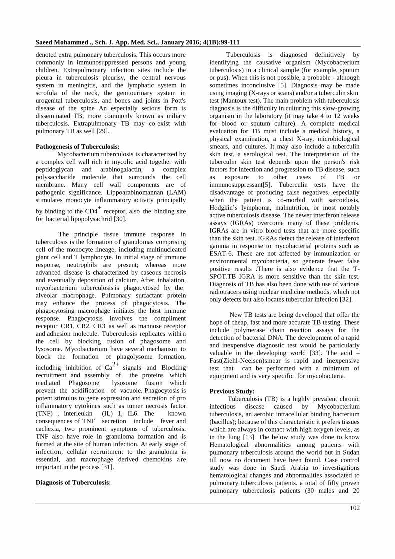
under microscope .These dye differentiate the different
component of blood cells as show in figure1.
Procedure:
Clean labeled slide was placed on flat bench then
one drop of well mixed EDTA blood was added,
spreader slide was possessed at angel 45 degree and
moved back to touch blood drop, then the spreader was
bushed smooth with little pressure to make and ideal
thin blood film compose (head, body and feathered
sharp tail) and let to dry and do all stain stabs as show
below [37].
Fig-1: Normal Blood Cells Morphology [37].
Ethical Consideration:
The selection of subjects and the collection of
specimens from the subjects were done after prior
notice and approval from all protocols involved, was
obtained and informed the aim of the study.
Data Presentation:
Data obtain analyzed by Statistical Package of
Social Science (SPSS.version -11.5) software program
and obtain mean and p.values by Independent –
sample T test. The results presented in form of tables
and figures.
RESULTS:
Demographic details:
The study was conducted to measure of some
of hematological parameters among pulmonary
tuberculosis patients whom visit Abo Anjh hospital in
Khartoum state were investigated .The result were
compared with apparently healthy subjects (controls).
The study included sixty (60) subjects, 40 patients
selected were already diagnosed as acid- fast bacilli
positive by Z-N smear stain with mean age 41 years, 25
male (62.5%) and 15 female (37.5%) and 20 healthy
volunteers were selected as controls, these volunteers
are Z-N Stain Negative and free from dieses or
medication therapy in the last three month before
sample collection the mean age of control was 39 years
as show in Table1.
Table2 Show the mean ± SD of Hemoglobin
level in T.B patients was ( 9.6 ± 2.01g/dl ) and in
control group the mean of Hemoglobin was (14.06 ±
.80 g/dl) ,when compared with controls group showed
significant decrease in hemoglobin level (P.value =
0.00) . the mean of Red Blood Cell in T.B patients was
(3.7± .64 x106
/ul ) and in control group was (4.7 ± .77
x106
/ul) also showed significant decrease when
compared with controls (P.value = 0.00), the packed
cell volume was found significant decrease in T.B
patients mean (31.0± 6.2%) compared with control
group (39.4 ± 2.7 % ) ,the mean of Mean Cell Volume
was found in study group (82.7 ± 8.7 fl ) and in control
group was (83.4± 2 fl ) when compared with controls
group showed no significant difference wich is normal
as controls (P.value= 0.78), the mean of Mean Cell
Hemoglobin was found in study group (26.2 ± 4.3 pg )
and in control group was (30.1 ± 1.2 pg ) was found
significant different (P.value = 0.00) , the mean of
Mean Cell Hemoglobin Concentration was found in
study group (30.6± 2.1 g/dl) and in control group was
(36.1± 1.1 g/dl) , when compared with control showed
decrease MCHC in patients with significant difference
(P.value = 0.00) .
Table 3 show the mean ± SD of Total leukocyte
count in T.B patients was ( 10±3.9 x10⁹/L) and in
control group was (6.7±1.9 x10⁹/L) was found
significant increase total leukocyte count when
compared with controls group (P.value =0.00).the
absolute neutrophils count in study group was found
increase in count when compared with controls found
significant different (P.value=0.008) ,the mean of
patients (6.8±3.9 x10⁹/L) and mean in control group
was (3.9 ±1.5 x10⁹/L), the mean of absolute
lymphocytes count in study group was found (1.8±0.85
x109/L ) and in control group was (2.2±0.57x10
9/L)
with no significant difference (P.value=0.06), the
Saeed Mohammed ., Sch. J. App. Med. Sci., January 2016; 4(1B):99-111
106
absolute monocytes count was increase in T.B patients
when compared with controls and theirs significant
difference (P-value0.00),the mean of absolute
Eosinophils count in study group was found
(0.23±0.17x10⁹/L) and in control was (0.11±0.1 x10
9/L
), the mean of absolute Basophiles count in study group
was (0.04±0.06 x10⁹/L) and in control group was
(0.15±0.03x10⁹/L).when compared absolute eosinophil
and basophil count of patients to controls was found no
significant difference (P.value>0.05).
Table-4 Show the mean ± SD of platelet count in
study group was found ( 425.5±22.9 x10⁹/L) and in
control group was (268.7±12.1 x10⁹/L) which is
significant increase platelet count in T.B patients
compared with control group (p.value=0.00).
Table-1: Frequency of Gender & Mean of Age among Study population:
Frequency Percent% Mean of years ± SD
Male 25 62.5 41.2±7.3
Female 15 37.5 42.4±7.9
Total 40 100.0 41.7±7.3
Table-2: Some of Hematologic Parameters among Study Population:
Parameter Variable Number 9.67 ± 2.01 P.values
Hb ( g/dL)
PTB patient 40 14.06± 0.8 0.00
Control 20 3.728± 0.64
RBC(x106/ul)
PTB patient 40 4.682± 0.37 0.00
Control 20 31.02± 6.2
PCV ( %)
PTB patient 40 39.41± 2.69 0.00
Control 20 82.75± 8.7
MCV ( FL )
PTB patient 40 83.45± 2.02 0.78
Control 20 26.23± 1.2
MCH (Pg)
PTB patient 40 30.16± 4.3 0.00
Control 20 30.66± 2.1
MCHC (g/dL)
PTB patient 40 36.14±1.1 0.00
Control
* The mean difference is significant at the P.value ≤ 0.05
* PTB (Pulmonary Tuberculosis Patient)
Tables-3: Total WBCs and Absolute Leukocytes Count in T.B patients:
Parameter( x109/L) Variable Number Means ± SD P.values
TWBCs PTB patient 40 10.06±5.09 0.00
Control 20 6.71±1.9
Neutrophil PTB patient 40 6.84±3.9 0.008
Control 20 3.92±1.5
Lymphocyte PTB patient 40 1.83±0.8 0.06
Control 20 2.23±0.5
Monocyte PTB patient 40 1.14±0.89 0.00
Control 20 0.39±0.16
Eosinophil PTB patient 40 0.23±0.17 0.29
Control 20 0.11±0.1
Basophile PTB patient 40 0.04±0.06 0.12
Control 20 0.015±0.03
* The mean difference is significant at the P.value ≤ 0.05
* PTB (Pulmonary Tuberculosis Patient)
Table-4: Platelet Count among Study population:
Parameter x109/L Variable Number Means ± SD P.values
platelet PTB patient 40 425.4±145 0.00
Control 20 268.7±54.2
The mean difference is significant at the P.value ≤ 0.05
* PTB (Pulmonary Tuberculosis Patient)
Saeed Mohammed ., Sch. J. App. Med. Sci., January 2016; 4(1B):99-111
107
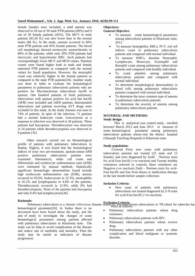
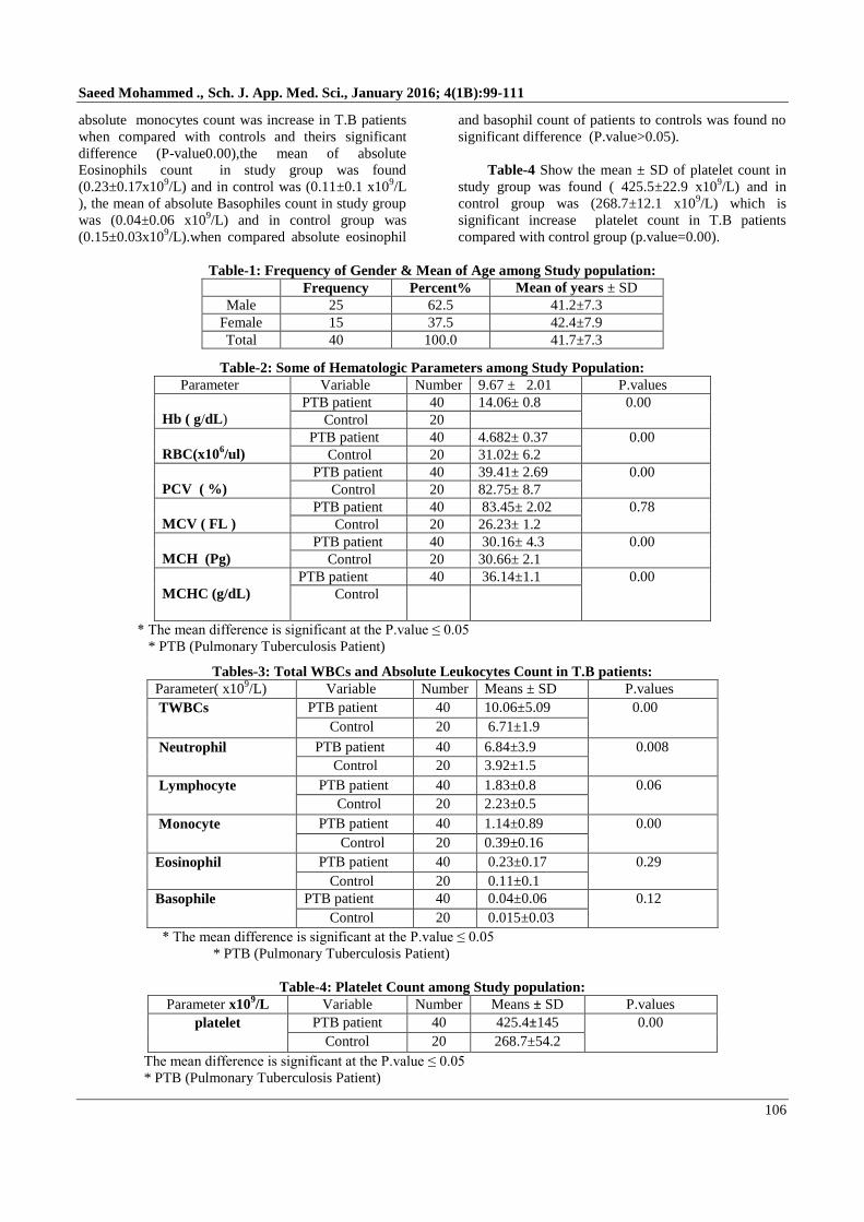
Fig. -2: Type of anemia among study population:
Type of anemia was found in patients: The definition of anemia is degrease in
hemoglobin concentration less than 13 g/dl in men and
12 g/dl in women anemia was identified in 34) 85%)
patient at the time of diagnosis of tuberculosis . 22
(64.7%) men and 12 (35.3%) women had anemia,
normocytic anemia was the most common, and was
identified in 29 (72.5%) of patient and microcytic
anemia was next common, 11(27.5%) patients were
identified with microcytic as show in figure 2.
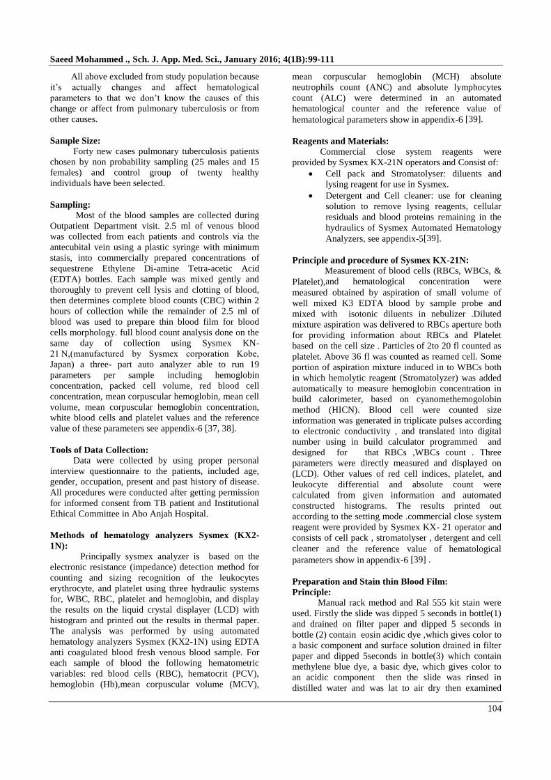
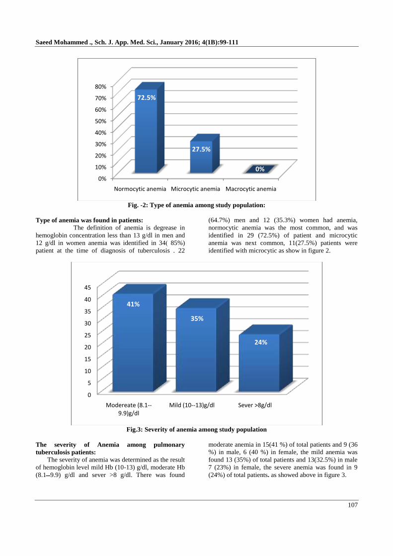
Fig.3: Severity of anemia among study population
The severity of Anemia among pulmonary
tuberculosis patients:
The severity of anemia was determined as the result
of hemoglobin level mild Hb (10-13) g/dl, moderate Hb
g/dl and sever >8 g/dl. There was found (9.9ـــ8.1)
moderate anemia in 15(41 %) of total patients and 9 (36
%) in male, 6 (40 %) in female, the mild anemia was
found 13 (35%) of total patients and 13(32.5%) in male
7 (23%) in female, the severe anemia was found in 9
(24%) of total patients. as showed above in figure 3.
0%
10%
20%
30%
40%
50%
60%
70%
80%
Normocytic anemia Microcytic anemia Macrocytic anemia
72.5%
27.5%
0%
0
5
10
15
20
25
30
35
40
45
Modereate (8.1--9.9)g/dl
Mild (10--13)g/dl Sever >8g/dl
41%
35%
24%
Saeed Mohammed ., Sch. J. App. Med. Sci., January 2016; 4(1B):99-111
108
Normal Leucocytosis Leucopenia
(7±3 x10⁹ /ul) ( >10 x10
9 /ul) ( <4 x10
9 /ul)
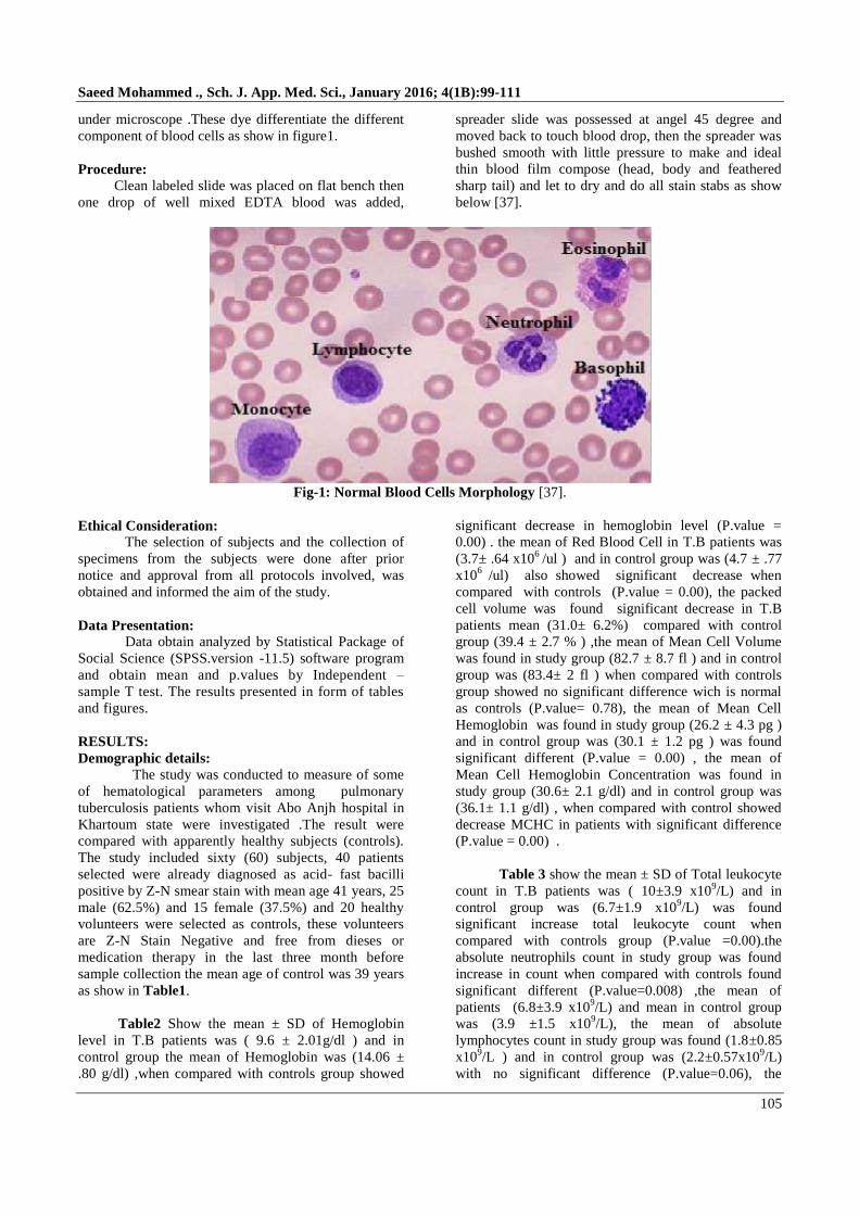
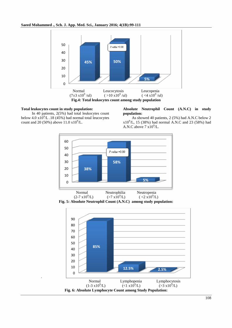
Fig.4: Total leukocytes count among study population
Total leukocytes count in study population:
In 40 patients, 2(5%) had total leukocytes count
below 4.0 x10⁹/L .18 (45%) had normal total leucocytes
count and 20 (50%) above 11.0 x10⁹/L.
Absolute Neutrophil Count (A.N.C) in study
population:
As showed 40 patients, 2 (5%) had A.N.C below 2
x109/L, 15 (38%) had normal A.N.C and 23 (58%) had
A.N.C above 7 x10⁹/L.
Normal Neutrophilia Neutropenia
(2-7 x10⁹/L) (>7 x10
9/L) ( <2 x10
9/L)
Fig. 5: Absolute Neutrophil Count (A.N.C) among study population:
.
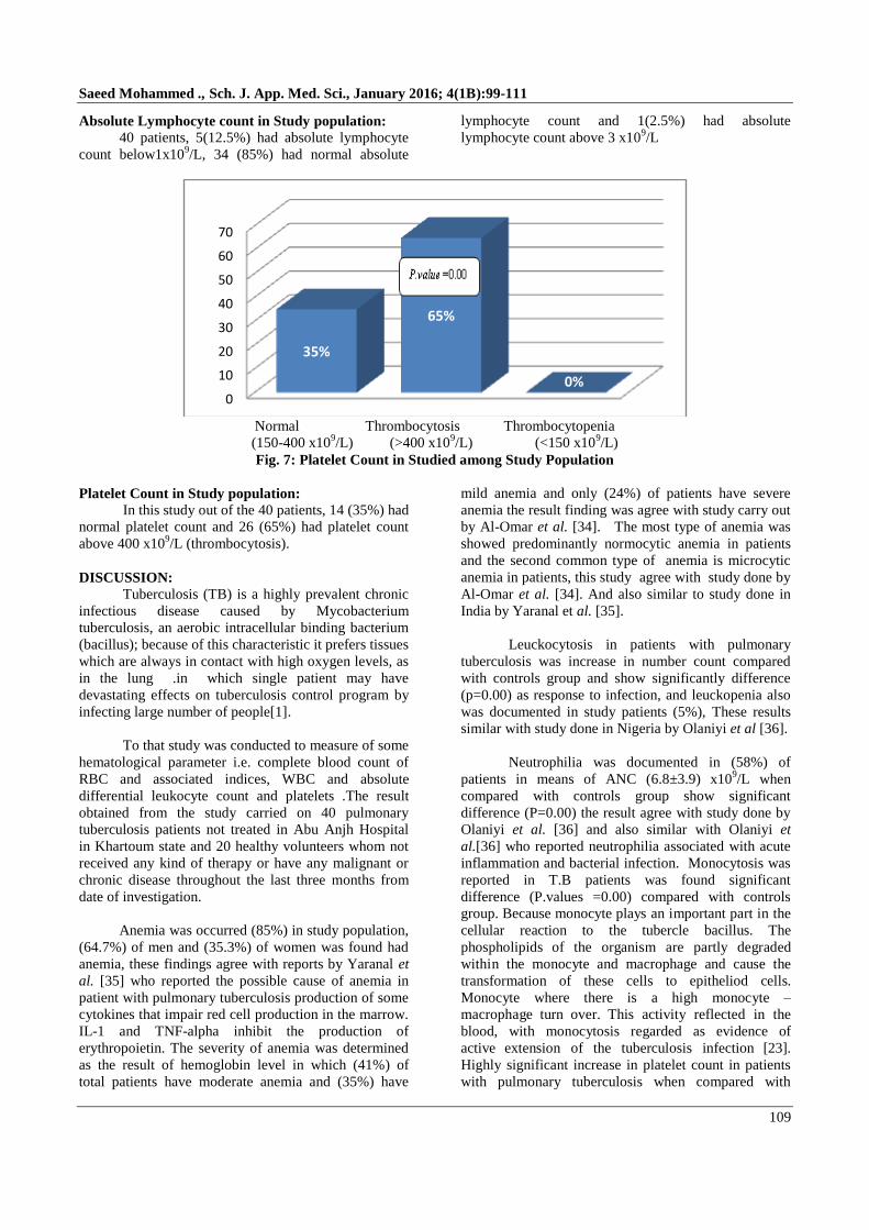
Normal Lymphopenia Lymphocytosis
(1-3 x10⁹/L) (<1 x10
9/L) (>3 x10
9/L)
Fig. 6: Absolute Lymphocyte Count among Study Population:
0
10
20
30
40
50
45% 50%
5%
0
10
20
30
40
50
60
38%
58%
5%
0
10
20
30
40
50
60
70
80
90
85%
12.5% 2.5%
Saeed Mohammed ., Sch. J. App. Med. Sci., January 2016; 4(1B):99-111
109
Absolute Lymphocyte count in Study population: 40 patients, 5(12.5%) had absolute lymphocyte
count below1x109/L, 34 (85%) had normal absolute
lymphocyte count and 1(2.5%) had absolute
lymphocyte count above 3 x10⁹/L
Normal Thrombocytosis Thrombocytopenia
(150-400 x109/L) (>400 x10
9/L) (<150 x10
9/L)
Fig. 7: Platelet Count in Studied among Study Population
Platelet Count in Study population:
In this study out of the 40 patients, 14 (35%) had
normal platelet count and 26 (65%) had platelet count
above 400 x109/L (thrombocytosis).
DISCUSSION:
Tuberculosis (TB) is a highly prevalent chronic
infectious disease caused by Mycobacterium
tuberculosis, an aerobic intracellular binding bacterium
(bacillus); because of this characteristic it prefers tissues
which are always in contact with high oxygen levels, as
in the lung .in which single patient may have
devastating effects on tuberculosis control program by
infecting large number of people[1].
To that study was conducted to measure of some
hematological parameter i.e. complete blood count of
RBC and associated indices, WBC and absolute
differential leukocyte count and platelets .The result
obtained from the study carried on 40 pulmonary
tuberculosis patients not treated in Abu Anjh Hospital
in Khartoum state and 20 healthy volunteers whom not
received any kind of therapy or have any malignant or
chronic disease throughout the last three months from
date of investigation.
Anemia was occurred (85%) in study population,
(64.7%) of men and (35.3%) of women was found had
anemia, these findings agree with reports by Yaranal et
al. [35] who reported the possible cause of anemia in
patient with pulmonary tuberculosis production of some
cytokines that impair red cell production in the marrow.
IL-1 and TNF-alpha inhibit the production of
erythropoietin. The severity of anemia was determined
as the result of hemoglobin level in which (41%) of
total patients have moderate anemia and (35%) have
mild anemia and only (24%) of patients have severe
anemia the result finding was agree with study carry out
by Al-Omar et al. [34]. The most type of anemia was
showed predominantly normocytic anemia in patients
and the second common type of anemia is microcytic
anemia in patients, this study agree with study done by
Al-Omar et al. [34]. And also similar to study done in
India by Yaranal et al. [35].
Leuckocytosis in patients with pulmonary
tuberculosis was increase in number count compared
with controls group and show significantly difference
(p=0.00) as response to infection, and leuckopenia also
was documented in study patients (5%), These results
similar with study done in Nigeria by Olaniyi et al [36].
Neutrophilia was documented in (58%) of
patients in means of ANC (6.8±3.9) x109/L when
compared with controls group show significant
difference (P=0.00) the result agree with study done by
Olaniyi et al. [36] and also similar with Olaniyi et
al.[36] who reported neutrophilia associated with acute
inflammation and bacterial infection. Monocytosis was
reported in T.B patients was found significant
difference (P.values =0.00) compared with controls
group. Because monocyte plays an important part in the
cellular reaction to the tubercle bacillus. The
phospholipids of the organism are partly degraded
within the monocyte and macrophage and cause the
transformation of these cells to epitheliod cells.
Monocyte where there is a high monocyte –
macrophage turn over. This activity reflected in the
blood, with monocytosis regarded as evidence of
active extension of the tuberculosis infection [23].
Highly significant increase in platelet count in patients
with pulmonary tuberculosis when compared with
0
10
20
30
40
50
60
70
35%
65%
0%
Saeed Mohammed ., Sch. J. App. Med. Sci., January 2016; 4(1B):99-111
110
healthy individual (p.value=0.00). The thrombocytosis
was reported and no patient had thrombocytopenia in
study population these findings agree with study done
by Yaranal et al.; [35] and disagree with him and with
Olaniyi et al.; [36]. In thrombocytopenia, who reported
thrombocytopenia in the study population. was
documented Platelets have been suggested to play a
role in the inflammatory response, including defense
against bacteria and in the evolution of inflammatory
response against mycobacterium, Various
inflammatory cells, cytokines and mediators are
involved in the formation of granulomatous lesions
encountered in tuberculosis. Of variety of cytokines,
interleukin-6 (IL-6) has been known to promote
platelet production.
CONCLUSION:
This study was concluded:
Moderate normocytic normochromic anemia was
found in the majority of pulmonary tuberculosis
patients.
Total WBCs count significantly increased in
pulmonary tuberculosis patients with significant
increase in absolute neutrophil and monocyte count
compared to controls group.
The majority of pulmonary tuberculosis patients
were significantly moderate increase platelet count
(Thrombocytosis) compared to the controls group.
Recommendation:
This study recommended:
That physicians treat patients suffering from
pulmonary tuberculosis not only of pulmonary
tuberculosis but underlining hematological
disorders as a result of pulmonary tuberculosis.
Further studies should also focus on finding
the extent of damage caused by these abnormal
changes as a co-infection with pulmonary
tuberculosis. This will help establish whether
these changes significantly affects the progress
of pulmonary tuberculosis or the vice versa in
pulmonary tuberculosis patients, it will help
establish the relation between pulmonary
tuberculosis and abnormal hematological
profile in pulmonary T.B patients.
REFERENCES 1. Sauders E; “Robbins Basic Pathology”, 8th
Edition Cambridge Press, London. 2007;
516 – 520.
2. Oliva VM, CezarioGAC, CactoRA; (Ed0
Pulmonary tuberculosi Heamtology Serum
Biochemistry and relationship with the
disease condition . J Venom. Anim.Toxins
incl.Trop.Dis. 2008; 14: 71-78.
3. Southwick L; “Testing Tuberculosis”, 4th
Edition Australian Publishing, 2007; 1218.
4. Ben-Kahka I, Wnd J. Ben - Selma M.
Marzouk, A. Ferjeni, S. Ghezal, Boukadida;
Evaluattion of asimplified IS6110 PCR for
the rapid Diadnosis of mycobacterium
tuberculosis I an area with high
tuberculosis incidence. Payhol Biol. (Paris),
2009; 22: 121-108, PMID; 19477082.
5. Konstantinos E.T; “Infectious Diseases: A
Clinical Course” 2nd
edition McGraw Hill
Medical Publishing Division. 2010; 104.
6. Hoffbrand A.V, Petit J.E; “Clinical
Haematology”, 3rd
Edn.Mosby, London. 2005;
56.
7. Morris CDW; The Radiography,
Haematology and Biochemistry of pulmonary
tuberculosis in the aged. Q J Med.; 1989;
266:529-535.
8. Morris CDW. Bird AR, Nell H; The
h a e ma t o l o g i c a l and biochemical change
in severe pulmonary tuberculosis. Q J Med.
1989; 272:1151-1159.
9. Single R N.Al sharif, M. AL. Sayegh;
aprevalence of resistance of anti tuberculosis
changes in Riyadh an areview of previous
report. Am .saudi Med ,2003; 23:143-147.
10. Paul J S, Abdul Kadir TA; Blood disorder and
tuberculosis Indian Journal of Tuberculosis,
2004; 28:200.
11. Frank Firkin, Colin Chesterman, David
penmington and Bryan Rush; De Gruchy
Clinical Hematology in medical practice. 5 Th
ed London .Blackwell, 2002.
12. Job C J Calis, Kamija S Phiri, E. Brian
Faragher; .severe anemia. In Malawian
Children volume 358 New England Journal of
Medicine. Medical society 2008; 888-899.
13. Cartwright GE; The anemia of chronic
disorders. Semin Hemato1966; l3: 351.
14. Cash JM, Sears DA; The anemia of chronic
disease: Spectrum of associated diseases in a
series of unselected hospitalized patients. Am
J Med 1989; 87: 639.
15. Atkins MB, Kappler K, Mier J W , Isaacs RE,
Berkman EM; Interleukin-6-associated
anemia: determination of the underlying
mechanism. Blood 1995; 86:1288 – 1291.
16. Roberts PD, Hoffbrand AV, Mollin
DL; Br Med J Iron and folate
metabolism in tuberculosis. 1966; 5507: 198-
202.
17. Robert T, Mean JR; the anemia of chronic
disorder. Wintobes haematology, 2005.
18. Weiss G, Bogdan C, Hentze MW; Pathways
for the regulation of macrophage Iron
metabolism by the anti- inflammatory
cytokines IL-4 and IL- 13. J Immunol 1997;
158: 420-425
19. Robert T, Mean JR; the anemia of chronic
Saeed Mohammed ., Sch. J. App. Med. Sci., January 2016; 4(1B):99-111
111
disorder. Wintobes haematology, 2005.
20. William RC, Koster FI, Kilpatrik KA;
Alteration in lymphocyte cell surface
markers during various infections. Am Med J
1983; 75:807.
21. Mitchell, Richard Sheppard; Kumar, Vinay;
Abbas, Abul K.; Fausto, Nelson. Robbins
Basic Pathology Philadelphia: Saunders. 8 th
edition, 2006.
22. Goldenberg AS; Hematologic abnormalities
and mycobacterial infection In;.Williams NR,
Stuart GM (ed) .Tuberculosis .littile- brown
company , Boston Hits SV,ShawCC,.
Leukemoid blood reactions. N Eng Med J
1953; 249: 434.
23. Schmitt E, Meuret G, StixL; Monocyte
recruitment in tuberculosis and sarcoidosis.
Brit J Haematol 1977; 35:1.
24. Bayokarirt Y, Soylu B, SoyluAc, Ozecebe O
C a n b e k S; In v i v o p l a t e l e t a n t T
lymphocyte Elevation during Pulmonary
Tuberculosis. Eur Respir J. 1998; 12:1375-
1379.
25. Tozkoparan E. Omar D, Ergan U, Hayti B,
Kurdet E; changes in platelet count and
indices in pulmonary tuberculosis. Clin Chem
Lab Med 2007; 45:1009–13.
26. Vijayan V. K, Sagal Das (Ed); Pulmonary
tuberculosis In: Suredra sharma Tuberculosis
1st
editon, Jaypee Publishers Delhi: 2009;
217-227.
27. Comstock G; "The International Tuberculosis
Campaign: a pioneering venture in mass
vaccination and research". Clin Infect Dis
1994; 19 (3): 528–40.
28. Danek SJ, Bower JS; Diagnosis of pulmonary
tuberculosis by flexible fiberoptic
bronchoscopy .Am Rev Respir Dis. 1979;
119(4): 677-9.
29. Madison B; “Application of stains in clinical
microbiology". Biotech Histochem 2001; 76
(3): 119–25.
30. Arruda S, Bomfim G, Knights R, T Huima-
Byron, Riley LW; Cloning of an M; Science
tuberculosis DNA fragment associated with
entry and survival inside Cells 1993;
261(5127): 1454 – 1457.
31. John S fridland. (Ed), London; Tuberculosis:
In Donald A, Jonathan C. Infectious
disease, Vol 1, Mosby, 1995; 2.30.1.
32. Yuen K Y, Chan K S; use for PCR in routine
diagnosis of Treated and untreated
pulmonary tuberculosis J clin pathol 1996;
46:318-22.
33. Philip CH; USA Tuberculosis and other
mycobacteriual Disease IN: Murray and
Nadel’s Text book of respiratory medicine
4t h
ed i t io n , E lsevier , 2005; 979-997.
34. Al-Omar RM, Al-Ashban, A.H.Shah;
Hematological Abnormalities in Saudis
Suffering from Pulmonary Tuberculosis and
Their Reponce to the Treatment.Research
Journal of Pharmacology, 2009; 3:78-85.
35. Yaranal PJ, Umashankar T Harish SG;
Hematological Profile in Pulmonary
Tuberculosis. Int J Health Rehabil Sci. [cited
July 03, 2013] 2013; 2(1): 50-55.
36. Olaniyi JA, Aken'Ova YA; Afr J Med Sci.
2003; 32(3):239-42.
37. Lewis SM, Bain, Bates I; Dacie and lewis
practical hematology, 10 th
ed England:
Elsevier ltd, 2006; 35.77.
38. Lewis Mitchell S, Barbara J Bain, Imelda
Bates; Practical haematology 10th edition
.Philadelphia, USA. Elsevier Ltd, 2006.
39. Diamond Diagnostic, Sysmex KX- 21
operator manual, 1999.

![Computerization of the Students’ Industrial Work …saspublisher.com/wp-content/uploads/2017/04/SJET53104-116.pdf · national development objectives [4]. Okpor and Hassan [5] stated](https://img.pdfslide.us/doc/110x75/5aa7e8237f8b9a294b8ca9e2/computerization-of-the-students-industrial-work-development-objectives-4.jpg)


![41b. Protecting Cyberspace as a National Asset Act of 2010 [Excerpt 1-2, 76-77, 79]](https://img.pdfslide.us/doc/110x75/577d28ca1a28ab4e1ea541ec/41b-protecting-cyberspace-as-a-national-asset-act-of-2010-excerpt-1-2-76-77.jpg)