Embed Size (px)
Citation preview
ORIGINAL CONTRIBUTION Open Access
Phytochemical studies, antiangiogenic, anti-inflammatory and antioxidant activities ofScyphocephalium ochocoa Warb.(Myristicaceae), medicinal plant fromGabonRick-Leonid Ngoua-Meye-Misso1,2,5* , Jean De La Croix Ndong3, Cédric Sima-Obiang1,2, Joseph Privat Ondo1,2,Guy Roger Ndong-Atome1,2, Felix Ovono Abessolo4 and Louis-Clément Obame-Engonga1,2
Abstract
Background: The search for new anti-cancer molecules is one of the main concerns of oncology researchers.Scyphocephalium ochocoa is a plant of Myristicaceae family, used in traditional medicine against inflammatorydiseases and several types of cancer. It is well established that free radicals, chronic inflammation and angiogenesisplay an important role in initiation, tumor progression and metastasis formation. The aim of this study was to carryout a phytochemical screening, to determine the phenolic compounds content, to investigate the antiangiogenic,anti-inflammatory and antioxidant activities of water, water-ethanol and ethanol extracts of S. ochocoa.
Methods: Phytochemical screening and determination of phenolic compounds content were performed usingstandard methods. Antiangiogenic activity was assessed using chick chorioallantoic membrane (CAM) model andDrabkin test. Anti-inflammatory activity was estimated by protein denaturation and erythrocyte membranestabilization method. Finally the antioxidant activity was appreciated by DPPH radical inhibition andphosphomolybdenum assay.
Results: The results of phytochemical studies show that extracts of bark of S. ochocoa are rich in polyphenols,tannins, flavonoids, proantocyanidins, saponosides, flavonols, flavanonols, sterol and triterpenes. The water extractshowed good antiangiogenic activity (IC50 = 1.153 μg/mL). Strong anti-inflammatory activity was observed with allextracts, IC50 ranging from 34.775 ± 2.543 μg/mL to 74.577 ± 3.456 μg/mL for protein denaturation inhibition testand IC50 values ranging from 36.793 ± 0.529 μg/mL at 48.912 ± 0.957 μg/mL for antihemolytic activity. In addition,all extracts showed good antioxidant activity marked by a strong inhibition of the DPPH radical (IC50 ranging from4.969 ± 0.263 μg/mL to 16.188 ± 0.336 μg/mL and AAI ranging from 3.090 ± 0.065 to 10.080 ± 0.517) and by greatertotal antioxidant capacity (with contents ranging from 37.654 ± 0.995 to 131.302 ± 1.102 VtCE (mg)/g dry extract).
(Continued on next page)
* Correspondence: [email protected]; [email protected] of Research in Biochemistry (LAREBIO), University of Sciences andTechnology of Masuku, Franceville, Gabon2Laboratory of Natural Substances and Organometallic Synthesis, Universityof Sciences and Technology of Masuku, Franceville, GabonFull list of author information is available at the end of the article
© The Author(s). 2018, corrected publication July 2018. Open Access This article is distributed under the terms of the CreativeCommons Attribution 4.0 International License (http://creativecommons.org/licenses/by/4.0/), which permits unrestricted use,distribution, and reproduction in any medium, provided you give appropriate credit to the original author(s) and the source,provide a link to the Creative Commons license, and indicate if changes were made.
Ngoua-Meye-Misso et al. Clinical Phytoscience (2018) 4:15 https://doi.org/10.1186/s40816-018-0075-x

(Continued from previous page)
Conclusion: Ultimately, these results could justify the use of S. ochocoa extracts in traditional medicine in thetreatment of diseases related to angiogenesis and cancer, inflammatory diseases and diseases due to oxidativestress. A phyto-product with such a pharmacological profile could be a good candidate for the development ofanticancer.
Keywords: Scyphocephalium ochocoa, Anti-inflammatory, Antiangiogenesis, Antioxidants, Chick Chorioallantoicmembrane (CAM), Cancer
BackgroundScyphocephalium ochocoa (S. ochocoa) belongs to thefamily of Myristicaceae and widespread in the littoralforests of Niger, Cameroon and Gabon [1]. Its vernacularnames in Gabon are: Soghe, Sogho (Fang), Ossoko(Myene), Musuku (Nzebi, Punu-Shira), Otsoko, Soko(Tsogo) [2]. For its therapeutic and nutritional virtues,several populations of Central Africa use this plant. InCongo, a bark decoction is used for vaginal injectionagainst female infertility. In a steam bath, mixing itsleaves with those of Microdermis puberula, Costus afer,Macaranga barteri and Chorophora excelsa is usedagainst febrile body aches [3]. It is also used as a decoc-tion against gonorrhea or with salt and chili pepper [2].In Gabon, the seeds of S. ochocoa are eaten grilled orlooted. In traditional Gabonese medicine, a bark decoc-tion is used in the treatment of anemia. As a drink, thisdecoction is used against rheumatism, joint pain, bodyaches and disorders of ovulation. The bark, dried andkneaded with white clay, serves to stop excessive milksecretions. This plant is also used in the treatment ofseveral cancers: breast, stomach, skin and liver cancer.The search for new anti-cancer molecules is one of
the main concerns of researchers in oncology because,despite the advancement of medical technologies oncancer, this pathology remains among the main publichealth problems in the world with 14 million newcases and 8.8 million deaths in 2015 [4]. In Gabon,incomplete data available due to the lack of actualregistration of the cancer population indicate that thenumber of new patients is constantly increasing from183 new cases in 2000 to more than 1000 new pa-tients in 2008 constituting a real public health prob-lem [5]. According to the World Health Organization,metastases are the leading cause of cancer-relateddeaths [4]. Many studies have shown that reactiveoxygen species (ROS), chronic inflammation andangiogenesis play an important role in initiation,tumor progression and metastasis formation [6, 7].ROS, in cancer, are involved in the progression and
proliferation of the cell cycle, cell survival and apop-tosis, energy metabolism, cell morphology, cell-celladhesion, cell mobility, angiogenesis, maintenance ofthe tumor and the formation of metastases [7, 8].
Other research has shown that there is a relation-ship between inflammation and cancer. In 1863, Vir-chow hypothesized that the origin of the cancer wasat sites of chronic inflammation [9]. Inflammationacts as a tumor promoter via the pro-inflammatorycytokine TNFα (tumor necrosis factor-α) which is im-portant in the early stages of tumors, regulating acascade of cytokines, chemokines, adhesions, matrixmetalloproteinases (MMPs), and pro-biogenic [9, 10].In addition, there is strong evidence that the use ofnonsteroidal anti-inflammatory drugs (NSAIDs) suchas aspirin reduces the risk of colon cancer and maybe preventative for the respiratory tract, esophagealcancer and stomach cancer [11, 12]. Other NSAIDs,such as flurbiprofen, may have strong antimetastaticeffects because of their inhibition of platelet aggrega-tion [13].Angiogenesis corresponds to all the processes leading
to the formation of new blood capillaries by the out-growth or budding of preexisting vessels [14]. Via theproangiogenic factor VEGF (vascular endothelial growthfactor), angiogenesis is essential for tumor growth andfor the formation of metastases because it supplies can-cer cells with oxygen and nutrients [15].ROS neutralization, inhibition of inflammation and
angiogenesis may therefore be one of the strategies forfinding anti-cancer products. Thus, in order to searchfor potential phyto-anticancer substances, this prelimin-ary work consists in carrying out a phytochemicalscreening, in determining the content of phenolic com-pounds, in researching the antiangiogenic,anti-inflammatory and antioxidant activities of water,water-ethanol and ethanol extracts from S. ochocoa.
MethodsChemicalsButylated hydroxytoluene (BHT); 2,2-diphenyl-1-picryl--hydrazyl (DPPH); sorafenib p-toluenesulfonate salt (LCLaboratories; Woburn, MA, USA); Diclofenac (Combi--Blocks, Silverton, San Diego, CA, USA); quercetin, etha-nol; ferric chloride; sulfuric acid; methanol; sodiumphosphate; ammonium molybdate; chloridric acid; ben-zene; ammonium hydroxide; sodium chloride;Folin-Ciocalteu reagent; sodium bicarbonate; gallic acid
Ngoua-Meye-Misso et al. Clinical Phytoscience (2018) 4:15 Page 2 of 13

and ascorbic acid were obtained from Sigma ChemicalCo. (St. Louis, MO, USA).
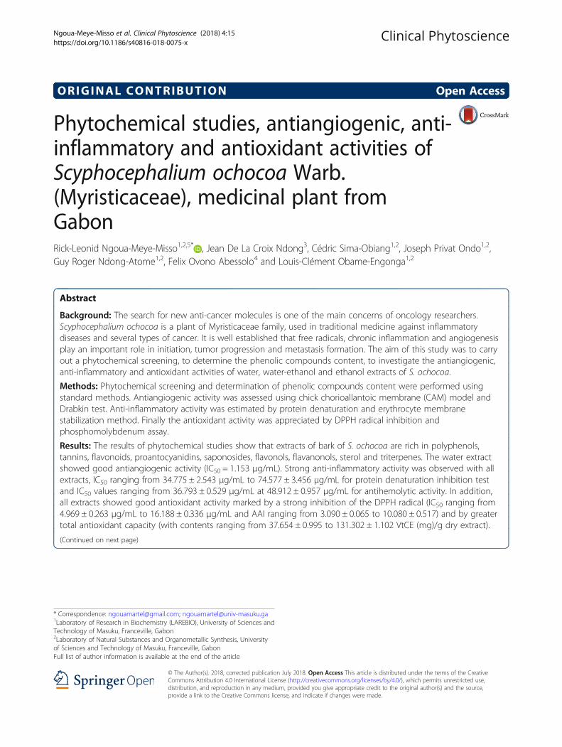
Plant materialThe barks of S. ochocoa were collected August 2015 inMitzic, Woleu-Ntem (Northern of Gabon) (Fig. 1). Theywere identified at National Herbarium of IPHAMETRA,Libreville (Gabon). Voucher specimen has been depos-ited in the Herbarium of IPHAMETRA and at Labora-tory of Biochemistry Research (LAREBIO) Departmentof Chemistry-Biochemistry, Faculty of Sciences of USTMin Franceville.
Preparation of plant extractBarks were dried at ambient temperature of the Labora-tory (20–30 °C) and protected from light for severaldays. After drying, barks were crushed using a grinder(Laboratory Blender, Torrington, CT. USA). This powderwas used for extractions by maceration method. Briefly,200 g of powder was mixed with 2000 ml of solvent(water, water-ethanol (50/50, v/v) and ethanol). After72 h, the obtained extract was filtered using WhatmanN°1 filter paper. Ethanol and water-ethanol extracts wereconcentrated under reduced pressure at rotavapor(Büchi, Labortechnik, Switzerland) at 40 °C and 60 °C,respectively. Water extract was lyophilized using a ly-ophilizer (Alpha 1–2 LDplus, Germany). All crude ex-tracts obtained were stored at 4 °C until analysis.
Phytochemical screeningEach extract was then tested for the presence of fla-vonoids, coumarins, tannins, total phenolic, sapono-sids, cardiac glycosides, reducing sugar, sterols andtriterpenes, oses and holosides, anthracenics, antho-cyans, alkaloids and anthracenosids as described else-where [16, 17].
Phenolic compound contentTotal phenolic contentThe Folin-Ciocalteu method [18] with minor modifica-tions was used to determine the total phenolic contentsof the different extracts using gallic acid as standard.The absorbance was measured at 735 nm using a Spec-trophotometre (Evolution 60S, USA). Results wereexpressed as gallic acid equivalent per gram of lyophi-lized sample (average of the triplicate analysis).
Total flavonoid contentThe aluminum chloride (AlCl3) colorimetric assaymethod [19] was used to determine total flavonoid con-tents, using quercetin as a standard. The absorbance wasmeasured at 415 nm and total flavonoid contents wereexpressed as quercetin equivalents in milligrams pergram sample (average of the triplicate analysis).
Tannins contentTannins content was determined according the referencemethod of European community [20]. The absorbancewas measured at 525 nm and tannic acid was used asstandard.
Proanthocyanidins (PAs) contentThe proanthocyanidins content was determined accord-ing the method described by Prigent [21]. The absor-bances were read at 550 nm. Apple procyanidins wereused as a standard. Results were expressed as apple pro-cyanidins equivalent (APE).
Antiangiogenic activityAntiangiogenic activity was evalued using Chick Chorio-allantoic Membrane (CAM) Model acording to previ-ously reported method [22, 23] with minorsmodifications. Brefly, fertilized chicken eggs were pur-chased from a local poultry farm, were sterilized with70° ethanol and incubated at 37 °C in incubator (Ecocell,LSIS-B2V/EC55, Germany), with 80% relative humidity.On day 2 of post incubation, 3 mL of albumin werewithdrawn to minimize adhesion of the shell membranewith CAM. A square window of 1 cm2 was opened inthe egg shell at the opposite to blunt edge and sealedwith an adhesive tape. The eggs were returned for fur-ther incubation.
Drug administrationAt the 8th day, the experimental groups were dividedinto 5 of each containing 50 numbers of eggs. Group 1,2, 3 and 4 were treated with water extracts and sorafenib(positive control). Sterile discs (diameter: 10 mm) ofWhatman N°1 soaked of 10 μL of the water extract fromS. ochocoa and sorafenib at concentrations ranging from60, 125, 250 to 500 μg/mL was applied to the CAM. In
Fig. 1 Shrub and trunk of S. ochocoa. Photos taken at Mitzic,Woleu-Ntem (Northern of Gabon) by Ngoua-Meye-Misso with digitalcamera Cannon 16 M pixel made in China
Ngoua-Meye-Misso et al. Clinical Phytoscience (2018) 4:15 Page 3 of 13

parallel Group 5 treated with phosphate buffered saline(PBS) alone as negative control, a paper disc WhatmanN°1 soaked of 10 μL PBS at pH 7.4 was placed on theCAM of egg. The treated CAM samples were incubatedfor 48 h.
Angiogenesis quantificationAfter 48 h of incubation at 37 °C and 80% relative hu-midity, a volume of 10 μL of formaldehyde at 4% was ap-plied to the CAM. 5 min later, the CAM was cut aroundthe disk and all (disc and CAM) was placed in a Petri.Then the photos were taken with a Nikon digital cameraD5100 (made in Thailand) and the images were subse-quently analyzed with the software Image J. The num-bers of vessel branch points contained in a circularregion (equal to the area of each filter disk) werecounted manually. The percentage of vascularization(density) is measured relative to a normal controlvascularization. The ability to inhibition angiogenesiswas calculated by the following equation:
%AIA ¼ Nbvcontrol‐ Nbvsample� �
=Nbvcontrol� �� 100
Nbv = number of blood vessel branch points. The IC50
(concentration providing 50% inhibition) of extracts andstandards was determinate using regression curves inthe linear range of concentrations. The experiments havebeen repeated at least four times, and the results werereproducible.
Drabkin reagent testThe determination of hemoglobin in CAM sections wasdone by the Drabkin and Austin [24] assay method. Thedissected CAM sections were crushed with a pestle andmortar. The pilate was mixed with 5 ml of cooled nor-mal salt solution and then homogenized. The resultingsolution was centrifuged at 2500 rpm for 30 min. 3 mLof Drabkin reagent was added to 1 mL of supernatant.The reaction was incubated at room temperature(27 ± 3 °C) for 20 min and the absorbance measuredat 580 nm using a UV spectrophotometer (Evolution60S, USA). All tests were performed in triplicate toensure reproducibility of the result.
Toxicity of extractsToxicity of extracts was determined by observation ofembryos statue after extracts actions on vascularizationaccording to a previously reported methods [25].
Anti-inflammatory activityThe anti-inflammatory activity was evaluated by proteindenaturation method and membrane stabilizationmethod.
Protein anti-denaturation testProtein denaturation methods have been used [26, 27]with slight modifications mentioned byNgoua-Meye-Misso et al. [28]. 0.1 mL of fresh chickenegg albumin was mixed with 1.9 mL of phosphate buff-ered saline (PBS, pH 6.4) and 1 mL of varying concen-tration of extract (6.25, 12.5, 25, 50 and 100 μg/mL). Asimilar volume of distilled water served as a negativecontrol. Then the mixtures were incubated at 37 °C inan incubator (Ecocell, LSIS-B2V/EC55, Germany) for20 min and then heated at 70 °C for 5 min. After cool-ing, the absorbances were measured at 660 nm on thespectrophotometer (Evolution 60S, USA). Sodium diclo-fenac in the final concentrations of 31.25, 62.5, 125, 250and 500 μg/mL, was used as a reference drug and simi-larly treated for the determination of absorbance.
Inhibition %ð Þ ¼ Abssample ‐ Abscontrol� �
=Abscontrol� �� 100
Abs = absorbance. The concentration of the extract for50% inhibition (IC50) was determined by thedose-response curve.
Membrane stabilization testThe membrane stabilization test was evaluated by themethod of hemolysis of red blood cells. This hemolysiswas induced on the one hand by heat on the other handby distilled water [29] with some modifications.
Preparation of the suspension of erythrocytesFresh whole blood (3 mL) collected from healthy volun-teers in EDTA tubes was centrifuged at 2500 rpm for10 min at 4 °C. A volume of normal saline equivalent tothat of supernatant was used to dissolve the red bloodcells. The volume of dissolved red blood cells obtainedwas measured and reconstituted in the form of a 40% v/v suspension with isotonic buffer solution (10 mM so-dium phosphate buffer, pH 7.4). The buffer solution con-tained 0.2 g of NaH2PO4, 1.15 g of Na2HPO4 and 9 g ofNaCl in 1 L of distilled water. Reconstituted red bloodcells (supernatant resuspended) were used as such. Thestudy protocol was performed according to the Helsinkideclaration and approved by the Ethics Committee ofGabon (N° 009 March 2013).
Hemolysis induced by heatSamples of used extracts were dissolved in isotonic phos-phate buffer solution. A set of 5 centrifugation tubes con-taining respectively 2 mL of extracts at increasingconcentrations (15.625, 31.25, 62.5, 125 and 250 μg/mL).Sodium diclofenac, with the same concentration rangewas used as the reference medicine. The negative controlcontained 2 mL of distilled water. A suspension of 0.1 mLof red blood cells was added to each of the tubes and
Ngoua-Meye-Misso et al. Clinical Phytoscience (2018) 4:15 Page 4 of 13

mixed gently. The tubes were incubated at 54 °C for20 min in a water bath. After incubation, tubes were cen-trifuged at 2500 rpm for 10 min at 4 °C and thehemoglobin content of the supernatant was estimatedusing the spectrophotometer (Evolution 60S, USA) at540 nm. The percentage of inhibition by the extract wascalculated as follows:
% Inhibition of hemolysis ¼ 1−ODsample=DOcontrol� �� 100
Where OD sample = absorbance of the sample; DO con-
trol = absorbance of the control. The concentration of theextract for 50% inhibition (IC50) was determined by thedose-response curve.
Hemolysis induced by hypotonicityThe extract samples were dissolved in distilled water(hypotonic solution) at different concentrations obtainedby double dilution (15.625, 31.25, 62.5, 125 and 250 μg/mL). Sodium diclofenac, at the same concentrations, wasused as a reference medicine. Distilled water was used asa negative control. 2 ml of sample were mixed with0.1 mL of a suspension of erythrocytes and then themixtures were incubated for 1 h at 37 °C. The tubeswere then centrifuged at 2500 rpm for 10 min at 4 °C.the hemoglobin content of the supernatant was esti-mated using the spectrophotometer (Evolution 60S,USA) at 540 nm. The percentage of hemolysis was cal-culated assuming hemolysis produced in the presence ofdistilled water as 100%. The percentage inhibition ofhemolysis by the extract was calculated as follows:
% Inhibition of hemolysis ¼ 1−ODsample=DOcontrol� �� 100
Where OD sample = absorbance of the sample; DO con-
trol = absorbance of the control. The concentration of theextract for 50% inhibition (IC50) was determined by thedose-response curve.
Observation of erythrocyte membranesA blood smear was performed before and after treat-ment with the extracts to observe the appearance oferythrocyte membranes. 5 μL of sample was depositedand spread on the slide. After drying, fixing with metha-nol and stained with Giemsa, the cells were observedunder an optical microscope (Motic Digital Microscope)coupled to a computer using the Motic image plus 2.0software and the photos were taken.
Antioxidant activityDPPH testThe method described by Scherer and Godoy [30], basedon DPPH radical test, was used to determine the Anti-oxidant Activity Index (AAI). Briefly, DPPH solutionwas prepared by dissolving 10 mg of DPPH powder in
100 mL methanol. Graded concentrations of extractsranging from 3.125 to 100 μg/mL obtained by two-folddilutions were prepared and 1 mL of each dilution weremixed with 1 mL of the working solution of DPPH(100 μg/mL). Absorbencies were measured at 517 nmafter 15 min incubation at room temperature in thedark. Ascorbic acid (vitamin C), Butylated hydroxytolu-ene (BHT) and quercetin were used as references. Theability to scavenge DPPH radical was calculated by thefollowing equation:
%RSA ¼ Acontrol−Asample=Acontrol� �� 100
A = absorbance at 517 nm. The IC50 (concentrationproviding 50% inhibition) of extracts and standards wasdeterminate using regression curves in the linear rangeof concentrations. The AAI was then calculated as fol-lows: AAI = [DPPH] f (μg.mL− 1)/IC50 (μg.mL− 1),[DPPH] f is the final concentration of DPPH (50 μg/mL).We considered criteria of Scherer and Godoy (2009) ac-cording to which plant extracts show poor antioxidantactivity when AAI < 0.5, moderate antioxidant activitywhen AAI between 0.5 and 1.0, strong antioxidant activ-ity when AAI between 1.0 and 2.0, and very strong whenAAI > 2.0.
Total antioxidant capacityThe assay was based on the reduction of Mo (VI) to Mo(V) and subsequent formation of a green phosphate/ Mo(V) complex in acid pH [31]. A total volume of 0.3 mLextract dissolved in methanol was added to 3 mL of re-agent solution (0.6 mol/L sulphuric acid, 28 mmol/L so-dium phosphate and 4 mmol/L ammonium molybdate).The mixtures were incubated at 70 °C for 90 min thencooled to room temperature. The absorbance was mea-sured at 695 nm. The total antioxidant activity wasexpressed as the number of equivalence of ascorbic acid,BHT and quercetin.
Statistical analysisThe data were expressed as the mean ± standard devi-ation (SD) of three independent experiments and ana-lyzed using one-way analysis of variance, Student’s t-testand XLSTAT 2015.4.01 software. P-values of < 0.05 wereconsidered to be statistically significant.
ResultsPhytochemical screeningThe results of the phytochemical screening are shown inTable 1 and show that the extracts of Scyphocephaliumochocoa bark are rich in bioactive compounds. All ex-tracts are rich in phenolic compounds (total polyphe-nols, tannins, total flavonoids and proantocyanidins).The water extract has an abundance of saponosides,
Ngoua-Meye-Misso et al. Clinical Phytoscience (2018) 4:15 Page 5 of 13

alkaloids, anthracenics and digitoxigenins while thewater-ethanol extract has a strong presence of flavonols,flavanonols and digitoxins. In particular, the ethanol ex-tract has an abundance of sterol, triterpenes, oses andholosides.
Phenolic compound contentThe Table 2 presents the contents of total phenolic, totalflavonoids, total tannins and total proanthocyanidins ofextracts from Scyphocephalium ochocoa.
Total phenolicThe contents of total phenolic in terms of gallic acidequivalent (standard curve equation: Y = 0.0012X -0.0004, R2 = 0.990) ranged from 1885.611 ± 31.356 to
6284.667 ± 3.333 mgGAE/ 100 g of drug and were abun-dants in water extracts.
Total flavonoidTotal flavonoids (standard curve equation: Y = 0.0032X+ 0.0077, R2 = 1) ranged from 159.365 ± 2.228 to480.406 ± 51.498 mgEQ/ 100 g and were abundants inwater extracts.
Total tanninsLevels of tannins were expressed in terms of tannic acidequivalent (TAE). The equation of the right-hand side ofthe proportioning of the total tannins by the referencemethod of European Community gave Y = 0.0009X +0.2088 with R2 = 1. Total tannins are ranged from 194.667± 7.286 to 1898.000 ± 62.568 mg EQ/ 100 g and wereabundant in water extracts. There were abundant in waterextracts than water-ethanol and ethanol extracts.
Total proanthocyanidins contentsLevels of proanthocyanidins were expressed in terms ofapple proanthocyanidins equivalent (APE). The equationof the right-hand side of the proportioning of theproanthocyanidins by the HCl-Butanol method gave Y =0.0006X + 0.0024 with R2 = 0.986. Proanthocyanidin con-tents had ranged between 94.944 ± 2.158 to 1234.111 ±39.457 mg APE/ 100 g of drug and are very abundant inall extracts of S. ochocoa.
Antiangiogenic activityThe antiangiogenic potential of the extracts was evalu-ated in vivo with the chicken chorioallantoic membrane(CAM) the eighth embryonic day. The fertilized eggswere treated with aqueous extracts (60, 125, 250 and500 μg/mL). The degree of vessel branches formation onCAM was scored 48 h later. The vessel density is thepercentage of blood supply to the analysis area. It is in-versely proportional to the degree of inhibition. In thepresence of phosphate buffered saline (PBS) used as anegative control, the target area has a vascularizationpercentage of 100%, corresponding to a normal vascula-ture with a number of vessels branches equal to 15. Theinhibitory potential of extracts was expressed throughtheir inhibitory concentration fifty (IC50). In the
Table 1 Results of the preliminary phytochemical screening
Chemical groups Wat Wat-eth Eth
Saponosids +++ ++ –
polyphenols +++ +++ +++
Sterols and triterpenes ++ ++ +++
Oses and holosides + ++ +++
Tannins Gallics +++ +++ +++
Catechics +++ +++ +++
Alkaloids +++ + +
Cyanidins Flavons +++ – +
Flavanons – – –
Flavonols – +++ –
Flavanonols – +++ +++
Total flavonoids +++ +++ +++
Anthocyans – – –
Proanthocyanidins +++ +++ +++
Anthracenics +++ +++ –
Coumarins – – –
Cardiac glycosides Digitoxins – +++ –
Digitoxigenins – – –
Gitoxins – – ++
Gitoxigenins +++ – –
Reducing sugar – – –
+++ = Very abundant; ++ = Abundant; + = Not abundant; − = Not detected;Wat =Water; Eth = Ethanol; Wat-Eth =Water-ethanol
Table 2 Results of phenolic compounds dosage
Phenolic compounds Water Water-ethanol Ethanol
TPCa (mgGAE/ 100 g of extract) 6284.667 ± 3.333 1885.611 ± 31.356 3123.444 ± 1.156
TFC (mgEQ/ 100 g of extract) 480.406 ± 51.498 159.365 ± 2.228 364.781 ± 0.722
TTC (mgTAE/ 100 g of extract) 1898.000 ± 62.568 194.667 ± 7.286 392.815 ± 1.301
TPC (mgAPE/ 100 g of extract) 1234.111 ± 39.457 94.944 ± 2.158 254.944 ± 0.893
TPCa = Total phenolic content; TFC = Total flavonoid content; TTC = Total tannins content; TPC = Total proanthocyanidins content
Ngoua-Meye-Misso et al. Clinical Phytoscience (2018) 4:15 Page 6 of 13

presence of sorafenib (positive control), the number ofblood vessels branches is reduced to 6, 2, 1 and 0, re-spectively to a concentration of 60, 125, 250 and 500 μg/mL and respectively an ability to inhibition angiogenesisto 60%, 86.667%, 93.333% and 100% with IC50 =0.197 μg/mL. In the presence of the water extract of S.ochocoa, the number of blood vessels branches is re-duced to 8, 6, 4 and 0, respectively to a concentration of60, 125, 250 and 500 μg/mL and respectively an abilityto inhibition angiogenesis to 60%, 86.667%, 93.333% and100% with IC50 = 1.153 μg/mL. All results are reportedin Table 3 and Fig. 2. Compared to the reference mol-ecule, the water extract of S. ochocoa has good antian-giogenic activity although this activity is lower than thatof the reference.
Drabkin reagent testDrabkin’s reagent test determined the hemoglobin con-tent of the dissected CAM sections. The results aresummarized in Fig. 3. Considering that the hemoglobincontent is 100% for the sections of CAM treated withthe PBS (negative control), it is shown that in the pres-ence of the variable concentrations of the drug of 60,125, 250 and 500 μg/mL, the hemoglobin content is55%, 35%, 20% and 2%, respectively for water extract.This content is 40%, 14%, 8% and 0%, respectively forsorafenib. These data indicate that the hemoglobin con-tent decreases with the number of branches of the bloodvessels and is dependent on the concentration of thedrug. This test therefore confirms the antiangiogenic ac-tivity observed.
Toxicity of extractsThe status of embryos after extracts action onvascularization inform on toxicity (Table 3). No embryodeath was recorded in the concentration range tested in-dicating that the observed antiangiogenic activity is notdue to the toxicity of the extracts.
Anti-inflammatory activityThe anti-inflammatory activity was assessed on the onehand by protein denaturation method [26, 27] and onthe other hand by membrane stabilization method [29].
Protein anti-denaturation testThe results of protein anti-denaturation test are shownin Table 4. These results indicate that S. ochocoa extractshave good anti-denaturation activities with IC50 proteinsranging from 34.775 ± 2.543 μg/mL to 74.577 ± 3.456 μg/mL. Inhibition of protein denaturation is stronger withwater-ethanol extract (IC50 = 34.775 ± 2.543 μg/mL).This anti-denaturation activity is similar to that of thereference drug, sodium diclofenac (IC50 = 35.746 ±2.374 μg/mL) (p value = 0.582). In addition this activityis dependent on the concentration. Thus for extract con-centrations of 6.25, 12.5, 25, 50 and 100 μg/mL, the per-centages of inhibitions are 17.588 ± 1.553%, 33.040 ±2.442%, 38.317 ± 1.797%, 46.357 ± 1.599% and 65.201 ±1.178%, respectively for water-ethanol extract.
Membrane stabilization testThe anti-inflammatory activity of S. ochocoa extract ob-served by inhibition of albumin denaturation is con-firmed by membrane stabilization test. The results aresummarized in Table 5. The extracts showed good inhib-ition of hemolysis of red blood cells.For heat-induced hemolysis, all the extracts showed
a very strong anti-hemolytic activity (IC50 rangingfrom 36.793 ± 0.529 μg/mL to 48.912 ± 0.9573 μg/mL)compared to the reference drug, sodium diclofenac(IC50 = 180.911 ± 2.205 μg/mL) (p value = 0.001). Theinhibition percentages of water extract are 46.850 ±0.059%, 57.261 ± 0.102%, 56.985 ± 0.120%, 33.639 ±0.137% and 7.710 ± 0.120% at respective concentra-tions of 250, 125, 62.5, 31.25 and 15.625 μg/mL.For hemolysis induced by a hypotonic solution, only
the water extract was more active (IC50 = 56.713 ±0.492 μg/mL) than the other two extracts.
Table 3 Antiangiogenic effect of S. ochocao and Sorafenib
Drugs Dose μg/mLper œuf
Tested eggs (n) Embryos statusafter 48 h
Branches vesselsnombers
%AIA IC50 (μg/mL) P value
PBS (negative control) – 5 living 15 ± 0.954 – – 0.002
Sorafenib (positive control) 62.5 5 living 6 ± 0.753 60 ± 1.056 0.197 ± 0.062
125 5 living 2 ± 0.954 86.667 ± 1.045
250 5 living 1 ± 0.976 93.333 ± 0.965
500 5 living 0 100
Water extract of S. ochocao 62.5 5 living 8 ± 0.834 26.667 ± 1.634 1.153 ± 0.089
125 5 living 5 ± 0.850 60 ± 0.507
250 5 living 3 ± 0.500 73.333 ± 0.910
500 5 living 0 100
A significant difference is observed between the antiangiogenic effect of sorafenib and that of water extract of S. ochocoa (p value = 0.002)
Ngoua-Meye-Misso et al. Clinical Phytoscience (2018) 4:15 Page 7 of 13

Anti-hemolytic activity exhibited by the water extractis similar to that of sodium diclofenac (58.590 ±1.021 μg/mL) (p value = 0.471).The observation of the blood smears performed
after the various treatments shows that red bloodcells have a normal form in presence of an isotonicsolution, in presence of water extract (250 μg/mL)and in presence of sodium diclofenac (250 μg/mL). Inpresence of a hypotonic solution, the haemolysis iscomplete and the limbs are disorganized in presenceof low concentration of water extract (15.625 μg/mL)and sodium diclofenac (15.625 μg/mL) (Fig. 4).
Antioxidant activityThe antioxidant activity was evaluated by DPPH rad-ical inhibition test and phosphomolybdenum method.The results are shown in Table 6. Compared with
the other crude extracts, water-ethanol extract has agreater antioxidant activity marked by a stronger in-hibition of DPPH radical (IC50 = 4.969 ± 0.263 μg/mL,AAI = 10.080 ± 0.517) and by a greater total antioxi-dant capacity (87.766 ± 0.852 EQ (mg)/g, 131.302 ±1.102 VtCE (mg)/g and 570.364 ± 5.307 BHTE (mg)/gdry extract). Comparing this antioxidant activity withthose of the reference molecules, it appears thatwater-ethanol extract has an antioxidant activitythree times stronger than that of BHT (IC50 = 13.759± 0.029 μg/mL, AAI = 3.634 ± 0.008), twice as strongas that of quercetin (IC50 = 11.224 ± 0.611 μg/mL, IAA= 4.463 ± 0.245) and similar to that of vitamin C (IC50 =4.597 ± 0.095 μg/mL, AAI = 10.880 ± 0.228). Other extractsalso show good antioxidant activity with IC50 ranging from8.373 ± 0.242 μg/mL to 16.188 ± 0.336 μg/mL and AAI ran-ging from 3.090 ± 0.065 to 5.975 ± 0.172.
DiscussionCancer correspond to a rapid proliferation of abnormalcells that, beyond their usual boundaries, can invade ad-jacent parts of the body and then form metastases,which according to WHO are the leading cause of can-cer deaths [4]. It is clearly established that reactive oxy-gen species (ROS), chronic inflammation andangiogenesis play an important role in initiation, tumorprogression and metastasis formation [6, 7]. In order tocontribute to the search for potential phyto-anticancerdrugs, this preliminary study aimed to perform a phyto-chemical screening, to determine the content of phen-olic compounds, to evaluate the antiangiogenic,anti-inflammatory and antioxidant activities of ethanol,water-ethanol and water extracts of S. ochocoa barks.This African medicinal plant is used in the treatment ofseveral diseases such as female infertility, febrile bodyaches, gonorrhea, anemia, rheumatism, joint pain,
Fig. 3 Haemoglobin content of CAM sections detected by Drabkin’sassay. The hemoglobin content in the CAM sections was determinedby Drabkin method. As in the case of macroscopic observationof CAM that showed that the number of blood vessel brancheswas concentration dependent, so the present figure shows ahemoglobin content dependent on the concentration of S.Ochocoa water extract and sorafenib
Fig. 2 Inhibitory effects of water extracts of plants on angiogenesis. The CAM of a 8 days old chick embryo was separately exposed to PBS(control). Extracts were introduced on top of the CAMs. After 48 h of incubation, the CAM tissue directly beneath each filter disk was resected,and digital images of the CAM sections were captured
Ngoua-Meye-Misso et al. Clinical Phytoscience (2018) 4:15 Page 8 of 13

ovulation disorders, breast cancer, stomach cancer, skincancer and liver cancer.The results of the phytochemical screening revealed that
the extracts of S. ochocoa are rich in total polyphenols,tannins, total flavonoids and proantocyanidins, sapono-sides, alkaloids, anthracenics and digitoxigenins, flavonols,flavanonols, digitoxins, sterol, triterpenes, oses and holo-sides. High levels of phenolic compounds (total polyphe-nol) were recorded. The polyphenol content of S. ochocoais higher than that recorded in Oncoba welwitschii plants(with contents ranging from 232.050 ± 2.200 to 735.150 ±23.650 mgGAE/g) and Tetrorchidium oppositifolium (withquantities ranging from 126.400 ± 0.750 to 718.100). ±0.100) [32]. Several researchers have shown that polyphe-nols are highly active compounds against cancer, inflam-matory diseases, cardiovascular, neuro-degenerative
(Parkinson’s and Alzheimer’s disease), are powerful antiox-idants and antivirals agents [33].The results also indicate that water extract has a
strong anti-angiogenic activity marked by the inhibitionof the formation of new blood vessels in the CAM. Thisactivity is confirmed by Drabkin test which makes it pos-sible to determine the hemoglobin level in the sectionsof CAM after treatment with the extracts. Thehemoglobin level is proportional to the number of bloodvessels. By comparison of IC50, the antiangiogenic activ-ity of S ochocoa (IC50 = 1.153 ± 0.089 μg/mL) is lowerthan that of the reference drug, sorafenib (IC50 = 0.197 ±0.062 μg/mL). However, this activity is greater than thatof Oncoba welwitschii and Tetrorchidium oppositifoliumwhich at 500 μg/mL exhibit an antiangiogenic activity of83.334% [32] while at this same concentration, S.
Table 5 Effect of S. ochocoa extracts and diclofenac sodium against membrane stabilization
Conc. (μg/mL)
% Inhibition of extracts from S. ochocoa Sodium diclofenac
Water Water-ethanol Ethanol % Inhibition
Hemolysis induced by hypotonicity 250 66.144 ± 1.663 56.867 ± 1.948 76.348 ± 0.017 100.000 ± 0.000
125 56.532 ± 0.744 50.626 ± 0.452 51.937 ± 0.296 100.000 ± 0.000
62.5 53.436 ± 0.603 43.873 ± 0.152 30.573 ± 0.163 99.583 ± 1.078
31.25 32.633 ± 0.452 21.256 ± 1.285 14.720 ± 0.311 99.132 ± 1.068
15.625 10.874 ± 0.149 10.539 ± 1.095 7.672 ± 1.976 0.885 ± 0.121
IC50 (μg/mL) 56.713 ± 0.492 71.200 ± 1.833 133.375 ± 0.661 58.590 ± 1.021
P value 0.471 0.016 0.471
Hemolysis induced by heat 250 46.850 ± 0.059 56.867 ± 2.948 69.822 ± 2.791 71.039 ± 0.112
125 57.261 ± 0.102 41.033 ± 1.826 56.118 ± 3.369 32.971 ± 0.640
62.5 56.985 ± 0.120 33.077 ± 1.442 35.542 ± 2.841 13.788 ± 3.527
31.25 33,639 ± 0.137 20.704 ± 2.170 12.777 ± 1.437 6.430 ± 1.453
15.625 7.710 ± 0.120 1.667 ± 0.995 7.553 ± 1.878 2.911 ± 0.147
IC50 (μg/mL) 36.793 ± 0.529 39.526 ± 0.710 48.912 ± 0.9573 180.911 ± 2.205
P value 0.273 0.001
For hypotonicity-induced hemolysis, the ability to stabilize the membrane is similar for water extract, ethanol and diclofenac (p value = 0.471). A significantdifference is observed with ethanol extract (p value = 0.016)For heat-induced hemolysis, all extracts have the same effect on membrane stabilization (p value = 0.273). However, their effect is significantly different from thatof diclofenac (p value = 0.001)
Table 4 Effect of S. ochocoa extracts and diclofenac sodium against protein denaturation
% Inhibition of S. ochocoa extracts Sodium diclofenac
Conc. (μg/mL) Water Water-Ethanol Ethanol Conc. (μg/mL) % Inhibition
100 66.332 ± 1.777 65.201 ± 1.178 56.281 ± 1.264 500 129.481 ± 1.508
50 31.281 ± 1.244 46.357 ± 1.599 39.573 ± 1.954 250 103.015 ± 2.970
25 17.714 ± 1.178 38.317 ± 1.797 32.161 ± 2.132 125 96.566 ± 2.609
12.5 13.693 ± 1.178 33.040 ± 2.442 24.497 ± 2.152 62.5 71.106 ± 3.048
6.25 4.899 ± 1.533 17.588 ± 1.553 10.678 ± 1.244 31.25 43.300 ± 1.002
IC50 (μg/mL) 74.577 ± 3.456 34.775 ± 2.543 40.345 ± 3.567 35.746 ± 2.374
P value 0.014 0.582
There is no significant difference between the IC50 of the water-ethanol, ethanol extract and diclofenac (p value = 0.582). On the other hand the difference ismade with the water extract (p value = 0.014)
Ngoua-Meye-Misso et al. Clinical Phytoscience (2018) 4:15 Page 9 of 13

ochocoa has a 100% activity. It is similar to the activityof Lophira procera, which has an inhibition of angiogen-esis of 100% at 500 μg/mL [28]. From these results, itappears that water extract of S. ochocoa could be used inthe treatment of angiogenesis-related diseases such asdiabetic proliferative retinopathy, psoriasis, rheumatoidarthritis, atherosclerosis and cancers. The use of S. ocho-coa as anticancer in traditional medicine could thereforebe justified.In addition to having antiangiogenic activity, S. ocho-
coa exhibited strong anti-inflammatory activities markedby inhibition of protein denaturation. Water-ethanol ex-tract was more active than the other extracts and activitywas similar to that of sodium diclofenac. It is well estab-lished that denaturation of tissue proteins leads to in-flammatory and arthritic diseases [34]. As a result,
phyto-substances that can inhibit protein denaturationmay be useful for the research and development ofanti-inflammatory drugs. The anti-inflammatory activityof S. ochocoa extracts was confirmed by erythrocytemembrane stabilization test. The results showed that atdifferent concentrations of extracts, human erythrocytemembranes were protected against hypotonicsolution-induced lysis and heat. Indeed, during the in-flammatory reaction, lysosomes release their acidic lyticenzymes and pro-inflammatory mediators that will de-grade most of the proteins [35]. The beneficial effect ofnonsteroidal anti-inflammatory drugs (NSAIDs) is theirability to inhibit the release of lysosomal enzymes ortheir ability to stabilize lysosomal membranes [36]. Ex-posure of red blood cells to harmful substances such ashypotonic media and heat leads to membrane lysis,
Table 6 Results of antioxidant activity of S. ochocoa extracts
Samples Antioxidant activity DPPH assay Total antioxidant capacities
IC50 (μg/mL) AAI QE (mg)/g dry extract VtCE (mg)/g dry extract BHTE (mg)/g dry extract
Eth ext 8.373 ± 0.242 5.975 ± 0.172 77.207 ± 0.704 117.654 ± 0.909 504.606 ± 4.382
Wat-Eth ext 4.969 ± 0.263 10.080 ± 0.517 87.766 ± 0.852 131.302 ± 1.102 570.364 ± 5.307
Wat ext 16.188 ± 0.336 3.090 ± 0.065 15.309 ± 0.770 37.654 ± 0.995 119.152 ± 4.792
BHT 13.759 ± 0.029 3.634 ± 0.008
Quercetin 11.224 ± 0.611 4.463 ± 0.245
Vitamin C 4.597 ± 0.095 10.880 ± 0.228
Wat ext. = Water extract; Eth ext. = Ethanol extract; Wat-Eth extract =Water-ethanol extract; QE = Quercetin equivalent; VtCE = Vitamin C equivalent;BHTE = BHT equivalent
Fig. 4 Appearance of erythrocytes before and after the treatment with the extract and sodium diclofenac. The smear was performed before andafter treatment with extracts and sodium diclofenac to observe the appearance of erythrocytes. Here it is the case of hemolysis induced by ahypotonic solution. It is well shown that at 250 μg/mL of drugs, erythrocytes have a normal form. However at low concentration (15.625 μg/mL),these cells have an irregular contour testifying to disorganization of the membrane but the cells are not lysed
Ngoua-Meye-Misso et al. Clinical Phytoscience (2018) 4:15 Page 10 of 13

followed by hemolysis and oxidation of hemoglobin [37].Human erythrocyte membranes have been shown to beidentical to lysosome membranes [36], so the antihemo-lytic effect of S. ochocoa extracts observed on erythro-cytes can be transposed to lysosomes. The observation ofblood smears shows that erythrocyte membranes are morestable in the presence of a concentration of 250 μg/mL ofwater extract and sodium diclofenac. Thus, S. ochocoa ex-tracts can exert anti-inflammatory effect by membranesstabilization and prevent lytic enzymes release andpro-inflammatory active mediators.Finally, all extracts of S. ochocoa showed strong antioxi-
dant activities manifested by the inhibition of DPPH radicaland by the reduction of Mo (VI) in Mo (V) leading to theformation of a complex Mo (V) of green phosphate.Water-ethanol extract showed greater antioxidant activitycompared to the other extracts. It was shown thatwater-ethanol extract of S. ochocoa had an antioxidant ac-tivity half as much as that of vitamin C with the respectiveIC50 values of IC50 = 0.169 ± 0.019 μg / ml and IC50 = 0.267± 0.009 μg/mL [38]. However, the results of this study showthat water-ethanol extract of S. ochocoa has antioxidant ac-tivity similar to that of vitamin C. This extract has an anti-oxidant activity slightly above that of water-acetone extractof Englerina gabonensis (IC50 = 5.67 ± 0.32 μg/mL) [39]. S.ochocoa has a strong activity as the methanol extract ofSyzygium rowlandii (IC50 = 9.22 ± 0.02 μg/mL) [40] and Eu-calyptus citriodora (IC50 = 34.2 ± 2.3 μg/mL) [41].It is well known that reactive oxygen species play an
important role in angiogenesis, tumor progression andmetastasis formation [6, 7]. Once formed, the cancer cellsecretes reactive oxygen species (ROS). These ROS willactivate the hypoxia-inducing factor (HIF-1α) which willlead, on the one hand, to the secretion of vascular endothe-lial growth factors (VEGFs) whose role is to stimulate theproliferation and migration of endothelial cells to increasethe microvascular permeability. These VEGFs will interactwith their receptors (VEGFR2) and induce neovasculariza-tion [8]. On the other hand, HIF-1α will induce the produc-tion of matrix metalloproteinases 2 and 9 (MMP 2 and 9),which will degrade membranes and cause tumor expansion.ROS can also interact directly with VEGF receptors and in-duce angiogenesis or oxidize lipids that will interact withToll receptors and activate nuclear kappa B (NF-kB) factorsthat are a transcription factor involved in immune responseand response in cell stress, its activation triggers the tran-scription of anti-apoptotic genes in the nucleus [42]. ROScan also induce the production of thioredoxin whichleads to the synthesis of matrix metalloproteinases 9[8]. As a result, a phyto-product with a high contentof polyphenols and flavonoids, with good anti-angiogenic,anti-inflammatory and antioxidant activities such asextracts of S. ochocoa could be an ideal candidate for thedevelopment of anticancer drugs.
ConclusionIn conclusion, this study provides convincing evidencethat bark extracts of Scyphocephalium ochocoa havebeneficial health effects. Water, water-ethanol and etha-nol extracts of barks showed high levels of polyphenols,flavonoids, tannins and proanthocyanidins. Thephyto-constituents identified in water, water-ethanol andethanol extracts of S. ochocoa were found to be biologic-ally active by their antiangiogenic ability manifested bythe inhibition of the news branches vessels formation onCAM model. Then by their powerful anti-inflammatoryeffects marked by the inhibition of the denaturation ofthe proteins and by the stabilization of the erythrocytemembranes. Finally by their strong antioxidant activitiesin terms of reducing power and a significant ability totrap the DPPH radicals. The water extract is richer inphenolic compounds, has a stronger anti-inflammatoryactivity than the other extracts and very active on angio-genesis. These results could justify the use of S. ochocoaextracts in traditional medicine in the treatment of can-cers, other diseases related to angiogenesis (diabetic ret-inopathy, rheumatoid arthritis, plaque ofatherosclerosis), inflammatory diseases due to pathogen-icity microbial and oxidative stress caused by the over-production of radicals.
AbbreviationsAAI: Antioxidant activity index; AIA: Ability to inhibition angiogenesis;APE: Apple procyanidins equivalent; BHT: Butylated hydroxytoluene;BHTE: BHT equivalent; CAM: Chicken chorioallantoic membrane; DPPH: 2,2-diphenyl-1-picryl-hydrazyl; EDTA: Ethylene diamine tetraacetic acid;GAE: Gallic acid equivalent; HIF-1α: Inducing factor of hypoxia; MMP: Matrixmetalloproteinases; NF-kB: Kappa B nuclear factors; NSAIDs: Non-steroidalanti-inflammatory drugs; PBS: Phosphate-buffered saline; QE: Quercetinequivalent; TAE: Tannic acid equivalent; TFC: Total flavonoid content;TNFα: Tumor necrosis factor-α; TPC *: Total phenolic content; TPC: Totalproanthocyanidins content; TTC: Total tannins content; VEGF: Vascularendothelial growth factor; VtCE: Vitamin C equivalent
AcknowledgementsThe authors are very much thankful to Shell Gabon for the financial supportof materials in Laboratory of Research in Biochemistry (LAREBIO) USTM,Franceville-Gabon. We are very much grateful to local informants and Mr.Meye Misso Paul-Edouard who shared their knowledge on the use of medi-cinal plants with us. We also thank Mr. Meye M’Ella Antoine, Mr. AlloghoEssono Judicaël and Ms. Ada Mengome Meredith for their participation.
FundingLaboratory of Research in Biochemistry (LAREBIO), Department of Chemistry-Biochemistry, Faculty of Sciences of USTM in Franceville has partially sup-ported this research work. The authors have no other affiliation or financialinvolvement with any organization.
Availability of data and materialsAll data and materials are contained and described in the manuscript. Thedataset has been deposited in publicly available repositories. The barks of S.ochocoa were collected August 2015 in Mitzic, Woleu-Ntem (Northern ofGabon). They have been identified at the National Herbarium of IPHAMETRA,Libreville (Gabon). Voucher specimen has been deposited in the Herbariumof IPHAMETRA and Laboratory of Biochemistry Research (LAREBIO), Depart-ment of Chemistry-Biochemistry, Faculty of Sciences of USTM, Franceville.
Ngoua-Meye-Misso et al. Clinical Phytoscience (2018) 4:15 Page 11 of 13

Authors’ contributionsRLNMM is the lead author, designed the study, developed the protocols,analyzed the data and drafted the manuscript. JDLCN reviewed theprotocols, provided material support, and made corrections to themanuscript. CSO participated in all the experiments and the manuscript. JPOand GRNA participated in the interpretation and analysis of the data. FOA isHead of Laboratory of Biochemistry and the Mixed Unit for BiomedicalResearch, University of Health Sciences, Libreville, Gabon. LCOE, HeadLaboratory of Research in Biochemistry, oversaw this work and made thenecessary editorial corrections and gave final approval for the submission ofthe revised version. All authors have read and approved the final version.
Authors’ informationRick-Léonid Ngoua-Meye-Misso is a PhD student in Biochemistry-Pharmacology-Oncology at Doctoral School of Fundamental and Applied Sci-ences, University of Science and Technology of Masuku, Franceville, Gabon.Jean De La Croix NDONG as Research Professor at Ear, Nose and Throat La-boratory, Department of Otolaryngology-Head and Neck Surgery, EmorySchool of Medicine, Atlanta, USA. Sima-Obiang Cedric, Ondo Joseph-Privatand Ndong-Atome Guy-Roger work as a Research Professor in the Depart-ment of Chemistry-Biochemistry, University of Science and TechnologyMasuku, Franceville, Gabon. Ovono-Abessolo Felix is a Doctor, Associate Pro-fessor in Department of Chemistry-Biochemistry, University of Health Sci-ences, Libreville, Gabon. Finally, Obame-Engonga Louis-Clément is aProfessor in Department of Chemistry-Biochemistry, University of Scienceand Technology Masuku, Franceville, Gabon.
Ethics approval and consent to participateThe study protocol was performed according to the Helsinki declaration andAll volunteers have completed informed consent. The study protocol wasapproved by the Ethics Committee of Gabon (N° 009 March 2013).
Competing interestsThe authors declare that there are no competing interests.
Author details1Laboratory of Research in Biochemistry (LAREBIO), University of Sciences andTechnology of Masuku, Franceville, Gabon. 2Laboratory of Natural Substancesand Organometallic Synthesis, University of Sciences and Technology ofMasuku, Franceville, Gabon. 3Department of Otolaryngology-Head and NeckSurgery, Ear, Nose and Throat Laboratory, Emory School of Medicine, Atlanta,USA. 4Laboratory of Biochemistry, Joint Unit of Biomedical Research,University of Health Sciences, Libreville, Gabon. 5Department of Chemistryand Biochemistry, Faculty of Science, University of Science and Technologyof Masuku, Franceville, Gabon.
Received: 15 February 2018 Accepted: 21 May 2018
References1. Letouzey R. Manuel de botanique forestière, Afrique Tropicale, Centre
technique forestier d’Afrique tropicale Tome 2, Botanique Générale, 2è éd.Paris; 1972, 423.
2. Walker R, Sillans S. Plantes utiles du Gabon. Ed Lechevalier. Sepia; 1961.3. Bouquet A. Féticheurs et médecines traditionnelles du congo (Brazzaville).
Paris: Mémoires O.R.S.T.O.M. N°36; 1969.4. OMS. Cancer Aide-mémoire N°297. In: OMS-Centre des médias. 2018. http://
www.who.int/fr/news-room/fact-sheets/detail/cancer. Accessed 29 Apr 2018.5. Ferlay J, Shin HR, Bray F, Forman D, Mathers C, Parkin DM. Les estimations
de la charge mondiale du cancer en 2008: Globocan 2008. Int J Cancer.2010;127:2893–917.
6. Coussens LM, Werb Z. Inflammation and cancer. Nature. 2002;420:860–7.7. Liou GY, Storz P. Reactive oxygen species in cancer. Free Radic Res. 2010;
44(5):1–31.8. Tonissen K, Bhatia M, Karlenius T, Trapani GD. The interaction between
redox and hypoxic Signalling pathways in the dynamic oxygenenvironment of Cancer cells. In: Tonissen K, editor. Carcinogenesis. Rijeka:InTech; 2013. https://doi.org/10.5772/55185.
9. Balkwill F, Mantovani A. Inflammation and cancer: back to Virchow? Lancet.2001;357:539–45.
10. Balkwill F. Tumor necrosis factor or tumor promoting factor? CytokineGrowth Factor Rev. 2002;13:135–41.
11. Baron JA, Sandler RS. Nonsteroidal anti-inflammatory drugs and cancerprevention. Annu Rev Med. 2000;51:511–23.
12. Garcia-Rodriguez LA, Huerta-Alvarez C. Reduced risk of colorectal canceramong long-term users of aspirin and nonaspirin nonsteroidalantiinflammatory drugs. Epidemiology. 2001;12:88–93.
13. Rossi D, Zlotnik A. The biology of chemokines and their receptors. Annu RevImmunol. 2000;18:217–42.
14. Folkman J, Shing Y. Angiogenesis. J Biol Chem. 1992;267(16):10931–4.15. Folkman J. Tumor angiogenesis: therapeutic implications. N Engl J Med.
1971;285:1182–6.16. Ciulei, I. Methodology for Analysis of Vegetable Drugs. Practical Manual on
the Industrial Utilisation of Medicinal and Aromatic Plants. Romania:Bucharest. 1982;1–62.
17. Sofowora A. Medicinal plants and traditional. Ibadan Nigeria: Medicine inAfrica Spectrum books Ltd; 1993. p. 289.
18. Vernon LS, Orthofer R, Raventos LRM. Analysis of total phenols and otheroxidation substrates and antioxidants by means of Folin-Ciocalteu reagent.Methods Enzymol. 1999;299:152–78.
19. Quettier-Deleu C, Gressier B, Vasseur J, Dine T, Brunet C. Phenoliccompounds and antioxidant activities of buckwheat (Fagopyrum esculentumMoench) hulls and flour. J Ethnopharmacol. 2000;72:35–42.
20. Standard method for determining the tannins in sorghum. Official Journalof the European Communities N° L 197/ 19; 1984. https://eur-lex.europa.eu/legal-content/EN/TXT/PDF/?uri=CELEX:31984R2159&from=FR.
21. Prigent S. Interactions of phenolics compounds with globular proteins andtheir effects on food related functional properties. PhD Thesis, WageningenUniversity, Wageningen, The Netherlands. 2005; 131–133.
22. Ribatti D, Nico B, Vacca A, Presta M. The gelatin sponge-chorioallantoicmembrane assay. Nat Protoc. 2006;1(1):85–91.
23. Ribatti D. Chick embryo chorioallantoic membrane as a useful tool to studyangiogenesis. Int Rev Cell Mol Biol. 2008;270:181–224.
24. Drabkin DL, Austin JH. Spectrophotometric studies II. Preparations fromwashed blood cells; nitric oxide hemoglobin and sulfhemoglobin. J BiolChem. 1935;112:51–65.
25. Yi J-M, Bang OS, Kim NS. An evaluation of the anti-angiogenic effect of theKorean medicinal formula Sa-mi-yeon-geon-tang in vitro and in ovo. BMCComplement Altern Med. 2015;15:42.
26. Williams LAD, O'Connar A, Latore L, Dennis O, Ringer S, Whittaker JA, et al.The in vitro Anti-denaturation effects induced by natural products and non-steroidal compounds in heat treated (Immunogenic) Bovine SerumAlbumin is proposed as a screening assay for the detection of anti-inflammatory compounds, without the use of animals, in the early stages ofthe drug discovery process. West Indian Med J. 2008;57(4):327–31.
27. Chatterjee P, Chandra S, Dey P, Bhattacharya S. Evaluation of anti-inflammatory effects of green tea and black tea: a comparative in vitrostudy. J Adv Pharm Technol Res. 2012;3(2):136–8.
28. Ngoua-Meye-Misso RL, Sima-Obiang C, Ndong JDLC, Ondo JP, Abessolo FO,Obame-Engonga LC. Phytochemical screening, antioxidant, anti-inflammatoryand antiangiogenic activities of Lophira procera a. Chev. (Ochnaceae) medicinalplant from Gabon. Egyp Jour Bas App Sci. 2018;5(1):80–6.
29. Shinde UA, Phadke AS, Nair AM, Mungantiwar AA, Dikshit VJ, Sarsf MN.Membrane stabilization activity a possible mechanism of action for the anti-inflammatory activity of Cedrus deodara wood oil. Fitoterapia. 1989;70:251–7.
30. Scherer R, Godoy HT. Antioxidant activity index (AAI) by 2,2-diphenyl- 1-picrylhydrazyl method. Food Chem. 2009;112:654–8.
31. Kubola J, Siriamornpun S. Phenolic content and antioxidant activities ofbitter gourd (Momordica charantia L.) leaf stem and fruit fraction extracts invitro. Food Chem. 2008;110:881–90.
32. Ngoua-Meye-Misso RL, Ondo JP, Assam EJN, Orango BJO, Sima-Obiang C,Ndong JDLC, et al. Phytochemical screening, antioxidant andantiangiogenic properties of Oncoba welwitschii (Oliv.) Gilgn. AndTetrorchidium oppositifolium (Pax. And Khoffm.), medicinal plants fromGabon. Int J Innov Res Sci Eng Technol. 2017;6:1–10.
33. Lacaille-Dubois MA, Wagner H. Importance pharmacologique des dérivéspolyphénoliques. Acta Botanica Gallica. 1996;143(6):555–62.
34. Opie-EL. On the relation of necrosis and inflammation to denaturation ofproteins. J Exp Med 1962; 115(3): 597–608.
35. Chayen J, Bitensky L. Lysosomal enzymes and inflammation with particularreference to rheumatoid diseases. Ann Rheum Dis. 1971;30:522–36.
Ngoua-Meye-Misso et al. Clinical Phytoscience (2018) 4:15 Page 12 of 13

36. Mounnissamy VM, Kavimani S, Balu V, Quine SD. Evaluation of anti-inflammatory and membrane stabilizing properties of ethanol extract ofCansjera reheedii J.Gmelin (Opiliaceae). Iranian J Pharmacol Therapeut. 2007;6(2):235–0.
37. Feirrali M, Signormi C, Ciccolili L, Comporti M. Iron release and membranedamage in erythrocytes exposed to oxidizing agents, phenylhydrazine,devicene and iso-uranil. Biochem J. 1992;285:295–301.
38. Feuya TGR, Foundikou H, Lebibi J, Choudhary MI, Menkem EZ, Nantia EA,et al. Phytochemical analysis, antioxidant and antimicrobial properties of theleaves and stem bark of Scyphocephalium ochocoa Warb (Myristicaceae).J Pharmacogn Nat Prod. 2015;1:111.
39. Sima-Obiang C, Ngoua- Meye-Misso RL, Ndong-Atome GR, Ondo JP, Obame-Engonga LC, Nsi-Emvo E. Chemical composition, antioxidant and antimicrobialactivities of stem barks of Englerina gabonensis Englerand Sterculia tragacanthaLindlfrom Gabon. Int J Phytomedicine. 2017;9(3):501–10.
40. Obame-Engonga LC, Abdoul-Latif-Fatouma M, Ondo JP, Sima-Obiang C,Ngoua-Meye-Misso RL, Traoré A, et al. phytochemical screening, antioxidantand antibacterial activities of Guibourtia ehie and Syzygium rowlandiimedicinal plants from Gabon. Int J Curr Res. 2017;9(8):56354–60.
41. Obame-Engonga LC, Sima-Obiang C, Ngoua-Meye-Misso RL, Orango BJO,Ondo JP, Ndong-Atome GR et al. Larvicidal and ovicidal properties againstAnopheles gambiae, antioxidant and antibacterial activities of thecombination of essential oils Eucalyptus citriodora, Cymbopogon giganteusand Cymbopogon nardus from Gabon. J Multidiscip Eng Sci Technol 2017;4(8): 7887–7894.
42. Kim YW, Byzova TV. Oxidative stress in angiogenesis and vascular disease.Blood. 2014;123:625–31.
Ngoua-Meye-Misso et al. Clinical Phytoscience (2018) 4:15 Page 13 of 13