Embed Size (px)
Citation preview

Original Article
Delayed Enhancement Cardiac Magnetic Resonance Imaging can Identify the Risk for Ventricular Tachycardia in Chagas’ cardiomyopathyRonaldo Peixoto de Mello1, Gilberto Szarf3, Paulo Roberto Schvartzman5, Edson Minoru Nakano3, Mariano Martinez Espinosa2, Denis Szejnfeld3, Verônica Fernandes6, João A. C. Lima6, Claudio Cirenza4, Angelo A.V. De Paola4 Departamento de Cardiologia - Hospital e Pronto Socorro Municipal de Cuiabá1; Departamento de Departamento de Estatística - Universi-dade Federal de Mato Grosso2, Cuiabá; Departamento de Radiologia - Hospital São Paulo - Escola Paulista de Medicina3; Departamento de Eletrofisiologia - Hospital São Paulo - Escola Paulista de Medicina4, São Paulo, SP; Serviço de Radiologia - Hospital Moinhos de Vento5, Porto Alegre, Brasil; Divisão de Cardiologia, Hospital Johns Hopkins6, Baltimore, EUA
AbstractBackground: Invasive and non-invasive tests have been used to identify the risk of ventricular tachycardia (VT) in patients with chagas’ cardiomyopathy (CCM). Cardiac magnetic resonance imaging (CMRI) using the delayed enhancement (DE) technique can be useful to select patients with global or segmentary ventricular dysfunction, with high degree of fibrosis and at higher risk for clinical VT.
Objective: To improve the identification of predictors of VT in patients with CCM.
Methods: This study assessed 41 patients with CCM [30 (72%) males; mean age, 55.1 ± 11.9 years]. Twenty-six patients had history of VT (VT group), and 15 had no VT (NVT group). All patients enrolled had DE and segmentary ventricular dysfunction. In each case, the following variables were determined: left ventricular volume; percentage of ventricular wall thickness impairment in each segment; and DE distribution.
Results: No statistical difference regarding the DE volume between both groups was observed: VT group = 30.0 ± 16.2%; NVT group = 21.7 ± 15.7%; p = 0.118. The probability of VT was greater in the presence of two or more contiguous transmural fibrosis areas, and that was a predictive factor of clinical VT (RR 4.1; p = 0,04). Agreement between observers was 100% regarding that criterion (p < 0.001).
Conclusion: The identification of two or more segments of transmural DE by use of CMRI is associated with the occurrence of clinical VT in patients with CCM. Thus, CMRI improved risk stratification in the population studied. (Arq Bras Cardiol 2012;98(5):421-430)
Keywords: Tachycardia ventricular; chagas’ cardiomyopathy; cardiac magnetic resonance imaging.
Mailing Address: Ronaldo Peixoto de Mello • Rua Diogo Domingos Ferreira, 234, Apto 101, Bandeirantes – 78010090 – Cuiabá, MT, BrazilE-mail: [email protected], [email protected] received April 17, 2011; revised manuscript received August 09, 2011; accepted November 01, 2011.
as electrophysiological study (EPS), have shown unsatisfactory inducibility and reproducibility to identify individuals at a higher risk for VT13-15. On the other hand, the images provided by CMRI using the DE technique allow delimitating and quantifying myocardial fibrosis, estimating ventricular function16-18, and assessing regional wall motility, with a strong correlation with anatomical fibrosis19-21. In this context, CMRI can be an important tool for risk stratification, identifying a group of patients with a high degree of DE and ventricular dysfunction, who are at a higher risk for VT and arrhythmic cardiac death22. Our hypothesis is that the distribution pattern and extension of DE assessed by use of CMRI can be useful to identify predictors of clinical VT in CCM.
Method
PopulationThis study prospectively assessed 50 patients (minimum
age of 18 years, both genders) with positive serology for previous Trypanosoma cruzi infection and CCM characterized by the presence of ventricular dysfunction and/or typical
IntroductionVentricular tachycardia (VT) is a common complication of
chagas’ cardiomyopathy (CCM), resulting from ventricular fibrosis and dysfunction1-2. Up to 70% of the patients with CCM have sudden cardiac death (SCD) (annual mortality rate between 0.2% and 19.2%)1,3-5. In such patients, recurrence of VT is considered the cause of death2-3.
Cardiac magnetic resonance imaging (CMRI) using the technique of delayed enhancement (DE) has proved to be an important marker of risk in both ischemic (IHD) and non-ischemic (NIHD) heart disease6,7. In such patients with structural heart disease, clinical VT and left ventricular ejection fraction (LVEF) ≤ 40% are two important independent predictors of cardiac death8-12. On the one hand, non-invasive and invasive tests, such
421

Original Article
Mello et alDelayed enhancement and risk of ventricular tachycardia
Arq Bras Cardiol 2012;98(5):421-430
electrocardiographic alterations of the disease [right bundle branch block (RBBB) associated with left anterior hemiblock (LAHB)], referred to the service between April 2004 and November 2007. Individuals with the undetermined form of the disease were excluded from the study. The study protocol was reviewed and approved by the ethics committee of the local institution and all patients provided written informed consent. Patients cared for at the emergency unit due to clinical VT and resuscitated from SCD due to VT were included in the VT group. Patients with no history of events associated with VT, asymptomatic or with symptoms of heart failure, were included in the group no VT (NVT). Patients complaining of either unexplained palpitations or syncope were assessed by use of non-invasive tests and underwent EPS. Those who showed no VT induction were also included in the NVT group. Ischemic heart disease was investigated
in all patients by use of coronary angiography or 64-slice multidetector computed tomography (CT)23,24.
From the initial sample, six patients were excluded due to claustrophobia or poor-quality imaging, and three others were excluded due to significant coronary artery disease. The final sample comprised 41 patients with segmentary abnormality of the left ventricle (LV) and positive DE in CMRI. Twenty-six patients comprised the VT group, and 15 patients the NVT group. All VT group patients were referred to our service due to clinical VT for VT radiofrequency ablation and/or prevention of SCD by use of implantable cardiodefibrillator (ICD). Seventeen patients complained of palpitations (13 in the VT group and four in the NVT group) and 15 patients had dyspnea (nine in the VT group and six in the NVT group). Five patients in the NVT group were asymptomatic. The patients’ characteristics and variables are shown in Table 1.
Table 1 – Clinical data*
Variables VT group (26) NVT group (15) pAge (years) 56.8 ± 13.2 52.1 ± 9.4 0.24
Male sex (%) 20 (67) 10(66) 0.49
Syncope (%) 22 (82) 5 (33) <0.01
Hypertension (%) 6 (23) 4 (27) 1.00
Current smoking (%) 3 (12) 0
Dyslipidemia (%) 0 3 (20)
Diabetes (%) 4 (15) 0
Dyspnea (%) 9 (35) 6 (40) 0.73
Βeta-blockers (%) 17 (71) 9 (60) 0.73
ACEI or ARB II (%)† 23 (88) 8 (58) 0.02
Furosemide (%) 13 (50) 7 (50) 0.81
Amiodarone or sotalol (%) 23 (89) 0
Digoxin (%) 7 (27) 2 (17) 0.45
Spironolactone (%) 10 (38) 3 (20) 0.31
FC I (%) 18 (69) 9 (35)
FC II (%) 6 (23) 3 (25)
FC III (%) 2 (8)
QRS duration (ms) 464 ± 40 438 ± 45 0.734
RBBB (%) 20 (77) 7 (58) 0.272
LAHB (%) 18 (69) 9 (75) 1.000
LBBB (%) 1 (4) 0
LVEF (%) 36.2 ± 14.5 45.5 ± 12.7 0.046
DE (%) 30.0 ± 16.2 21.7 ± 15.7 0.161
LV mass (g) 158 ± 44 137 ± 30 0.205
Stroke volume (ml) 74 ± 26 78 ± 20 0.703
LVEDV (mm) 222 ± 94 198 ± 48 0.461
LVESV (mm) 150 ± 92 121 ± 55 0.351
*Data are shown as means (± SD) or absolute values (%); ACEI - angiotensin-converting-enzyme inhibitor; ARB II - angiotensin receptor blocker; FC - functional class; LVEF – LV ejection fraction; DE - delayed enhancement; LVEDV - LV end-diastolic volume; LVESV - LV end-systolic volume; RBBB - right bundle branch block; LAHB - left anterior hemiblock; LBBB - left bundle branch block .
422

Original Article
Arq Bras Cardiol 2012;98(5):421-430
Mello et alDelayed enhancement and risk of ventricular tachycardia
CMRIThe patients in this study underwent CMRI for functional
assessment and DE analysis after intravenous infusion of 0.2 mmol/kg of gadolinium tetraazacyclododecane-tetraacetic acid (Dotarem®, gadoteric acid, Guerbet Produtos Médicos, Rio de Janeiro, Brazil) to detect areas of fibrosis. The procedures were performed on a 1.5 Tesla system (Sonata, Siemens, Erlanger, Germany) with the patient in the supine position. Images were recorded in two steps by using repeated 10-20-second sequences of apnea. Images of long and short axes were obtained from the apex to the base for the cardiac function study by using the following parameters: repetition time, 3.0 ms; echo time, 1.5 ms; imaging matrix, 256 x 128; field of view, 300-400 mm; slice thickness, 8 mm; slice spacing, 2.0 mm; and flip angle, 60°. The DE images were performed with 8 mm of thickness on a short axis, from the LV base to the apex, 5-20 minutes after the administration of gadolinium using the following parameters adjusted for each case aiming at nullifying the normal myocardial signal: repetition time, two RR intervals; excitation time, 4 to 4.8 ms; imaging matrix, 256 x 192; field of view, 300-400 mm; slice spacing, 2.0 mm; flip angle, 30°; and inversion time, 200-300 ms.
Analysis of the images Data were analyzed in an off-line work station (Leonardo,
Siemens Inc, Berlin). The myocardial wall underwent planimetry on all short-axis images to determine LV volume, diameter, ejection fraction, and mass (density = 1.05 g/cm3). The CMRI determined the size of infarction by use of the DE images, and that was demonstrated as the percentage of LV mass (%LV). That distribution of the segmentary and transmural measure has also been recorded. The images were analyzed by two independent observers separately and blinded to clinical data. The LV was divided into 17 segments by using a standard method25. The DE images were classified by use of visualization techniques with short and long axes. The DE protocol was visually classified as subendocardial, endocardial, subepicardial or transmural. The total mass was estimated by using two methods: score and planimetry. At first, to facilitate the extension of the transmural DE, the mass of fibrosis was calculated by use of the score method described by Comte et al26. Each segment was attributed a degree (from 0 to 4) according to the transmural DE extension as follows: 0 = 0%; 1 = 1% - 25%; 2 = 26% - 50%; 3 = 51% - 75%; 4 = 76% - 100%. Transmural DE was defined as the presence of DE in 75% of the cardiac segment. The resulting score was the sum of the degrees attributed to each segment. The maximum possible score of the participants in this study was 68. The total DE score for each patient was later divided by 68 and multiplied by 100, providing the percentage of ventricular fibrosis. Later, the second technique of enhancement quantification by use of planimetry was also applied to all short-axis images. Dynamic images of cine MRI were used to determine or exclude the presence of apical fibrosis. Consequently, DE images were assessed on the long axis for better visualization and quantification of apical fibrosis, and added to the result of the standard planimetry. Finally, the final result was automatically expressed in grams by the program and manually converted to percentage in relation to total ventricular mass. The results of the DE masses were later statistically compared. In parallel, the distribution and extension of transmural lesions were considered.
Statistical analysisDescriptive statistics are presented for quantitative and
qualitative variables as follows: mean ± standard deviation (SD) for quantitative variables, and percentages for qualitative or categorical variables. Regarding the quantitative variables, differences between the groups were assessed by use of analysis of variance (ANOVA). The chi-square (χ2) and Fisher exact tests were considered for the categorical data. Linear regression was used to assess the agreement between observers in quantifying DE, while the planimetry and score methods were compared by use of the Bland-Altman analysis. The variables were adjusted by using the multivariate regression model to determine the independent predictors for the occurrence of VT. Then, the hazard rates (HR) for each significant factor were calculated with 95% confidence interval (CI). The statistical significance level adopted was P < 0.05.
The authors had full access to data and were responsible for their integrity. All authors read the written text and agreed with it. No funding was provided to this study.
Results
Clinical characteristicsThe clinical characteristics of the participants in this study
are shown in Table 1. Of the 41 patients with CCM, 30 (73.2%) were men and the mean age was 55.1 ± 11.9 years. Twenty-nine (70.7%) patients were grade I of the New York Heart Association functional classification (NYHA-FC). Syncope was reported by 22 (82%) patients in the VT group, and by only five (33%) patients in the NVT group. In those NVT five patients, the EPS was negative (no antiarrhythmic effects).
Findings of CMRIAll patients had areas of DE. The DE volume showed no
significant difference between both groups: VT group, 30.0 ± 16.2%; NVT group, 21.7 ± 15.7%; p = 0.118. On rest electrocardiography (ECG), the DE showed no correlation with the following: QRS prolongation; RBBB; LAHB; and left bundle branch block (LBBB) (tab. 1). A difference was found in LVEF: VT group, 36.2 ± 14.5%; NVT group, 45.5 ± 12.7%; p = 0.046. A typical rest ECG and morphology of clinical VT for CCM are shown in Figure 1. An EPS was performed in 23 (88%) VT group patients. However, in only five (19%) patients, mapping could confirm that the site of VT origin coincided with the transmural lesion. In the other cases, no hemodynamic stability was sufficient to allow appropriate mapping of the VT origin site. The measurements of the DE volumes determined by the two observers using the score (R2 = 0.40; p < 0.001) and planimetry (R2 = 0.74; p < 0.001) methods were compatible. The Bland-Altman analysis showed correlation between the planimetry and score methods (R2 = 0.46; p < 0.001; fig. 2).
Distribution of DE Figure 3 shows the typical findings of the DE distribution in
our case series. Delayed enhancement predominated in both inferolateral basal and apical segments of the LV in 29 (70.1%) patients. In the VT group, DE was apical in five (19%) patients
423
Original Article
Arq Bras Cardiol 2012;98(5):421-430
Mello et alDelayed enhancement and risk of ventricular tachycardia
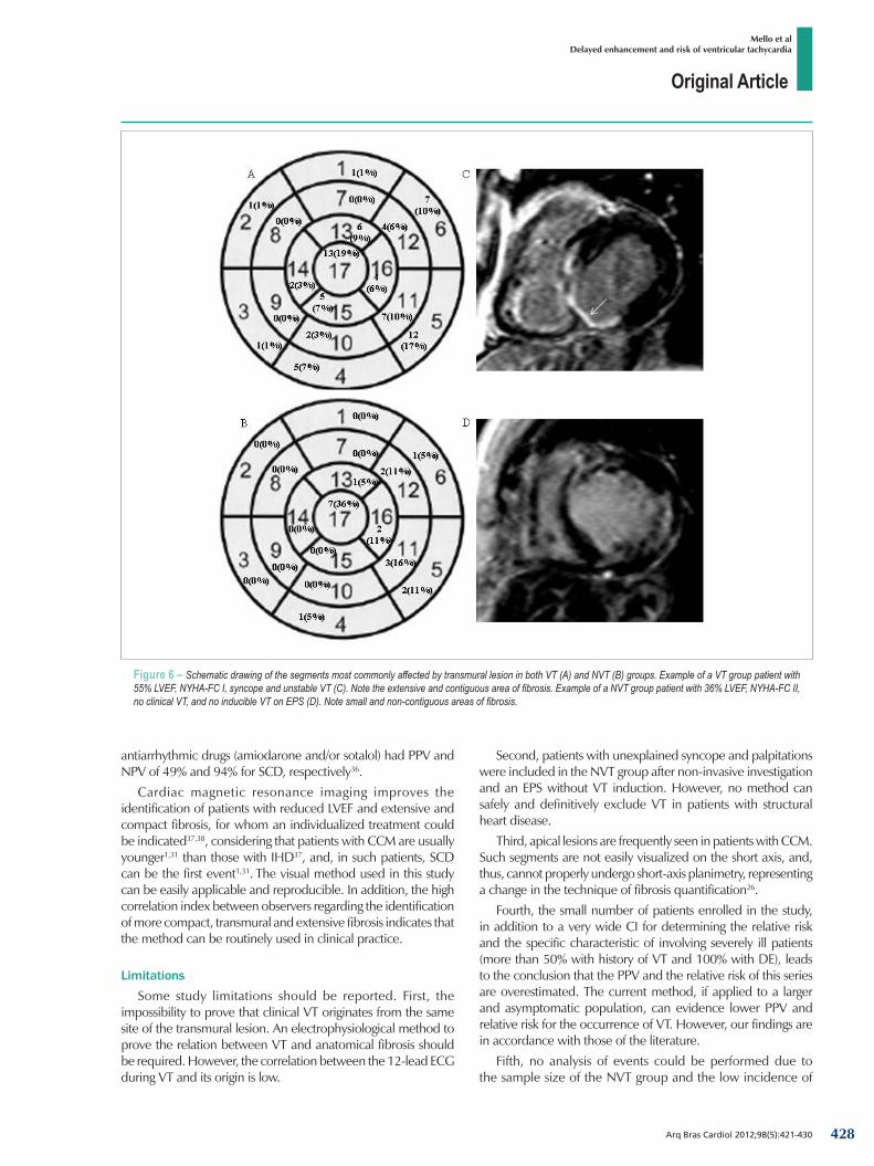
transmural lesions. On univariate analysis, that new variable significantly correlated with the occurrence of clinical VT. Such pattern of distribution was found in 19 VT group patients (73.1%) and six NVT group patients (40.0%) (p = 0.036). The multivariate regression model adjusted for LVEF ≤ 40%, age, sex, and DE percentage showed that two or more transmural segments were predictors of VT (RR 4.1; 95% CI: 1.06 - 15.68; p = 0.04; table 2). The observers agreed regarding the presence or absence of that distribution pattern in 41 (100%) patients with significant reproducibility (p < 0.001). This contiguous and compact distribution pattern of DE and transmural fibrosis was present in the apical segment of 13 VT group patients (50.0%) and five NVT group patients (33.3%), and in the infero-lateral basal segments of ten VT group patients (38.5%) and eight NVT group patients (53.3%) (p = 0.385). Figure 6 depicts the
Figure 1 – (A) Typical electrocardiogram of a patient with CCM. Note the right bundle branch block pattern in V1. (B) Common morphology of VT in a patient with CCM. Note the right bundle branch block pattern in V1, negative QRS in DII, and abrupt inversion of the R wave from V1 to V2 associated with QS in V6, suggesting origin in the left ventricular lateral-apical segment. Two-chamber view (C). Late diastole (C) and late systole showing apical aneurysm (red arrow) (D). Delayed enhancement image on the same plane showing intracavitary thrombus (white arrow) and extensive transmural fibrosis corresponding to the VT pattern of the ECG (E). In this case, the origin of the VT was confirmed by use of mapping and well-succeeded radiofrequency ablation.
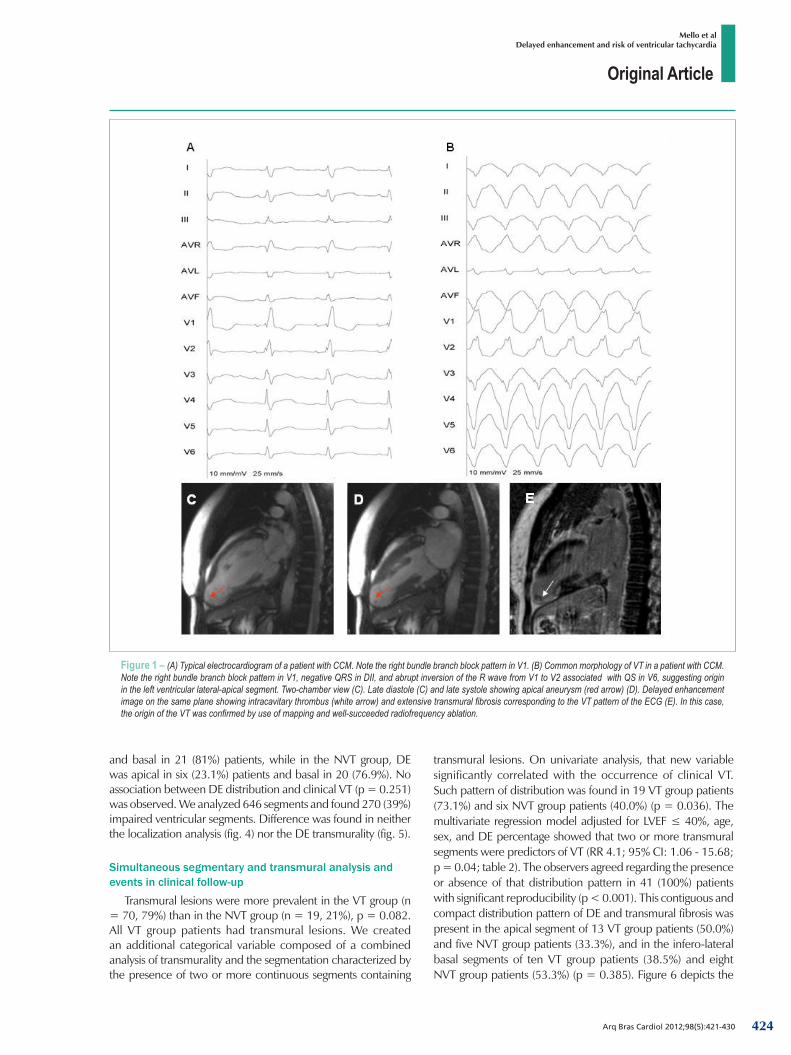
and basal in 21 (81%) patients, while in the NVT group, DE was apical in six (23.1%) patients and basal in 20 (76.9%). No association between DE distribution and clinical VT (p = 0.251) was observed. We analyzed 646 segments and found 270 (39%) impaired ventricular segments. Difference was found in neither the localization analysis (fig. 4) nor the DE transmurality (fig. 5).
Simultaneous segmentary and transmural analysis and events in clinical follow-up
Transmural lesions were more prevalent in the VT group (n = 70, 79%) than in the NVT group (n = 19, 21%), p = 0.082. All VT group patients had transmural lesions. We created an additional categorical variable composed of a combined analysis of transmurality and the segmentation characterized by the presence of two or more continuous segments containing
424

Original Article
Arq Bras Cardiol 2012;98(5):421-430
Mello et alDelayed enhancement and risk of ventricular tachycardia
frequency and preferential distribution of the transmural lesions of the patients studied.
For a mean 500 ± 303-day follow-up, NVT group patients with no transmural lesions and those with less than 6% of ventricular segments myocardial fibrosis showed no events. However, three patients of that same group with at least one transmural lesion had SCD during clinical follow-up, and two of them were totally asymptomatic. The third patient had undergone EPS, during which no VT was induced.
DiscussionThe result of our study shows that the presence of two or more
contiguous segments with transmural fibrosis was an independent predictor of VT in CCM. Such finding was associated with a 4.1-fold greater risk of developing VT. This indicates that CMRI with the DE technique can improve the identification of patients with CCM and at high risk for VT. To our knowledge, this is the first study comparing the transmural extension by segments and the occurrence of VT.
Substrate analysisVentricular fibrillation and VT are considered the most
common causes of SCD in CCM1,3. Fibrosis and reentry are the
most important substrates and mechanisms for VT in that group, respectively 2,27. The CMRI with the DE technique provides an excellent correlation with fibrosis18,19, including the identification of the inflammatory process due to CCM via endocardial biopsy28.
Such findings support the hypothesis that CMRI can be an important tool for assessing fibrosis in CCM. The applicability of CMRI for risk stratification in clinical practice has been studied by other authors. In IHD, Wu et al29 have shown an increase in the occurrence of events with an increase and greater extension of DE in patients with more than 18% of DE. In IHD, Kwong et al8 have shown, even in patients with low DE volumes (mean of 1.4 ± 1.1%), a seven-fold increment in the rate of events as compared to that of patients without DE. A DE volume > 15%, infarction area > 100 cm2 30 and DE involving 26% to 75% of the ventricular myocardial thickness31 were predictors of VT inducibility.
However, specifically in CCM, transmural fibrosis areas seem to be necessary for the occurrence of clinical VT. Rochitte et al6 have studied 51 patients with Chagas’ disease by use of CMRI and included patients with the undetermined form of the disease. In that study, 68% of the patients had DE, which was more marked in VT group patients.
The present study included only patients with myocardial fibrosis, segmentary abnormality, and compared patients with
Figure 2 – Linear regression analysis with results of the two observers: (A) DE according to the score method; (B) DE volume according to the planimetry method; and (C) Bland-Altman method to measure agreement between the results of the score and planimetry methods.
Scor
e meth
od: o
bser
ver I
Score method: observer II
Fit curve (13.06 + 0.7234x)95% Prediction interval
Plan
imetr
y meth
od: o
bser
ver I
Planimetry method: observer II
Fit curve (+1.382x)
95% Prediction interval
Diffe
renc
e: sc
ore -
plan
imetr
y
Bias (14.94)95% limit of agreement (-14.73 to 44.60)
R2 = 0.46
R2 = 0.40 R2 = 0.74
A
C
B
425

Original Article
Arq Bras Cardiol 2012;98(5):421-430
Mello et alDelayed enhancement and risk of ventricular tachycardia
Figure 4 – Mean fibrosis (SD) according to preferential location in the left ventricular wall. Subendocardial and transmural fibrosis was the most prevalent lesion. No significant difference was found between the VT and NVT groups regarding that criterion.
Mean
s of t
he le
sions
(SD)
Preferential distribution of fibrosisSubepicardial Mesocardial Subendocardial Transmural
VT NO VT
P = 0.250
P = 0.768
P = 0.378
P = 0.082
0.88 ± 1.82
0.25 ± 0.62
1.12 ± 2.10
1.33 ± 2.10
2.92 ± 3.16
2.00 ± 2.45
2.54 ± 1.73
1.5 ± 1.51
Figure 3 – Typical findings in patients with CCM. Image on the short axis: late diastole (A) and late systole (B). Hypokinesis and thinning of the inferolateral basal wall (red arrow). Delayed enhancement image on the same plane shows transmural fibrosis in that site (arrow) (C). Four-chamber view: late diastole (D) and late systole (E) showing left ventricular dilation, thinning of the inferolateral basal wall, and apical hypokinesis (red arrow). Delayed enhancement image on the same plane shows transmural fibrosis and small apical transmural thrombus (arrow) (F). Left ventricular outflow tract image (G, H, I) during diastole, systole and using the DE, with better demonstration of the same findings.
426
Original Article
Arq Bras Cardiol 2012;98(5):421-430
Mello et alDelayed enhancement and risk of ventricular tachycardia
Table 2 – Predictors of VT by use of multivariate regression analysis*
Variables RR CI p
DE % 0.994 0.92 – 1.07 0.87
Male sex 1.03 0.17 – 6.09 0.98
Age ≥ 54 years 2.9 0.70 – 12.08 0.14
LVEF ≤ 40% 2.8 0.53 –15.00 0.23
2 or more contiguous transmural segments 4.1 1.06 – 15.68 0.04
* Logistic regression analysis identified the variable “2 or more contiguous transmural segments” as an independent predictor of clinical VT; RR - relative risk; CI - confidence interval; DE – delayed enhancement; LVEF - left ventricular ejection fraction.
Figure 5 – Segmentary analysis of DE distribution according to the percentage of ventricular wall impairment. Lesions affecting 1% - 25% of the wall thickness were more prevalent in both groups. The second most common lesion was transmural. The analysis of the fibrosis pattern regarding its distribution along the ventricular wall in isolation showed no difference.
Percentage of ventricular wall thickness impairment
Mean
s of t
he le
sions
(SD)
p = 0.901
2.62 ± 2.352.5 ± 3.21
VT NO VT
p = 0.627
0.96 ± 1.31
0.75 ± 1.06
p = 0.152
1.58 ± 1.79
0.75 ± 1.14
p = 0.115
2.54 ± 1.77
1.58 ± 1.51
and without clinical VT. One hundred per cent of the patients had detectable DE, and all patients with VT had transmural lesion, confirming the findings by Rochitte et al6. In addition, one typical pattern of DE, expressed by the presence of two or more contiguous transmural segments was significantly associated with VT. Such findings have shown that the DE analysis, myocardial fibrosis quantification, and identification of transmural lesions, especially when such lesions are coalescent, forming extensive fibrosis plaques that occupy at least two contiguous segments, can identify a group at higher risk for VT.
Clinical impact
Until the present date, no diagnostic test in isolation has been able to identify patients at high risk for the occurrence
of VT. Clinical VT and LVEF ≤ 40% are known to be the most important independent predictors of cardiac death9-12. Recently, a new score method for risk stratification has been validated to identify patients at high risk for death by using the following clinical data: NYHA-FC > II; male sex; abnormalities of wall motility on echocardiography; presence of non-sustained VT on 24-h Holter; cardiomegaly on chest radiography; and low voltage on rest ECG32. On the other hand, invasive tests, such as EPS, have shown a low power of inducibility, reproducibility, and positive predictive value (PPV)13,33,34. In a study with patients with CCM, the EPS showed PPV and negative predictive value (NPV) of 24% and 98% for clinical VT, respectively. Inducibility had PPV and NPV of 46% and 85% for cardiac mortality, respectively35. In another study, inducibility of non-tolerated sustained VT in a patient with CCM after previous impregnation with class III
427
Original Article
Arq Bras Cardiol 2012;98(5):421-430
Mello et alDelayed enhancement and risk of ventricular tachycardia
Figure 6 – Schematic drawing of the segments most commonly affected by transmural lesion in both VT (A) and NVT (B) groups. Example of a VT group patient with 55% LVEF, NYHA-FC I, syncope and unstable VT (C). Note the extensive and contiguous area of fibrosis. Example of a NVT group patient with 36% LVEF, NYHA-FC II, no clinical VT, and no inducible VT on EPS (D). Note small and non-contiguous areas of fibrosis.
antiarrhythmic drugs (amiodarone and/or sotalol) had PPV and NPV of 49% and 94% for SCD, respectively36.
Cardiac magnetic resonance imaging improves the identification of patients with reduced LVEF and extensive and compact fibrosis, for whom an individualized treatment could be indicated37,38, considering that patients with CCM are usually younger1,31 than those with IHD37, and, in such patients, SCD can be the first event1,31. The visual method used in this study can be easily applicable and reproducible. In addition, the high correlation index between observers regarding the identification of more compact, transmural and extensive fibrosis indicates that the method can be routinely used in clinical practice.
LimitationsSome study limitations should be reported. First, the
impossibility to prove that clinical VT originates from the same site of the transmural lesion. An electrophysiological method to prove the relation between VT and anatomical fibrosis should be required. However, the correlation between the 12-lead ECG during VT and its origin is low.
Second, patients with unexplained syncope and palpitations were included in the NVT group after non-invasive investigation and an EPS without VT induction. However, no method can safely and definitively exclude VT in patients with structural heart disease.
Third, apical lesions are frequently seen in patients with CCM. Such segments are not easily visualized on the short axis, and, thus, cannot properly undergo short-axis planimetry, representing a change in the technique of fibrosis quantification26.
Fourth, the small number of patients enrolled in the study, in addition to a very wide CI for determining the relative risk and the specific characteristic of involving severely ill patients (more than 50% with history of VT and 100% with DE), leads to the conclusion that the PPV and the relative risk of this series are overestimated. The current method, if applied to a larger and asymptomatic population, can evidence lower PPV and relative risk for the occurrence of VT. However, our findings are in accordance with those of the literature.
Fifth, no analysis of events could be performed due to the sample size of the NVT group and the low incidence of
428

Original Article
Arq Bras Cardiol 2012;98(5):421-430
Mello et alDelayed enhancement and risk of ventricular tachycardia
References1. Rassi A Jr, Rassi A, Rassi SG. Predictors of mortality in chronic Chagas disease: a
systematic review of observational studies. Circulation. 2007;115(9):1101-8.
2. Távora MZ, Mehta N, Silva RM, Gondim FA, Hara VM, De Paola AA. Characteristics and identification of sites of chagasic ventricular tachycardia by endocardial mapping. Arq Bras Cardiol. 1999;72(4):451-74.
3. Luu M, Stevenson WG, Stevenson LW, Baron K, Walden J. Diverse mechanisms of unexpected cardiac arrest in advanced heart failure. Circulation. 1989;80(6):1675-80.
4. Kelly P, Coats A. Variation in mode of sudden cardiac death in patients with dilated cardiomyopathy. Eur Heart J. 1997;18(5):879-80.
5. Tamburro P, Wilber D. Sudden death in idiopathic dilated cardiomyopathy. Am Heart J. 1992;124(4):1035-45.
6. Rochitte CE, Oliveira PF, Andrade JM, Ianni BM, Parga JR, Avila LF, et al. Myocardial delayed enhancement by magnetic resonance imaging in patients with Chagas’ disease: a marker of disease severity. J Am Coll Cardiol. 2005;46(8):1553-8.
7. Wu KC, Weiss RG, Thiemann DR, Kitagawa K, Schmidt A, Dalal D, et al. Late gadolinium enhancement by cardiovascular magnetic resonance heralds an adverse prognosis in nonischemic cardiomyopathy. J Am Coll Cardiol. 2008;51(25):2414-21.
8. Kwong RY, Chan AK, Brown KA, Chan CW, Reynolds HG, Tsang S, et al. Impact of unrecognized myocardial scar detected by cardiac magnetic resonance imaging on event-free survival in patients presenting with signs or symptoms of coronary artery disease. Circulation. 2006;113(23):2733-43.
9. Bigger JT Jr, Fleiss JL, Kleiger R, Miller JP, Rolnitzky LM. The relationships among ventricular arrhythmias, left ventricular dysfunction, and mortality in the 2 years after myocardial infarction. Circulation. 1984;69(2):250-8.
10. Moss AJ, DeCamilla JJ, Davis HP, Bayer L. Clinical significance of ventricular ectopic beats in the early posthospital phase of myocardial infarction. Am J Cardiol. 1977;39(5):635-40.
11. Vasan RS, Larson MG, Benjamin EJ, Evans JC, Reiss CK, Levy D. Congestive heart failure in subjects with normal versus reduced left ventricular ejection fraction: prevalence and mortality in a population-based cohort. J Am Coll Cardiol. 1999;33(7):1948-55.
12. Brugada P, Talajic M, Smeets J, Mulleneers R, Wellens HJ. The value of the clínical history to assess prognosis of patients with ventricular tachycardia or ventricular fibrillation after myocardial infarction. Eur Heart J. 1989;10(8):747-52.
13. Grimm W, Hoffmann J, Menz V, Luck K, Maisch B. Programmed ventricular stimulation for arrhythmia risk prediction in patients with idiopathic dilated
cardiomyopathy and nonsustained ventricular tachycardia. J Am Coll Cardiol. 1998;32(3):739-45.
14. Chen X, Shenasa M, Borggrefe M, Block M, Hindricks G, Martinez-Rubio A, et al. Role of programmed ventricular stimulation in patients with idiopathic dilated cardiomyopathy and documented sustained ventricular tachyarrhythmias: inducibility and prognostic value in 102 patients. Eur Heart J. 1994;15(1):76-82.
15. Turitto G, Ahuja RK, Caref EB, el-Sherif N. Risk stratification for arrhythmic events in patients with nonischemic dilated cardiomyopathy and nonsustained ventricular tachycardia: role of programmed ventricular stimulation and the signal-averaged electrocardiogram. J Am Coll Cardiol. 1994;24(6):1523-8.
16. Jaochim Nesser H, Sugeng L, Corsi C, Weinert L, Niel J, Ebner C, et al. Volumetric analysis of regional left ventricular function with real-time three-dimensional echocardiography: validation by magnetic resonance and clinical utility testing. Heart. 2007;93(5):572-8.
17. Maniar HS, Cupps BP, Potter DD, Moustakidis P, Camillo CJ, Chu CM, et al. Ventricular function after coronary artery bypass grafting: evaluation by magnetic resonance imaging and myocardial strain analysis. J Thorac Cardiovasc Surg. 2004;128(1):76-82.
18. Kim RJ, Chen EL, Lima JA, Judd RM. Myocardial Gd-DTPA kinetics determine MRI contrast enhancement and reflect the extent and severity of myocardial injury after acute reperfused infarction. Circulation. 1996;94(12):3318-26.
19. Lima JA, Judd RM, Bazille A, Schulman SP, Atalar E, Zerhouni EA. Regional heterogeneity of human myocardial infarcts demonstrated by contrast-enhanced MRI. Potential mechanisms. Circulation. 1995;92(5):1117-25.
20. Judd RM, Lugo-Olivieri CH, Arai M, Kondo T, Croisille P, Lima JA, et al. Physiological basis of myocardial contrast enhancement in fast magnetic resonance images of 2-day-old reperfused canine infarcts. Circulation. 1995;92(7):1902-10.
21. Goldman MR, Brady TJ, Pykett IL, Burt CT, Buonanno FS, Kistler JP, et al. Quantification of experimental myocardial infarction using nuclear magnetic resonance imaging and paramagnetic ion contrast enhancement in excised canine hearts. Circulation. 1982;66(5):1012-6.
22. Fernandes VR, Wu KC, Rosen BD, Schmidt A, Lardo AC, Osman N, et al. Enhanced infarct border zone function and altered mechanical activation predict inducibility of monomorphic ventricular tachycardia in patients with ischemic cardiomyopathy. Radiology. 2007;245(3):712-9.
23. Scheffel H, Leschka S, Plass A, Vachenauer R, Gaemperli O, Garzoli E, et al. Accuracy of 64-slice computed tomography for the preoperative detection of coronary artery disease in patients with chronic aortic regurgitation. Am J Cardiol. 2007;100(4):701-6.
short-term events in CCM. Further studies are required to confirm our findings.
ConclusionThis study indicates that CMRI improves risk stratification
of patients with CCM and identifies those more susceptible to VT. In this group, the identification of two or more contiguous segments of transmural DE is associated with greater probability of clinical VT. Such finding evidences CMRI as an important tool to identify patients with CCM at higher risk for VT, characterized by the presence of ventricular dysfunction and an extensive area of transmural fibrosis occupying at least two coalescent segments. Further studies are required to confirm such findings and
should include asymptomatic patients in the long-term clinical follow-up.
Potential Conflict of InterestNo potential conflict of interest relevant to this article
was reported.
Sources of FundingThere were no external funding sources for this study.
Study AssociationThis article is part of the thesis of doctoral submitted by Ronaldo
Peixoto de Mello, from Escola Paulista de Medicina - UNIFESP.
429
Original Article
Arq Bras Cardiol 2012;98(5):421-430
Mello et alDelayed enhancement and risk of ventricular tachycardia
24. Shabestari AA, Abdi S, Akhlaghpoor S, Azadi M, Baharjoo H, Pajouh MD, et al. Diagnostic performance of 64-channel multislice computed tomography in assessment of significant coronary artery disease in symptomatic subjects. Am J Cardiol. 2007;99(12):1656-61.
25. Cerqueira MD, Weissman NJ, Dilsizian V, Jacobs AK, Kaul S, Laskey WK, et al. Standardized myocardial segmentation and nomenclature for tomographic imaging of the heart: a statement for healthcare professionals from the Cardiac Imaging Committee of the Council on Clínical Cardiology of the American Heart Association. Circulation. 2002;105(4):539-42.
26. Comte A, Lalande A, Walker PM, Cochet A, Legrand L, Cottin Y, et al. Visual estimation of the global myocardial extent of hyperenhancement on delayed contrast-enhanced MRI. Eur Radiol. 2004;14(12):2182-7.
27. Stevenson WG, Khan H, Sager P, Saxon LA, Middlekauff RR, Natterson PD, et al. Identification of reentry circuit sites during catheter mapping and radiofrequency ablation of ventricular tachycardia late after myocardial infarction. Circulation. 1993;88(4 Pt 1):1647-70.
28. Kalil R, Bocchi EA, Ferreira BM, Higuchi ML, Lopes NH, Magalhães AC, et al. [Magnetic resonance imaging in chronic Chagas cardiopathy: correlation with endomyocardial biopsy findings]. Arq Bras Cardiol. 1995;65(5):413-6.
29. Wu KC, Zerhouni EA, Judd RM, Lugo-Olivieri CH, Barouch LA, Schulman SP, et al. Prognostic significance of microvascular obstruction by magnetic resonance imaging in patients with acute myocardial infarction. Circulation. 1998;97(8):765-72.
30. Bello D, Fieno DS, Kim RJ, Pereles FS, Passman R, Song G, et al. Infarct morphology identifies patients with substrate for sustained ventricular tachycardia. J Am Coll Cardiol. 2005;45(7):1104-8.
31. Nazarian S, Bluemke DA, Lardo AC, Zviman MM, Watkins SP, Dickfeld TL, et al. Magnetic resonance assessment of the substrate for inducible
ventricular tachycardia in nonischemic cardiomyopathy. Circulation. 2005;112(18):2821-5.
32. Rassi A Jr, Rassi A, Little WC, Xavier SS, Rassi SG, Rassi AG, et al. Development and validation of a risk score for predicting death in Chagas’ heart disease. N Engl J Med. 2006;355(8):799-808.
33. Brilakis ES, Shen WK, Hammill SC, Hodge DO, Rea RF, Lexvold NY, et al. Role of programmed ventricular stimulation and implantable cardioverter defibrillators in patients with idiopathic dilated cardiomyopathy and syncope. Pacing Clin Electrophysiol. 2001;24(11):1623-30.
34. Bansch D, Antz M, Boczor S, Volkmer M, Tebbenjohanns J, Seidl K, et al. Primary prevention of sudden cardiac death in idiopathic dilated cardiomyopathy: the Cardiomyopathy Trial (CAT). Circulation. 2002;105(12):1453-8.
35. Silva RM, Távora MZ, Godim FA, Metha N, Hara VM, Paola AA. Predictive value of clinical and electrophysiological variables in patients with chronic chagasic cardiomyopathy and nonsustained ventricular tachycardia. Arq Bras Cardiol. 2000;75(1):33-47.
36. Leite LR, Fenelon G, Simoes A Jr, Silva GG, Friedman PA, de Paola AA. Clinical usefulness of electrophysiologic testing in patients with ventricular tachycardia and chronic chagasic cardiomyopathy treated with amiodarone or sotalol. J Cardiovasc Electrophysiol. 2003;14(6):567-73.
37. Moss AJ, Zareba W, Hall WJ, Klein H, Wilber DJ, Cannom DS, et al. Multicenter Automatic Defibrillator Implantation Trial II Investigators. Prophylactic implantation of a defibrillator in patients with myocardial infarction and reduced ejection fraction. N Engl J Med. 2002;346(12):877-83.
38. Bardy GH, Lee KL, Mark DB, Poole JE, Packer DL, Boineau R, et al.; Sudden Cardiac Death in Heart Failure Trial (SCD-HeFT) Investigators. Amiodarone or an implantable cardioverter-defibrillator for congestive heart failure. N Engl J Med. 2005;352(3):225-37.
430