Embed Size (px)
Citation preview
EXCLI Journal 2017;16:265-277 – ISSN 1611-2156 Received: January 03, 2017, accepted: March 03, 2017, published: March 14, 2017
265
Original article:
SCHISANDRAE FRUCTUS ETHANOL EXTRACT AMELIORATES INFLAMMATORY RESPONSES AND ARTICULAR CARTILAGE
DAMAGE IN MONOSODIUM IODOACETATE-INDUCED OSTEOARTHRITIS IN RATS
Jin-Woo Jeong1,2, Jongsik Kim3, Eun Ok Choi1,2, Da Hye Kwon1,2, Gyu Min Kong4, Il-Whan Choi5, Bum Hoi Kim6, Gi-Young Kim7, Ki Won Lee8, Ki Young Kim8, Sung Goo Kim8, Young Whan Choi9, Su Hyun Hong1, Cheol Park10, Yung Hyun Choi1,2,*
1 Department of Biochemistry, Dongeui University College of Korean Medicine,
Busan 614-052, Republic of Korea 2 Anti-Aging Research Center, Dongeui University, Busan 614-714, Republic of Korea 3 Department of Anatomy, Kosin University College of Medicine, Busan 602-702, Republic
of Korea 4 Department of Orthopaedic Surgery, College of Medicine, Inje University, Busan,
47392, Republic of Korea 5 Department of Microbiology, College of Medicine, Inje University, Busan,
47392, Republic of Korea 6 Department of Anatomy, Dongeui University College of Korean Medicine, Busan 614-052,
Republic of Korea 7 Laboratory of Immunobiology, Department of Marine Life Sciences, Jeju National
University, Jeju, 690-756, Republic of Korea 8 Research Institute, Bio-Port Korea INC, MarineBio-industry Development Center, Busan
619-912, Republic of Korea 9 Department of Horticultural Bioscience, College of Natural Resource and Life Sciences,
Pusan National University, Miryang 627-706, Republic of Korea 10 Department of Molecular Biology, College of Natural Sciences and Human Ecology,
Dongeui University, Busan 614-714, Republic of Korea * Corresponding author: Dr. Y. H. Choi, Department of Biochemistry, Dongeui University
College of Korean Medicine, 52-57, Yangjeong-ro, Busanjin-gu, Busan 47227, Republic of Korea, E-mail: [email protected]
http://dx.doi.org/10.17179/excli2017-119
This is an Open Access article distributed under the terms of the Creative Commons Attribution License (http://creativecommons.org/licenses/by/4.0/).
ABSTRACT
Schisandrae Fructus, the fruit of Schisandra chinensis (Turcz.) Baill., is widely used in traditional medicine for the treatment of a number of chronic diseases. Although, Schisandrae Fructus was recently reported to attenuate the interleukin (IL)-1β-induced inflammatory response in chondrocytes in vitro, its protective and therapeutic po-tential against osteoarthritis (OA) in an animal model remains unclear. Therefore, we investigated the effects of the ethanol extract of Schisandrae Fructus (SF) on inflammatory responses and cartilage degradation in a mono-sodium iodoacetate (MIA)-induced OA rat model. Our results demonstrated that administration with SF had a tendency to attenuate MIA-induced damage of articular cartilage as determined by a histological grade of OA. SF significantly suppressed the production of pro-inflammatory cytokines such as interleukin (IL)-1β, IL-6, and tumor necrosis factor-α in MIA-induced OA rats. SF also effectively inhibited expression of inducible nitric oxide (NO)
EXCLI Journal 2017;16:265-277 – ISSN 1611-2156 Received: January 03, 2017, accepted: March 03, 2017, published: March 14, 2017
266
synthase and cyclooxygenase-2, thereby inhibiting the release of NO and prostaglandin E2. In addition, the elevated levels of matrix metalloproteinases-13 and two biomarkers for diagnosis and progression of OA, such as cartilage oligomeric matrix protein and C-telopeptide of type II collagen, were markedly ameliorated by SF administration. These findings indicate that SF could be a potential candidate for the treatment of OA. Keywords: Schisandrae Fructus, osteoarthritis, MIA, inflammatory responses, cartilage degradation
INTRODUCTION
Osteoarthritis (OA) is the most common musculoskeletal disease causing chronic pain and joint disability. OA is characterized by the loss of articular cartilage, involving in-creased subchondral bone remodeling, osteo-phyte formation, weakening of the periarticu-lar muscles, and thickening of the capsule and synovial membrane, which lead to functional joint limitations (Guilak, 2011; Mobasheri, 2013; Speziali et al., 2015). In particular, OA is the result of mechanical and biological events that cause the degradation of articular cartilage. These processes are mediated by excessive synthesis and release of catabolic tissue proteinases, such as matrix metallopro-teinases (MMPs), collagenases and aggre-canases, that are upregulated by inflammatory stimuli and oxidative stress, including inflam-matory cytokines and mediators, and reactive oxygen species (Goldring and Otero, 2011; Makki and Haqqi, 2015; Lepetsos and Papa-vassiliou, 2016). Currently, pharmacological treatment for patients with OA is based on some steroidal and non-steroidal anti-inflam-matory drugs for alleviation of pain as well as inflammation. However, they can not per-fectly prevent the progressive cartilage degra-dation and repair the impaired cartilage of OA patients, and long-term use of these drugs can lead to severe side effects or toxicity, includ-ing gastrointestinal disturbances and cardio-vascular risk (Mobasheri, 2013; Goldring and Berenbaum, 2015). Therefore, ideal agents that inhibit cartilage degradation with better safety and efficacy may represent an attrac-tive strategy to treat OA.
Herbal sources have been widely and safely consumed for centuries. Many studies indicate that these herbs have a wide range of
diverse biological activities with few side ef-fects (Chen et al., 2015; Hou et al., 2015). Therefore, traditional herbal medicinal sources have been investigated widely as ad-juvant therapeutic agents in the treatment of OA (Cameron and Chrubasik, 2013; Dhip-payom et al., 2015). Schisandrae Fructus, the dried fruit of Schisandra chinensis (Turcz.) Baill. (Magnoliaceae), has been used in tradi-tional medicine for the treatment of various medicinal purposes (Panossian and Wikman, 2008; Chun et al., 2014). The diverse pharma-cological effects of Schisandrae Fructus in-clude antioxidant (Kang et al., 2014a), anti-tumor (Lv et al., 2015), hepatoprotective (Wat et al., 2016), anti-septic (Kook et al., 2015), neuroprotective (Lee et al., 2012), anti-in-flammatory (Bae et al., 2012; Kang et al., 2014b), anti-atherosclerotic (Jeong et al., 2015a), and anti-atrophic (Kim et al., 2015a, b), and anti-diabetic effects (Kwon et al., 2011). Recently we reported that Schisandrae Fructus possessed potential chondroprotec-tive effects of the collagen matrix breakdown in a pro-inflammatory cytokine interleukin (IL)-1β-induced model in vitro (Jeong et al., 2015b). However, to our knowledge, the in vivo therapeutic effects of this compound and its effects on the molecular mechanisms of OA have not been investigated. Therefore, as a part of our on-going research program for finding novel anti-osteoarthritic substances from traditional medicinal sources, the pre-sent study investigated the anti-osteoarthritic potential and underlying mechanism of an ethanol extract of Schisandrae Fructus (SF) in a rat model of metabolic-inhibitor-monoso-dium iodoacetate (MIA)-induced OA.
EXCLI Journal 2017;16:265-277 – ISSN 1611-2156 Received: January 03, 2017, accepted: March 03, 2017, published: March 14, 2017
267
MATERIALS AND METHODS
Preparation of SF The dried fruits of S. chinensis were col-
lected from Mungyeong city (Gyeongsang-buk-do, Republic of Korea) and washed three times with tap water before storage at -80 °C. Frozen samples were lyophilized and homog-enized using a grinder before extraction with 20 % ethanol at room temperature for 4 h, fil-tered, and concentrated using a rotary vacuum evaporator (BÜCHI Labortechnik, Flawil, Switzerland). The extract (SF) was dissolved in dimethyl sulfoxide (DMSO; Sigma-Al-drich Chemical Co., St. Louis, MO, USA) as a 50 mg/mL stock solution and stored at 4 °C and diluted with physiological saline to the desired concentration prior to use.
Animals
Male Sprague-Dawley rats weighing 180~240 g (5 weeks of age) at the start of the experiment were purchased from Samtako Inc. (Osan, Republic of Korea). Two animals were housed per polycarbonate cage in a room under controlled-temperature condi-tions (20~24 °C, humidity 40~70 %) with controlled lighting (12 h light and/or 12 h dark cycle) and had access to sterile food and R/O water (Lee et al., 2016). This study was car-ried out in strict accordance with the recom-mendations of the Guide for the Care and Use of Laboratory Animals of the National Insti-tutes of Health. In addition, the animal proto-col used in this study was reviewed by the Dongeui University - Institutional Animal Care and Use Committee on their ethical pro-cedures and scientific care, and it has been ap-proved (Approval Number: A2015-019).
Development of OA with MIA injection and administration with SF
The rats were randomized and assigned to treatment groups before the initiation of the study (n = 8 per group). For induction of OA, rats were anesthetized using isoflurane and then given a single injection of 50 μL sterile 0.9 % saline containing 3 mg/kg MIA (Sigma-Aldrich Chemical Co.) using a 0.3 mL insulin syringe (BD Medical-Diabetes
Care, Franklin Lakes, NJ, USA) through the patellar ligament into the articular cavity of the right knee. Control rats were injected with an equivalent volume of saline. SF was ad-ministered orally once per day for 3 weeks at a dose of 100 mg/kg.
Measurement of knee joint swelling
After the rats were killed at 3 weeks post-MIA injection, the right knee was isolated, and the femur, tibia, and patella were dis-sected free of muscle. Knee diameter was measured using a calibrated digital caliper (Mitutoyo, Kawasaki, Japan) to assess the de-velopmental stages of OA on 3 weeks post-MIA injection (Fernihough et al., 2004).
Serum analysis
At the end of the 3 weeks, the samples of whole blood were collected from the ab-dominal vein. Blood was allowed to clot for 30 min. Then, the serum was separated via centrifugation at 1,500 g for 10 min and stored at -80 °C. Concentrations of nitric ox-ide (NO) in the serum samples were deter-mined by measuring nitrite, which is a major stable product of NO, using the Griess rea-gent. Briefly, 50 μL of serums were mixed with 50 μL of Griess reagent (Sigma-Aldrich Chemical Co.), followed by incubation for 10 min at 37 °C. Optical density was measured at 540 nm using an enzyme-linked immuno-sorbent assay (ELISA) reader (Dynatech La-boratories, Chantilly, VA, USA) (Kwon et al., 2016). Serum levels of prostaglandin E2 (PGE2) were determined according to the manufacturer’s instructions (ELISA kit, R&D Systems, Minneapolis, MN, USA) (Kim et al., 2016). The pro-inflammatory cytokines including IL-1β, IL-6 and TNF-α, and carti-lage degeneration mediators such as cartilage oligomeric matrix protein (COMP) and C-telopeptide of type II collagen (CTX-II) were also determined using ELISA kits (R&D Sys-tems, Minneapolis, MN, USA) according to the manufacturer’s recommendations.
EXCLI Journal 2017;16:265-277 – ISSN 1611-2156 Received: January 03, 2017, accepted: March 03, 2017, published: March 14, 2017
268
Joint histological examination Histological changes were assessed to
confirm the effects of SF on cartilage degen-eration in the knee joints of MIA-induced OA rats. After the rat sacrifice at 3 weeks, each knee joint was resected, fixed in 10 % forma-lin (Sigma-Aldrich Chemical Co.) for 24 h at 4° C, and decalcified with 5 % hydrochloric acid (Sigma-Aldrich Chemical Co.) for 4 days at 4° C. After decalcification, the specimens were embedded in paraffin. Sections (2~3 μm) were stained with hematoxylin and eosin (H&E), safranin O-fast green and toluidine blue (Sigma-Aldrich Chemical Co.), respec-tively, and then observed under Carl Zeiss Axio-plan 2 imaging microscope (Carl Zeiss, Deisenhofen, Germany). All stained slides were histologically evaluated and statistically graded on a scale of 0~13 by double-blind ob-servation, according to the modified Mankin scoring system (Mankin et al., 1971).
Immunohistochemical analysis
The sections were depleted of endogenous peroxidase activity by treatment with 3 % H2O2 for 15 min. They were then were blocked with normal goat serum (Sigma-Al-drich Chemical Co.) for 30 min and incubated at 4 °C overnight with primary antibodies fol-lowed by the appropriate biotinylated second-ary antibodies and horseradish peroxidase-conjugated streptavidin-biotin staining, and finally with a 3,3′-diaminobenzidine (DAKO, Glostrup, Denmark). Primary antibodies against the following proteins were used: in-ducible NO synthase (iNOS, 1:100; SC-7271, mouse monoclonal, Santa Cruz Biotechnol-ogy, Inc., Santa Cruz, CA, USA), cyclooxy-genase-2 (COX-2, 1:50; SC-19999, mouse monoclonal; Santa Cruz Biotechnology, Inc.), MMP-13 (1:50; ab51072, rabbit monoclonal; Abcam, Cambridge, UK), COMP (1:50; ab11056, rabbit monoclonal; Abcam) and CTX-II (1:50; PAA686Hu01, rabbit polyclo-nal; Cloud-Clone Corp., Houston, TX, USA ). Histological changes were examined by a Carl Zeiss Axio-plan 2 imaging microscope (Carl Zeiss) and photographed.
Statistical analysis Data were expressed as mean ± standard
deviation (SD) for at least three separate de-terminations for each group. The differences between the groups were examined for statis-tical significance using the Student’s t-test and one-way ANOVA with GraphPad soft-ware (GraphPad Inc., La Jolla, CA, USA). A value of p < 0.05 was considered as being sig-nificant.
RESULTS
SF reduced MIA-induced knee swelling in rats
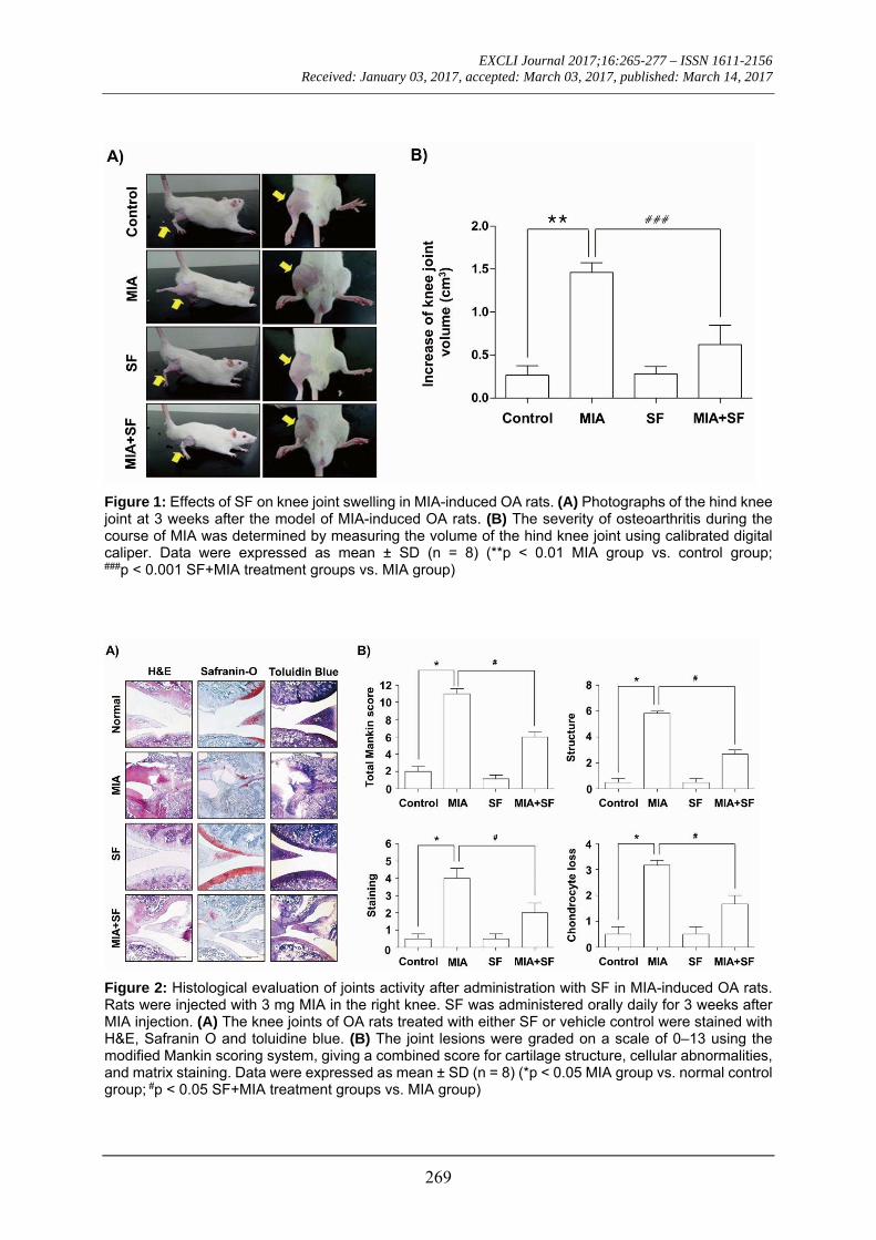
Knee diameters were measured to deter-mine the degree of joint swelling, an index of inflammation that occurred after intra-articu-lar injection of MIA. As shown in Figure 1, the knee diameter of in MIA groups increased significantly after an intra-articular injection of MIA as compared with normal control group. However, SF treatment significantly reduced the joint swelling at 3 weeks after MIA injection.
SF ameliorated the histological evaluation of the articular damage in MIA-induced OA rats
Because, the cartilage degeneration is the main histologic feature of OA, we investi-gated the effects of SF on the morphological changes and severity of the particular damage using H&E, Safranin O-fast green, and tolui-dine blue staining in the MIA-induced OA rat. Our findings showed that MIA-injected group showed the severity of surface irregularity and surface cleft, and matrix loss of articular cartilage associated (Figure 2A). However, SF alone had little effect on the structural, morphological changes in the joints, and ad-ministration with SF attenuated damages in the articular cartilages compared with those of the MIA-treated group. Therefore, the sever-ity of OA lesion was graded using the modi-fied Mankin scoring system, and we found that the overall modified Mankin’s scores were significantly recovered by SF treatment compared with a MIA-treated group (Figure 2B).
EXCLI Journal 2017;16:265-277 – ISSN 1611-2156 Received: January 03, 2017, accepted: March 03, 2017, published: March 14, 2017
269
Figure 1: Effects of SF on knee joint swelling in MIA-induced OA rats. (A) Photographs of the hind knee joint at 3 weeks after the model of MIA-induced OA rats. (B) The severity of osteoarthritis during the course of MIA was determined by measuring the volume of the hind knee joint using calibrated digital caliper. Data were expressed as mean ± SD (n = 8) (**p < 0.01 MIA group vs. control group; ###p < 0.001 SF+MIA treatment groups vs. MIA group)
Figure 2: Histological evaluation of joints activity after administration with SF in MIA-induced OA rats. Rats were injected with 3 mg MIA in the right knee. SF was administered orally daily for 3 weeks after MIA injection. (A) The knee joints of OA rats treated with either SF or vehicle control were stained with H&E, Safranin O and toluidine blue. (B) The joint lesions were graded on a scale of 0–13 using the modified Mankin scoring system, giving a combined score for cartilage structure, cellular abnormalities, and matrix staining. Data were expressed as mean ± SD (n = 8) (*p < 0.05 MIA group vs. normal control group; #p < 0.05 SF+MIA treatment groups vs. MIA group)
EXCLI Journal 2017;16:265-277 – ISSN 1611-2156 Received: January 03, 2017, accepted: March 03, 2017, published: March 14, 2017
270
SF inhibited the production of pro-inflam-matory cytokines in MIA-induced OA rats
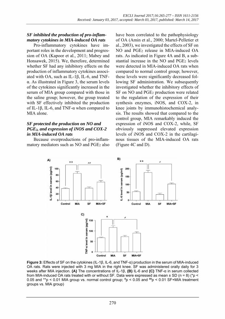
Pro-inflammatory cytokines have im-portant roles in the development and progres-sion of OA (Kapoor et al., 2011; Mabey and Honsawek, 2015). We, therefore, determined whether SF had any inhibitory effects on the production of inflammatory cytokines associ-ated with OA, such as IL-1β, IL-6, and TNF-α. As illustrated in Figure 3, the serum levels of the cytokines significantly increased in the serum of MIA group compared with those in the saline group; however, the group treated with SF effectively inhibited the production of IL-1β, IL-6, and TNF-α when compared to MIA alone.
SF protected the production on NO and PGE2, and expression of iNOS and COX-2 in MIA-induced OA rats
Because overproductions of pro-inflam-matory mediators such as NO and PGE2 also
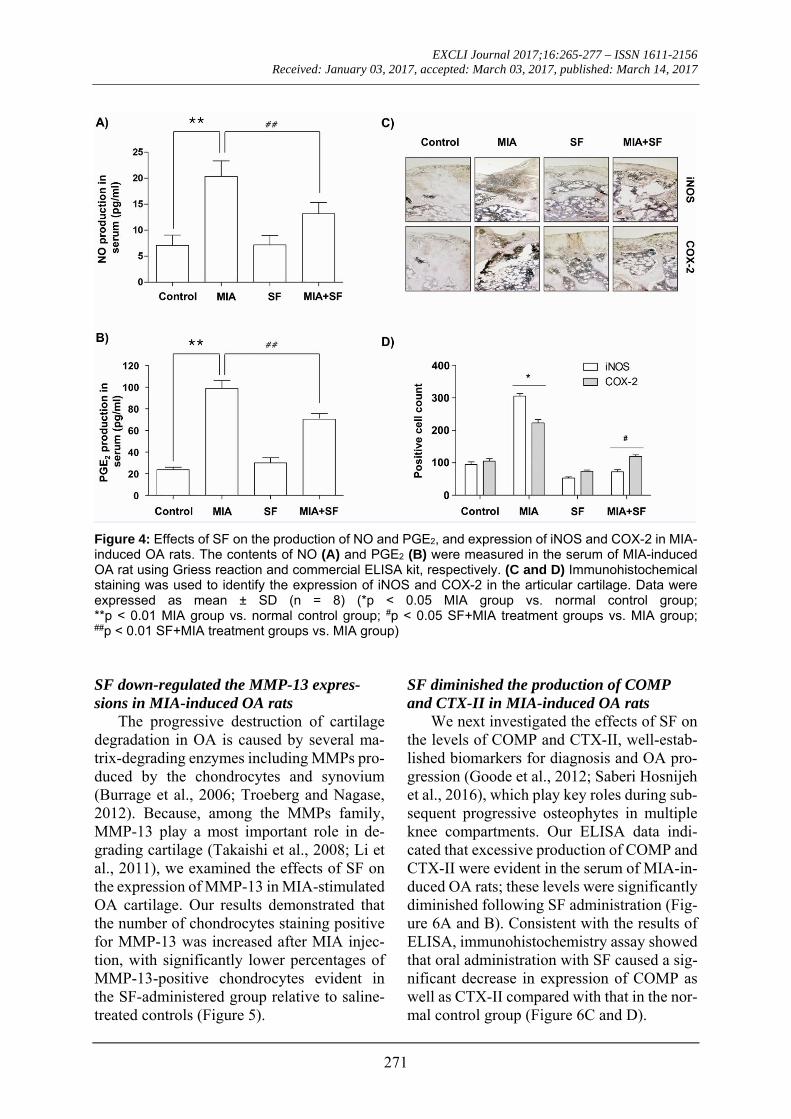
have been correlated to the pathophysiology of OA (Amin et al., 2000; Martel-Pelletier et al., 2003), we investigated the effects of SF on NO and PGE2 release in MIA-induced OA rats. As indicated in Figure 4A and B, a sub-stantial increase in the NO and PGE2 levels were detected in MIA-induced OA rats when compared to normal control group; however, these levels were significantly decreased fol-lowing SF administration. We subsequently investigated whether the inhibitory effects of SF on NO and PGE2 production were related to the regulation of the expression of their synthesis enzymes, iNOS, and COX-2, in knee joints by immunohistochemical analy-sis. The results showed that compared to the control group, MIA remarkably induced the expression of iNOS and COX-2, while, SF obviously suppressed elevated expression levels of iNOS and COX-2 in the cartilagi-nous tissues of the MIA-induced OA rats (Figure 4C and D).
Figure 3: Effects of SF on the cytokines (IL-1β, IL-6, and TNF-α) production in the serum of MIA-induced OA rats. Rats were injected with 3 mg MIA in the right knee. SF was administered orally daily for 3 weeks after MIA injection. (A) The concentrations of IL-1β, (B) IL-6 and (C) TNF-α in serum collected from MIA-induced OA rats treated with or without SF. Data were expressed as mean ± SD (n = 8) (*p < 0.05 and **p < 0.01 MIA group vs. normal control group; #p < 0.05 and ##p < 0.01 SF+MIA treatment groups vs. MIA group)
EXCLI Journal 2017;16:265-277 – ISSN 1611-2156 Received: January 03, 2017, accepted: March 03, 2017, published: March 14, 2017
271
Figure 4: Effects of SF on the production of NO and PGE2, and expression of iNOS and COX-2 in MIA-induced OA rats. The contents of NO (A) and PGE2 (B) were measured in the serum of MIA-induced OA rat using Griess reaction and commercial ELISA kit, respectively. (C and D) Immunohistochemical staining was used to identify the expression of iNOS and COX-2 in the articular cartilage. Data were expressed as mean ± SD (n = 8) (*p < 0.05 MIA group vs. normal control group; **p < 0.01 MIA group vs. normal control group; #p < 0.05 SF+MIA treatment groups vs. MIA group; ##p < 0.01 SF+MIA treatment groups vs. MIA group) SF down-regulated the MMP-13 expres-sions in MIA-induced OA rats
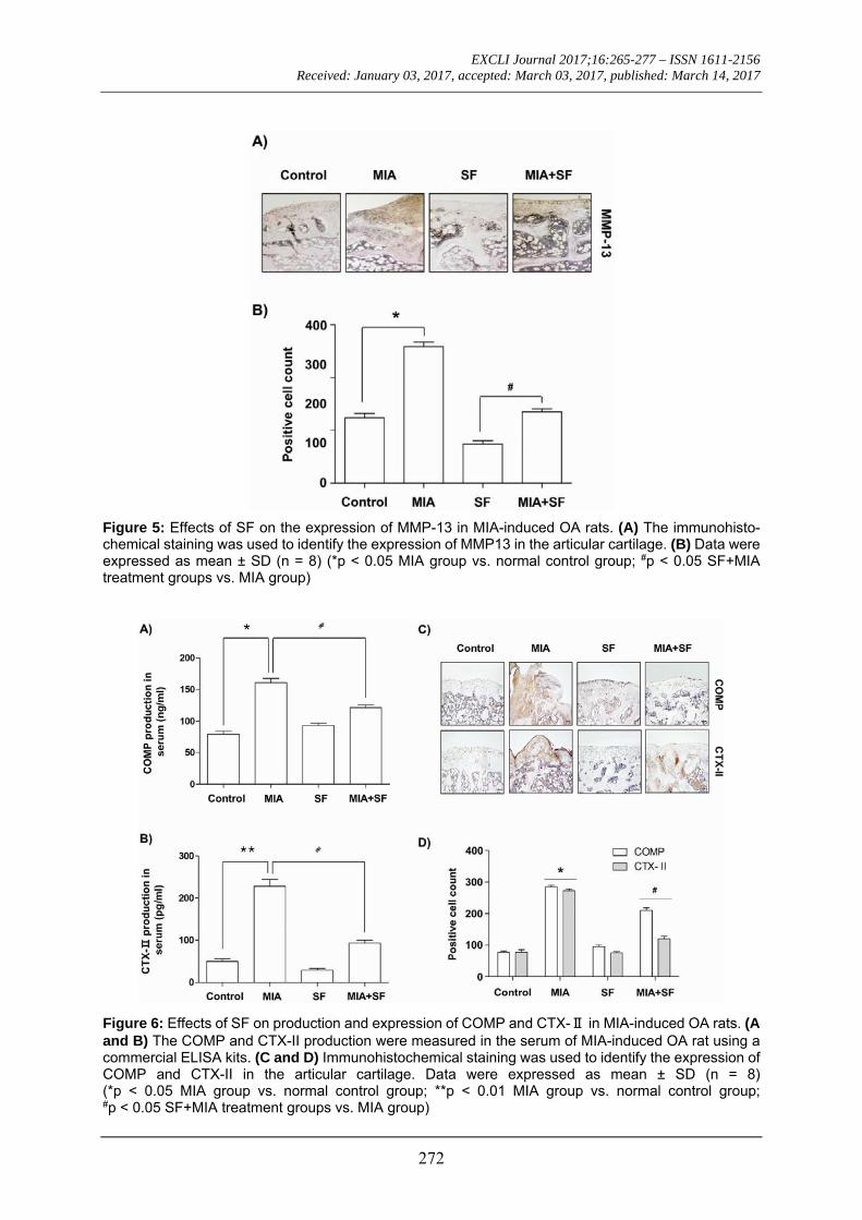
The progressive destruction of cartilage degradation in OA is caused by several ma-trix-degrading enzymes including MMPs pro-duced by the chondrocytes and synovium (Burrage et al., 2006; Troeberg and Nagase, 2012). Because, among the MMPs family, MMP-13 play a most important role in de-grading cartilage (Takaishi et al., 2008; Li et al., 2011), we examined the effects of SF on the expression of MMP-13 in MIA-stimulated OA cartilage. Our results demonstrated that the number of chondrocytes staining positive for MMP-13 was increased after MIA injec-tion, with significantly lower percentages of MMP-13-positive chondrocytes evident in the SF-administered group relative to saline-treated controls (Figure 5).
SF diminished the production of COMP and CTX-II in MIA-induced OA rats
We next investigated the effects of SF on the levels of COMP and CTX-II, well-estab-lished biomarkers for diagnosis and OA pro-gression (Goode et al., 2012; Saberi Hosnijeh et al., 2016), which play key roles during sub-sequent progressive osteophytes in multiple knee compartments. Our ELISA data indi-cated that excessive production of COMP and CTX-II were evident in the serum of MIA-in-duced OA rats; these levels were significantly diminished following SF administration (Fig-ure 6A and B). Consistent with the results of ELISA, immunohistochemistry assay showed that oral administration with SF caused a sig-nificant decrease in expression of COMP as well as CTX-II compared with that in the nor-mal control group (Figure 6C and D).
EXCLI Journal 2017;16:265-277 – ISSN 1611-2156 Received: January 03, 2017, accepted: March 03, 2017, published: March 14, 2017
272
Figure 5: Effects of SF on the expression of MMP-13 in MIA-induced OA rats. (A) The immunohisto-chemical staining was used to identify the expression of MMP13 in the articular cartilage. (B) Data were expressed as mean ± SD (n = 8) (*p < 0.05 MIA group vs. normal control group; #p < 0.05 SF+MIA treatment groups vs. MIA group)
Figure 6: Effects of SF on production and expression of COMP and CTX-Ⅱ in MIA-induced OA rats. (A and B) The COMP and CTX-II production were measured in the serum of MIA-induced OA rat using a commercial ELISA kits. (C and D) Immunohistochemical staining was used to identify the expression of COMP and CTX-II in the articular cartilage. Data were expressed as mean ± SD (n = 8) (*p < 0.05 MIA group vs. normal control group; **p < 0.01 MIA group vs. normal control group; #p < 0.05 SF+MIA treatment groups vs. MIA group)
EXCLI Journal 2017;16:265-277 – ISSN 1611-2156 Received: January 03, 2017, accepted: March 03, 2017, published: March 14, 2017
273
DISCUSSION
In this study, we investigated whether or not SF exerts a chondroprotective effect in a rat OA model by injection with MIA, which is known to induce OA through interruption of chondrocyte metabolism (Barve et al., 2007). Our data showed that oral administra-tion of SF led to a significant decrease of structural changes such as joint space narrow-ing and cartilage destruction in MIA-induced OA rats, which was associated with a reduc-tion of pro-inflammatory molecules, MMP-13 and both biomarkers of cartilage and bone metabolism such as COMP and CTX-II.
The compelling evidence demonstrated that pro-inflammatory cytokines are signifi-cantly elevated in synovial fluid from OA pa-tients and play critical roles in the promotion of the catabolic processes in OA, causing car-tilage degradation (Kapoor et al., 2011; Rah-mati et al., 2016). High levels of pro-inflam-matory cytokines have been found in synovial fluid from OA patients and several models of cartilage degradation (Goldring and Otero, 2011; Kellesarian et al., 2016). Among these cytokines, IL-1β is highly over-expressed in the cartilage as well as in the synovial tissue while the expression of IL-1Rα, a receptor an-tagonist of the IL-1 family (Jotanovic et al., 2012). This cytokine inhibits proliferation and triggers apoptosis of chondrocytes and blocks the extracellular matrix (ECM) structural compounds synthesis by activating MMPs in-cluding MMP-13 (Mabey and Honsawek, 2015; Rahmati et al., 2016). IL-6 has also been reported to act as one of the main pro-inflammatory cytokines involved in the path-ophysiology of OA. IL-6 induces destruction of joint and cartilage by stimulating the acti-vation of osteoclasts and differentiation of mesenchymal cells into chondroblasts (Doss et al., 2007; Jotanovic et al., 2012). In addi-tion, previous studies revealed that TNF-α has similar to or synergistic with IL-1β and IL-6 in the production of matrix-degrading en-zymes and inhibition of proteoglycan synthe-sis, resulting in loss of cartilage and bone re-sorption during the process of OA develop-ment (Jotanovic et al., 2012; Kellesarian et
al., 2016). In accordance with, many studies demonstrated that anti-inflammatory agents capable of inhibiting the production of those cytokines may have the potential to control or treatment of OA (Kapoor et al., 2011; Mabey and Honsawek, 2015). Hence, we investi-gated the anti-inflammatory effects of SF by measurements of the serum levels of pro-in-flammatory cytokines, such as IL-1β, IL-6, and TNF-α, in MIA-induced OA rats, and found that SF administration decreased these cytokines, the MIA increased these parame-ters. Taken together, the present results indi-cate that SF has a potential of prevention against inflammatory responses, and subse-quently might reduce the damage of articular cartilage.
In addition to the roles of inflammatory cytokines, pro-inflammatory mediators such as NO and PEG2 plays an extremely im-portant role in the development of inflamma-tion in OA (Amin et al., 2000; Rahmati et al., 2016). These pro-inflammatory mediators can induce cell death of chondrocytes and loss of cartilage matrix in the pathogenesis of OA, and they are also significantly elevated in car-tilage and synovial tissues from OA patients (Notoya et al., 2000; Park et al., 2006). More-over, the pro-inflammatory cytokines can stimulate the production of pro-inflammatory mediators, such as NO and PEG2, through ac-tivation of chondrocytes (Martel-Pelletier et al., 2006; Rahmati et al., 2016). Therefore, we next investigated the effects of SF on the re-lease of NO and PEG2 in MIA-induced OA rat model. Our results clearly demonstrated that the serum levels of NO and PEG2 were signif-icantly higher in the MIA group compared with the control group. However, SF effec-tively reduced MIA-induced elevation of NO and PEG2 production by suppressing up-stream molecules iNOS and COX-2 expres-sion, consistent with a previous our report that SF reduced production of NO and PEG2 in IL-1β-stimulated human chondrocytes (Jeong et al., 2015b) and lipopolysaccharide-activated murine macrophage (Kang et al., 2014b).
EXCLI Journal 2017;16:265-277 – ISSN 1611-2156 Received: January 03, 2017, accepted: March 03, 2017, published: March 14, 2017
274
Accumulated evidence suggested that MMPs are important metalloproteases in-volved in tissue remodeling including the turnover, catabolism, and degradation of the ECM (Burrage et al., 2006; Troeberg and Na-gase, 2012). MMPs expression could be up-regulated by pro-inflammatory cytokines in a variety of tissues and cell types, including ar-ticular chondrocytes (Goldring and Otero, 2011; Takaishi et al., 2008). Among the MMPs, MMP-13 is critical for degrading col-lagens, proteoglycans and other ECM macro-molecules in the osteoarthritic pathological process (Takaishi et al., 2008; Li et al., 2011). We then analyzed the expression of MMP-13, to assess the effects of SF on the catabolic ac-tivity of chondrocytes. Consistent with previ-ous studies (Andereya et al., 2006; Barve et al., 2007), the serum levels of MMP-13 and the percentages of MMP-13-positive chon-drocytes were significantly higher in the MIA-treated group than in the saline-treated controls. However, our results demonstrated that the SF-administrated rats had fewer MMP-13-producing cells than did the MIA-treated rats, which was connected with lower-ing MMP-13 production. These observations support the fact that SF might have a chondro-protective effect by reducing the production and activation of MMP-13.
Several clinical studies in OA patients and OA animal models demonstrated that the ele-vated levels of COMP and CTX-II are corre-lated with increased risk and progression of OA. COMP, a pentameric glycoprotein, is one of the essential components of the extra-cellular matrix of the cartilage (Goode et al., 2012; Saberi Hosnijeh et al., 2016). COMP functions as a regulator in governing the as-sembly of type II collagen fibers in cartilage, thereby this glycoprotein stabilizes the colla-gen network in cooperation with other matrix proteins (Christgau et al., 2001). However, its levels during the development of OA and un-der inflammatory condition are obviously in-creased in serum and synovial fluid and posi-tively correlated with joint damage in knee OA (Vilím et al., 2002; Verma and Dalal, 2013). In addition, CTX-II is produced by
degradation of type II collagen through the action of proteases with cartilage injury or de-generation and finally excreted in the urine (Christgau et al., 2001; Freeston et al., 2011). CTX-II contents were also elevated in OA pa-tients as compared with normal individuals, which levels are associated with both the prevalence and progression of OA (Jansen et al., 2009; Freeston et al., 2011). These obser-vations indicated that these two factors have the potential to be prognostic biomarkers for monitoring cartilage degradation in patients with OA (Goode et al., 2012; Saberi Hosnijeh et al., 2016). In our ELISA study, both serum COMP and CTX-II levels in the MIA-induced OA group were highly increased compared to the control group; however, treatment with SF significantly prevented the increase. In agree-ment with the result, the immunohistochemis-try data also showed that rats injected with MIA dramatically increased COMP and CTX-II expression in articular cartilage, and besides administration with SF significantly reduced their expression in MIA-induced OA rats. Therefore, it is possible that the reduc-tion in COMP and CTX-II serum levels by SF most likely represents suppressed MIA-in-duced degradation of cartilage, as cartilage is a major contributor to circulating COMP and CTX-II levels.
CONCLUSIONS
In conclusion, we demonstrated that ad-ministration with SF effectively attenuated the severity of articular cartilage destruction in MIA-induced OA of the knee joint in rats. To the best of our knowledge, this is the first report to demonstrate the antiarthritic effects of SF on MIA-induced OA model. The anti-arthritic effects of SF were associated with the decreased production of pro-inflammatory cytokines, such as IL-1β, IL-6 and TNF-α, and mediators including NO and PGE2
through reducing their corresponding genes expression. SF also protected the articular cartilage damage by suppression of MMP-13 and two representative biomarkers for diag-nosis of OA, COMP, and CTX-II, in the OA
EXCLI Journal 2017;16:265-277 – ISSN 1611-2156 Received: January 03, 2017, accepted: March 03, 2017, published: March 14, 2017
275
animal model induced by MIA. Based on the results of this study, we suggest that Schisan-drae Fructus has excellent potential as a ther-apeutic modality for treating OA.
Conflicts of interest
The authors declare that there is no con-flict of interest.
Acknowledgement
This work was supported by the High Value-added Food Technology Development Program (314043-3), Ministry of Agriculture, Food and Rural Affairs, Republic of Korea
REFERENCES
Amin AR, Dave M, Attur M, Abramson SB. COX-2, NO, and cartilage damage and repair. Curr Rheumatol Rep. 200;2:447-53.
Andereya S, Streich N, Schmidt-Rohlfing B, Mumme T, Müller-Rath R, Schneider U. Comparison of modern marker proteins in serum and synovial fluid in patients with advanced osteoarthrosis and rheumatoid arthritis. Rheumatol Int. 2006;26:432-8.
Bae H, Kim R, Kim Y, Lee E, Jin Kim H, Pyo Jang Y, et al. Effects of Schisandra chinensis Baillon (Schizan-draceae) on lipopolysaccharide induced lung inflam-mation in mice. J Ethnopharmacol. 2012;142:41-7.
Barve RA, Minnerly JC, Weiss DJ, Meyer DM, Aguiar DJ, Sullivan PM, et al. Transcriptional profiling and pathway analysis of monosodium iodoacetate-induced experimental osteoarthritis in rats: relevance to human disease. Osteoarthritis Cartilage. 2007;15:1190-8.
Burrage PS, Mix KS, Brinckerhoff CE. Matrix metal-loproteinases: role in arthritis. Front Biosci. 2006;11: 529-43.
Cameron M, Chrubasik S. Topical herbal therapies for treating osteoarthritis. Cochrane Database Syst Rev. 2013;31:CD010538.
Chen B, Zhan H, Chung M, Lin X, Zhang M, Pang J, et al. Chinese herbal bath therapy for the treatment of knee osteoarthritis: Meta-analysis of randomized con-trolled trials. Evid Based Complement Alternat Med. 2015;2015:949172.
Christgau S, Garnero P, Fledelius C, Moniz C, Ensig M, Gineyts E, et al. Collagen type II C-telopeptide fragments as an index of cartilage degradation. Bone. 2001;29:209-15.
Chun JN, Cho M, So I, Jeon JH. The protective effects of Schisandra chinensis fruit extract and its lignans against cardiovascular disease: a review of the molec-ular mechanisms. Fitoterapia. 2014;97:224-33.
Dhippayom T, Kongkaew C, Chaiyakunapruk N, Dilokthornsakul P, Sruamsiri R, Saokaew S, et al. A. clinical effects of Thai herbal compress: a systematic review and meta-analysis. Evid Based Complement Alternat Med. 2015;2015:942378.
Doss F, Menard J, Hauschild M, Kreutzer HJ, Mitt-lmeier T, Müller-Steinhardt M, et al. Elevated IL-6 lev-els in the synovial fluid of osteoarthritis patients stem from plasma cells. Scand J Rheumatol. 2007;36:136-9.
Fernihough J, Gentry C, Malcangio M, Fox A, Rediske J, Pellas T, et al. Pain related behaviour in two models of osteoarthritis in the rat knee. Pain. 2004;112:83-93.
Freeston JE, Garnero P, Wakefield RJ, Hensor EM, Conaghan PG, Emery P. Urinary type II collagen C-terminal peptide is associated with synovitis and pre-dicts structural bone loss in very early inflammatory arthritis. Ann Rheum Dis. 2011;70:331-3.
Goldring MB, Berenbaum F. Emerging targets in oste-oarthritis therapy. Curr Opin Pharmacol. 2015;22:51-63.
Goldring MB, Otero M. Inflammation in osteoarthritis. Curr Opin Rheumatol. 2011;23:471-8.
Goode AP, Marshall SW, Kraus VB, Renner JB, Stürmer T, Carey TS, et al. Association between serum and urine biomarkers and lumbar spine individual radi-ographic features: the Johnston County Osteoarthritis Project. Osteoarthritis Cartilage. 2012;20:1286-93.
Guilak F. Biomechanical factors in osteoarthritis. Best Pract Res Clin Rheumatol. 2011;25:815-23.
Hou PW, Fu PK, Hsu HC, Hsieh CL. Traditional Chi-nese medicine in patients with osteoarthritis of the knee. J Tradit Complement Med. 2015;5:182-96.
Jansen NW, Roosendaal G, Lundin B, Heijnen L, Mau-ser-Bunschoten E, Bijlsma JW, et al. The combination of the biomarkers urinary C-terminal telopeptide of type II collagen, serum cartilage oligomeric matrix protein, and serum chondroitin sulfate 846 reflects car-tilage damage in hemophilic arthropathy. Arthritis Rheumatol. 2009;60:290-8.
Jeong JW, Kim JW, Ku SK, Kim SG, Kim KY, Kim GY, et al. Essential oils purified from Schisandrae se-men inhibits tumor necrosis factor-α-induced matrix metalloproteinase-9 activation and migration of human aortic smooth muscle cells. BMC Complement Altern Med. 2015a;15:7.
EXCLI Journal 2017;16:265-277 – ISSN 1611-2156 Received: January 03, 2017, accepted: March 03, 2017, published: March 14, 2017
276
Jeong JW, Lee HH, Choi EO, Lee KW, Kim KY, Kim SG, et al. Schisandrae Fructus inhibits IL-1β-induced matrix metalloproteinases and inflammatory mediators production in SW1353 human chondrocytes by sup-pressing NF-κB and MAPK activation. Drug Dev Res. 2015b;76:474-83.
Jotanovic Z, Mihelic R, Sestan B, Dembic Z. Role of interleukin-1 inhibitors in osteoarthritis: an evidence-based review. Drugs Aging. 2012;29:343-58.
Kang JS, Han MH, Kim GY, Kim CM, Kim BW, Hwang HJ, et al. Nrf2-mediated HO-1 induction con-tributes to antioxidant capacity of a Schisandrae Fruc-tus ethanol extract in C2C12 myoblasts. Nutrients. 2014a;6:5667-78.
Kang YS, Han MH, Hong SH, Park C, Hwang HJ, Kim BW, et al. Anti-inflammatory effects of Schisandra chinensis (Turcz.) Baill fruit through the inactivation of nuclear factor-κB and mitogen-activated protein ki-nases signaling pathways in lipopolysaccharide-stimu-lated murine macrophages. J Cancer Prev. 2014b;19: 279-87.
Kapoor M, Martel-Pelletier J, Lajeunesse D, Pelletier JP, Fahmi H. Role of proinflammatory cytokines in the pathophysiology of osteoarthritis. Nat Rev Rheumatol. 2011;7:33-42.
Kellesarian SV, Al-Kheraif AA, Vohra F, Ghanem A, Malmstrom H, Romanos GE, et al. Cytokine profile in the synovial fluid of patients with temporomandibular joint disorders: A systematic review. Cytokine. 2016; 77:98-106.
Kim JW, Ku SK, Han MH, Kim KY, Kim SG, Kim GY, Hwang HJ, et al. The administration of Fructus Schisandrae attenuates dexamethasone-induced mus-cle atrophy in mice. Int J Mol Med. 2015a;36:29-42.
Kim JW, Ku SK, Kim KY, Kim SG, Han MH, Kim GY, et al. Schisandrae Fructus supplementation ame-liorates sciatic neurectomy-induced muscle atrophy in mice. Oxid Med Cell Longev. 2015b;2015:872428.
Kim MS, Kim JE, Yoon YS, Seo JG, Chung MJ, Yum DY. A probiotic preparation alleviates atopic dermati-tis-like skin lesions in murine models. Toxicol Res. 2016;32:149-58.
Kook M, Lee SK, Kim SD, Lee HY, Hwang JS, Choi YW, et al. Anti-septic activity of α-cubebenoate iso-lated from Schisandra chinensis. BMB Rep. 2015;48: 336-41.
Kwon DY, Kim DS, Yang HJ, Park S. The lignan-rich fractions of Fructus Schisandrae improve insulin sen-sitivity via the PPAR-γ pathways in in vitro and in vivo studies. J Ethnopharmacol. 2011;135:455-62.
Kwon HO, Lee M, Kim OK, Ha Y, Jun W, Lee J. Effect of Hijikia fusiforme extracts on degenerative osteoar-thritis in vitro and in vivo models. Nutr Res Pract. 2016; 10:265-73.
Lee TH, Jung CH, Lee DH. Neuroprotective effects of Schisandrin B against transient focal cerebral ischemia in Sprague-Dawley rats. Food Chem Toxicol. 2012;50: 4239-45.
Lee TH, Song HK, Jang JY, Kim DY, Park HK, Choi EA, et al. Anti-inflammatory effect of egg white-chal-canthite and purple bamboo salts mixture on arthritis induced by monosodium iodoacetate in Sprague-Daw-ley rats. Lab Anim Res. 2016;32:91-8.
Lepetsos P, Papavassiliou AG. ROS/oxidative stress signaling in osteoarthritis. Biochim Biophys Acta. 2016;1862:576-91.
Li NG, Shi ZH, Tang YP, Wang ZJ, Song SL, Qian LH, et al. New hope for the treatment of osteoarthritis through selective inhibition of MMP-13. Curr Med Chem. 2011;18:977-1001.
Lv XJ, Zhao LJ, Hao YQ, Su ZZ, Li JY, Du YW, et al. Schisandrin B inhibits the proliferation of human lung adenocarcinoma A549 cells by inducing cycle arrest and apoptosis. Int J Clin Exp Med. 2015;8:6926-36.
Mabey T, Honsawek S. Cytokines as biochemical markers for knee osteoarthritis. World J Orthop. 2015; 6:95-105.
Makki MS, Haqqi TM. miR-139 modulates MCPIP1/ IL-6 expression and induces apoptosis in human OA chondrocytes. Exp Mol Med. 2015;47:e189.
Mankin HJ, Dorfman H, Lippiello L, Zarins A. Bio-chemical and metabolic abnormalities in articular car-tilage from osteo-arthritic human hips. II. Correlation of morphology with biochemical and metabolic data. J Bone Joint Surg Am. 1971;53:523-37.
Martel-Pelletier J, Pelletier JP, Fahmi H. Cyclooxygen-ase-2 and prostaglandins in articular tissues. Semin Ar-thritis Rheum. 2003;33:155-67.
Martel-Pelletier J, Lajeunesse D, Fahmi H, Tardif G, Pelletier JP. New thoughts on the pathophysiology of osteoarthritis: one more step toward new therapeutic targets. Curr Rheumatol Rep. 2006;8:30-6.
Mobasheri A. The future of osteoarthritis therapeutics: targeted pharmacological therapy. Curr Rheumatol Rep. 2013;15:364.
EXCLI Journal 2017;16:265-277 – ISSN 1611-2156 Received: January 03, 2017, accepted: March 03, 2017, published: March 14, 2017
277
Notoya K, Jovanovic DV, Reboul P, Martel-Pelletier J, Mineau F, Pelletier JP. The induction of cell death in human osteoarthritis chondrocytes by nitric oxide is re-lated to the production of prostaglandin E2 via the in-duction of cyclooxygenase-2. J Immunol. 2000;165: 3402-10.
Panossian A, Wikman G. Pharmacology of Schisandra chinensis Bail.: an overview of Russian research and uses in medicine. J Ethnopharmacol. 2008;118:183-212.
Park JY, Pillinger MH, Abramson SB. Prostaglandin E2 synthesis and secretion: the role of PGE2 synthases. Clin Immunol. 2006;119:229-40.
Rahmati M, Mobasheri A, Mozafari M. Inflammatory mediators in osteoarthritis: A critical review of the state-of-the-art, current prospects, and future chal-lenges. Bone. 2016;85:81-90.
Saberi Hosnijeh F, Siebuhr AS, Uitterlinden AG, Oei EH, Hofman A, Karsdal MA, et al. Association be-tween biomarkers of tissue inflammation and progres-sion of osteoarthritis: evidence from the Rotterdam study cohort. Arthritis Res Ther. 2016;18:81.
Speziali A, Delcogliano M, Tei M, Placella G, Chillemi M, Tiribuzi R, et al Chondropenia: current concept review. Musculoskelet Surg. 2015;99:189-200.
Takaishi H, Kimura T, Dalal S, Okada Y, D'Armiento J. Joint diseases and matrix metalloproteinases: a role for MMP-13. Curr Pharm Biotechnol. 2008;9:47-54.
Troeberg L, Nagase H. Proteases involved in cartilage matrix degradation in osteoarthritis. Biochim Biophys Acta. 2012;1824:133-45.
Verma P, Dalal K. Serum cartilage oligomeric matrix protein (COMP) in knee osteoarthritis: a novel diag-nostic and prognostic biomarker. J Orthop Res. 2013; 31:999-1006.
Vilím V, Olejárová M, Machácek S, Gatterová J, Kraus VB, Pavelka K. Serum levels of cartilage oligomeric matrix protein (COMP) correlate with radiographic progression of knee osteoarthritis. Osteoarthritis Carti-lage. 2002;10:707-13.
Wat E, Ng CF, Wong EC, Koon CM, Lau CP, Cheung DW, et al. The hepatoprotective effect of the combina-tion use of Fructus Schisandrae with statin - A preclin-ical evaluation. J Ethnopharmacol. 2016;178:104-14.


![Extracts of Immature Orange (Aurantii fructus …consumed drugs. Immature orange [Aurantii fructus immaturus (IO)] and citrus unshiu peel [Citri unshiu pericarpium (CP)] are common](https://img.pdfslide.us/doc/110x75/5e5e87e454e63818f659446d/extracts-of-immature-orange-aurantii-fructus-consumed-drugs-immature-orange-aurantii.jpg)


![INDEX [botanicalink.com]botanicalink.com/SGHCatalogue.pdf · Fructus Capsici Hot Pepper/Chilli Fructus Chaenomelis Floweringquince Fruit Fructus Corni Dogwood Fruit/Asiatic cornelian](https://img.pdfslide.us/doc/110x75/5cdf42a688c9938b288e092b/index-fructus-capsici-hot-pepperchilli-fructus-chaenomelis-floweringquince.jpg)