Embed Size (px)
Citation preview
J Med Dent Sci 2010; 57:55-64
Original Article
Corresponding Author: Kenya SatoDepartment of Microbiology and Immunology, Graduate School of Health Care Sciences, Tokyo Medical and Dental University, Bunkyo-ku, Tokyo 113-8510, Japan.Tel: +81-3-5803-5368 Fax: +81-3-5689-5375E-mail: [email protected] , [email protected] September 29;Accepted November 13, 2009
ompA variants affecting the adherence of ulcerative colitis-derived Bacteroides vulgatus.
Kenya Sato1, Wakako Kumita1, Takashi Ode1, Shizuko Ichinose2, Akira Ando3, Yoshihide Fujiyama3, Toshio Chida1 and Noboru Okamura1
1) Department of Microbiology and Immunology, Graduate School of Health Care Sciences, Tokyo Medical and Dental University, Bunkyo-ku, Tokyo 113-8510, Japan.2) Research Center for Frontier Life Sciences, Tokyo Medical and Dental University, Bunkyo-ku, Tokyo 113-8510, Japan3) Department of Medicine, Shiga University of Medical Science, Seta-Tsukinowa, Otsu, Shiga 520-2192, Japan.
To investigate the adhesion factor of Bacteroides vulgatus derived from ulcerative colitis (UC), we isolated B. vulgatus strains from the large intestinal mucosa of UC patients and non-UC individuals, and examined their adherence to tissue-cultured cells. The adherence to tissue-cultured cells in UC-derived strains (36.5 ± 7.9 %) was higher than that in non-UC-derived strains (13.2 ± 7.7 %). PCR and sequencing analysis of outer membrane proteins revealed that the strains derived from five of seven (71.4 %) UC patients had ompA genes belonging to either ompA variant type A or B. The adherence rates in Escherichia coli DH5α transformants with ompA type A variants (33.3 ± 4.6 %) and type B variants (34.6 ± 7.1 %) were higher than the rate in those with non-UC ompA (16.4 ± 4.0 %). Our results suggest that B. vulgatus isolates in the mucosal flora of the large intestine of UC patients have a high frequency of ompA variants and that the variation of ompA variants is one of the factor causing an increase in the adherence of the bacterium.
Keywords: Bacteroides vulgatus, adherence, ompA
Introduction
A number of studies have examined the bacterial flora of UC patients. Among them, many recent studies have analysed mucosa-associated bacterial flora closely adhering to intestinal epithelial cells rather than common bacterial flora, since they implicate direct immunological and physiological effects on the host 1-3. In fact, mucosa-associated bacterial flora have been chosen as samples in studies investigating bacterial flora from inflammatory bowel disease patients, and in them, Bacteroides spp. have often been studied, especially Bacteroides vulgatus 4,5. Our previous study demonstrated that both the number and prevalence of B. vulgatus were higher in the large intestinal mucosa of UC patients than in those of non-UC individuals6. B. vulgatus is a numerically dominant group of bacteria among the bacterial flora, and its involvement in the onset of UC has been suggested7-9. Bacterial adherence, the basic process for the establishment of infection, is mediated by expression of potential adherence factors. Outer membrane protein A (OmpA) is a major protein of the outer membrane of Escherichia coli K-12 strains10. A previous study has shown that the ompA knockout mutant of E. coli K1 showed decreased invasion of brain microvascular endothelial cells and suggested that E. coli K1-OmpA-specific receptors on brain microvascular endothelial cel ls contributed to E. col i K1 adherence and invasion11. Simi lar ly , f indings for the ompA of Aeromonas veronii suggest that it is be involved in adherence to the carp intestinal tract12. Furthermore,
56 J Med Dent SciK. Sato et al.
the Ng-OmpA of Neisseria gonorrhoeae, which is highly similar to OmpA of E. coli K1, facilitates adherence to and invasion of bacteria in host cells13. These studies findings strongly suggest that ompA is associated with bacterial adhesion and invasion into host cells. For Bacteroides spp., ompA homologue has been also identified in Bacteroides fragilis 14. More recent study has revealed the whole genome sequence of B. vulgatus, including identification of ompA 15. However, no studies have been performed on the function of ompA of B. vulgatus. In the present study, we examined adherence to tissue-cultured cells and investigated the related factors for B. vulgatus strains in mucosa-associated bacterial flora, which had been isolated from UC patients and non-UC individuals in our previous study, by determining whether a pili structure was present or absent under an electron microscope. We also studied ompA with regard to its involvement in mucosal adhesion by comparing and analysing the ompA gene derived from UC and non-UC patients. This is the first description of a correlation between adherence and ompA in B. vulgatus.
Materials and Methods
Preparation of bacterial strains for experiments The mucosal tissue samples were obtained from patients who underwent resection of the large intestine for colon cancer or UC at the Hospital of Hyogo College of Medicine6. The protocol was approved by the Clinical Research Committee of Hyogo College of Medicine. Mucosal tissue of an area apparently free of cancer and inflamed mucosal tissue that had been removed surgically from 10 non-UC (colon cancer) individuals and seven UC patients, respectively, were washed and then the mucosa were detached. Bacteria strains were isolated from the detached mucosa according to previously described methods6 and stored at -80 ℃ in 20 % skim milk. Some colonies were obtained from each patient, and a total of 75 strains of B. vulgatus of each colony origin were randomly selected and used for our investigations. The B. vulgatus ATCC 8482 strain was used as a control. The strains were cultured in GAM agar (Nissui) at 36 ℃ under an anaerobic condition for 48 h, and one of the colonies obtained was inoculated into brain heart infusion broth (BHI broth; Becton Dickinson), followed by stationary culture at 36 ℃ under an anaerobic condition for 48 h. The whole genome for gene manipulation was extracted from a 2-day culture
suspension using the Bact/Yeast kit (Qiagen) according to the manufacturer’s instructions. Escherichia coli DH5α strain (Takara Bio) was used as a cloning host.
Preparation of tissue-cultured cells Caco-2 cells, a human colon cancer-derived tissue-cultured cell line, were cultured in Dulbecco’s modified Eagle’s medium (D-MEM; Invitrogen) supplemented with 1 % non-essential amino acid (Invitrogen) and 10 % fetal bovine serum (FBS; Hyclone). The monolayer of the cells was prepared by treating the cells with 0.25 % trypsin (Invitrogen/GIBCO) to prepare a 3.0×10³ cells ml -1 suspension in D-MEM, transferring the suspension into a 24-well t issue culture plate (Falcon; BD Biosciences) at 1 ml per well, and culturing at 36 ℃ under 10 % CO2 for 14 days. For the adherence assay, the cell surface was washed three times with 10 mM phosphate-buffered saline (PBS) prior to the assay.
Adherence assay The adherence assay was carried out by a modified version of the method described previously16.Preparation: Bacterial suspension was prepared by di lut ing the BHI broth with PBS to adjust the concentration to 1×10⁷ cells ml -1, washing three times and resuspending in D-MEM supplemented with 10 % fetal calf serum. The bacterial suspension was plated into a 24-well tissue culture plate at 1 ml per well, centrifuged at 210 g and incubated at 36 ℃ under an anaerobic condition for 2 h. After incubation, each well of the plate was washed three times with PBS to remove bacteria not adhering to the surface of the cells. Counting adherent bacteria under light microscope: The specimens used for the adherence assay were fixed with methanol for 20 min and then stained with 20 % Giemsa stain solution (Wako Chemical) at 37 ℃ for 20 min. The stained specimens were dehydrated with acetone, cleared in xylene, and examined under light microscope. The number of adherent bacteria on monolayered cells in one microscopic field at 1000-fold magnification (mean number of Caco-2 cells: 124 ± 12.1 cells) was counted. Ten microscopic fields were randomly selected. For convenience of determining differences in the number of adherent bacteria, results were evaluated according to three categories of number of adherent bacteria per microscopic field at 1000-fold magnification: one plus (<50), two plus (50-100), and three plus (>100). Counting viable adherent bacteria: Following the adherence assay, a few drops of 0.3 % trypsin were

57ompA variants and adherence of Bacteroides vulgatus.
added to each well of the 24-well tissue culture plate to detach and separate cells. The cells were then serially diluted in PBS, spread on GAM agar, cultured at 37 ℃ overnight and subjected to viable counting. The percentage of the number of bacteria obtained after the adherence assay relative to that of bacteria added to the wells was calculated as the adherence rate. The adherence assay was usually performed three times. The significance of differences was statistically assessed using the Student’s t test.
Transmission electron microscopy (TEM) TEM was performed for five strains of B. vulgatus randomly selected from non-UC-derived strains and for all UC-derived strains. A 2-day culture in BHI broth was centrifuged at 8000 g for 5 min and the pellet obtained was washed three times with PBS. The bacterial suspension was fixed with 2.5 % glutaraldehyde (EM science) for 2 h, then fixed with 1 % osmium for 2 h, dehydrated and embedded in Epon resin. The Epon-embedded samples were cut into thin sections using an
ultramicrotome and examined closely under the transmission electron microscopy (H-7100; Hitachi).
DNA sequencing of ompA Bacterial whole genomes were extracted from all of the isolates. The ompA gene was amplified using gene-specific primers designed on the basis of ompA sequence (DDBJ/EMBL/Genbank database accession number NC009614, gene position 5000850-5001944, gene ID BVU_4065) using the primer 3 programme available at http://frodo.wi.mit.edu/cgi-bin/primer3/primer3_www.cgi (Table 1, Fig. 1). Polymerase chain reaction (PCR) was carried out using Premix Taq enzyme (Takara Bio) as recommended by the manufacturer. PCR products were sequenced using a BigDye Terminator v3.1 Cycle Sequence Kit (Applied Biosystems) and an automated DNA sequencing system (ABI PRISM 3130xl, Applied Biosystems). A similarity search for the deduced amino acid sequences against DDBJ/EMBL/GenBank sequence databases was conducted using the BLAST programme at the DNA
Name Sequence (5'-3') * or characteristic Use
Primers
Outer-up8 ATGAAAAATCAGAGCTCTTCCAA Sequence
Inner-dn1 CTGCAGCGCTAGGAACTTCT Sequence
Inner-up1 AATGGGCTATCGATGTTGAA Sequence
Outer-dn1 GGAAATACGCTTTGGGTTGA Sequence
Link-up ATGGAATTCATGAAATATAAATTTTTAAC RT-PCR, cloning
Link-up-typeA ATGGAATTCATGAAATATAAATTCTTAAC RT-PCR, cloning
Link-up-typeB ATGGAATTCATGAAATATAAAGTATTAAC RT-PCR, cloning
Link-dn TTAGGATCCTTATTTTACTTCCAGAATAA RT-PCR, cloning
Link-dn-typeA.B TTAGGATCCTTATTTTACTTCCAAGATAA RT-PCR, cloning
Vul-16s-up AACCTGCCGTCTACTCTT RT-PCR
Vul-16s-dn CAACTGACTTAAACATCCAT RT-PCR
Plasmids
pUC18 E. coli cloning vector, source of LacZ and MCS, Amp r.
pUC18-non-UC ompA gene from non-UC (ATCC) B. vulgatus cloned into pUC18 BamHI/EcoRI sites. Transformation
pUC18-type A ompA gene from type A B. vulgatus cloned into pUC18 BamHI/EcoRI sites. Transformation
pUC18-type B ompA gene from type B B. vulgatus cloned into pUC18 BamHI/EcoRI sites. Transformation
* Underlined sequences indicate the linker positions including restriction sites of BamHI (GGATCC) and EcoRI (GAATTC). Boldface: point mutation in ompA.
Table 1. Primers and plasmids used in this study.
58 J Med Dent SciK. Sato et al.
Databank of Japan (Shizuoka, Japan). Alignment of amino acid sequence was carried out using the CLUSTAL W2 multiple sequence alignment programme available at http://www.ebi.ac.uk/Tools/clustalw2/.
Analysis of ompA gene expression by RT-PCR The transcription of ompA was analysed with reverse transcriptase-PCR (RT-PCR). Based on the viable count results, one strain, showing the median of adherence rate, was chosen as the sample from each case (TMD1, 2, 17, 27, 34, 37, 44, 56). Mid-logarithmic phase cultures in BHI broth were pelleted by centrifugation at 13000 g, and RNA was isolated using ISOGEN (Nippon gene) according to the manufacturer’s instructions. Total RNA(1 µg)was reverse transcribed into single-stranded cDNA using SuperScript Ⅲ First-Strand Synthesis Supermix Kit (Invitrogen) according to the manufacturer’s instructions. PCR amplification of cDNA was performed using ompA gene-specific primer sets with an annealing temperature of 55 ℃ (Table 1, Fig. 1). The number of PCR cycles was 21, and constitutive expression of 16S rRNA assessed from the same cDNA preparation was used as a standard. PCR product was separated by electrophoresis in 1.5 % agarose and visualised by ethidium bromide staining. The RT-PCR experiment was repeated at least twice.
Adherence assay of transformants A linker primer containing the restriction sites for BamHI and EcoRI (Takara Bio) was prepared for cloning of the ompA gene into the same restriction sites of pUC18 vector (Takara Bio) (Table 1, Fig. 1). Whole genomes were used as the samples for the cloning. PCR was performed using the Premix Taq enzyme with an annealing temperature of 55 ℃. After the samples were subjected to electrophoresis, bands were cut out of the gel and purified with the QIA quick Gel extraction
Kit (Qiagen). The purified products were then digested with BamHI and EcoRI. The pUC18 vector was treated with the same enzyme used for the insert fragments and ligated using the Ligation mix (Takara Bio) according to the manufacturer’s instructions. The ligated plasmid was transformed into E. coli DH5α (Takara Bio) and transformants were selected on LB agar plates (Merk) supplemented with 50µg ampicillin ml -1. The selected strains were cultured in LB broth containing 50µg ampicil l in ml -1, and a bacterial suspension diluted to 1×10 7 CFU ml -1 was used for adherence assay. Adherence assay was performed at three times, and the significance of differences was assessed using the Student’s t test.
Results
Adherence assay For B. vulgatus isolated from non-UC individual and UC patients and those of the ATCC 8482 strain adhering to Caco-2 cells, the number of bacteria showing Giemsa staining was counted by screening based on light microscopy (Fig 2a, b). The number of bacteria of the UC-derived strains observed on the Caco-2 cells was greater than that of the non-UC strains. The strains isolated from non-UC controls adhered to Caco-2 cells at category levels one plus and two plus, whereas the strains isolated from the UC patients adhered at category levels two plus and three plus (Table 2). The adherence rate indicated the number of the bacteria that were adherent after washing, and was calculated based on the initial number of bacteria (100%) added to the Caco-2. The adherence rate based on viable count showed that the adhesion of B. vulgatus strains isolated from UC patients (36.5 ± 7.9%) was significantly greater than those derived from non-UC
Sequence primers
5’ 3’
Cloning and RT-PCR primers BamHI EcoRI
Outer-dn1
Link-up Link-dn
Inner-up1
Outer-up8
960bp1094bp333bp44bp
848bp
hypothetical protein OmpA conserved hypothetical protein
Inner-dn1
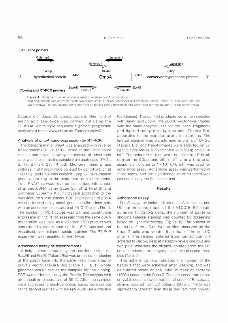
Figure 1 : Schema of primer positions used to analyse ompA in this study.DNA sequencing was performed with two primer sets; Outer-up8 and Inner-dn1 set (black arrow), Inner-up1 and Outer-dn1 set (white arrow). Link-up having BamHI and Link-dn having EcoRI restriction site were used for cloning and RT-PCR (grey arrow).

59ompA variants and adherence of Bacteroides vulgatus.
strains from non-UC and UC patients are shown (Fig. 2c - e). There were some differences in appearance, in particular in the outer surfaces of the UC-derived strains which appeared to be rough and wavy compared to those of strains isolated from the non-UC controls.
Characterisation of ompA The nucleotide sequences of the ompA gene in all of the non-UC-derived strains and in all of the strains derived from two UC patients (TMD48 and TMD73) were identical to that of B. vulgatus ATCC 8482 strain (Table 2). On the other hand, ompA in B. vulgatus strains derived from five out of seven UC patients
individuals (13.2 ± 7.7%) (Table 3).
Morphological observation by TEM TEM morphological findings for the representative
Case (patients) Strain no. * Disease Adherence category † Adherence rate (%) ‡ ompA variant
TMD1 1 Colon cancer + 6.8 ± 2.1 -TMD2 2 Colon cancer ++ 27.7 ± 9.5 -TMD4 3, 5
Colon cancer+ 11.8 ± 7.9 -
4 ++TMD7 6, 7 Colon cancer + 3.1 ± 1.8 -TMD9 8 - 11 Colon cancer + 2.4 ± 2.0 -TMD12 12 -14
Colon cancer+
16.6 ± 11.2-
15 ++TMD17 16
Colon cancer++
19.2 ± 10.8-
17 +TMD19 18 - 20 Colon cancer + 13.7 ± 5.3 -TMD21 21 Colon cancer + 17.5 ± 3.9 -TMD22 22 Colon cancer + 13.3 ± 5.1 -TMD27 23, 27
24 -26, 28Ulcerative colitis
++46.0 ± 13.9 A
+++TMD34 29 - 38 Ulcerative colitis +++ 40.6 ± 14.4 BTMD37 39 - 43 Ulcerative colitis ++ 29.9 ± 7.2 ATMD44 44 - 49 Ulcerative colitis +++ 45.5 ± 9.2 BTMD48 50 - 55 Ulcerative colitis ++ 36.1 ± 7.2 -
TMD5656, 59, 70
57, 58, 60 - 69Ulcerative colitis
++25.6 ± 17.2 A
+++TMD73 71 - 75 Ulcerative colitis ++ 31.5 ± 10.5 -
B. vulgatus ATCC 8482 + 9.1 ± 2.5 -
*The number of B. vulgatus isolated from each case.† The number of the adherent bacteria per 10 view randomly selected under light microscopy was averaged, and classified according to category; + (<50) ,++ (50-100), +++ (>100).‡ Mean±SD calculated followed by the experiment performed three times.
Adherence rate (%)*
Non-UC (n = 10) † UC (n = 7) † p ‡
13.2 ± 7.7 36.5 ± 7.9 < 0.01
* Mean±SD. Experiments were done three times.† The number of case.‡ Student’s t test.
Table 2. Profiles of B. vulgatus strains.
Table 3. Adherence of B. vulgatus strains.

60 J Med Dent SciK. Sato et al.
Figure 2 : Adherence to Caco-2 and TEM-based analysis of B. vulgatus.Part of one microscopic field at 1000-fold magnification examined under light microscopy. (a) UC-derived strain isolated from TMD34 belonging to three plus of adherence category. (b) Non-UC-derived strain isolated from TMD1 belonging to one plus of adherence category. TEM-based analysis of B. vulgatus ATCC 8482 (c), non-UC-derived strain isolated from TMD1 (d) and UC-derived strain isolated from TMD34 (e).
61ompA variants and adherence of Bacteroides vulgatus.
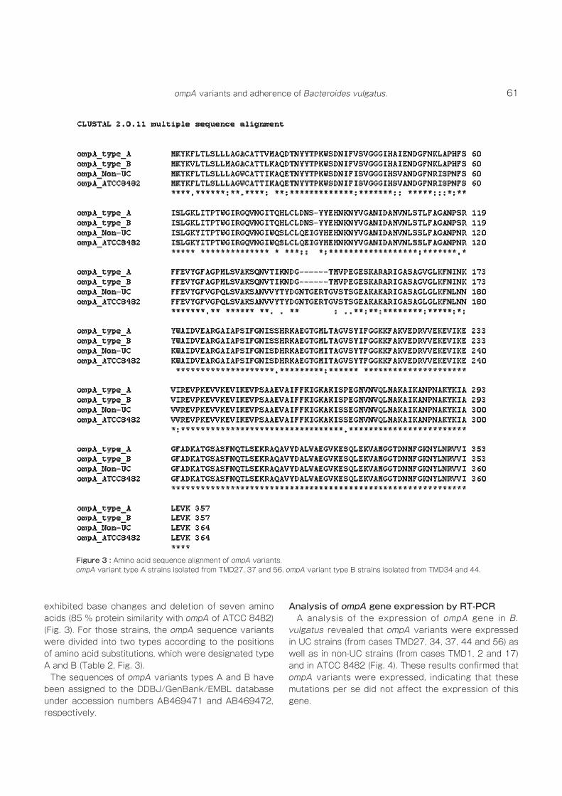
exhibited base changes and deletion of seven amino acids (85 % protein similarity with ompA of ATCC 8482) (Fig. 3). For those strains, the ompA sequence variants were divided into two types according to the positions of amino acid substitutions, which were designated type A and B (Table 2, Fig. 3). The sequences of ompA variants types A and B have been assigned to the DDBJ/GenBank/EMBL database under accession numbers AB469471 and AB469472, respectively.
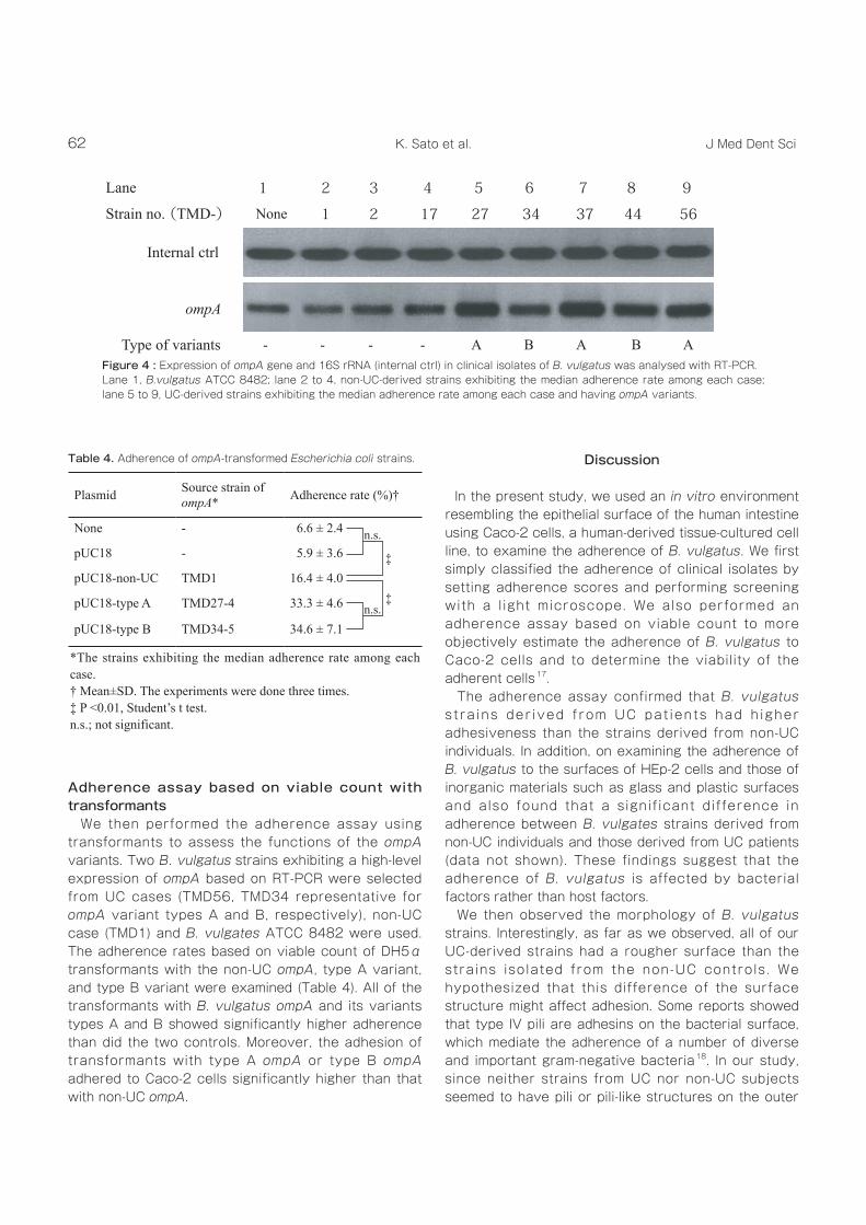
Analysis of ompA gene expression by RT-PCR A analysis of the expression of ompA gene in B. vulgatus revealed that ompA variants were expressed in UC strains (from cases TMD27, 34, 37, 44 and 56) as well as in non-UC strains (from cases TMD1, 2 and 17) and in ATCC 8482 (Fig. 4). These results confirmed that ompA variants were expressed, indicating that these mutations per se did not affect the expression of this gene.
Figure 3 : Amino acid sequence alignment of ompA variants.ompA variant type A strains isolated from TMD27, 37 and 56. ompA variant type B strains isolated from TMD34 and 44.
62 J Med Dent SciK. Sato et al.
Adherence assay based on viable count with transformants We then performed the adherence assay using transformants to assess the functions of the ompA variants. Two B. vulgatus strains exhibiting a high-level expression of ompA based on RT-PCR were selected from UC cases (TMD56, TMD34 representative for ompA variant types A and B, respectively), non-UC case (TMD1) and B. vulgates ATCC 8482 were used. The adherence rates based on viable count of DH5α transformants with the non-UC ompA, type A variant, and type B variant were examined (Table 4). All of the transformants with B. vulgatus ompA and its variants types A and B showed significantly higher adherence than did the two controls. Moreover, the adhesion of transformants with type A ompA or type B ompA adhered to Caco-2 cells significantly higher than that with non-UC ompA.
Discussion
In the present study, we used an in vitro environment resembling the epithelial surface of the human intestine using Caco-2 cells, a human-derived tissue-cultured cell line, to examine the adherence of B. vulgatus. We first simply classified the adherence of clinical isolates by setting adherence scores and performing screening with a l ight microscope. We also performed an adherence assay based on viable count to more objectively estimate the adherence of B. vulgatus to Caco-2 cells and to determine the viability of the adherent cells17. The adherence assay confirmed that B. vulgatus s t ra ins der ived f rom UC pat ients had h igher adhesiveness than the strains derived from non-UC individuals. In addition, on examining the adherence of B. vulgatus to the surfaces of HEp-2 cells and those of inorganic materials such as glass and plastic surfaces and also found that a signif icant difference in adherence between B. vulgates strains derived from non-UC individuals and those derived from UC patients (data not shown). These findings suggest that the adherence of B. vulgatus is affected by bacterial factors rather than host factors. We then observed the morphology of B. vulgatus strains. Interestingly, as far as we observed, all of our UC-derived strains had a rougher surface than the strains isolated from the non-UC controls. We hypothesized that this difference of the surface structure might affect adhesion. Some reports showed that type IV pili are adhesins on the bacterial surface, which mediate the adherence of a number of diverse and important gram-negative bacteria18. In our study, since neither strains from UC nor non-UC subjects seemed to have pili or pili-like structures on the outer
Lane 1 2 3 4 5 6 7 8 9
Strain no. (TMD-) None 1 2 17 27 34 37 44 56
Internal ctrl
ompA
Type of variants - - - - A B A B A
Plasmid Source strain of ompA* Adherence rate (%)†
None - 6.6 ± 2.4
pUC18 - 5.9 ± 3.6
pUC18-non-UC TMD1 16.4 ± 4.0
pUC18-type A TMD27-4 33.3 ± 4.6
pUC18-type B TMD34-5 34.6 ± 7.1
*The strains exhibiting the median adherence rate among each case.† Mean±SD. The experiments were done three times.‡ P <0.01, Student’s t test.n.s.; not significant.
Figure 4 : Expression of ompA gene and 16S rRNA (internal ctrl) in clinical isolates of B. vulgatus was analysed with RT-PCR. Lane 1, B.vulgatus ATCC 8482; lane 2 to 4, non-UC-derived strains exhibiting the median adherence rate among each case; lane 5 to 9, UC-derived strains exhibiting the median adherence rate among each case and having ompA variants.
Table 4. Adherence of ompA-transformed Escherichia coli strains.
n.s.
n.s.
‡
‡
63ompA variants and adherence of Bacteroides vulgatus.
surface under TEM, we hypothesized that they had another adhesion factor. ompA is a major factor in the adherence of bateria to the host cell and their invasion into it and although the identification of ompA has been conducted, no studies have investigated a correlation between adherence and ompA in B. vulgatus. In B.vulgatus ATCC 8482, two major outer membrane proteins, BVU_0414 ompA and BVU_4065 ompA have been reported to date. We analyzed the sequences of these two ompA genes and found that all of our isolates had no mutations in the BVU_0414 locus (data not shown). On the other hand, five of seven UC-derived strains (excepting TMD48 and 73) had amino acid substitutions and deletions in ompA (BVU_4065) as compared with B.vulgatus ATCC 8482, and these strains had high adherence rates. The non-UC strains had no mutat ion in ompA . These f indings and differences of ompA variation in B. vulgatus have not yet been reported elsewhere. In addition, to our knowledge, there has been no mention of their relevance to adherence knowledge. An issue that arose in our study was that two UC-derived strains (TMD48 and TMD73) which had a non-variant ompA also showed high-level adherence rates based on the viable count (Table 2). However, we were unable to clarify the causal factor(s), suggesting that other adherence mechanisms were present in TMD48 and 73. Furthermore, some studies have noted that a rough outer membrane is associated with partial deletion of ompA in Porphyromonas gingivalis 19. Although we were unable to make an association between the rough outer membrane observed in TEM for ompA variants, there could be relationship like the one in P. gingivalis in B. vulgatus. As shown in Fig. 4, RT-PCR results for several strains performed in parallel also confirmed the expression of ompA variants, indicating that these mutations do not affect expression. Interestingly, the UC-derived strains had a greater quantity of ompA expression than all of the strains derived from non-UC (data not shown). A l though we d id no t per fo rm a quant i ta t i ve experimental investigation by real-time PCR or other means, these results suggest the possibility that quantity of ompA expression influences adhesion in B. vulgatus. We also briefly performed an adherence assay using transformants to investigate the functions of the ompA variants, in which transformants with ompA variants exhibited greatly increased adhesiveness, and transformants with non-variant ompA showed only a
small increase in adhesiveness. Additionally, in RT-PCR, there were no signif icant differences in ompA expression among transformants (data not shown). In this regard, ompA of E. coli has been shown to be associated with adherence to Caco-2 20. Additionally, ompA expression of each transformants showed no significant differences by RT-PCR (data not shown). Our findings for ompA suggest that it was associated with adherence of in several of our B. vulgatus strains and its mutation might be one of the factors to involve in a further increase in adherence. In this study, we demonstrated the adherence of our isolates using artificially cultured epithelial cells, and the number of subjects used for it was rather small. Further, our data showed that the ompA sequence types were identical among strains isolated from the same specimens. Therefore, the B. vulgatus strains, isolated from the same patients, might be from the identical clones. In addition, since all the UC-derived stains of B. vulgatus used in the present study were derived from patients diagnosed with moderate pancolitis-type UC, only one of the various types of UC, it is possible that the nature of these strains could be substantially different from that of strains derived from patients with other types of UC. Therefore, in the future studies, we will need to have much more samples from various types of UC and investigate the relationship between the ompA and adherence of B. vulgatus strains in vivo. We should also perform a detailed analysis of proteins produced by ompA variants and do one for other genes associated with ompA variants. In conclusion, our study revealed that B. vulgatus, which had been predominantly on the mucosa of UC patients, adhered to Caco-2 cells more efficiently and had a higher frequency of ompA variants. E. coli transformants carrying ompA variants also showed greater adherence. Our results therefore indicate the possibility that ompA genes are an adherence factor, although there may be others, and mutation of ompA might be cause higher number of colonized B. vulgatus on the mucosa of UC patients.
Acknowledgement
We thank Ikue Tsuru and Hiroyuki Abe for valuable comments on this study. We did not receive any financial support from third parties.

64 J Med Dent SciK. Sato et al.
References1. Fiocchi, C. (1998). Inflammatory bowel disease: Etiology
and pathogenesis. Gastroenterology 115; 182-205.2. Sartor, R. B. (1997). The influence of normal microbial
f lo ra on the deve lopment o f chron ic mucosa l inflammation. Res Immunol 148; 567-576.
3. Zhang, L. & Mitchell, H. (2006). The roles of mucus-associated bacteria in inflammatory bowel disease. Drugs Today 42; 605-616.
4. Conte, M. P., Schippa, S., Zamboni, I. & other authors (2006). Gut-associated bacterial microbiota in paediatric patients with inflammatory bowel disease. Gut 55; 1760-1767.
5. Lucke, K., Miehlke, S., Jacobs, E. & Schuppler, M. (2006). Prevalence of Bacteroides and Prevotella spp. in ulcerative colitis. J Med Microbiol 55; 617-624.
6. Okamura, N., Chida, T., Baba, C., Kobayashi, K., Okamura, T., Matsumura, T., Shimoyama, T. (2002). Mucosa-associated microbial flora of the large intestine from the patients with ulcerative colitis: A preliminary report. In Helicobacter meets inflammatory bowel disease, pp. 445-451. Edited by Shimoyama, T., Axon, A., Lee, A., Podolsky, D. K. & O’Morain, C. Tokyo: Axel Springer Japan Publishing.
7. Onderdonk, A. B., Franklin, M. L. & Cisneros, R. L. (1981). Production of experimental ulcerative col it is in gnotobiotic guinea pigs with simplified microflora. Infect Immun 32; 225-231.
8. Onderdonk, A. B., Cisneros, R. L. & Bronson, R. T. (1983). Enhancement of experimental ulcerative colitis by immunization with Bacteroides vulgatus. Infect Immun 42; 783-788.
9. Onderdonk, A. B., Bronson, R. & Cisneros, R. (1987). Comparison of Bacteroides vulgatus strains in the enhancement of experimental ulcerative colitis. Infect Immun 55; 835-836.
10. Chai, T.-J., and Foulds, J. (1977). Purification of protein A, an outer membrane component missing in Escherichia coli K-12 ompA mutants. Biochim Biophys Acta 493; 210–215.
11. Khan, N. A., Shin, S., Chung, J. W., Kim, K. J., Elliott, S., Wang, Y. & Kim, K. S. (2003). Outer membrane protein A
and cytotoxic necrotizing factor-1 use diverse signaling mechanisms for Escherichia coli K1 invasion of human brain microvascular endothelial cells. Microb Pathog 35; 35-42.
12. Namba, A., Mano, N., Takano, H., Beppu, T., Ueda, K. & Hirose, H. (2008). OmpA is an adhesion factor of Aeromonas veroni i , an optimist ic pathogen that habituates in carp intestinal tract. J Appl Microbiol 105; 1441-1451.
13. Serino, L., Nesta, B., Leuzzi, R. & other authors (2007). Identification of a new OmpA-like protein in Neisseria gonorrhoeae involved in the binding to human epithelial cells and in vivo colonization. Mol Microbiol 64; 1391-1403.
14. Wexler, H. M., Read, E. K. & Tomzynski, T. J. (2002). Identification of an OmpA protein from Bacteroides fragil is: ompA gene sequence, OmpA amino acid sequence and predictions of protein structure. Anaerobe 8; 180-191.
15. Xu, J., Mahowald, M. A., Ley, R. E. & other authors (2007). Evolution of symbiotic bacteria in the distal human intestine. Plos Biology 5; 1574-1586.
16. Sasaki, M., Sitaraman, S., Babbin, B., & other authors (2007) Invasive Escherichia coli are a feature of Crohn’s disease. Lab Invest 87; 1042–1054.
17. Hauri, H. P., Sterchi, E. E., Bienz, D., Fransen, J. A. M. & Marxer, A. (1985). Expression and intracellular transport of microvillus membrane hydrolases in human intestinal epithelial cells. J Cell Biol 101; 838-851.
18. Donnenberg, M. S., Kaper, J. B. & Finlay, B. B. (1997). Interactions between enteropathogenic Escherichia coli and host epithelial cells. Trends Microbiol 5; 109-114.
19. Iwami, J., Murakami, Y., Nagano, K., Nakamura, H. & Yoshimura, F. (2007). Further evidence that major outer m e m b r a n e p r o t e i n s h o m o l o g o u s t o O m p A i n Porphyromonas gingivalis stabilize bacterial cells. Oral Microbiol Immunol 22; 356-360.
20. Torres, A. G. & Kaper, J. B. (2003). Multiple elements controlling adherence of enterohemorrhagic Escherichia coli O157 : H7 to HeLa cells. Infect Immun 71; 4985-4995.