Embed Size (px)
Citation preview
Diabetic mice exhibited a peculiar alteration in bodycomposition with exaggerated ectopic fat depositionafter muscle injury due to anomalous celldifferentiation
Masaki Mogi1*, Katsuhiko Kohara2, Hirotomo Nakaoka1, Harumi Kan-no1, Kana Tsukuda1, Xiao-Li Wang1,Toshiyuki Chisaka1,3, Hui-Yu Bai1, Bao-Shuai Shan1, Masayoshi Kukida1,4, Jun Iwanami1, Tetsuro Miki2
& Masatsugu Horiuchi1
1Department of Molecular Cardiovascular Biology and Pharmacology, Ehime University, Graduate School of Medicine, Tohon, Ehime, 791-0295, Japan; 2Department ofNeurology and Geriatric Medicine, Ehime University, Graduate School of Medicine, Tohon, Ehime, 791-0295, Japan; 3Department of Pediatrics, Ehime University, GraduateSchool of Medicine, Tohon, Ehime, 791-0295, Japan; 4Department of Cardiology, Pulmonology, Hypertension and Nephrology, Ehime University, Graduate School ofMedicine, Tohon, Ehime, 791-0295, Japan
Abstract
Background Sarcopenic obesity, age-related muscle loss, which is compensated by an increase in fat mass, impairs quality oflife in elderly people. Although the increase in intramuscular fat is associated with decreased insulin sensitivity and increasedmetabolic risk factors, the origin of diabetes-associated intramuscular fat has not been elucidated. Here, we investigated in-tramuscular fat deposition using a muscle injury model in type 2 diabetic mice.
Methods Male 8-week-old C57BL/6 and 8-week-old and 26-week-old KKAy underwent intramuscular injection of cardiotoxin(Ctx) (100μL/10μM) into the tibialis anterior (TA) muscles. After 2weeks, the muscles were removed and evaluated.
Results KKAy exhibited impaired muscle regeneration and ectopic fat deposition. Such impairment was more marked inolder KKAy. These changes were also observed in another diabetic mouse model, db/db and diet-induced obese mice butnot in streptozocin-induced diabetic mice. Deposited fat was platelet-derived growth factor (PDGF) receptor alpha positiveand its cytoskeleton was stained with Masson’s trichrome, indicating it to be of fibro-adipocyte progenitor cell origin. Expres-sion of a myogenic marker, myoD, was lower and that of PDGF receptor alpha and CCAAT/enhancer binding protein (CEBP)alpha was higher in Ctx-injured TA of KKAy compared with that of C57BL/6. Peroxisome proliferator-activated receptorγ (PPARγ) was highly expressed in fat-forming lesions in older KKAy. Treatment with all-trans retinoic acid prevented the forma-tion of intramuscular fat; however, treatment with GW9662, a PPARγ antagonist, increased the fibrotic change in muscle.
Conclusions Diabetic mice showed impaired muscle regeneration with fat deposition, suggesting that diabetes may enhancesarcopenic obesity through a mechanism involving anomalous fibro-adipocyte progenitor cell differentiation.
Keywords Diabetes mellitus; Muscle regeneration; Ectopic fat deposition; Fibro-adipocyte progenitor cell; Sarcopenic obesity
Received: 19 November 2014; Revised: 4 April 2015; Accepted: 23 April 2015*Correspondence to: Masaki Mogi, Department of Molecular Cardiovascular Biology and Pharmacology, Ehime University, Graduate School of Medicine, Shitsukawa, Tohon,Ehime 791-0295, Japan: Tel: + 81-89-960-5249, Fax: + 81-89-960-5251, Email: [email protected]
Introduction
Sarcopenia is the degenerative loss of skeletal muscle mass,which is related to frailty and the geriatric syndrome1 and im-pairs quality of life in elderly people.2,3 On the other hand,
sarcopenic obesity (SO) has been defined as a combinationof excess weight gain and reduced muscle mass and/orstrength.4 SO is also highlighted and reported to worsencardio-metabolic outcome.5,6 Interestingly, SO is modestly as-sociated with increased risk in cardiovascular disease (CVD),
OR IG INAL ART ICLE
© 2015 The Authors. Journal of Cachexia, Sarcopenia and Muscle published by John Wiley & Sons Ltd on behalf of the Society of Sarcopenia, Cachexia and Wasting Disorders
Journal of Cachexia, Sarcopenia and Muscle (2015)Published online in Wiley Online Library (wileyonlinelibrary.com) DOI: 10.1002/jcsm.12044
This is an open access article under the terms of the Creative Commons Attribution-NonCommercial-NoDerivs License, which permits use and distribution in any medium,provided the original work is properly cited, the use is non-commercial and no modifications or adaptations are made.
while sarcopenia or obesity alone is not sufficient to increasethe risk of CVD.7 Moreover, SO is independently associatedwith and precedes the onset of disability in instrumentalactivities of daily living, more than in lean sarcopenia ornon-sarcopenic obesity, in the community-dwelling elderlypopulation.8 Recently, we observed that one-leg standingtime and sway are significantly impaired in subjects withsarcopenic visceral obesity compared with those in subjectswith sarcopenia or visceral obesity alone.9 These results indi-cate that SO is a high risk for impairment of quality of life inelderly people.
Certain conditions have strong potential to coexist withsarcopenia to accelerate the progression of muscle atrophyin elderly people.10 The prevalence of sarcopenia is higherin patients with type 2 diabetes mellitus (T2DM) than innon-diabetic subjects.11 DM may accelerate the developmentof age-associated changes in body composition through anumber of mechanisms. T2DM is associated with excessiveloss of skeletal muscle and increased trunk fat mass incommunity-dwelling older adults.12 Although multiple mech-anisms, such as oxidative injury, subclinical inflammation andinsulin resistance, have been proposed to be involved in ac-celeration of sarcopenia in diabetic patients, the detailedmechanism is not yet well understood.
Excessive intramuscular fat (IMF) in skeletal muscle is ob-served in T2DM patients13 and is associated with decreasedinsulin sensitivity,14–17 indicating that an increase in IMFcauses a vicious cycle of glucose metabolism. Recently, IMFhas been shown to be also associated with a wide range ofmetabolic risk factors such as dysglycemia, dyslipidemia andhypertension.18 These results indicate that prevention of ex-cessive IMF may ameliorate sarcopenic obesity and contributeto the improvement of the quality of life in T2DM patients.However, the detailed mechanism of diabetes-inducedsarcopenia and the correlation between muscle atrophy andexcessive IMF in diabetic patients are enigmas. Here, we in-vestigated the possible mechanism of intramuscular fat depo-sition using a muscle injury model in diabetic mice, KKAy.
Materials and methods
This study was performed in accordance with the NationalInstitutes of Health guidelines for the use of experimentalanimals. All animal studies were reviewed and approved bythe Animal Studies Committee of Ehime University.
Animals
C57BL6, wild-type (WT) mice (CLEA, Tokyo, Japan), type 2 di-abetes model mice, KKAy (CLEA, Tokyo, Japan) and db/db anddiet-induced obesity (DIO) mice (Charles River LaboratoriesInc., Kanagawa, Japan) were used in the following
experiments. Some WT mice and DIO mice were fed withhigh-cholesterol diet (HCD) (high-fat diet (HFD)-60, OrientalYeast Co., Ltd., Tokyo, Japan). Some WT mice were intraperi-toneally injected with streptozocin at 250mg/kg/day to in-duce experimental diabetes. A blood glucose levelexceeding 300mg/dL was considered to indicate diabetes.Mice were kept in a room in which lighting was controlled(12 h on and 12 h off) and temperature was kept at 25°C.They were given a standard diet (MF, Oriental Yeast, Tokyo,Japan) and water ad libitum. Some mice were treated withall-trans retinoic acid (ATRA) intraperitoneally at 1mg/kg/daywith or without GW9662, a peroxisome proliferator-activatedreceptor γ (PPARγ) antagonist, in water at 0.0005% (w/w).
Magnetic resonance imaging
Muscle in the lower limbs of 18-week-old WT and KKAywas evaluated with a magnetic resonance imaging (MRI)system, MRmini SA (DS Pharma Biomedical, Osaka, Japan),consisting of a 1.5-Tesla permanent magnet made of neo-dymium magnets (Nd-Fe-B). After appropriate positioningwas confirmed on localizer images, axial and sagittal MRimages were obtained using a T1-weighted multi-slice se-quence. MRI parameters were set with average = 3 andthickness = 1.5mm.
Muscle injury
Eight-week-old (young) and 26-week-old (older) miceunderwent intramuscular injection of cardiotoxin (Ctx)(100μL/10μM) into either the tibialis anterior (TA) or gas-trocnemius (GA) muscle as described previously.19 After2weeks, the muscles were removed and histological evalua-tion was performed with haematoxylin and eosin staining fol-lowing fixation in 4% paraformaldehyde. Some mice wereperfused with Indian ink (Kuretake Co. Ltd., Nara, Japan)(Indian ink: phosphate buffer saline (PBS) = 2:1) followinginjection of PBS with 0.4% NaNO3 for vascular staining.
Generation of chimeric mice
To analyse the functional role of bone marrow cells (BMC) inmuscle repair, we generated chimeric mice as described pre-viously, with minor modification.20 Briefly, 8-week-old maleKKAy was exposed to a total dose of 8Gy whole-body irradi-ation and used as recipients. BMC were isolated from sixcrushed bones (bilateral tibias, femurs and iliac bones) from8-week-old male mice overexpressing green fluorescent pro-tein (GFP) (green mouse FM131: provided by Dr MasaruOkabe, Osaka University). Bulk BMC (1.0 × 10⁶ cells) dilutedin PBS (200μL) were injected via the tail vein immediately
2 M. Mogi et al.
Journal of Cachexia, Sarcopenia and Muscle (2015)DOI: 10.1002/jcsm.12044
after irradiation. The mice were used for the experiments6weeks after transplantation.
Immunohistochemical staining
Formalin-fixed, paraffin-embedded sections were preparedfrom tibialis anterior muscle 2weeks after Ctx injection. En-dogenous peroxidase was blocked by incubation in 3% H2O2
for 15min, and nonspecific protein binding was blocked byincubation for 10min in a blocking reagent (Nichirei Biosci-ence Inc., Tokyo, Japan). Then, the sections were incubatedovernight at 4°C with the primary antibody as shown inSupporting information Table S1. Primary antibody bindingwas visualized with specific secondary fluorescent antibodiesas shown in Table S1. Samples were examined with a fluores-cence microscope (Keyence BZ-9000, Osaka, Japan) equippedwith a computer-based imaging system.
Real time reverse transcription polymerase chainreaction
RNA was extracted from tibialis anterior muscles. Real-timequantitative reverse-transcription polymerase chain reaction(PCR) was performed with a SYBR Premix Ex Taq (Takara BioInc., Japan). PCR primers were as shown in Table S2.
Materials
Reagents not listed earlier were purchased from Sigma–Aldrich Inc. (St. Louis, MO, USA).
Statistical analysis
All values are expressed as mean ± standard deviation inthe text and figures. Data were evaluated by ANOVA. If astatistically significant effect was found, post hoc analysiswas performed to detect the difference between thegroups. Values of P< 0.05 were considered statisticallysignificant.
Results
Characterization of intramuscular fat deposition inKKAy using magnetic resonance imaging
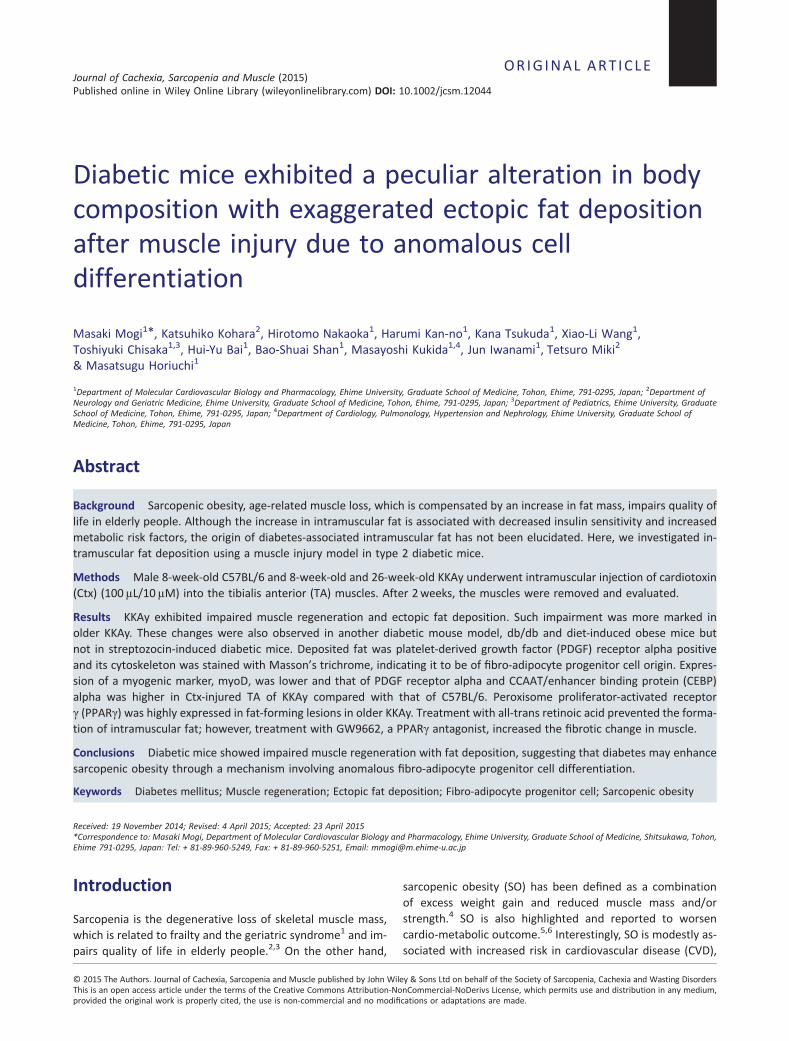
First, we compared the characteristics of skeletal muscle andintramuscular fat between 18-week-old C57BL6 (as WT mice)and KKAy using MRI. As shown in Figure 1A, in coronal T1images of the lower limbs, an increase in subcutaneous andintramuscular fat deposition was observed in KKAy. Moreover,
Figure 1 Magnetic resonance imaging of muscles in lower limbs of 18-week-old C57BL6 (wild-type) and KKAy. 1.5 T magnetic resonance imaging T1images in coronal (A) and sagittal (B) planes were obtained as described in Methods. Representative photos from three mice in each group areshown. Red arrows show muscle atrophy. (C) Haematoxylin-eosin staining of tibialis anterior muscle of 18-week-old WT and KKAy at ×100 magni-fication. White triangles show ectopic fat deposition. Black triangles show blood vessels.
Ectopic fat exaggerated in diabetic mice 3
Journal of Cachexia, Sarcopenia and Muscle (2015)DOI: 10.1002/jcsm.12044
the diameter of the lower limbs tended to be smaller in KKAycompared with WT. Sagittal T1 images suggested the pres-ence of muscle atrophy in the soleus of KKAy (Figure 1B). His-tological analysis of the lower limbs of 18-week-old KKAyshowed slight ectopic fat deposition around blood vessels(Figure 1C).
Muscle repair after cardiotoxin injection
Impairment of muscle repair following injury is one of thefactors contributing to the pathogenesis of sarcopenia.21–23
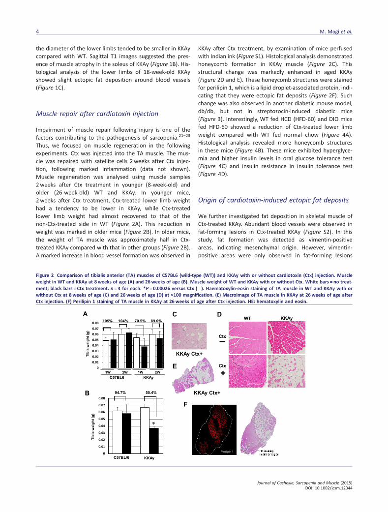
Thus, we focused on muscle regeneration in the followingexperiments. Ctx was injected into the TA muscle. The mus-cle was repaired with satellite cells 2weeks after Ctx injec-tion, following marked inflammation (data not shown).Muscle regeneration was analysed using muscle samples2weeks after Ctx treatment in younger (8-week-old) andolder (26-week-old) WT and KKAy. In younger mice,2weeks after Ctx treatment, Ctx-treated lower limb weighthad a tendency to be lower in KKAy, while Ctx-treatedlower limb weight had almost recovered to that of thenon-Ctx-treated side in WT (Figure 2A). This reduction inweight was marked in older mice (Figure 2B). In older mice,the weight of TA muscle was approximately half in Ctx-treated KKAy compared with that in other groups (Figure 2B).A marked increase in blood vessel formation was observed in
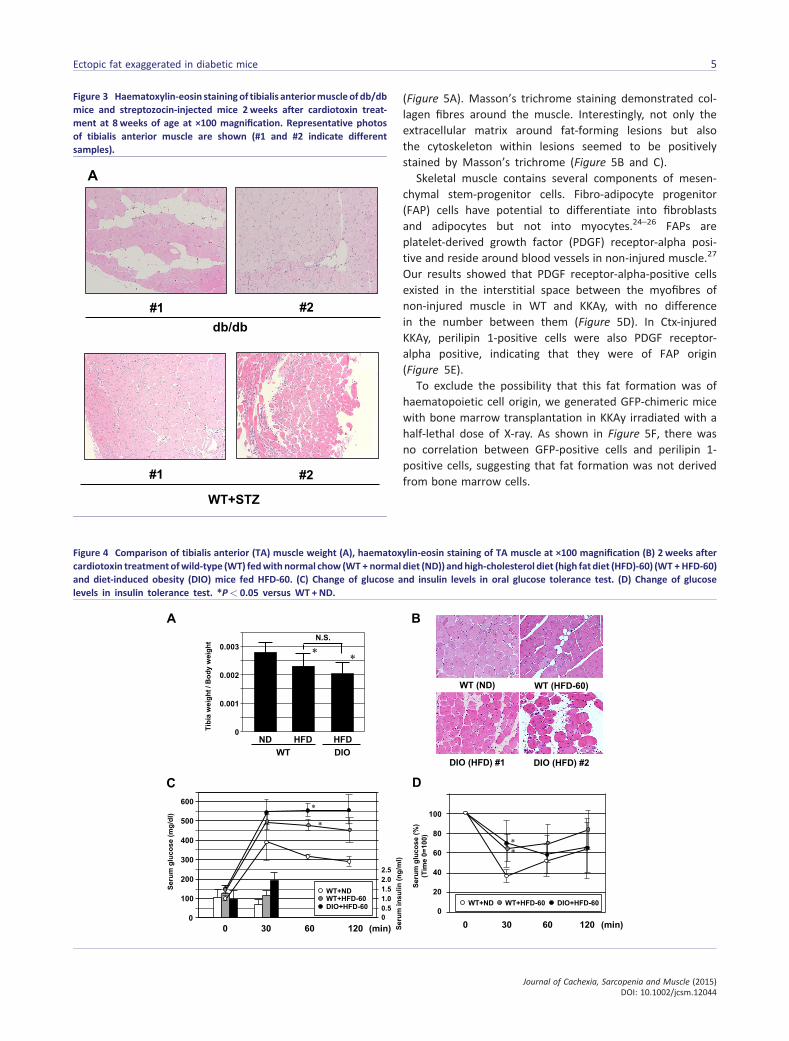
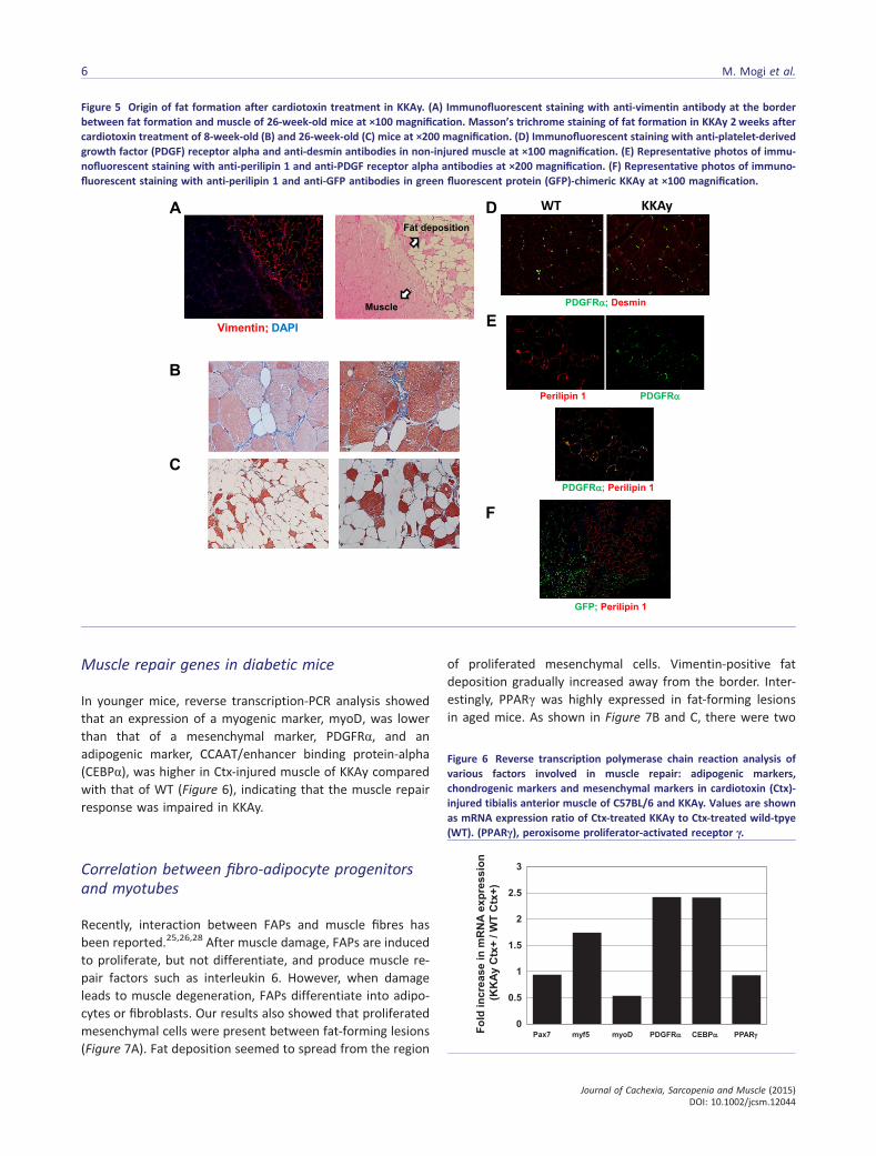
KKAy after Ctx treatment, by examination of mice perfusedwith Indian ink (Figure S1). Histological analysis demonstratedhoneycomb formation in KKAy muscle (Figure 2C). Thisstructural change was markedly enhanced in aged KKAy(Figure 2D and E). These honeycomb structures were stainedfor perilipin 1, which is a lipid droplet-associated protein, indi-cating that they were ectopic fat deposits (Figure 2F). Suchchange was also observed in another diabetic mouse model,db/db, but not in streptozocin-induced diabetic mice(Figure 3). Interestingly, WT fed HCD (HFD-60) and DIO micefed HFD-60 showed a reduction of Ctx-treated lower limbweight compared with WT fed normal chow (Figure 4A).Histological analysis revealed more honeycomb structuresin these mice (Figure 4B). These mice exhibited hyperglyce-mia and higher insulin levels in oral glucose tolerance test(Figure 4C) and insulin resistance in insulin tolerance test(Figure 4D).
Origin of cardiotoxin-induced ectopic fat deposits
We further investigated fat deposition in skeletal muscle ofCtx-treated KKAy. Abundant blood vessels were observed infat-forming lesions in Ctx-treated KKAy (Figure S2). In thisstudy, fat formation was detected as vimentin-positiveareas, indicating mesenchymal origin. However, vimentin-positive areas were only observed in fat-forming lesions
Figure 2 Comparison of tibialis anterior (TA) muscles of C57BL6 (wild-type (WT)) and KKAy with or without cardiotoxin (Ctx) injection. Muscleweight in WT and KKAy at 8 weeks of age (A) and 26weeks of age (B). Muscle weight of WT and KKAy with or without Ctx. White bars = no treat-ment; black bars = Ctx treatment. n = 4 for each. *P = 0.00026 versus Ctx (�). Haematoxylin-eosin staining of TA muscle in WT and KKAy with orwithout Ctx at 8 weeks of age (C) and 26weeks of age (D) at ×100 magnification. (E) Macroimage of TA muscle in KKAy at 26 weeks of age afterCtx injection. (F) Perilipin 1 staining of TA muscle in KKAy at 26 weeks of age after Ctx injection. HE: hematoxylin and eosin.
4 M. Mogi et al.
Journal of Cachexia, Sarcopenia and Muscle (2015)DOI: 10.1002/jcsm.12044
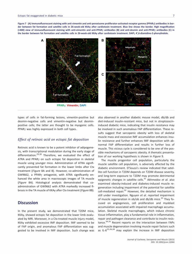
(Figure 5A). Masson’s trichrome staining demonstrated col-lagen fibres around the muscle. Interestingly, not only theextracellular matrix around fat-forming lesions but alsothe cytoskeleton within lesions seemed to be positivelystained by Masson’s trichrome (Figure 5B and C).
Skeletal muscle contains several components of mesen-chymal stem-progenitor cells. Fibro-adipocyte progenitor(FAP) cells have potential to differentiate into fibroblastsand adipocytes but not into myocytes.24–26 FAPs areplatelet-derived growth factor (PDGF) receptor-alpha posi-tive and reside around blood vessels in non-injured muscle.27
Our results showed that PDGF receptor-alpha-positive cellsexisted in the interstitial space between the myofibres ofnon-injured muscle in WT and KKAy, with no differencein the number between them (Figure 5D). In Ctx-injuredKKAy, perilipin 1-positive cells were also PDGF receptor-alpha positive, indicating that they were of FAP origin(Figure 5E).
To exclude the possibility that this fat formation was ofhaematopoietic cell origin, we generated GFP-chimeric micewith bone marrow transplantation in KKAy irradiated with ahalf-lethal dose of X-ray. As shown in Figure 5F, there wasno correlation between GFP-positive cells and perilipin 1-positive cells, suggesting that fat formation was not derivedfrom bone marrow cells.
Figure 3 Haematoxylin-eosin stainingof tibialis anteriormuscleof db/dbmice and streptozocin-injected mice 2 weeks after cardiotoxin treat-ment at 8 weeks of age at ×100 magnification. Representative photosof tibialis anterior muscle are shown (#1 and #2 indicate differentsamples).
Figure 4 Comparison of tibialis anterior (TA) muscle weight (A), haematoxylin-eosin staining of TA muscle at ×100 magnification (B) 2 weeks aftercardiotoxin treatmentofwild-type (WT) fedwith normal chow (WT + normal diet (ND)) andhigh-cholesterol diet (high fat diet (HFD)-60) (WT + HFD-60)and diet-induced obesity (DIO) mice fed HFD-60. (C) Change of glucose and insulin levels in oral glucose tolerance test. (D) Change of glucoselevels in insulin tolerance test. *P< 0.05 versus WT + ND.
Ectopic fat exaggerated in diabetic mice 5
Journal of Cachexia, Sarcopenia and Muscle (2015)DOI: 10.1002/jcsm.12044
Muscle repair genes in diabetic mice
In younger mice, reverse transcription-PCR analysis showedthat an expression of a myogenic marker, myoD, was lowerthan that of a mesenchymal marker, PDGFRα, and anadipogenic marker, CCAAT/enhancer binding protein-alpha(CEBPα), was higher in Ctx-injured muscle of KKAy comparedwith that of WT (Figure 6), indicating that the muscle repairresponse was impaired in KKAy.
Correlation between fibro-adipocyte progenitorsand myotubes
Recently, interaction between FAPs and muscle fibres hasbeen reported.25,26,28 After muscle damage, FAPs are inducedto proliferate, but not differentiate, and produce muscle re-pair factors such as interleukin 6. However, when damageleads to muscle degeneration, FAPs differentiate into adipo-cytes or fibroblasts. Our results also showed that proliferatedmesenchymal cells were present between fat-forming lesions(Figure 7A). Fat deposition seemed to spread from the region
of proliferated mesenchymal cells. Vimentin-positive fatdeposition gradually increased away from the border. Inter-estingly, PPARγ was highly expressed in fat-forming lesionsin aged mice. As shown in Figure 7B and C, there were two
Figure 5 Origin of fat formation after cardiotoxin treatment in KKAy. (A) Immunofluorescent staining with anti-vimentin antibody at the borderbetween fat formation and muscle of 26-week-old mice at ×100 magnification. Masson’s trichrome staining of fat formation in KKAy 2 weeks aftercardiotoxin treatment of 8-week-old (B) and 26-week-old (C) mice at ×200 magnification. (D) Immunofluorescent staining with anti-platelet-derivedgrowth factor (PDGF) receptor alpha and anti-desmin antibodies in non-injured muscle at ×100 magnification. (E) Representative photos of immu-nofluorescent staining with anti-perilipin 1 and anti-PDGF receptor alpha antibodies at ×200 magnification. (F) Representative photos of immuno-fluorescent staining with anti-perilipin 1 and anti-GFP antibodies in green fluorescent protein (GFP)-chimeric KKAy at ×100 magnification.
Figure 6 Reverse transcription polymerase chain reaction analysis ofvarious factors involved in muscle repair: adipogenic markers,chondrogenic markers and mesenchymal markers in cardiotoxin (Ctx)-injured tibialis anterior muscle of C57BL/6 and KKAy. Values are shownas mRNA expression ratio of Ctx-treated KKAy to Ctx-treated wild-tpye(WT). (PPARγ), peroxisome proliferator-activated receptor γ.
6 M. Mogi et al.
Journal of Cachexia, Sarcopenia and Muscle (2015)DOI: 10.1002/jcsm.12044
types of cells in fat-forming lesions, vimentin-positive butdesmin-negative cells and vimentin-negative but desmin-positive cells; the latter are thought to be myogenic cells.PPARγ was highly expressed in both cell types.
Effect of retinoic acid on ectopic fat deposition
Retinoic acid is known to be a potent inhibitor of adipogene-sis, with transcriptional modulation during the early stage ofdifferentiation.29,30 Therefore, we evaluated the effect ofATRA and PPARγ on such ectopic fat deposition in skeletalmuscle using younger mice. Administration of ATRA signifi-cantly prevented fat formation in the lower limbs after Ctxtreatment (Figure 8A and B). However, co-administration ofGW9662, a PPARγ antagonist, with ATRA significantly en-hanced the white area in macroscopic images of TA muscle(Figure 8A). Histological analysis demonstrated that co-administration of GW9662 with ATRA markedly increased fi-brosis in the TA muscle of KKAy after Ctx treatment (Figure 8B).
Discussion
In the present study, we demonstrated that T2DM mice,KKAy, showed ectopic fat deposition in the lower limb evalu-ated by MRI. Moreover, in a Ctx-treated muscle injury model,KKAy exhibited excessive IMF deposition. IMF seemed to beof FAP origin, and anomalous FAP differentiation was sug-gested to be involved in IMF deposition. Such change was
also observed in another diabetic mouse model, db/db anddiet-induced insulin-resistant mice, but not in streptozocin-induced diabetic mice, indicating that insulin resistance maybe involved in such anomalous FAP differentiation. These re-sults suggest that sarcopenic obesity with loss of skeletalmuscle mass and excessive IMF accumulation enhances insu-lin resistance and further enhances IMF deposition with ab-normal FAP differentiation and results in further loss ofmuscle. This vicious cycle is considered to be one of the pos-sible mechanisms of sarcopenic obesity. A thematic presenta-tion of our working hypothesis is shown in Figure 9.
The muscle progenitor cell population, particularly themuscle satellite cell population, is adversely affected by thediabetic environment. D’Souza’s review indicated that satel-lite cell function in T2DM depends on T2DM disease severity,and long-term exposure to T2DM may promote detrimentalepigenetic changes in satellite cells.31 Akhmedov et al. alsoexamined obesity-induced and diabetes-induced muscle re-generation including impairment of the potential for satellitecell-mediated repair.32 However, the detailed mechanism isstill under investigation. Nguyen et al. reported impairmentof muscle regeneration in ob/ob and db/db mice.33 They fo-cused on angiogenesis, cell proliferation and myoblastaccumulation associated with impaired macrophage accumu-lation. Skeletal muscle macrophages, which induce chronictissue inflammation, play a fundamental role in inflammation,repair and pathogen clearance and contribute to insulin resis-tance.34,35 Recent reports on the interaction between FAPsand muscle degeneration involving muscle repair factors suchas IL-625,26,28 may explain the increase in IMF deposition
Figure 7 (A) Immunofluorescent staining with anti-vimentin and anti-peroxisome proliferator-activated receptor gamma (PPARγ) antibodies in bor-der between fat formation and satellite cells in 26-week-old KKAy after cardiotoxin treatment. Blue line shows the border. High magnification(×400) view of immunofluorescent staining with anti-vimentin and anti-PPARγ antibodies (B) and anti-desmin and anti-PPARγ antibodies (C) inthe border between fat formation and satellite cells in 26-week-old KKAy after cardiotoxin treatment. DAPI, 4’,6-diamidino-2-phenylindole.
Ectopic fat exaggerated in diabetic mice 7
Journal of Cachexia, Sarcopenia and Muscle (2015)DOI: 10.1002/jcsm.12044
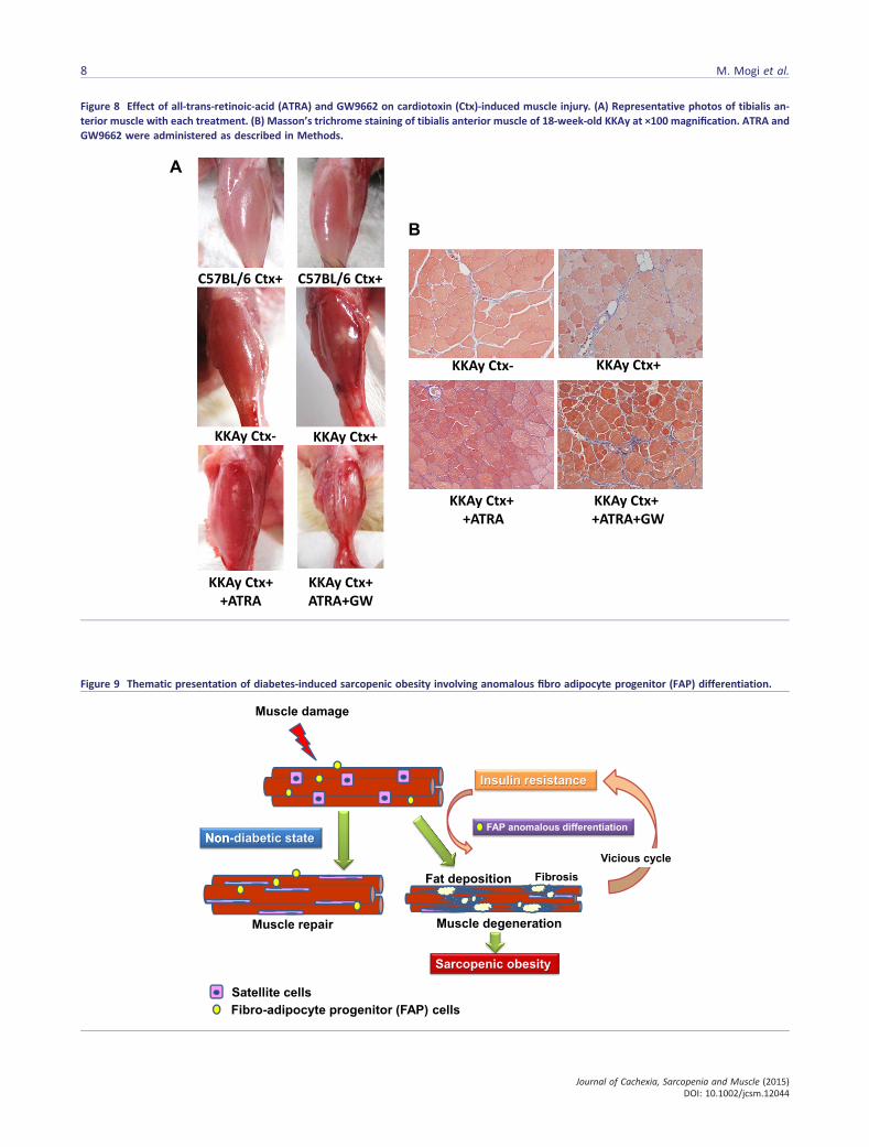
Figure 8 Effect of all-trans-retinoic-acid (ATRA) and GW9662 on cardiotoxin (Ctx)-induced muscle injury. (A) Representative photos of tibialis an-terior muscle with each treatment. (B) Masson’s trichrome staining of tibialis anterior muscle of 18-week-old KKAy at ×100 magnification. ATRA andGW9662 were administered as described in Methods.
Figure 9 Thematic presentation of diabetes-induced sarcopenic obesity involving anomalous fibro adipocyte progenitor (FAP) differentiation.
8 M. Mogi et al.
Journal of Cachexia, Sarcopenia and Muscle (2015)DOI: 10.1002/jcsm.12044
because of anomalous FAP differentiation into adipocytes orfibroblasts via such diabetes-induced impairment of musclerepair. IMF accumulation in obese patients is positively corre-lated with insulin resistance and reduced muscle perfor-mance.36,37 Lipid overload causes impaired skeletal musclefunction because of a reduction of muscle mass and ultra-structural damage38; therefore, lipotoxic species induced viaIMF deposition also interfere with insulin signalling and mus-cle repair. Moreover, muscle regenerative capacity declineswith ageing.39 These reports and our findings suggest a vi-cious cycle of reduced satellite cell function related to ageing,inflammation, insulin resistance and IMF in patients withsarcopenic obesity. Because the present study did not com-pare macrophage filtration in injured skeletal muscle be-tween diabetic and non-diabetic mice, we shouldinvestigate the interaction between inflammatory cells andsatellite cell function as a key determinant of diabetes-induced muscle degeneration in future experiments.
Fibro-adipocyte progenitor cells have been highlighted as akey determinant in the pathogenesis of muscular diseases, in-cluding Duchenne muscular dystrophy. Dong et al. alsoshowed that glucocorticoids stimulate FAPs to differentiateinto adipocytes in injured muscle and that IL-4 inhibited theirdifferentiation process.40 Moreover, Cordani et al. suggesteda preventive effect of nitric oxide on FAP differentiation intoadipocytes via increased expression of miR-27b, leading todownregulation of PPARγ expression.41 Very recently,Saccone et al. reported that the dystrophic muscle environ-ment causes FAPs to adopt a chromatin state that impartsthese cells with myogenic potential.42 Interestingly, suchmyogenic potential of FAPs is limited to cells derived frommuscle in young mdx mice. Moreover, they demonstratedthat treatment of muscle with a histone deacetylase inhibitorblocked adipogenesis and driving muscle differentiation.These findings support that ageing, cytokines, nuclear recep-tor signalling and epigenetic changes may synergisticallyinduce FAP differentiation. In the present study, we usedhalf-a-year-old mice to study sarcopenia. Mice at this ageare young and not close to the sarcopenic threshold; there-fore, we should perform similar experiments, especiallytherapeutic analysis, using more aged mice to confirm age-related sarcopenia as a geriatric phenomenon.
A recent report demonstrated that a PPARγ-activating mi-croenvironment, such as treatment with fatty acids, causedfibroblasts to differentiate into adipocytes.43 In contrast,myogenic cells did not undergo adipogenesis. Our resultsdemonstrated that aged KKAy exhibited higher expressionof PPARγ in injured muscle, while the expression of PPARγdid not change in young KKAy (Figures 4 and 5). Therefore,high PPARγ expression may markedly induce differentiationof FAPs into adipocytes in aged KKAy. Wu et al. reported thatvisceral adipose tissue of old C57BL mice showed signifi-cantly higher mRNA expression of proinflammatory cytokinesand lower expression of anti-inflammatory factors such as
PPARγ than those in young mice.44 Moreover, relative PPARγexpression is increased in omental fat in obesity,45 and ahighly significant negative correlation between adipocytePPARγ expression and BMI was observed.46 These reportssuggest that relative PPARγ expression is increased in agedobese diabetic mice. This PPARγ-activating microenviron-mental change may enhance adipocyte differentiation intoFAP cells.
To maintain the quality of skeletal muscle, prevention ofmuscle fibrosis is also important as well as intramuscularfat deposition. Satellite cells from aged mice tend to convertfrom a myogenic to a fibrogenic lineage associated with Wntsignalling pathway.47 Very recently, Fry et al. reported thatthe loss of satellite cells may contribute to age-related mus-cle fibrosis using a genetic mouse model that allows for thespecific, inducible depletion of satellite cells in adult skeletalmuscle.48 Moreover, Krause et al. demonstrated the impair-ment of satellite cell infiltration in diabetic skeletal mus-cle.49 Therefore, fibrotic change may be involved in musclequality of diabetic skeletal muscle. On the other hand, in-creased collagen content is observed in insulin-resistantskeletal muscles of humans.50 Inoue et al. reported that lackof thrombospondin 1 state protects mice from HFD-inducedmuscle fibrosis and insulin resistance.51 Moreover, extracel-lular matrix remodelling is an important factor for determin-ing muscle insulin resistance in the presence of HFD.52
Although we have not assessed insulin resistance in KKAymice with or without fibrosis in skeletal muscle, these re-ports suggest that muscle fibrosis may also enhance insulinresistance. Therefore, impaired muscle quality with an in-crease in not only fat but also fibrosis of skeletal muscle af-t1er injury may cause vicious cycle of insulin resistance.Further investigation of muscle fibrosis involving satellitecells is necessary in the future.
Treatment with retinoic acid (RA) has partly prevented fatdeposition; however, co-administration with a PPARγ antago-nist has enhanced fibrosis. RA is reported to inhibit differen-tiation of 3T3-F442A cells into adipocytes53 and upregulatepreadipocyte genes such as cellular RA binding protein typeII and nuclear RA receptors to block adipogenesis and sup-press diet-induced obesity.54 Recently, it was reported thatcombined treatment with three ligands, PPARs, ATRA andthe retinoic X receptor, 9-cis, prevented liver fibrosis in ratprimary hepatic stellate cells,55 indicating that cooperationof several factors including PPARs, RA and nuclear receptorsregulates FAP cell differentiation after muscle injury.
Some of the limitations of this study are as follows. The re-sults were mainly obtained from immunohistological analysisand lacked quantitative analysis. It is necessary to identifyconcrete mechanisms to address the connection between in-tramuscular lipid and muscle degeneration. In the presentstudy, we used obese diabetic mice. It is not clear which intra-muscular fat deposition is induced by obesity or diabetes. Toclarify the relation, we could use animal models of obesity
Ectopic fat exaggerated in diabetic mice 9
Journal of Cachexia, Sarcopenia and Muscle (2015)DOI: 10.1002/jcsm.12044
such as diet-induced obesity or ob/ob mice in the future.Moreover, in a previous study, 16-week-old KKAy exhibitedan increase in creatinine level, which suggests early onsetof kidney failure.56 Therefore, it cannot be excluded thatmuscle atrophy is associated with nephropathy. Furthermore,it is not known how ‘acute muscle injury’ is involved insarcopenic obesity as a chronic disease. Our findings shouldbe investigated from a clinical perspective in patients withdiabetes.
In conclusion, the mice with T2DM showed significantlyincreased IMF deposition, possibly due to anomalous celldifferentiation. To prevent sarcopenic obesity, prevention ofIMF deposition with consideration of muscle degenerationshould be one of the critical targets and its mechanism shouldbe investigated, focusing on anomalous FAP differentiation.
Acknowledgements
The authors certify that they comply with the ethical guidelines for
authorship and publishing of the Journal of Cachexia, Sarcopenia
and Muscle (von Haehling S, Morley JE, Coats AJS, Anker SD. Ethical
guidelines for authorship and publishing in the Journal of Cachexia,
Sarcopenia and Muscle. J Cachexia Sarcopenia Muscle. 2010;1:7-8.)
We thank Dr Masaru Okabe from the Department of Experimental
Genome Research, Osaka University for generously providing the
Green mouse, FM131. We also thank Takeshi Kiyoi from the the
Integrated Center for Sciences, Ehime University for providing exper-
imental technological assistance.
Funding
JSPS KAKENHI Grant Numbers 25293310 (M.H.), 25462220(M.M.). Research grants from pharmaceutical companies:
Astellas Pharma Inc., Bayer Yakuhin, Ltd., Daiichi-SankyoPharmaceutical Co. Ltd., Nippon Boehringer Ingelheim Co.Ltd., Novartis Pharma K. K., Shionogi & Co., Ltd. and TakedaPharmaceutical Co. Ltd.
Supporting information
Supporting information may be found in the online version ofthis article.
Table S1. List of antibodies.
Table S2. List of primer sequences used for RT-PCR analysis.
Figure S1. Comparison of tibialis anterior (TA) muscles ofC57BL6 (WT) and KKAy with or without Ctx injection. Repre-sentative photos of TA muscle with or without Indian ink.
Figure S2. Fat formation and blood vessels. Hematoxylin-eosinstaining (upper panel) and immunofluorescent staining withanti-CD31 and anti-perilipin antibodies (lower panel) of tibialisanterior muscles in 26-week-old KKAy after Ctx treatment.
Conflict of interest
Masatsugu Horiuchi has received research grants from pharma-ceutical companies: Astellas Pharma Inc., Bayer Yakuhin, Ltd.,Daiichi-Sankyo Pharmaceutical Co. Ltd., Nippon BoehringerIngelheim Co. Ltd., Novartis Pharma K. K., Shionogi & Co., Ltd.and Takeda Pharmaceutical Co. Ltd. All other authors havedeclared no conflict of interest.
References
1. Kinney JM. Nutritional frailty, sarcopeniaand falls in the elderly. Curr Opin Clin NutrMetab Care 2004;7:15–20.
2. Cruz-Jentoft AJ, Baeyens JP, Bauer JM,Boirie Y, Cederholm T, Landi F, et al.Sarcopenia: European consensus ondefinition and diagnosis: Report of theEuropean Working Group on Sarcopeniain Older People. Age Ageing 2010;39:412–423.
3. Fielding RA, Vellas B, Evans WJ, Bhasin S,Morley JE, Newman AB, et al. Sarcopenia:an undiagnosed condition in older adults.Current consensus definition: prevalence,etiology, and consequences. InternationalWorking Group on Sarcopenia. J Am MedDir Assoc 2011;12:249–256.
4. Roubenoff R. Sarcopenic obesity: the con-fluence of two epidemics. Obes Res2004;12:887–888.
5. Kohara K, Ochi M, Tabara Y, Nagai T,Igase M, Miki T. Arterial stiffness insarcopenic visceral obesity in the elderly:J-SHIPP study. Int J Cardiol 2012;158:146–148.
6. Dominguez LJ, Barbagallo M. The cardio-metabolic syndrome and sarcopenic obe-sity in older persons. J Cardiometab Syndr2007;2:183–189.
7. Stephen WC, Janssen I. Sarcopenic-obesityand cardiovascular disease risk in the elderly.J Nutr Health Aging 2009;13:460–466.
8. Baumgartner RN, Wayne SJ, Waters DL,Janssen I, Gallagher D, Morley JE.Sarcopenic obesity predicts instrumentalactivities of daily living disability in the el-derly. Obes Res 2004;12:1995–2004.
9. Ochi M, Tabara Y, Kido T, Uetani E, Ochi N,Igase M, et al. Quadriceps sarcopenia andvisceral obesity are risk factors for postural
instability in the middle-aged to elderlypopulation. Geriatr Gerontol Int 2010;10:233–243.
10. Buford TW, Anton SD, Judge AR, MarzettiE, Wohlgemuth SE, Carter CS, et al.Models of accelerated sarcopenia: criticalpieces for solving the puzzle of age-related muscle atrophy. Ageing Res Rev2010;9:369–383.
11. Kim TN, Park MS, Yang SJ, Yoo HJ, Kang HJ,Song W, et al. Prevalence and determinantfactors of sarcopenia in patients with type2 diabetes: the Korean Sarcopenic ObesityStudy (KSOS). Diabetes Care 2010;33:1497–1499.
12. Park SW, Goodpaster BH, Lee JS, Kuller LH,Boudreau R, de Rekeneire N, et al. Exces-sive loss of skeletal muscle mass in olderadults with type 2 diabetes. Diabetes Care2009;32:1993–1997.
10 M. Mogi et al.
Journal of Cachexia, Sarcopenia and Muscle (2015)DOI: 10.1002/jcsm.12044
13. van Loon LJ, Goodpaster BH. Increased in-tramuscular lipid storage in the insulin-resistant and endurance-trained state.Pflugers Arch 2006;451:606–616.
14. Komiya H, Mori Y, Yokose T, Kurokawa N,Horie N, Tajima N. Effect of intramuscularfat difference on glucose and insulin reac-tion in oral glucose tolerance test. JAtheroscler Thromb 2006;13:136–142.
15. Pigeon E, Couillard E, Tremblay A, BouchardC, Weisnagel SJ, Joanisse DR. Mid-thighsubcutaneous adipose tissue and glucosetolerance in the Quebec family study. ObesFacts 2008;1:310–318.
16. Ingram KH, Lara-Castro C, Gower BA,Makowsky R, Allison DB, Newcomer BR,et al. Intramyocellular lipid and insulinresistance: differential relationships inEuropean and African Americans. Obesity(Silver Spring) 2011;19:1469–1475.
17. Ingram KH, Hill H, Moellering DR, Hill BG,Lara-Castro C, Newcomer B, et al. Skeletalmuscle lipid peroxidation and insulin resis-tance in humans. J Clin Endocrinol Metab2012;97:E1182–E1186.
18. Therkelsen KE, Pedley A, Speliotes EK,Massaro JM, Murabito J, Hoffmann U,et al. Intramuscular fat and associationswith metabolic risk factors in the Framing-ham Heart Study. Arterioscler Thromb VascBiol 2013;33:863–870.
19. Garry DJ, Yang Q, Bassel-Duby R, WilliamsRS. Persistent expression of MNF identifiesmyogenic stem cells in postnatal muscles.Dev Biol 1997;188:280–294.
20. Iwanami J, Mogi M, Tsukuda K, Min LJ,Sakata A, Jing F, et al. Effect of angiotensinII type 2 receptor deletion in hematopoi-etic cells on brain ischemia-reperfusion in-jury. Hypertension 2011;58:404–409.
21. Carlson ME, Suetta C, Conboy MJ, AagaardP, Mackey A, Kjaer M, et al. Molecular agingand rejuvenation of human muscle stemcells. EMBO Mol Med 2009;1:381–391.
22. Conboy IM, Conboy MJ, Smythe GM,Rando TA. Notch-mediated restoration ofregenerative potential to aged muscle.Science 2003;302:1575–1577.
23. Lee CE, McArdle A, Griffiths RD. The role ofhormones, cytokines and heat shock pro-teins during age-related muscle loss. ClinNutr 2007;26:524–534.
24. Giordani L, Puri PL. Epigenetic control ofskeletal muscle regeneration: integratinggenetic determinants and environmentalchanges. FEBS J 2013;280:4014–4025.
25. Uezumi A, Fukada S, Yamamoto N, TakedaS, Tsuchida K. Mesenchymal progenitorsdistinct from satellite cells contribute to ec-topic fat cell formation in skeletal muscle.Nat Cell Biol 2010;12:143–152.
26. Joe AW, Yi L, Natarajan A, Le Grand F, So L,Wang J, et al. Muscle injury activates residentfibro/adipogenic progenitors that facilitatemyogenesis. Nat Cell Biol 2010;12:153–163.
27. Pretheeban T, Lemos DR, Paylor B, ZhangRH, Rossi FM. Role of stem/progenitor cellsin reparative disorders. Fibrogenesis TissueRepair 2012;5:20.
28. Rodeheffer MS. Tipping the scale: muscleversus fat. Nat Cell Biol 2010;12:102–104.
29. Xue JC, Schwarz EJ, Chawla A, Lazar MA.Distinct stages in adipogenesis revealedby retinoid inhibition of differentiation af-ter induction of PPARgamma. Mol Cell Biol1996;16:1567–1575.
30. Marchildon F, St-Louis C, Akter R, RoodmanV, Wiper-Bergeron NL. Transcription factorSmad3 is required for the inhibition of adi-pogenesis by retinoic acid. J Biol Chem2010;285:13274–13284.
31. D’Souza DM, Al-Sajee D, Hawke TJ. Diabeticmyopathy: impact of diabetes mellitus onskeletal muscle progenitor cells. FrontPhysiol 2013;4:379.
32. Akhmedov D, Berdeaux R. The effects ofobesity on skeletal muscle regeneration.Front Physiol 2013;4:371.
33. NguyenMH, ChengM, Koh TJ. Impairedmus-cle regeneration in ob/ob and db/db mice.Scientific World Journal 2011;11:1525–1535.
34. Pillon NJ, Bilan PJ, Fink LN, Klip A. Cross-talk between skeletal muscle and immunecells: muscle-derived mediators and meta-bolic implications. Am J Physiol EndocrinolMetab 2013;304:E453–E465.
35. Osborn O, Olefsky JM. The cellular and sig-naling networks linking the immune systemand metabolism in disease. Nat Med2012;18:363–374.
36. Goodpaster BH, Thaete FL, Kelley DE. Thighadipose tissue distribution is associatedwith insulin resistance in obesity and intype 2 diabetes mellitus. Am J Clin Nutr2000;71:885–892.
37. Hilton TN, Tuttle LJ, Bohnert KL, MuellerMJ, Sinacore DR. Excessive adipose tissueinfiltration in skeletal muscle in individualswith obesity, diabetes mellitus, and periph-eral neuropathy: association with perfor-mance and function. Phys Ther 2008;88:1336–1344.
38. Tamilarasan KP, Temmel H, Das SK, AlZoughbi W, Schauer S, Vesely P, et al. Skel-etal muscle damage and impaired regener-ation due to LPL-mediated lipotoxicity. CellDeath Dis 2012;3:e354.
39. Jang YC, Sinha M, Cerletti M, Dall’Osso C,Wagers AJ. Skeletal muscle stem cells: ef-fects of aging and metabolism on muscleregenerative function. Cold Spring HarbSymp Quant Biol 2011;76:101–111.
40. Dong Y, Silva KA, Zhang L. Glucocorticoidsincrease adipocytes in muscle by affectingIL-4 regulated FAP activity. FASEB J 2014,in press
41. Cordani N, Pisa V, Pozzi L, Sciorati C,Clementi E. Nitric oxide controls fat deposi-tion in dystrophic skeletal muscle byregulating fibro-adipogenic precursor differ-entiation. Stem Cells 2014;32:874–885.
42. Saccone V, Consalvi S, Giordani L, MozzettaC, Barozzi I, Sandona M, et al. HDAC-regu-lated myomiRs control BAF60 variantexchange and direct the functional pheno-type of fibro-adipogenic progenitors indystrophic muscles. Genes Dev 2014;28:841–857.
43. Agley CC, Rowlerson AM, Velloso CP,Lazarus NR, Harridge SD. Human skeletalmuscle fibroblasts, but not myogeniccells, readily undergo adipogenic differ-entiation. J Cell Sci 2013;126:5610–5625.
44. Wu D, Ren Z, Pae M, Guo W, Cui X, MerrillA, et al. Aging up-regulates expression ofinflammatory mediators in mouse adiposetissue. J Immunol 2007;179:4829–4839.
45. Lefebvre AM, Laville M, Vega N, Riou JP,van Gaal L, Auwerx J, et al. Depot-specificdifferences in adipose tissue gene expres-sion in lean and obese subjects. Diabetes1998;47:98–103.
46. Montague CT, Prins JB, Sanders L, Zhang J,Sewter CP, Digby J, et al. Depot-relatedgene expression in human subcutaneousand omental adipocytes. Diabetes1998;47:1384–1391.
47. Brack AS, Conboy MJ, Roy S, Lee M, KuoCJ, Keller C, et al. Increased Wnt signalingduring aging alters muscle stem cell fateand increases fibrosis. Science 2007;317:807–810.
48. Fry CS, Lee JD, Mula J, Kirby TJ, Jackson JR,Liu F, et al. Inducible depletion of satellitecells in adult, sedentary mice impairs mus-cle regenerative capacity without affectingsarcopenia. Nat Med 2015;21:76–80.
49. Krause MP, Al-Sajee D, D’Souza DM,Rebalka IA, Moradi J, Riddell MC, et al.Impaired macrophage and satellite cell in-filtration occurs in a muscle-specific fash-ion following injury in diabetic skeletalmuscle. PLoS One 2013;8:e70971.
50. Berria R, Wang L, Richardson DK, FinlaysonJ, Belfort R, Pratipanawatr T, et al. In-creased collagen content in insulin-resistant skeletal muscle. Am J PhysiolEndocrinol Metab 2006;290:E560–E565.
51. Inoue M, Jiang Y, Barnes RH 2nd, TokunagaM, Martinez-Santibañez G, Geletka L, et al.Thrombospondin 1 mediates high-fat diet-induced muscle fibrosis and insulin resistancein male mice. Endocrinology 2013;154:4548–4559.
52. KangL1, Mayes WH, James FD, Bracy DP,Wasserman DH. Matrix metalloproteinase9 opposes diet-induced muscle insulin re-sistance in mice. Diabetologia 2014;57:603–613.
53. Kuri-HarcuchWDifferentiation of 3 T3-F442Acells into adipocytes is inhibited by retinoicacid. Differentiation 1982;23:164–169.
54. Berry DC, DeSantis D, Soltanian H, CronigerCM, Noy N. Retinoic acid upregulatespreadipocyte genes to block adipogenesisand suppress diet-induced obesity. Diabe-tes 2012;61:1112–1121.
55. Sharvit E, Abramovitch S, Reif S, Bruck R.Amplified inhibition of stellate cell activa-tion pathways by PPAR-gamma, RAR andRXR agonists. PLoS One 2013;8:e76541.
56. Ishizawa K, Izawa-Ishizawa Y, Yamano N,Urushihara M, Sakurada T, Imanishi M,et al. Nitrosonifedipine ameliorates theprogression of type 2 diabetic nephropathyby exerting antioxidative effects. PLoS One2014;9:e86335.
Ectopic fat exaggerated in diabetic mice 11
Journal of Cachexia, Sarcopenia and Muscle (2015)DOI: 10.1002/jcsm.12044


![Genome-wide association study of appendicular lean mass in ...Other LBM-related conditions include dysmobility syndrome [4], sarcopenic obesity [5], and cachexia [6]. Overall, sarcopenia](https://img.pdfslide.us/doc/110x75/60bd3388aca5cb169d372338/genome-wide-association-study-of-appendicular-lean-mass-in-other-lbm-related.jpg)