Embed Size (px)
Citation preview
Int J Clin Exp Pathol 2016;9(7):6896-6904www.ijcep.com /ISSN:1936-2625/IJCEP0020114
Original Article Fucoidan attenuates atherosclerosis in LDLR-/- mice through inhibition of inflammation and oxidative stress
Xin Wang1, Lili Pei2, Haibo Liu1, Kai Qv2, Weiwei Xian2, Jia Liu2, Guomin Zhang2
1Department of Emergency, The First Hospital of Jilin University, Changchun 130021, China; 2The Department of Cardiology, The First Affiliated Hospital of Qiqihar Medical University, Qiqihar, Heilongjiang, China
Received November 19, 2015; Accepted May 26, 2016; Epub July 1, 2016; Published July 15, 2016
Abstract: Background and purpose: Fucoidan is a major bioactive polysaccharide which contains substantial percent-ages of L-fucose and sulfate ester groups, mainly isolated from brown seaweed. For the past decade fucoidans have been extensively studied due to their varied biological activities, including anti-inflammatory, blood lipids reducing, antioxidant and anticomplementary properties. However, it is not known whether fucoidan can prevent experimental atherosclerosis. The present study was designed to investigate the protective effects of fucoidan on atherosclerosis and its molecular mechanisms of action. Methods: Low Density Lipoprotein Receptor-deficient (LDLR-/-) mice, fed an atherogenic diet, were dosed daily with Fucoidan (50, 100 mg/kg day) by oral gavage. In vitro studies were car-ried out in oxidized LDL (oxLDL)-stimulated RAW264.7 cells treated with or without Fucoidan. Results: Fucoidan significantly attenuated atherosclerotic plaque formation and enhanced plaque stability in LDLR-/- mice by decreas-ing the serum lipids and inhibiting the macrophage infiltration, as well as inhibiting reactive oxygen species (ROS) generation. Fucoidan treatment significantly decreased the expression of LOX-1 and the levels of pro-inflammatory mediators in vivo. In vitro, fucoidan decreased oxLDL-induced LOX-1, pro-inflammatory mediators as well as adhe-sion molecules (ICAM-1 and VCAM-1). Furthermore, fucoidan inhibited NADPH oxidase subunit 4 (NOX4)-mediated ROS generation. Conclusion: Fucoidan was shown to have anti-atherosclerotic activity, which was mediated through inhibition of the inflammation and oxidative stress. This suggests that Fucoidan is a vasculoprotective drug that has potential therapeutic value for the clinical treatment of atherosclerotic cardiovascular diseases.
Keywords: Fucoidan, atherosclerosis, inflammation, oxidative stress
Introduction
Atherosclerotic cardiovascular disease, driven and regulated by lipid retention (in the artery wall), lipid oxidation, persistent inflammation and immune response disturbances, is the le- ading cause of premature death in developed and developing countries [1, 2]. One critical event in the initiation of atherosclerosis is the adhesion of leukocytes to activated endotheli-um and their subsequent migration into the vessel wall. These cellular processes are medi-ated by the up-regulation of adhesion mole-cules in endothelial cells (ECs) and an increased expression of leukocyte chemotactic factors in the vascular wall [3]. One major determinant of this alteration could be oxidative stress. Modified LDL, in particular oxidized LDL (oxLDL; the pathological form of oxidatively modified LDL), exerts several pro-atherogenic effects to
facilitate atherosclerosis [4]. The plasma oxLDL level is transiently increased before the devel-opment of atherosclerotic lesions in apolipopro-tein E-deficient (LDLR-/-) mice [5], suggesting that oxLDL may play a crucial role in the early stages in the formation of atherosclerotic lesions. Previous studies have shown that lec-tin-like oxLDL receptor-1 (LOX-1), a primary sca- venger receptor expressed in ECs [6], is up-reg-ulated in atherosclerotic plaques of experimen-tal animals and humans [7]. LOX-1 facilitates the uptake of oxLDL by ECs and macrophages, thus mediating several of its biological effects [8-11]: oxLDL induces (i) apoptosis of ECs and phagocytosis of aged and apoptotic cells; (ii) the adhesion of monocytes to activated endo-thelium; and (iii) macrophage-derived foam cell formation. More recently, it has been demon-strated that LOX-1 is the main culprit that trans-duces the signal of the adverse effects of dys-
Fucoidan attenuates atherosclerosis in LDLR-/- mice
6897 Int J Clin Exp Pathol 2016;9(7):6896-6904
functional high-density lipoprotein (HDL; such as oxidized HDL and HDL from patients with coronary artery disease) [12]. In vitro, LOX-1 expression is up-regulated by various pro-ath-erogenic stimuli, including oxLDL, TNF-α, angio-tensin II, shear stress, homocysteine and high glucose [11]. Therefore, LOX-1 has recently been suggested as an attractive therapeutic target for atherosclerosis [11].
The brown seaweed, Laminaria japonica, is common seafood in China and many other countries, and documented as a drug in tradi-tional Chinese medicine for over a 1000 years. Fucoidan is a complex sulfated polysaccharide, derived from marine brown seaweed. Fucoidan has been reported to possess diverse biologi-cal activity of potential medicinal value, such as antioxidant, anticoagulant, antitumor, antiviral and anti-inflammatory activity [13, 14]. Notably, its anti-inflammatory has drawn much attention in recent years [15, 16]. However, there is no report on the potential anti-atherosclerotic effects of fucoidan in vivo.
Therefore, in the present study, we investigated the potential effects of fucoidan on atheroscle-rotic plaque development in LDLR-/- mice kept on a high cholesterol diet (HCD). Additionally, we examined the modulation of LOX-1 expres-sion by CTS in HUVECs stimulated with oxLDL.
Materials and methods
Materials
The test compound, fucoidan, was prepared from L. japonica cultured in Yantai, China and was identified by Prof. Benming Xu, School of Pharmacy, Yantai University. A voucher speci-men was deposited in the herbarium of the school of pharmacy, Yantai University. Fucoidan was isolated as described previously [17].
Animal experiment
Experimental protocols were approved by the Animal Care and Use Committee of Jilin
University (Jilin, China). All animal experimental procedures were performed in accordance with the Guide for the Care and Use of Laboratory Animals. All studies involving animals are re- ported in accordance with the ARRIVE guide-lines for reporting experiments involving ani-mals [18, 19]. Male LDLR-/- mice on C57BL/6J background were purchased from Peking Uni- versity Experimental Animal Center (Beijing, China). The mice were housed under a 12-h light/dark cycle in specific pathogen-free facili-ty at Institutional Experimental Animal Center. Starting from 6 weeks, the mice were fed with a HCD (10% fat, 1.25% cholesterol, 0% cholic acid) for 16 weeks. All LDLR-/- mice were dosed daily via intragastric gavage with 50 or 100 mg/kg Fucoidan dissolved in 0.5% carboxy-methyl-cellulose sodium (CMC-Na) or adminis-tered 0.5% CMC-Na alone (control) (n = 5 per group). All animals received food and water ad libitum. Body weight and food intake were mon-itored during the study.
Detection of serum lipid profile
For measurement of the lipids, blood samples were collected at baseline (6 week) and the end of the diet treatment period (22 week), by retro-orbital venous plexus bleeding, from animals that had been fasted overnight. Serum total cholesterol, HDL cholesterol, LDL cholesterol and triglycerides were measured by colorimet-ric assays as previously described [20].
Morphometric analysis of atherosclerotic lesions
The en face and aortic sinus cryosection tech-niques [20] were used to quantify atherosclero-sis development throughout the aorta as well as at the vessel origin. Oil Red O (ORO) staining was used to measure lesion area in en face aorta and aortic sinus. For quantitative analysis of the total lesion area in aortic sinus, eight separate cryosections (spacing 50 μm apart) from each mouse were manually analysed with the Leica Qwin PLUS Software (Leica Micro-
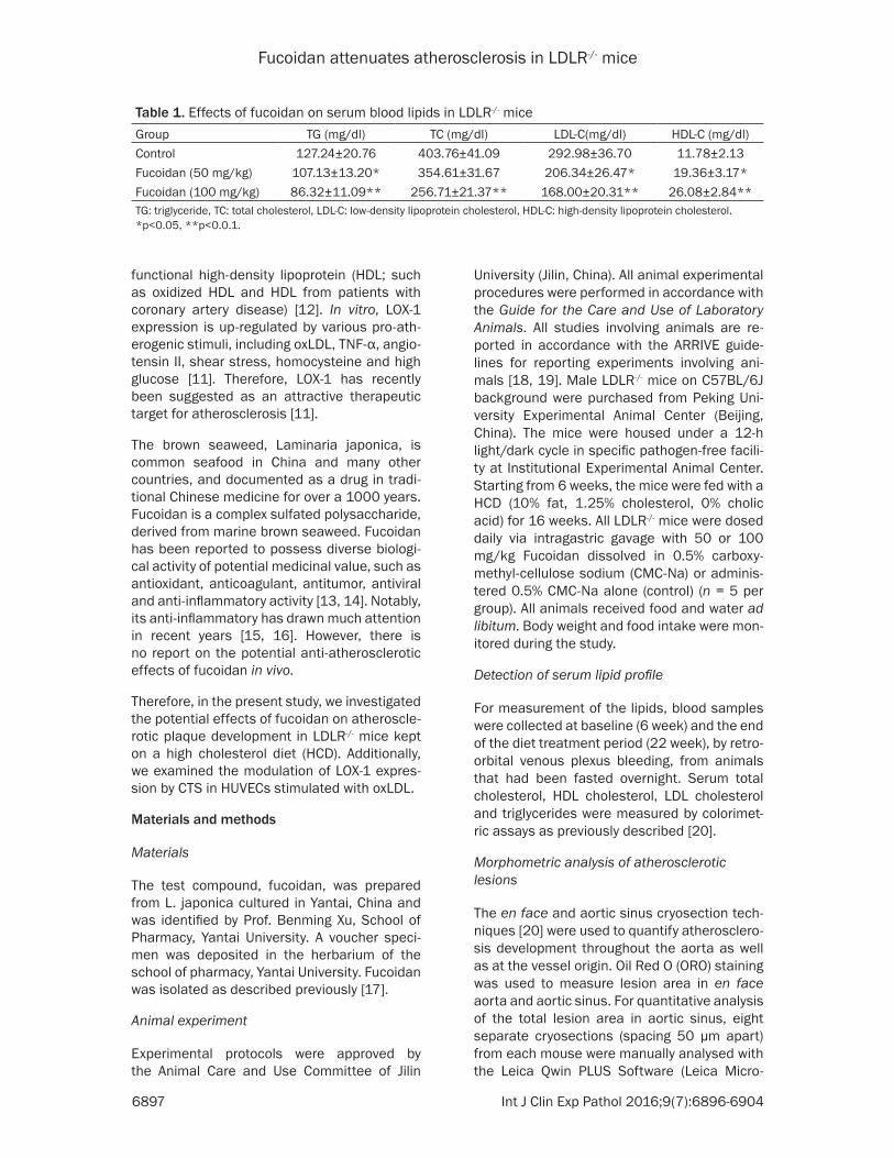
Table 1. Effects of fucoidan on serum blood lipids in LDLR-/- miceGroup TG (mg/dl) TC (mg/dl) LDL-C(mg/dl) HDL-C (mg/dl)Control 127.24±20.76 403.76±41.09 292.98±36.70 11.78±2.13Fucoidan (50 mg/kg) 107.13±13.20* 354.61±31.67 206.34±26.47* 19.36±3.17*Fucoidan (100 mg/kg) 86.32±11.09** 256.71±21.37** 168.00±20.31** 26.08±2.84**TG: triglyceride, TC: total cholesterol, LDL-C: low-density lipoprotein cholesterol, HDL-C: high-density lipoprotein cholesterol. *p<0.05, **p<0.0.1.
Fucoidan attenuates atherosclerosis in LDLR-/- mice
6898 Int J Clin Exp Pathol 2016;9(7):6896-6904
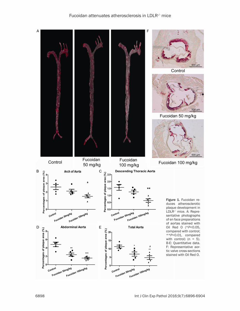
Figure 1. Fucoidan re-duces atherosclerotic plaque development in LDLR-/- mice. A: Repre-sentative photographs of en face preparations of aortas stained with Oil Red O (*P<0.05, compared with control; **P<0.01, compared with control) (n = 5). B-E: Quantitative data. F: Representative aor-tic valve cross-sections stained with Oil Red O.
Fucoidan attenuates atherosclerosis in LDLR-/- mice
6899 Int J Clin Exp Pathol 2016;9(7):6896-6904
systems, Heidelberg, Germany). For plaque area in whole aorta, the percentage of ORO-positive stained area in relation to total luminal surface area was quantified using computeras-sisted morphometry with NIH ImageJ software (http://imagej.nih.gov). Lesion size in the aortic sinus and en face arterial tree was measur- ed by two observers blinded to experimental groups.
Histology and immunohistochemistry analysis
Aortic sinus morphometric and immunohisto-chemical analysis was performed as described previously in detail [20]. Sections of 8 μm thick-ness were used for immunohistochemical staining with Mac-2 (Boster, Wuhan, China) and SM-22 antibodies (Abcam, Cambridge, MA, USA). Colour reaction was developed with dia- minobenzidine (Sigma-Aldrich, St. Louis, MO, USA). The remainders of the sections were uti-lized for hematoxylineosin (H&E) staining to examine basic lesion morphology.
Reactive oxygen species (ROS) production in situ
The production of ROS in aortic root cryosec-tions was assessed in situ by fluorescence microscopy of dihydroethidium (DHE)-stained sections as described, in detail, previously [20]. The fluorescence was quantified using NIH ImageJ software.
Cell culture
RAW264.7 murine macrophage cells (American Type Culture Collection, Rockville, MD, USA) were cultured in DMEM supplemented with 10% FBS and 1% penicillin and streptomycin at 37°C in the presence of 5% CO
2. RAW 264.7
cells were treated with 100 ng/mL LPS with or without different concentrations of fucoidan (50 and 100 μg/mL) for 24 h. Cells treated with LPS alone were utilised as the control. After 24 h of treatment, the cell culture supernatants were collected for cytokine and nitrite assays.
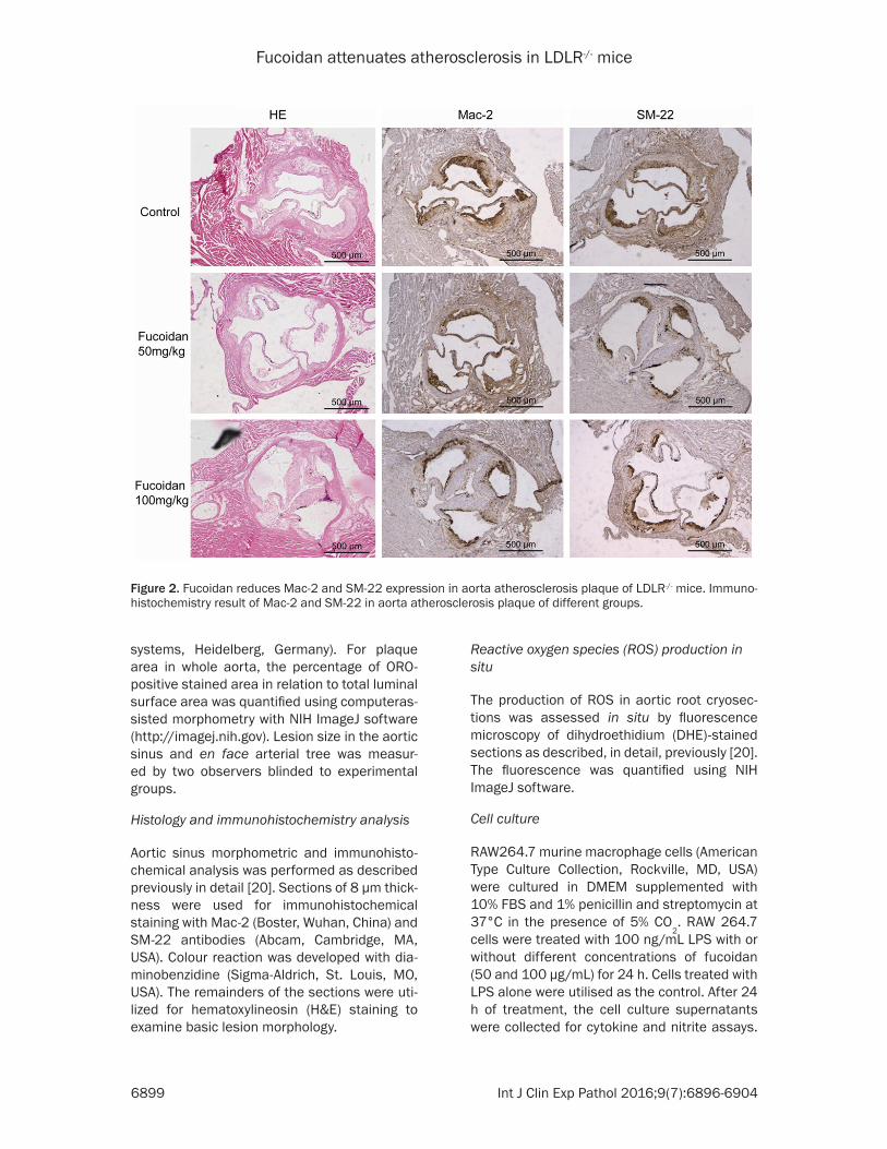
Figure 2. Fucoidan reduces Mac-2 and SM-22 expression in aorta atherosclerosis plaque of LDLR-/- mice. Immuno-histochemistry result of Mac-2 and SM-22 in aorta atherosclerosis plaque of different groups.
Fucoidan attenuates atherosclerosis in LDLR-/- mice
6900 Int J Clin Exp Pathol 2016;9(7):6896-6904
The cell lysates were collected for reverse tran-scriptase-polymerase chain reaction (RT-PCR) assays.
Realtime PCR
Real-time PCR was conducted as previously described [20]. Sequences for the oligonucle-otide primers used were designed with Primer 5.0 software (Premier Biosoft, Palo Alto, CA, USA) and were custom synthesized by Invi- trogen. mRNA levels of PAI1, NOX-2, p67, LOX-1, IL-1b, IL-6, TNF-α, intracellular adhesion mole-
Fifteen LDLR-/- mice were randomly allocated to groups and equal group sizes were obtained (n = 5 per group). Data are presented as mean ± SEM unless specified otherwise. Images shown are representative of five or more independent experiments. Statistical significance of differ-ences was calculated using one-way ANOVA with Bonferroni post hoc for multiple group comparison or Student’s unpaired t-test for two-group comparison where appropriate. The analyses were performed using GraphPad Prism Software version 5.02 (GraphPad Inc., La
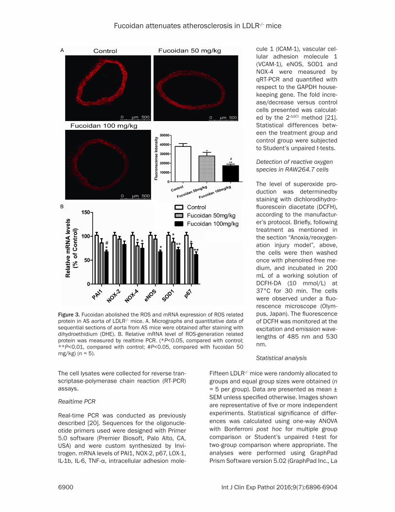
Figure 3. Fucoidan abolished the ROS and mRNA expression of ROS related protein in AS aorta of LDLR-/- mice. A. Micrographs and quantitative data of sequential sections of aorta from AS mice were obtained after staining with dihydroethidium (DHE). B. Relative mRNA level of ROS-generation related protein was measured by realtime PCR. (*P<0.05, compared with control; **P<0.01, compared with control; #P<0.05, compared with fucoidan 50 mg/kg) (n = 5).
cule 1 (ICAM-1), vascular cel-lular adhesion molecule 1 (VCAM-1), eNOS, SOD1 and NOX-4 were measured by qRT-PCR and quantified with respect to the GAPDH house-keeping gene. The fold incre- ase/decrease versus control cells presented was calculat-ed by the 2-ΔΔCt method [21]. Statistical differences betw- een the treatment group and control group were subjected to Student’s unpaired t-tests.
Detection of reactive oxygen species in RAW264.7 cells
The level of superoxide pro-duction was determinedby staining with dichlorodihydro-fluorescein diacetate (DCFH), according to the manufactur-er’s protocol. Briefly, following treatment as mentioned in the section “Anoxia/reoxygen-ation injury model”, above, the cells were then washed once with phenolred-free me- dium, and incubated in 200 mL of a working solution of DCFH-DA (10 mmol/L) at 37°C for 30 min. The cells were observed under a fluo-rescence microscope (Olym- pus, Japan). The fluorescence of DCFH was monitored at the excitation and emission wave-lengths of 485 nm and 530 nm.
Statistical analysis
Fucoidan attenuates atherosclerosis in LDLR-/- mice
6901 Int J Clin Exp Pathol 2016;9(7):6896-6904
Jolla, CA, USA). A P value<0.05 was considered to be statistically significant.
Results
Fucoidan decreases the serum lipid level in LDLR-/- mice
Similar to previous study, fucoidan notably reduced the concentration of serum total cho-lesterol (TC), triglyceride (TG), and low-density lipoprotein cholesterol (LDL-C) of hyperlipid-emic mice and increased the concentration of high-density lipoprotein cholesterol (HDL-C) (Table 1) [22].
Fucoidan reduces atherosclerotic plaque de-velopment in LDLR-/- mice
The efficacy of fucoidan in diet-induced athero-sclerosis in LDLR-/- mice was examined. Notably, compared with vehicle control group, fucoidan treatment (50 mg/kg, 100 mg/kg) significantly attenuated atherosclerotic lesion formation in the en face and prepared aorta and cross-sec-tions aortic valve (Figure 1A, 1F) of LDLR-/- mice fed a HCD for 16 weeks. These data suggest that fucoidan has potent anti-atherosclerotic effects in experimental atherosclerosis.
Fucoidan reduces macrophage infiltration and smooth muscle cell proliferation in atheroscle-rotic plaque development in LDLR-/- mice
Macrophage infiltration and smooth muscle cell proliferation are the key initiating and pri-
mary process in atherosclerosis. Macrophage infiltration and smooth muscle cells prolifera-tion in atherosclerosis plaque of different gro- ups were measured by immunohistochemistry of Mac-2 and SM-22 (Figure 2). Compared with control group, fucoidan reduces both the mac-rophage infiltration and smooth muscle cell proliferation.
Fucoidan inhibits ROS generation in LDLR-/- mice aorta
ROS in the plaque could modify LDL, react with endothelial-derived nitric oxide subsequently forming peroxynitrite, and amplify the expres-sion of various genes important for leukocyte recruitment within the arterial wall and leading to oxidant injury [23]. As compared to vehicle control, treatment with fucoidan abolished superoxide production as detected by dihydro-ethidium (DHE) fluorescence (Figure 3A). The following realtime PCR results of related pro-teins (PAI1, NOX-2, NOX-4, eNOS, SOD1, p67) revealed that 100 mg/kg fucoidan could inhibit the expression of ROS-generation related pro-tein significantly.
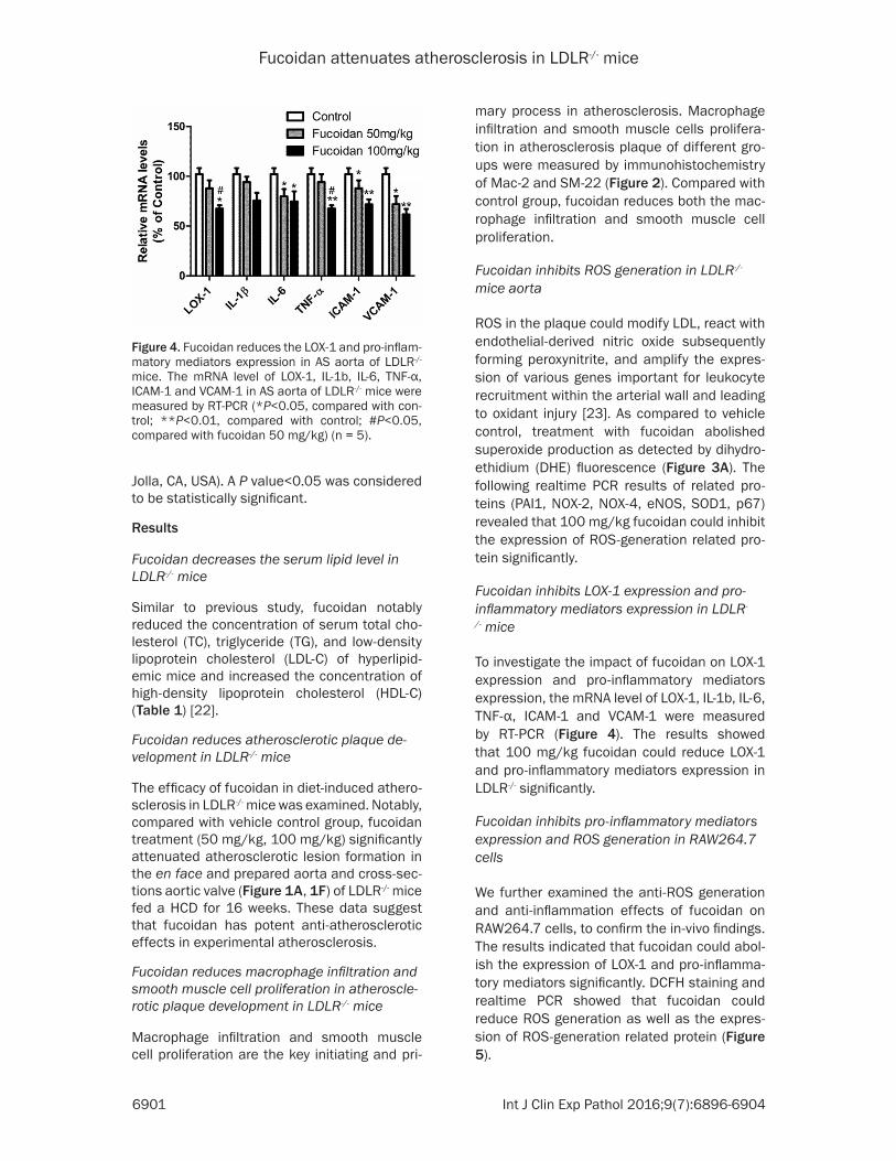
Fucoidan inhibits LOX-1 expression and pro-inflammatory mediators expression in LDLR-
/- mice
To investigate the impact of fucoidan on LOX-1 expression and pro-inflammatory mediators expression, the mRNA level of LOX-1, IL-1b, IL-6, TNF-α, ICAM-1 and VCAM-1 were measured by RT-PCR (Figure 4). The results showed that 100 mg/kg fucoidan could reduce LOX-1 and pro-inflammatory mediators expression in LDLR-/- significantly.
Fucoidan inhibits pro-inflammatory mediators expression and ROS generation in RAW264.7 cells
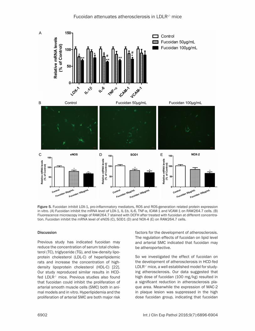
We further examined the anti-ROS generation and anti-inflammation effects of fucoidan on RAW264.7 cells, to confirm the in-vivo findings. The results indicated that fucoidan could abol-ish the expression of LOX-1 and pro-inflamma-tory mediators significantly. DCFH staining and realtime PCR showed that fucoidan could reduce ROS generation as well as the expres-sion of ROS-generation related protein (Figure 5).
Figure 4. Fucoidan reduces the LOX-1 and pro-inflam-matory mediators expression in AS aorta of LDLR-/- mice. The mRNA level of LOX-1, IL-1b, IL-6, TNF-α, ICAM-1 and VCAM-1 in AS aorta of LDLR-/- mice were measured by RT-PCR (*P<0.05, compared with con-trol; **P<0.01, compared with control; #P<0.05, compared with fucoidan 50 mg/kg) (n = 5).
Fucoidan attenuates atherosclerosis in LDLR-/- mice
6902 Int J Clin Exp Pathol 2016;9(7):6896-6904
Discussion
Previous study has indicated fucoidan may reduce the concentration of serum total choles-terol (TC), triglyceride (TG), and low-density lipo-protein cholesterol (LDL-C) of heperlipidemic rats and increase the concentration of high-density lipoprotein cholesterol (HDL-C) [22]. Our study reproduced similar results in HCD-fed LDLR-/- mice. Previous studies also found that fucoidan could inhibit the proliferation of arterial smooth muscle cells (SMC) both in ani-mal models and in vitro. Hyperlipidemia and the proliferation of arterial SMC are both major risk
factors for the development of atherosclerosis. The regulation effects of fucoidan on lipid level and arterial SMC indicated that fucoidan may be atheroportective.
So we investigated the effect of fucoidan on the development of atherosclerosis in HCD-fed LDLR-/- mice, a well established model for study-ing atherosclerosis. Our data suggested that high dose of fucoidan (100 mg/kg) resulted in a significant reduction in atherosclerosis pla- que area. Meanwhile the expression of MAC-2 in plaque lesion was suppressed in the high dose fucoidan group, indicating that fucoidan
Figure 5. Fucoidan inhibit LOX-1, pro-inflammatory mediators, ROS and ROS-generation related protein expression in vitro. (A) Fucoidan inhibit the mRNA level of LOX-1, IL-1b, IL-6, TNF-α, ICAM-1 and VCAM-1 on RAW264.7 cells. (B) Fluorescence microscopy image of RAW264.7 stained with DCFH after treated with fucoidan at different concentra-tion. Fucoidan inhibit the mRNA level of eNOS (C), SOD1 (D) and NOX-4 (E) on RAW264.7 cells.
Fucoidan attenuates atherosclerosis in LDLR-/- mice
6903 Int J Clin Exp Pathol 2016;9(7):6896-6904
could reduce the local inflammation. Further- more, we found that the expression of SM-22, a SMC marker, was significantly attenuated by fucoidan, suggesting that the anti-atheroscle-rosis effects of fucoidan may associated with the reduction of SMC proliferation. Moreover, the inhibitory effects of fucoidan on atheroscle-rosis may be involved in with its antioxidant activity [24], reflected by the decrease of ROS level in the aorta of high dose fucoidan group. Previous studies have demonstrated the pleio-tropic effects of fucoidan on cardiovascular dis-ease. Our results showed that the benefits effects of fucoidan on cardiovascular disease may be a combined results of various action.
To gain further insight into the molecular mech-anism by which fucoidan inhibits atherosclero-sis progress, we examined the expression of LOX-1, pro-inflammatory mediators (IL-1β, IL-6, TNF-α, ICAM-1, VACM-1) in LDLR-/- mice. LOX-1, a versatile scavenger receptor that is ubiqui-tously expressed in vascular cells is critical for the initiation and progression of atherosclero-sis [25]. Our study, for the first time, showed that fucoidan could attenuate the expression of LOX-1 with in plaque lesions. Further in-vitro experiment confirmed our in-vivo finding. Fu- coidan reduces the mRNA expression level of LOX-1 and pro-inflammatory mediators on RAW264.7 cells. Simultaneously, fucoidan abo- lished the ROS generation as well as the ROS-generation related protein expression.
Taken together, our present results have inden-tified a novel atheroprotective effect of fucoid-an. Fucoidan was shown to suppress the ROS related pathway and reduce the expression of LOX-1 and the diminishing the inflammation response. These observation indicate that fu- coidan could be exploited as an innovative car-diovascular drug to prevent or retard the pa- thogenesis of atherosclerotic cardiovascular diseases.
Acknowledgements
This study was supported by Qiqihar science and technology commission fundation (SPZD- 2013123).
Disclosure of conflict of interest
None.
Abbreviations
LDLR-/-, Low Density Lipoprotein Receptor-deficient; CD36, cluster of differentiation 36; HCD, high cholesterol diet; ICAM-1, intracellular adhesion molecule 1; NOX4, NADPH oxidase subunit 4; oxLDL, oxidized low-density lipopro-tein; ROS, reactive oxygen species; SMCs, sm- ooth muscle cells; SR-A, scavenger receptor-A; VCAM-1, vascular cellular adhesion molecule 1.
Address correspondence to: Xin Wang, Department of Emergency, The First Hospital of Jilin University, Changchun 130021, China. E-mail: [email protected]
References
[1] Ross R. Atherosclerosis--an inflammatory dis-ease. N Engl J Med 1999; 340: 115-126.
[2] Go AS, Mozaffarian D, Roger VL, Benjamin EJ, Berry JD, Borden WB, Bravata DM, Dai S, Ford ES, Fox CS, Franco S, Fullerton HJ, Gillespie C, Hailpern SM, Heit JA, Howard VJ, Huffman MD, Kissela BM, Kittner SJ, Lackland DT, Lichtman JH, Lisabeth LD, Magid D, Marcus GM, Marelli A, Matchar DB, McGuire DK, Mohler ER, Moy CS, Mussolino ME, Nichol G, Paynter NP, Schreiner PJ, Sorlie PD, Stein J, Turan TN, Virani SS, Wong ND, Woo D, Turner MB; American Heart Association Statistics Com- mittee and Stroke Statistics Subcommittee. Heart disease and stroke statistics--2013 up-date: a report from the American Heart Association. Circulation 2013; 127: e6-e245.
[3] Hansson GK and Libby P. The immune re-sponse in atherosclerosis: a double-edged sword. Nat Rev Immunol 2006; 6: 508-519.
[4] Li AC and Glass CK. The macrophage foam cell as a target for therapeutic intervention. Nat Med 2002; 8: 1235-1242.
[5] Kato R, Mori C, Kitazato K, Arata S, Obama T, Mori M, Takahashi K, Aiuchi T, Takano T and Itabe H. Transient increase in plasma oxidized LDL during the progression of atherosclerosis in apolipoprotein E knockout mice. Arterioscler Thromb Vasc Biol 2009; 29: 33-39.
[6] Sawamura T, Kume N, Aoyama T, Moriwaki H, Hoshikawa H, Aiba Y, Tanaka T, Miwa S, Katsura Y, Kita T and Masaki T. An endothelial receptor for oxidized low-density lipoprotein. Nature 1997; 386: 73-77.
[7] Kataoka H, Kume N, Miyamoto S, Minami M, Moriwaki H, Murase T, Sawamura T, Masaki T, Hashimoto N and Kita T. Expression of lectin-like oxidized low-density lipoprotein receptor-1 in human atherosclerotic lesions. Circulation 1999; 99: 3110-3117.
Fucoidan attenuates atherosclerosis in LDLR-/- mice
6904 Int J Clin Exp Pathol 2016;9(7):6896-6904
[8] Li D, Liu L, Chen H, Sawamura T, Ranganathan S and Mehta JL. LOX-1 mediates oxidized low-density lipoprotein-induced expression of ma-trix metalloproteinases in human coronary ar-tery endothelial cells. Circulation 2003; 107: 612-617.
[9] Li D and Mehta JL. Intracellular signaling of LOX-1 in endothelial cell apoptosis. Circ Res 2009; 104: 566-568.
[10] Xu S, Liu Z, Huang Y, Le K, Tang F, Huang H, Ogura S, Little PJ, Shen X and Liu P. Tanshinone II-A inhibits oxidized LDL-induced LOX-1 expres-sion in macrophages by reducing intracellular superoxide radical generation and NF-kappaB activation. Transl Res 2012; 160: 114-124.
[11] Xu S, Ogura S, Chen J, Little PJ, Moss J and Liu P. LOX-1 in atherosclerosis: biological func-tions and pharmacological modifiers. Cell Mol Life Sci 2013; 70: 2859-2872.
[12] Besler C, Heinrich K, Rohrer L, Doerries C, Riwanto M, Shih DM, Chroni A, Yonekawa K, Stein S, Schaefer N, Mueller M, Akhmedov A, Daniil G, Manes C, Templin C, Wyss C, Maier W, Tanner FC, Matter CM, Corti R, Furlong C, Lusis AJ, von Eckardstein A, Fogelman AM, Luscher TF and Landmesser U. Mechanisms underlying adverse effects of HDL on eNOS-activating pathways in patients with coronary artery dis-ease. J Clin Invest 2011; 121: 2693-2708.
[13] Feldman SC, Reynaldi S, Stortz CA, Cerezo AS and Damont EB. Antiviral properties of fucoid-an fractions from Leathesia difformis. Phyto- medicine 1999; 6: 335-340.
[14] Wang J, Zhang Q, Zhang Z, Song H and Li P. Potential antioxidant and anticoagulant capac-ity of low molecular weight fucoidan fractions extracted from Laminaria japonica. Int J Biol Macromol 2010; 46: 6-12.
[15] Bojakowski K, Abramczyk P, Bojakowska M, Zwolinska A, Przybylski J and Gaciong Z. Fucoidan improves the renal blood flow in the early stage of renal ischemia/reperfusion in-jury in the rat. J Physiol Pharmacol 2001; 52: 137-143.
[16] Li N, Zhang Q and Song J. Toxicological evalua-tion of fucoidan extracted from Laminaria ja-ponica in Wistar rats. Food Chem Toxicol 2005; 43: 421-426.
[17] Zhang Q, Li Z, Xu Z, Niu X and Zhang H. Effects of fucoidan on chronic renal failure in rats. Planta Med 2003; 69: 537-541.
[18] Kilkenny C, Browne W, Cuthill IC, Emerson M, Altman DG; National Centre for the Repla- cement, Refinement and Reduction of Amimals in Research. Animal research: reporting in vi- vo experiments: the ARRIVE guidelines. Br J Pharmacol 2010; 160: 1577-1579.
[19] McGrath JC, Drummond GB, McLachlan EM, Kilkenny C and Wainwright CL. Guidelines for reporting experiments involving animals: the ARRIVE guidelines. Br J Pharmacol 2010; 160: 1573-1576.
[20] Xu S, Little PJ, Lan T, Huang Y, Le K, Wu X, Shen X, Huang H, Cai Y, Tang F, Wang H and Liu P. Tanshinone II-A attenuates and stabilizes ath-erosclerotic plaques in apolipoprotein-E knock-out mice fed a high cholesterol diet. Arch Biochem Biophys 2011; 515: 72-79.
[21] Livak KJ and Schmittgen TD. Analysis of rela-tive gene expression data using real-time quantitative PCR and the 2(-Delta Delta C(T)) Method. Methods 2001; 25: 402-408.
[22] Huang L, Wen K, Gao X and Liu Y. Hypolipidemic effect of fucoidan from Laminaria japonica in hyperlipidemic rats. Pharm Biol 2010; 48: 422-426.
[23] Mugge A. The role of reactive oxygen species in atherosclerosis. Z Kardiol 1998; 87: 851-864.
[24] Religa P, Kazi M, Thyberg J, Gaciong Z, Swedenborg J and Hedin U. Fucoidan Inhibits Smooth Muscle Cell Proliferation and Reduces Mitogen-activated Protein Kinase Activity. Eur J Vasc Endovasc Surg 2000; 20: 419-426.
[25] Chen M, Masaki T and Sawamura T. LOX-1, the receptor for oxidized low-density lipoprotein identified from endothelial cells: implications in endothelial dysfunction and atherosclerosis. Pharmacol Ther 2002; 95: 89-100.