Embed Size (px)
Citation preview

139
Abstract
Purpose: This study investigated the flexural properties, shear bond strength (SBS) and interface to dentin of three recently developed self-adhesive bulk-fill materials. Methods: Bars of Surefil One (SO), Cention N (CN), Activa BioActive Restorative (AB) and EQUIA Forte HT Fil (EQUIA) were tested for flex-ural strength and flexural modulus in self-curing and light-curing modes. In addition, SBS to dentin was tested in specimens without pretreatment and after application of universal adhesive (Scotchbond Universal). EQUIA was used as the control material.Results: The flexural properties were significantly better in light-curing mode for all materials except CN. CN had the highest SBS values after universal adhesive application (33.8 MPa), and SO had the highest SBS without pretreatment (20.9 MPa). Conclusion: The mechanical and adhesive properties of these new materi-als varied widely.
Keywords; flexural strength, scanning electron microscopy, self-adhesive bulk-fill materials, shear bond strength
Introduction
Self-adhesive materials are a major development for direct restorative materials because the absence of a specific adhesive protocol makes them easier to use [1]. Bulk-fill techniques simplify procedures by limiting the number of increments needed to fill an entire cavity [2].
The glass ionomer cement (GIC) family is the most widely used self-adhesive bulk-fill material in direct restorations [3]. GICs have other interesting properties that are directly linked to their chemistry, such as moisture tolerance [4] and rechargeable fluoride release from fluoro-alumino-silicate (FAS) fillers, which leads to potential dental hard tissue remineralization [5] and cariostatic effects [6]. These materials can be resin-free, such as the GICs and their improved formulation—called high-viscosity glass ionomer cements (HV-GICs)—or may contain additional resinous content (mainly 2-hydroxyethylmethacrylate), such as resin-modified glass ionomer cement (RM-GIC) [3].
Conventional GICs are no longer used for definitive restorations because of their strong tendency to abrasion, fracture and debonding [7]. RM-GICs possess improved adhesion [8] and flexural characteristics but still have low abrasion resistance and must be laminated in accordance with the manufacturer’s instructions [9]. HV-GICs can be successfully used as definitive restorations of occlusal and limited proximal cavities; however, their enamel and dentinal bonding strength values are inferior to those of resin composites, [10,11] and bulk fracture may occur because
of low flexural strength [12]. Recently, some new self-adhesive resinous materials with claimed fluoride-releasing and “bulk-fill” properties were introduced as “bioactive materials” or “smart materials”. These materials differ in chemical composition from the GIC family [13] and could be an alternative that exceeds their performance [9].
Many parameters must be considered when restoring a large cavity, including the mechanical properties of the restorative material and dentin bond strength [14]. Few studies have evaluated the mechanical proper-ties and bonding ability of Activa BioActive Restorative (Pulpdent Corp., Watertown, MA, USA, launched in 2013), Cention N (Ivoclar-Vivadent AG, Schaan, Liechtenstein, launched in 2016) and Surefil One (Dentsply-Sirona, Konstanz, Germany, launched in 2019), which are putative competitors to HV-GICs and have extended indications. In contrast to HV-GICs, these materials are described as being able to restore all types of cavities when the tooth does not require cuspid coverage.
The aim of this study was, first, to study the flexural properties of these materials, with different curing protocols, and, second, to evaluate their dentin bond strength and the bonded interface. The null hypotheses tested were (i) that flexural properties would not differ in relation to material or curing mode, (ii) that there would be no differences in dentin bond strength between materials, with or without the use of an adhesive system and (iii) that there would be no differences in the interface pattern between tested groups.
Materials and Methods
Materials used and experimental proceduresSurefil One, Cention N and Activa BioActive Restorative were compared to HV-GICs. The materials, manufacturers, batch numbers, composition [9,13] and characteristics [15,16] are presented in Table 1. These materials were tested for flexural strength (FS), flexural modulus (E) and dentin bond strength and were compared to a recent HV-GIC (EQUIA Forte HT, GC Corp., Tokyo, Japan), used as a control group.
Flexural strength and flexural modulus testingOne hundred forty bars were made by using a 2 × 2 × 25 mm silicon mold (EXA’lence, GC Corp.) according to ISO 4049 [17]. Seven groups (n = 20) were evaluated: Surefil One in self-cure mode (SO-SC), Surefil One in light-curing mode (SO-LC), Cention N in self-cure mode (CN-SC), Cention N in light-curing mode (CN-LC), Activa BioActive Restorative in self-cure mode (AB-SC), Activa BioActive Restorative in light-curing mode (AB-LC) and EQUIA Forte HT Fil in self-cure mode followed by an application of coating material (EF-SC). Photopolymerization was performed with a polywave curing light at a minimum output of 950 mW/cm2 (Valo Grand Cordless, Ultradent Products, South Jordan, UT, USA).
The abbreviations, groups and detailed light-curing and self-curing protocols are presented in Table 2. Material bars were then stored in water for 2 weeks at 37°C before performing the mechanical tests, to ensure that the maturation process, which requires several hours for the GIC family, was mostly completed. For each group, the flexural strength and flexural modulus of samples were tested by a three-point bending test on a univer-sal testing machine (Shimadzu AGS-X, Shimadzu Corp., Kyoto, Japan). Each specimen was placed at the center of a universal tester between two
Journal of Oral Science, Vol. 63, No. 2, 139-144, 2021
Original article
Flexural properties and dentin adhesion in recently developed self-adhesive bulk-fill materialsPhilippe François1,2), Anis Remadi1), Stéphane Le Goff1), Sarah Abdel-Gawad1), Jean-Pierre Attal1,3), and Elisabeth Dursun1,4)
1) Innovative Dental Materials and Interfaces Research Unit (UR 4462), University of Paris, Montrouge, France2) Department of Restorative Dentistry, Bretonneau Hospital, Paris, France3) Department of Restorative Dentistry, Charles Foix Hospital, Ivry-sur-Seine, France4) Department of Pediatric Dentistry, Henri Mondor Hospital, Créteil, France
(Received September 2, 2020; Accepted November 24, 2020)
Correspondence to Dr. Elisabeth Dursun, Innovative Dental Materials and Interfaces Research Unit (UR 4462), University of Paris, 1 rue Maurice Arnoux, 92120 Montrouge, FranceE-mail: [email protected]
J-STAGE Advance Publication: February 17, 2021Color figures can be viewed in the online issue at J-STAGE.doi.org/10.2334/josnusd.20-0448DN/JST.JSTAGE/josnusd/20-0448
140
crossheads with a width of 20 mm, and the maximum load was measured by applying a vertical load to the center of the specimen at a crosshead speed of 0.5 mm/min until fracture.
Maximum load (fracture load) was recorded in newtons, and flexural strength (FS) was calculated in megapascals, as follows: FS = (3Pl)/(2wb2), where P is the applied load (in N), l is the test interval (in mm), w
is the width of the specimen (in mm) and b is the thickness of the specimen (in mm).
The flexural modulus (E) was calculated from the three-point bending test and expressed in gigapascals, as follows: E = (Pl3)/(4wb3d), where d is the deflection corresponding to load P.
Table 2 Abbreviations, sample preparation and curing protocol for testing of flexural strength and flexural modulus
Material and curing protocol used Bonding protocolSurefil One, self-curing protocol (SO-SC) For each sample, a SO capsule was mechanically mixed (Silver Mix 90 Mixer, GC Corp.) for 10 s according to the manufacturer’s
instructions, injected in excess inside a silicon mold and covered on the top surface by a Mylar band and a glass slab under finger pressure. After 10 min at room temperature, the sample bar was unmolded and stored in 37°C water for 2 weeks.
Surefil One, light-curing protocol (SO-LC) For each sample, a SO capsule was mechanically mixed (Silver Mix 90 Mixer, GC Corp.) for 10 s according to the manufacturer’s instructions, injected in excess inside a silicon mold and covered on the top surface by a Mylar band and a glass slab under finger pressure. The upper surface of the sample bar was light-cured for 60 s at three points and then unmolded and stored in 37°C water for 2 weeks.
Cention N, self-curing protocol (CN-SC) For each sample, CN was hand-mixed according to the manufacturer’s instructions, inserted in excess inside a silicon mold and covered on the top surface by a Mylar band and a glass slab under finger pressure. After 10 min at room temperature, the sample bar was unmolded and stored in 37°C water for 2 weeks.
Cention N, light-curing protocol (CN-LC) For each sample, CN was hand-mixed according to the manufacturer’s instructions, inserted in excess inside a silicon mold and covered on the top surface by a Mylar band and a glass slab under finger pressure. The upper surface of the sample bar was light-cured for 60 s at three points and then unmolded and stored in 37°C water for 2 weeks.
Activa Bioactive Restorative, self-curing protocol (AB-SC)
For each sample, AB was injected in excess inside a silicon mold and covered on the top surface by a Mylar band and a glass slab under finger pressure. After 24 h at room temperature (material was unset if removed earlier), the sample bar was unmolded and stored in 37°C water for 2 weeks.
Activa Bioactive Restorative, light-curing protocol (AB-LC)
For each sample, AB was injected in excess inside a silicon mold and covered on the top surface by a Mylar band and a glass slab under finger pressure. The upper surface of the sample bar was light-cured for 60 s at three points and then unmolded and stored in 37°C water for 2 weeks.
EQUIA Forte HT Fil, with self-curing protocol followed by application of coating material (EF-SC)
For each sample, an EF capsule was mechanically mixed (Silver Mix 90 Mixer, GC Corp.) for 10 s according to the manufacturer’s instructions, injected in excess inside a silicon mold and covered on the top surface by a Mylar band and a glass slab under finger pressure. After 10 min at room temperature, the coating material (EQUIA Forte Coat, GC Corp.) was applied on the top surface of the sample bar with a microbrush, rubbed for 20 s (not air-dried) and light-cured for 60 s at three points. Then, the bar was unmolded and stored in 37°C water for 2 weeks.
Table 1 Abbreviations, manufacturers, batch numbers and composition of the materials used
Materials Abbreviation Shade Manufacturer Batch number Composition / characteristics Properties and useNew resinous fluoride-releasing materials
Surefil One SO A3 Dentsply-Sirona,Konstanz, Germany
1907000695 Powder: silanated aluminum-phosphor-strontium-sodium-fluoro-silicate glass, dispersed silicon dioxide, ytterbium fluoride, pigmentsLiquid: acrylic acid, polycarboxylic acid, bifunctional acrylate, self-cure initiator, camphorquinone, stabilizer
Self-adhesive bulk-fill resinous restorative material with ionic release after polymerization.Does not require a bonding agent, regardless of the cavity
Cention N CN U Ivoclar-Vivadent,Schaan, Liechtenstein
X54163 X44732 Y15861
Powder: barium aluminum silicate glass, ytterbium trifluoride, isofiller, calcium barium aluminum fluorosilicate glass, calcium fluoro silicate glassLiquid: urethane dimethacrylate, tricyclodecan-dimethanol dimethacrylate, tetramethyl-xylylene diurethane dimethacrylate, polyethylene glycol 400 dimethacrylate, ivocerin, hydroxyperoxide
Non-adhesive bulk-fill resinous restorative material with ionic release after polymerization under acidic challengeBonding agent not required for retentive cavities but required for a non-retentive cavity
Activa Bioactive Restorative
AB A3 Pulpdent, Watertown,MA, USA
190307 Powder: silanated bioactive glass and calcium, silanated silica, sodium fluorideLiquid: diurethane modified by the insertion of a hydrogenated polybutadiene and other methacry-late monomers, modified polyacrylic acid
Self-adhesive bulk-fill resinous restorative material with ionic release after polymerization.Bonding agent mandatory for non-retentive cavities but optional for retentive cavities (initial manufacturer’s instructions)
Former non-resinous fluoride-releasing material(HV-GIC)
EQUIA Forte HT Fil
EF A3 GC Corp.,Tokyo, Japan
192051190710
Powder: fluoroaluminosilicate glass, polyacrylic acid, iron oxideLiquid: polybasic carboxylic acid, water
Self-adhesive bulk-fill resinous restorative material with ionic release.No bonding agent required, regardless of cavity
Dental adhesiveSystem
Scotchbond Universal
SBU 3M ESPE, St Paul,MN, USA
5271401 HEMA, Bis-GMA, decamethylene dimethacry-late, ethanol, silane treated silica, water, MDP, copolymer of acrylic and itaconic acid, dimeth-ylaminoethyl methacrylate, camphorquinone, dimethylaminobenzoate, 2,6-di-tert-butyl-p-cresol, photoinitiators
-
Etching product Scotchbond Universal Etchant
SE 3M ESPE 4651322 Orthophosphoric acid (37%), water, synthetic amorphous silica, polyethylene glycol, aluminum oxide
-
Cavity cleaning product
Dentin Conditioner
DC GC Corp. 1805111 Distilled water, polyacrylic acid -
HEMA, 2-hydroxyethyl methacrylate; Bis-GMA, bisphenol A-glycidyl methacrylate; MDP, 10-methacryloyloxydecyl dihydrogen phosphate

141
Dentin shear bond strength tests, failure mode determination and scanning electron microscopy examinationOne hundred and fifty-four freshly extracted human permanent molars were collected from adults after extraction, cleaned of soft tissues, stored at 4°C in a solution of 1% chloramine and used within 3 months. Teeth were obtained from the dental departments of AP-HP, France. All experiments were conducted in accordance with the principles articulated in the Decla-ration of Helsinki. All teeth were collected with the informed oral consent of all patients, in accordance with the ethical guidelines set by French law and with specific authorization by Paris university dental school (n°DC-2009-927, Cellule Bioéthique DGRI/A5, Paris, France).
The criterion for tooth selection was absence of cracks and decay. The greater portion of the roots was removed with sandpaper (80 grit) by using a polishing machine (Planopol 3, Struers, Kobenhavn, Denmark). The occlusal surface of the crowns was then abraded with water-cooled sand-paper (800 grit) to expose a flat dentin surface (>7 mm2) with a roughness corresponding to the finishing obtained with a red ring diamond bur. The residual crowns were embedded in self-curing acrylic resin (Plexcil-Escil, Chassieu, France) in a plastic cylinder (diameter: 25 mm, depth: 15 mm), with the flat dentin surface exposed. The surfaces were inspected under ×40 magnification to ensure that the enamel had been completely removed and that the dentin was cleared of debris.
These teeth were randomly assigned to seven groups (n = 22): Surefil One only, according to the manufacturer’s instructions (SO-Only); Surefil One, after universal adhesive application in self-etch mode (SO-SBU); Cention N only, according to the manufacturer’s instructions (CN-Only); Cention N, after universal adhesive application in self-etch mode, accord-ing to the manufacturer’s instructions (CN-SBU); Activa BioActive Restorative only, according to the former manufacturer’s instructions (AB-Only); Activa BioActive Restorative, after universal adhesive application in self-etch mode, according to the manufacturer’s instruction (AB-SBU); and EQUIA Forte HT Fil, after dentin conditioner application (EF-DC). A cylindrical Teflon mold was placed on each sample to build a 3 mm–high cylinder of material (diameter = 1.5 mm) with a flat base of 7 mm². The abbreviations, groups and detailed adhesive protocols are presented in Table 3. After the material set, the mold was removed, and any excess material was gently removed from around the base of the material cylinder with a scalpel. All the samples were stored in water at 37°C for 48 h.
For each group, 20 teeth were used for shear bond strength (SBS) test-ing and two were used for scanning electron microscopy (SEM) interfacial observation. SBS was determined in a universal testing machine (LRX, Lloyd Instruments, Fareham, UK). The shear force was applied at the
material cylinder/dentin interface, with a chisel-shaped blade parallel to the dentin surface. A crosshead speed of 0.5 mm/min was used.
The debonded specimens were observed under a binocular microscope (BZH10, Olympus, Hamburg, Germany) at ×30 magnification, and failure mode was classified as follows:
- Type CF-D: cohesive failure within the dentin- Type AF: adhesive failure at the interface between the material and
dentin- Type MF: mixed failure (adhesive and cohesive failure within the
material)- Type CF-M: cohesive failure within the restorative materialFor SEM examination, after the additional inclusion in self-cure acrylic
resin of the material cylinder, samples were sectioned perpendicularly to the bonded interface with a low-speed diamond saw (Isomet, Buehler, Coventry, UK) and water cooling, as near as possible to the center of the cylinder. The sections obtained were polished with abrasive disks of decreasing grit size (400, 800, 1,200, 2,400, and 4,000 SiC) and then with diamond particles of 3 μm and 1 μm. Finally, the sections were etched with orthophosphoric acid for 10 s (Scotchbond Universal Etchant, 3M ESPE, St. Paul, MN, USA). The samples were cleaned by ultrasonication after each step and then dehydrated in ethanol and metallized with gold (Sputter Coater, Bio-rad, Marnes-la-Coquette, France) for microscopy examination (JSM-6400, JEOL LTD, Tokyo, Japan).
Statistical analysisNormal distribution was confirmed by the Shapiro-Wilk test, and equal-ity of variance was assessed using Levene’s test before the tests were performed. SBS data were expressed as means and standard deviations. One-way ANOVA followed by Tukey’s post-hoc test was used to inves-tigate differences in flexural strength, flexural modulus and SBS between groups. Failure mode was analyzed by Fisher’s exact test for single com-parisons between groups and in pairwise analysis. In all tests, the chosen significance level was α = 0.05. R software (version 3.6.1; R Foundation for Statistical Computing, Vienna, Austria) was used for all statistical calculations.
Results
Flexural propertiesThe flexural properties for all experimental groups are summarized in Table 4. The Shapiro-Wilk test confirmed a normal distribution of flexural properties values among all groups (P > 0.05), and Levene’s test showed
Table 3 Sample preparation and dentin bonding protocol for the shear-tested groups
Group tested Bonding protocolSurefil One (SO-Only) After excess water was gently removed by drying with oil-free air, a Teflon mold was placed on dentin, and SO was injected in one
3-mm increment and light-cured for 20 s, according to the manufacturer’s instructions. After 15 min, the mold was removed, and the sample was stored in water at 37°C for 48 h.
Surefil One after SBU application with a self-etch protocol (SO-SBU)
After excess water was gently removed by drying with oil-free air, SBU was applied on dentin, rubbed for 20 s, gently air-dried for 5 s (until it no longer moved, i.e. complete solvent evaporation) and light-cured for 20 s. Then, a Teflon mold was placed on dentin, and SO was injected in one 3-mm increment and light-cured for 20 s. After 15 min, the mold was removed, and the sample was stored in water at 37°C for 48 h.
Cention N (CN-Only) After excess water was gently removed by drying with oil-free air, a Teflon mold was placed on dentin, and CN was inserted with a spatula in one 3-mm increment and light-cured for 20 s, according to the manufacturer’s instructions. After 15 min, the mold was removed, and the sample was stored in water at 37°C for 48 h.
Cention N after SBU application with a self-etch protocol (CN-SBU)
After excess water was gently removed by drying with oil free-air, SBU was applied on dentin, rubbed for 20 s, gently air-dried for 5 s (until it no longer moved, i.e. complete solvent evaporation) and light-cured for 20 s. Then, a Teflon mold was placed on dentin, and CN was inserted with a spatula in one 3-mm increment and light-cured for 20 s, according to the manufacturer’s instructions. After 15 min, the mold was removed, and the sample was stored in water at 37°C for 48 h.
Activa Bioactive Restorative (AB-Only) After excess water was gently removed by drying with oil-free air, orthophosphoric acid was applied on dentin for 5 s and then rinsed thoroughly for 15 s, and excess water was blotted with a cotton pellet, according to the manufacturer’s instructions. A Teflon mold was placed on dentin, and AB was injected in one 3-mm increment and light-cured for 20 s, according to the initial manufac-turer’s instructions. After 15 min, the mold was removed, and the sample was stored in water at 37°C for 48 h.
Activa Bioactive Restorative after SBU application with a self-etch protocol (AB-SBU)
After excess water was gently removed by drying with oil-free air, SBU was applied on dentin, rubbed for 20 s, gently air-dried for 5 s (until it no longer moved, i.e. complete solvent evaporation) and light-cured for 20 s. Then, a Teflon mold was placed on dentin, and AB was injected in one 3-mm increment and light-cured for 20 s, according to the manufacturer’s instructions. After 15 min, the mold was removed, and the sample was stored in water at 37°C for 48 h.
EQUIA Forte HT Fil after Dentin Conditioner application (EF-DC)
After excess water was air removed by drying with oil-free air, DC was applied on dentin for 20 s and then rinsed thoroughly for 15 s, and excess water was blotted with a cotton pellet. A Teflon mold was placed on dentin, and EF was injected in one 3-mm increment, according to the manufacturer’s instructions. After 15 min, the mold was removed, and the sample was stored in water at 37°C for 48 h.

142
equality of variance (P > 0.05). One-way ANOVA followed by Tukey’s post-hoc test revealed significant differences (P < 0.05).
Flexural strength significantly differed in relation to material and curing protocol. CN-SC and CN-LC had the highest flexural strength (86.3 ± 7.8 MPa and 83.3 ± 15.0 MPa, respectively). Flexural strength was higher with a light-curing protocol for SO-LC and AB-LC (45.3 ± 11.7 MPa and 67.7 ± 11.8 MPa, respectively) than with a self-curing protocol (24.5 ± 5.8 MPa and 45.7 ± 10.8 MPa, respectively). Flexural strength was significantly lower for EF-SC (22.7 ± 6.9 MPa) than for the other groups.
Flexural modulus significantly differed in relation to material. EF-SC had the highest flexural modulus (12.0 ± 2.1 GPa), whereas flexural modu-lus values were lowest for AB-LC and AB-SC, in light-curing (2.3 ± 0.3 GPa) and self-curing (1.4 ± 0.4 GPa) modes, respectively.
SBS and failure analysisSBS values for all experimental groups are summarized in Table 5. The Shapiro-Wilk test confirmed a normal distribution of SBS values in all groups (P > 0.05), and Levene’s test showed equality of variance (P > 0.05). One-way ANOVA followed by Tukey’s post-hoc test revealed sig-nificant differences in SBS values (P < 0.05).
SBS significantly different in relation to material and dentin treatment. All the materials tested with an adhesive had higher SBS values. SBS was significantly higher for CN-SBU (33.8 ± 5.7 MPa) and AB-SBU (28.9 ± 5.2 MPa) than for SO-SBU (20.9 ± 4.1 MPa). However, in the absence of adhesive, SBS was significantly higher for SO-Only (14.0 ± 3.4 MPa) than for EF-DC (8.0 ± 1.8 MPa), but these values were significantly higher than those for AB-Only (4.4 ± 2.5 MPa) and CN-Only (3.0 ± 1.0 MPa).
Failure modes are listed in Table 6. Fisher’s exact test indicated signifi-cant differences between groups. The most frequent failure for AB-SBU and CN-SBU was cohesive failure in dentin, and the frequency of such failures in these groups significantly differed from those in the other groups.
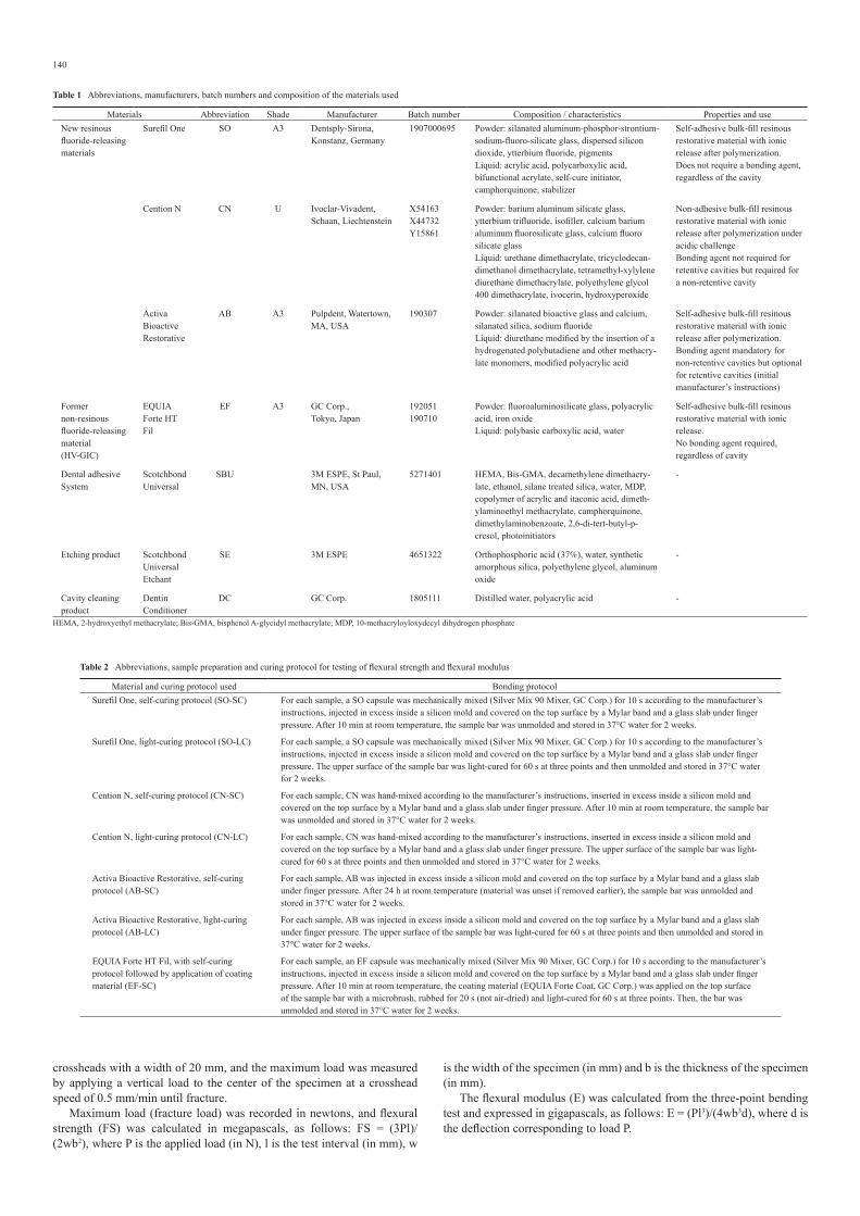
SEM examinationFigure 1 shows SEM images of Surefil One, Cention N and Activa BioAc-tive Restorative, with or without application of a universal adhesive, and Fig. 2 shows an SEM image of the control group, EQUIA Forte HT Fil, after application of Dentin Conditioner at ×1,000 magnification.
In the EF-DC group, HV-GIC/dentin appeared to be continuous. No material deposits are present inside dentin tubules. Many gaps are visible between FAS fillers.
In the CN-Only and AB-Only groups, the material/dentin interface appears discontinuous. No material is deposited inside dentin tubules. Gaps are visible between some fillers (especially for CN-Only) and the resin matrix. In the SO-Only, SO-SBU, CN-SBU and AB-SBU groups, the material/dentin interface appears continuous, and tags can be seen inside dentin tubules. Gaps are visible between some fillers (especially for CN-SBU) and the resin matrix.
Discussion
An ideal restorative material should be biocompatible, mechanically resis-tant and aesthetically pleasing. It should also yield spontaneous, durable adhesion to dental tissues and have bulk-fill properties and bioactivity.
HV-GICs, the present control material, are the most used self-adhesive bulk-fill material family for definitive restoration [3,12]. However, because of their tendency to debonding and bulk fracture, they have limited indi-cations as definitive materials [12] and lower dentin bond strength than resin composites [18], a limitation that cannot be improved by use of an adhesive system. Moreover, their aesthetic outcomes are worse than those of resin composites, and microleakage at restorative margins has been reported [19].
Resinous self-adhesive, bulk-fill, “hybrid materials” with putative bioactivity and enhanced indications for direct restorations were recently introduced. However, few studies have evaluated their chemical character-istics, clinical behavior and flexural and bonding properties. In this study, all materials underwent three-point bending testing with a self-curing and
Table 5 Mean (standard deviations) for shear bond strength (SBS) in the groups tested
Group SBS (±SD)
SO-Only 14.0d (±3.4)
SO-SBU 20.9c (±4.1)
CN-Only 3.0f (±1.0)
CN-SBU 33.8a (±5.7)
AB-Only 4.4f (±2.5)
AB-SBU 28.9b (±5.2)
EF-DC 8.0e (±1.8)Values with the same superscript letter are not significantly different (P > 0.05). DC, Dentin Conditioner; SBU, Scotchbond Universal
Table 6 Failure mode for the groups tested
Group No. of samples CF-D AF MF CF-FRM
1 SO-Only 20ab 0 15 4 1
2 SO-SBU 20b 2 7 8 3
3 CN-Only 20a 0 15 4 1
4 CN-SBU 20c 15 1 5 0
5 AB-Only 20a 0 20 0 0
6 AB-SBU 20c 13 4 3 0
7 EF-DC 20a 0 19 1 0Values with the same superscript letter are not significantly different (P > 0.05). CF-D, cohesive failure in dentin; CF-FRM, cohesive failure in fluoride-releasing material; MF, mixed failure; AF, adhesive failure
Table 4 Means (standard deviations) for flexural strength (MPa) and flexural modulus (GPa) in the groups tested
Surefil One Cention N Activa Bioactive RestorativeEQUIA
Forte HT Fil
SC LC SC LC SC LC SC
Flexural strength
24.5d
(±5.8)45.3c
(±11.7)86.3a
(±7.8)83.3a
(±15.0)45.7c
(±10.8)67.7b
(±11.8)22.7d
(±6.9)
Flexural modulus
7.7B
(±0.9)7.4B
(±0.8)7.6B
(±0.7)7.4B
(±0.8)1.4C
(±0.4)2.3C
(±0.3)12.0A
(±2.1)Values with the same superscript letter are not significantly different (P > 0.05). LC, light-cure; SC, self-cure

143
light-curing protocol, to determine how the polymerization reaction affects mechanical performance and the setting reaction of materials. Significant differences were observed in relation to material and curing mode; thus, the first null hypothesis was rejected.
When a definitive restorative material must be placed in a stress-bearing area, such as an occlusal posterior area, ISO 4049 standards recommend a flexural strength of at least 80 MPa [17]. However, many studies have reported that materials that satisfy this standard have increased fracture susceptibility, especially in proximal areas [20]. Flexural strength is there-fore an important predictor of the clinical success of a material.
Cention N satisfies the minimum ISO 4049 value. It exhibited the highest flexural strength in self-cured and light-cured reactions, and no significant difference between these modes, which suggests promising mechanical results in stress-bearing areas. Surefil One and Activa BioAc-tive Restorative had values below the ISO 4049 standard. Both had higher flexural strength in the light-curing mode, despite the absence of significant differences in surface microhardness and flexural modulus. These materi-als, in contrast to Cention N, are based on a double-setting reaction (an acid-base reaction and resinous polymerization) [21]. Competition between these reactions, as previously seen in RM-GICs [22], could explain the mechanical differences between curing modes. A decreased conversion rate in self-cure mode was also suspected in another study of Surefil One [9]. This hypothesis requires confirmation in more-comprehensive studies of polymerization.
As compared with HV-GIC, all the new hybrid materials had better flexural strength values in light-curing mode and some had better values in self-curing mode (Cention N and Activa BioActive Restorative). Thus, they may be stronger alternatives for posterior restorations with simplified protocols, even if the flexural strength needs to be improved to satisfy ISO standards [17]. The low flexural strength values observed for EF-SC could explain why bulk fracture was reported as the main cause of failure for large HV-GIC restorations [12,23].
Analysis of the flexural modulus showed important differences between groups. Many previous studies showed that flexural modulus was strongly associated with material composition [23,24]. The findings of this study suggest that the chemical characteristics of the three recently developed self-adhesive hybrid materials greatly differ from those of resin-free HV-GICs. Further studies of the chemical profiles of these new formulations
are thus warranted. SBS testing is preferred to other bonding tests, such as microtensile bond strength testing. In fact, because some materials have lower bond strengths than those of resin composites, other bonding tests would be difficult to perform. SBS is effective in overcoming this limita-tion [25].
Bonding values and failure patterns significantly differed in relation to material and adhesive procedure; thus, the second null hypothesis was rejected. All the materials tested with an adhesive had higher SBS values. Similarly, dentin bond strength of RM-GICs was reported to be higher when an adhesive was used [26].
SBS values were highest for the CN-SBU and AB-SBU groups. Most failures in these groups were cohesive within dentin, but there was no significant difference in failure pattern. Thus, although SBS was not an exact measure of the interfacial bond, it represented the cohesive strength of dentin. Evidence from several studies supports the hypothesis that cohe-sive failure in dentin is related to high bond strength [27], perhaps because of the strong bonding to dentin developed by universal adhesives [28]; the enhanced wettability of adhesives, which allows better micromechanical retention and chemical interaction between the acidic functional monomer contained in SBU (10-methacryloyloxydecyl dihydrogen phosphate) and calcium in dentin [29]; and the high resinous monomer content in CN and AB, which facilitates strong co-curing with the adhesive. The slight but significant difference in SBS between CN-SBU and AB-SBU, and the absence of a significant difference in failure pattern, may be attributable to their different flexural moduli (7.4 GPa for CN-LC vs 2.3 GPa for AB-LC). Several studies [30,31] reported that, under the same bonding protocol, SBS values were higher for stiffer materials than for more flexible materi-als.
When adhesive was not applied, SBS was highest for the SO-Only specimens, most likely because adhesion relies mostly on a functional-ized polyacrylic acid of high molecular weight, which is able to facilitate hybridization of the smear layer, and on ionic interactions between calcium contained in dentin and carboxyl groups of MOPOS (for the Modified Polyacid System), as has been reported in RM-GICs [21]. Moreover, even if there is no trace of functional monomer in this material, this hypoth-esis has been used to explain its good bonding properties [9]. Despite the presence of phosphate dimethacrylates and modified polyacrylic acids in Activa BioActive Restorative [21], which would be expected to provide micromechanical and chemical adhesion, this material had weak self-adhesion to dentin (4.4 MPa) and did not significantly differ from Cention N on untreated dentin (3.0 MPa). These findings accord with those of a previous clinical study reporting dramatic results without prior adhesive application [32]. However, the present results for Cention N are simpler to explain: this material does not contain polyacrylic acid or acidic mono-mers. These findings support the systematic use of an adhesive system for Activa BioActive Restorative and Cention N.
The present EF-DC (8.0 MPa) group was used to study the self-adhe-sive properties of an HV-GIC under ideal conditions, since polyacrylic acid was reported to improve dentin bond strength [33]. Interestingly, SBS was better for SO-Only without surface treatment than for EF-DC (even if no significant difference in failure pattern was observed). Indeed, improve-ment in the adhesion values of adhesive materials was associated with
Fig. 2 SEM image of the dentinal interface for EQUIA Forte HT Fil after application of dentin conditioner (×1,000 magnification)
Fig. 1 SEM images of the dentinal interface for resinous hybrid materials without (left) and after (right) the use of Scotchbond Universal in self-etch mode (×1,000 magnification). The orange arrows show resinous tags.

144
improved restoration reliability [34]. However, Surefil One is less attrac-tive when used with an adhesive, since Cention N or Activa BioActive Restorative offer higher SBS.
The groups differed in their interface profiles. CN-SBU, SO-SBU, AB-SBU, SO-Only and EF-DC specimens exhibited close contact between adhesive and dentin or between restorative material and dentin, whereas CN-Only and AB-Only specimens exhibited a gap between dentin and the material. Therefore, the third null hypothesis was rejected. These find-ings are probably linked to retraction of the acrylic resin in which dentin samples were included: the polymerization stress of this resin should have surpassed the weak bonding properties of these two materials on untreated dentin, as shown in SBS testing.
In CN-SBU, SO-SBU and AB-SBU specimens, some small resinous tags were observed within dentin tubules. These adhesive patterns have been described in many studies that used universal adhesive in self-etch mode and were linked to high bonding strength values [29]. More interest-ingly, SEM images revealed tags in the SO-Only group, which confirms the SBS values for this group and suggests that the MOPOS contained in these specimens infiltrated the smear layer, thereby partially demineralizing the underlying dentin. This effect could increase the specific surface area and improve microretention by forming a hybrid-like layer, as previously described for RM-GICs [35]. Previous studies reported that a hybrid-like layer was associated with the higher bond strengths of RM-GICs, as com-pared with HV-GICs [36].
For technical reasons, and to investigate the internal structure of the tested materials, polished samples were treated with orthophosphoric acid and dehydrated with ethanol before SEM analysis. Acid attacks were pre-viously shown to be sufficient to modify the surface state of samples [37] or even to dissolve the partially reactive FAS filler [38], which explains the irregular structure of EQUIA Forte HT Fil. However, this was not the case in studies using environmental SEM, which required no dehydration [39]. These observations are directly linked to aggressive acid and dehydration treatments conducted on resin-free GICs [37] that contain a substantial amount of water. Considerable filler degradation was observed in Cen-tion N samples, perhaps because of acid attack by highly reactive fillers, namely, calcium fluorosilicate glass fillers, which the manufacturer and some studies [15,40] claim are activated under acidic conditions.
The present results may be clinically relevant. As compared with HV-GIC, all these materials had better flexural strength in light-curing mode (and in self-curing mode, for Cention N and Activa BioActive Restorative) and thus may be stronger alternatives for posterior restorations with simplified protocols. None of the materials achieved the adhesive values obtained with an adhesive system and had higher SBS values when used with a universal adhesive. However, SBS was better for Surefil One without dentin surface treatment than for EQUIA Forte HT Fil. Surefil One might therefore be a promising material for use in difficult clinical situa-tions or for atraumatic restorative treatment. Further in vitro studies are needed to better understand the setting of these materials. In vivo studies of their clinical performance are warranted.
Conflict of interestThe authors declare no conflict of interest.
References
1. Van Meerbeek B, Frankenberger R (2019) Editorial: on our way towards self-adhesive restorative materials? J Adhes Dent 21, 295-296.
2. Van Ende A, De Munck J, Lise DP, Van Meerbeek B (2017) Bulk-fill composites: a review of the current literature. J Adhes Dent 19, 95-109.
3. Mickenautsch S (2016) High-viscosity glass-ionomer cements for direct posterior tooth restorations in permanent teeth: the evidence in brief. J Dent 55, 121-123.
4. Sidhu SK (2011) Glass-ionomer cement restorative materials: a sticky subject? Aust Dent J 56, 23-30.
5. Neves AB, Bergstrom TG, Fonseca-Gonçalves A, dos Santos TMP, Lopes RT, de Almeida Neves A (2019) Mineral density changes in bovine carious dentin after treatment with bioactive dental cements: a comparative micro-CT study. Clin Oral Investig 23, 1865-1870.
6. Raggio DP, Tedesco TK, Calvo AF, Braga MM (2016) Do glass ionomer cements prevent caries lesions in margins of restorations in primary teeth?: a systematic review and meta-
analysis. J Am Dent Assoc 147, 177-185. 7. Momoi Y, Hirosaki K, Kohno A, McCabe JF (1995) Flexural properties of resin-modified
“hybrid” glass-ionomers in comparison with conventional acid-base glass-ionomers. Dent Mater J 14, 109-119.
8. Pereira LC, Nunes MC, Dibb RG, Powers JM, Roulet JF, Navarro MF (2002) Mechanical properties and bond strength of glass-ionomer cements. J Adhes Dent 4, 73-80.
9. Yao C, Ahmed MH, Zhang F, Mercelis B, Van Landuyt KL, Huang C et al. (2020) Structural/chemical characterization and bond strength of a new self-adhesive bulk-fill restorative. J Adhes Dent 22, 85-97.
10. Manuja N, Pandit IK, Srivastava N, Gugnani N, Nagpal R (2011) Comparative evaluation of shear bond strength of various esthetic restorative materials to dentin: an in vitro study. J Indian Soc Pedod Prev Dent 29, 7-13.
11. Jurišić S, Jurišić G, Jurić H (2015) Influence of adhesives and methods of enamel pretreat-ment on the shear bond strength of orthodontic brackets. Acta Stomatol Croat 49, 269-274.
12. Hesse D, Bonifácio CC, Guglielmi Cde A, Bönecker M, van Amerongen WE, Raggio DP (2016) Bilayer technique and nano-filled coating increase success of approximal ART restorations: a randomized clinical trial. Int J Paediatr Dent 26, 231-239.
13. Klee JE, Renn C, Elsner O (2020) Development of novel polymer technology for a new class of restorative dental materials. J Adhes Dent 22, 35-45.
14. Papadopoulos C, Dionysopoulos D, Tolidis K, Kouros P, Koliniotou-Koumpia E, Tsitrou EA (2019) Structural integrity evaluation of large MOD restorations fabricated with a bulk-fill and a CAD/CAM resin composite material. Oper Dent 44, 312-321.
15. Tiskaya M, Al-Eesa NA, Wong FSL, Hill RG (2019) Characterization of the bioactivity of two commercial composites. Dent Mater 35, 1757-1768.
16. Porenczuk A, Jankiewicz B, Naurecka M, Bartosewicz B, Sierakowski B, Gozdowski D et al. (2019) A comparison of the remineralizing potential of dental restorative materials by analyzing their fluoride release profiles. Adv Clin Exp Med 28, 815-823.
17. International Organization for Standardization (2009) Dentistry—Polymerbased restor-ative materials. ISO 4049:2009, Geneve.
18. Kaup M, Dammann CH, Schäfer E, Dammaschke T (2015) Shear bond strength of bioden-tine, ProRoot MTA, glass ionomer cement and composite resin on human dentine ex vivo. Head Face Med 11, 14.
19. Meral E, Baseren NM (2019) Shear bond strength and microleakage of novel glass-ionomer cements: an in vitro study. Niger J Clin Pract 22, 566-572.
20. Heintze SD, Zimmerli B (2011) Relevance of in vitro tests of adhesive and composite dental materials, a review in 3 parts. Part 1: approval requirements and standardized testing of composite materials according to ISO specifications. Schweiz Monatsschr Zahnmed 121, 804-816.
21. Francois P, Fouquet V, Attal JP, Dursun E (2020) Commercially available fluoride-releasing restorative materials: a review and a proposal for classification. Materials (Basel) 13, 2313.
22. Lagarde M, Francois P, Goff SL, Attal JP, Dursun E (2018) Structural and long-term mechanical properties from a resin-modified glass ionomer cement after various delays of light-activation. Dent Mater J 37, 874-879.
23. Xie D, Brantley WA, Culbertson BM, Wang G (2000) Mechanical properties and micro-structures of glass-ionomer cements. Dent Mater 16, 129-138.
24. Alzraikat H, Burrow MF, Maghaireh GA, Taha NA (2018) Nanofilled resin composite properties and clinical performance: a review. Oper Dent 43, E173-E190.
25. Wang L, Sakai VT, Kawai ES, Buzalaf MA, Atta MT (2006) Effect of adhesive systems associated with resin-modified glass ionomer cements. J Oral Rehabil 33, 110-116.
26. Dursun E, Attal JP (2011) Combination of a self-etching adhesive and a resin-modified glass ionomer: effect of water and saliva contamination on bond strength to dentin. J Adhes Dent 13, 439-443.
27. Takamizawa T, Barkmeier WW, Tsujimoto A, Berry TP, Watanabe H, Erickson RL et al. (2016) Influence of different etching modes on bond strength and fatigue strength to dentin using universal adhesive systems. Dent Mater 32, e9-e21.
28. Kawazu M, Takamizawa T, Hirokane E, Tsujimoto A, Tamura T, Barkmeier WW et al. (2020) Comparison of dentin bond durability of a universal adhesive and two etch-and-rinse adhesive systems. Clin Oral Investig 24, 2889-2897.
29. Chen C, Niu LN, Xie H, Zhang ZY, Zhou LQ, Jiao K et al. (2015) Bonding of universal adhesives to dentine – old wine in new bottles? J Dent 43, 525-536.
30. Goracci C, Margvelashvili M, Apicella D, Sedda M, Magni E, Ferrari M (2011) Influence of resin composite mechanical properties on adhesive microtensile bond strength to dentin. J Adhes Dent 13, 323-331.
31. El Mourad AM (2018) Assessment of bonding effectiveness of adhesive materials to tooth structure using bond strength test methods: a review of literature. Open Dent J 12, 664-678.
32. van Dijken JWV, Pallesen U, Benetti A (2019) A randomized controlled evaluation of posterior resin restorations of an altered resin modified glass-ionomer cement with claimed bioactivity. Dent Mater 35, 335-343.
33. Avila WM, Hesse D, Bonifacio CC (2019) Surface conditioning prior to the application of glass-ionomer cement: a systematic review and meta-analysis. J Adhes Dent 21, 391-399.
34. Van Meerbeek B, Peumans M, Poitevin A, Mine A, Van Ende A, Neves A et al. (2010) Rela-tionship between bond-strength tests and clinical outcomes. Dent Mater 26, e100-e121.
35. Tanumiharja M, Burrow MF, Tyas MJ (2000) Microtensile bond strengths of glass ionomer (polyalkenoate) cements to dentine using four conditioners. J Dent 28, 361-366.
36. Abdalla AI (2000) Morphological interface between hybrid ionomers and dentin with and without smear-layer removal. J Oral Rehabil 27, 808-814.
37. Fuss J, Mount GJ, Makinson OF (1990) The effect of etching on a number of glass ionomer cements. Aust Dent J 35, 338-344.
38. Gao F, Matsuya S, Ohta M, Zhang J (1997) Erosion process of light-cured and conventional glass ionomer cements in citrate buffer solution. Dent Mater J 16, 170-179.
39. Francois P, Vennat E, Le Goff S, Ruscassier N, Attal JP, Dursun E (2018) Shear bond strength and interface analysis between a resin composite and a recent high-viscous glass ionomer cement bonded with various adhesive systems. Clin Oral Investig 23, 2599-2608.
40. Gupta N, Jaiswal S, Nikhil V, Gupta S, Jha P, Bansal P (2019) Comparison of fluoride ion release and alkalizing potential of a new bulk-fill alkasite. J Conserv Dent 22, 296-299.


![Adhesion of resin composite to enamel and dentin - A ... · study or used enamel as a control substrate when testing dentin adhesives [8,11]. Since the adhesive joints in clinical](https://img.pdfslide.us/doc/110x75/5ed1c5dbbcd0092f756bd1a4/adhesion-of-resin-composite-to-enamel-and-dentin-a-study-or-used-enamel-as.jpg)