Embed Size (px)
Citation preview

OR I G INA L ART I C L E
Epigenetic changes as a common trigger of muscleweakness in congenital myopathiesOri Rokach1, Marijana Sekulic-Jablanovic1, Nicol Voermans2, Jo Wilmshurst3,Komala Pillay4,5, Luc Heytens6, Haiyan Zhou7, Francesco Muntoni7,Mathias Gautel8, Yoram Nevo9, Stella Mitrani-Rosenbaum10, Ruben Attali10,Alessia Finotti11, Roberto Gambari11, Barbara Mosca12, Heinz Jungbluth13,14,†,Francesco Zorzato1,12,† and Susan Treves1,12,†,*1Department of Biomedicine and Anesthesia, Basel University Hospital, Basel, Switzerland, 2Department ofNeurology, Roadboud University Medical Center, Nijmegen, The Netherlands, 3Department of PaediatricNeurology and Child Health, 4Department of Paediatric Pathology, NHLS, Cape Town, South Africa, 5Departmentof Paediatrics andChild Health, University of Cape Town, Red Cross Children’s Hospital, Cape Town, SouthAfrica,6Department of Anesthesiology and Neurology, Antwerp University Hospital, Antwerp, Belgium, 7DubowitzNeuromuscular Centre and MRC Centre for Neuromuscular Diseases, Institute of Child Health, London, UK,8Randall Division of Cell and Molecular Biophysics and Cardiovascular Division, King’s College London, London,UK, 9The Unit of Neuropediatrics and Child Development, Division of Pediatrics, 10Goldyne Savad Institute ofGene Therapy, Hadassah Hebrew University Medical Center, Jerusalem, Israel, 11Department of Life Sciences,Section of Biochemistry and Molecular Biology, 12Department of Life Sciences, General Pathology Section,University of Ferrara, Ferrara, Italy, 13Department of Paediatric Neurology, Neuromuscular Service, EvelinaChildren’s Hospital, St. Thomas’ Hospital, London, UK and 14Department of Basic and Clinical Neuroscience,Institute of Psychiatry, Psychology and Neuroscience (IoPPN), King’s College London, London, UK
*To whom correspondence should be addressed at: LAB 408, Department of Biomedizin and Anesthesia, Hebelstrasse 20, 4031 Basel, Switzerland.Tel: +41 612652373; Fax: +41 612653702; Email: [email protected]
AbstractCongenital myopathies are genetically and clinically heterogeneous conditions causing severe muscle weakness, andmutations in the ryanodine receptor gene (RYR1) represent themost frequent cause of these conditions. A common feature ofdiseases caused by recessive RYR1mutations is a decrease of ryanodine receptor 1 protein content in muscle. The aim of thepresent investigation was to gain mechanistic insight into the causes of this reduced ryanodine receptor 1. We found thatmuscle biopsies of patients with recessive RYR1 mutations exhibit decreased expression of muscle-specific microRNAs,increased DNAmethylation and increased expression of class II histone deacetylases. Transgenic mousemuscle fibres over-expressing HDAC-4/HDAC-5 exhibited decreased expression of RYR1 and of muscle-specific miRNAs, whereas acute knock-down of RYR1 in mouse muscle fibres by siRNA caused up-regulation of HDAC-4/HDAC-5. Intriguingly, increased class IIHDAC expression and decreased ryanodine receptor protein and miRNAs expression were also observed in muscles ofpatients with nemaline myopathy, another congenital neuromuscular disorder. Our results indicate that a common
†H.J., F.Z. and S.T. contributed equally to this study.Received: March 27, 2015. Revised and Accepted: May 22, 2015
© The Author 2015. Published by Oxford University Press. All rights reserved. For Permissions, please email: [email protected]
Human Molecular Genetics, 2015, Vol. 24, No. 16 4636–4647
doi: 10.1093/hmg/ddv195Advance Access Publication Date: 27 May 2015Original Article
4636
pathophysiological pathway caused by epigenetic changes is activated in some forms of congenital neuromusculardisorders.
IntroductionCongenital myopathies constitute a genetically and phenotypic-ally broad spectrum of disorders characterized clinically bymus-cle weakness and atrophy, joint contractures, spinal deformitiesand variable cardiorespiratory involvement. Congenital myop-athies have been historically defined by their most predominanthistopathological feature, with major entities being central coredisease (CCD), multi-minicore disease (MmD), nemaline myop-athy (NM) and congenital fibre type disproportion (CFTD) (1–4).Their severe complications require patients to receive continualmedical attention, resulting in a substantial individual, familial,and social disease burden. Each congenital myopathy can becaused by mutations in more than one gene, and mutations inthe same gene can cause different pathological phenotypes.The prime examples are ryanodine receptor 1 (RYR1)-relatedmy-opathies, caused bymutations in the gene encoding the RyR1, thecalcium release channel of the skeletal muscle sarcoplasmic re-ticulum. Physiologically, activation of the RyR1 leads to releaseof calcium from the sarcoplasmic reticulum, leading to musclecontraction by a process called excitation–contraction coupling(ECC) (5). Excitation–contraction coupling occurs at the triad, astructure made up of two membrane compartments: the trans-verse tubules containing the voltage-gated dihydropyridine re-ceptors and the sarcoplasmic reticulum terminal cisternaecontaining the RyR1. ECC requires the proper distribution andassembly of sarcoplasmic reticulum proteins, and tight regula-tion of calcium homeostasis is critical for proper muscle func-tion. Indeed, mutations in RYR1 lead to calcium dysregulationand are the underlying cause of several neuromuscular disor-ders. While most dominant mutations associated with CCD andmalignant hyperthermia susceptibility are missense (6), reces-sive mutations associated with the pathological phenotypes ofMmD, centronuclear myopathy (CNM) and CFTD (1–4) are oftencompound heterozygous, with one allele presenting a non-sense, intronic splice site or a frameshift mutation, and theother allele presenting a missense mutation (3,4). As to theirmode of action, dominant missense mutations affect the bio-physical properties of the RyR Ca2+ channel (6), whereas for reces-sivemutations, themechanism is still elusive, though a commonfinding is the low levels of RyR1 and of other SR proteins in biop-sied muscles (2–4,7). Intriguingly, this decrease occurs only inmature muscle and not in other tissues expressing RyR1 suchas B-lymphocytes (8).
Because of their heterogeneity, one of the major aims ofresearch in congenital myopathies is to find a common targetin order to develop a pharmacological tool to help improvemuscle function and thus quality of life in this group of pa-tients. In fact, though the number of patients with a given gen-etic form of a disease is small (1:3000), the number of patientssuffering from inheritable congenital myopathies worldwide is∼286 million (9) with CCD accounting for 16% of cases, nema-line rod myopathy for 20%, CNM for 14% and multicore myop-athy for 10% (http://www.muscular-dystrophy.org/research/patient_registries) (1). Thus, discovering a common targetdownstream of the primary genetic defect could potentiallybenefit a large number of patients. The findings of the presentinvestigation indicate that common epigenetic changes conse-quent to the primary genetic defect are activated in differentcongenital myopathies.
ResultsCalcium homeostasis in myotubes from patientswith mutations leading to decrease RyR1 content
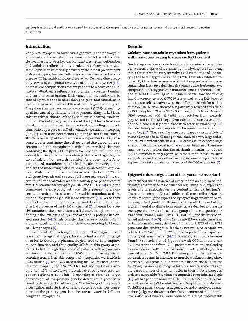
Our first approachwas to study calciumhomeostasis inmyotubesderived frombiopsies of four patients initially diagnosed ashavingMmD, three of whomcarry recessive RYR1mutations and one car-rying the heterozygous mutation p.G297D but who exhibited re-duced RyR1 protein on western blot. Subsequent whole-exomesequencing later revealed that the patient also harboured twocompound heterozygous NEB mutations and is therefore identi-fied as NEM UK06 in Figure 1. Figure 1 shows that the restingfura-2 fluorescence ratio (340/380 nm) as well as the KCl-depend-ent calcium release curves were not different, except for patientMinicore UK 07, who showed a significantly reduced sensitivityto KCl (EC50 for KCl was 55.3 ± 8.2 in myotubes from MinicoreUK07 compared with 13.9 ± 8.4 in myotubes from controls)(Fig. 1A and B). The KCl-dependent calcium release curve for pa-tient Minicore UK08 (dotted trace with asterisk symbol Fig. 1B)had also been previously reported to be similar to that of controlmyotubes (10). These results were surprising as western blots ofmuscle biopsies from all four patients showed a very large reduc-tion of RyR1 protein content (Fig. 1C) leading us to expect a largeeffect on calciumhomeostasis inmyotubes. Because of these rea-sons, we hypothesized that the mechanism leading to reducedRyR1 expression is only operative in more mature tissues, suchasmyofibres, andnot in culturedmyotubes, even though the latterexpress the main protein components of the ECC machinery (7).
Epigenetic down-regulation of the ryanodine receptor 1
We focussed the next series of experiments on epigenetic me-chanisms thatmay be responsible for regulating RyR1 expressionlevels and in particular on the content of microRNAs (miRs).These endogenous ∼22 nucleotide small non-coding RNAs areknown to control gene expression by repressing translation or en-hancing RNA degradation. Because of the limited amount of bio-logical material available from patients, we decided to measurethe expression levels of a selected group of muscle-specific miRtranscripts, namelymiR-1,miR-133,miR-206, and themuscle en-riched miR-486 (11–13). miR-22 and miR-124 were also measuredas bioinformatics analysis showed that the 3′ UTR of the RYR1gene contains binding sites for these two miRs. As controls, weselected miR-126 and miR-221 that are reported to be expressedin many different tissues (14,15). We analysed muscle biopsiesfrom 5–9 controls, from 4–5 patients with CCD with dominantRYR1mutations and from 12–16 patients with mutations leadingto a decrease of RyR1 protein expression with pathological fea-tures of either MmD or CNM. The latter patients are categorizedas ‘Minicore’, and in addition to muscle weakness, they showdecreased RyR1 protein in their muscle biopsy, and all have thefollowing common pathological features: several minicores andincreased number of internal nuclei in their muscle biopsy aswell as amyopathic face often accompanied by ophthalmoplegia(16). All but patients Minicore NL01, UK02, UK05 and UK09 har-boured recessive RYR1 mutations (see Supplementary Material,Table S1 for patient’s diagnosis, genotypic and phenotypic charac-teristics). Figure 2 shows that the relative contents ofmiR-22,miR-124, miR-1 and miR-133 were reduced to almost undetectable
Human Molecular Genetics, 2015, Vol. 24, No. 16 | 4637
levels in biopsies from >60% of Minicore patients with decreasedRyR1 protein expression,whereas the reductionwas not as consist-ent in biopsies fromCCDpatients. Our results also indicate that theobserved changes in miRs are specific to some muscle transcriptsandnot causedbyamore globaldefect in themiR-synthesizingma-chinery, as miR-206, miR486 as well as miR-126 and miR-221 werenot decreased (Fig. 2 and Supplementary Material, Fig. S1).
RYR1 methylation
The results obtained so far point to a potential role of regulationof skeletal muscle gene expression by additional factors.We nextverified whether changes of DNA methylation occur within theRYR1 gene. This experiment is essential as methylation-depend-ent expression of the RYR1 gene was not unexpected consideringthe presence of several CpG-rich regions. Genomic DNA was ex-tracted from biopsies of four controls and four Minicore patients,and the methylation of CpG regions was studied using the me-thyl-sensitive HpaII and the methyl-insensitive MspI restrictionenzymes. After cleavage of genomic DNA, quantitative real-time PCR was performed and compared with a control PCR amp-lifying a proximal RYR1 gene region lacking HpaII/MspI cleavagesites (see schematic representation in Fig. 3A). The resultsobtained clearly indicate that the CpG-III region of the RYR1gene (from nucleotide 6790 to nucleotide 7035) of all theMinicorepatients analysed is hypermethylated compared with that ofcontrols (Fig. 3B) suggesting that RYR1 mutations are associatedwithdeep changes in thepatternofDNAmethylation.Additionally,
we analysed biopsies fromMinicore patients and controls for DNAmethyltransferase (DNM) expression and found that in the formergroup DNMT1 and DNMT2 are significantly up-regulated (Fig. 3C),whereas the expression of DNMT3 did not vary significantly be-tween controls and patients (results not shown).
HDAC expression levels in muscle biopsies of patientswith minicores with recessive RYR1 mutations
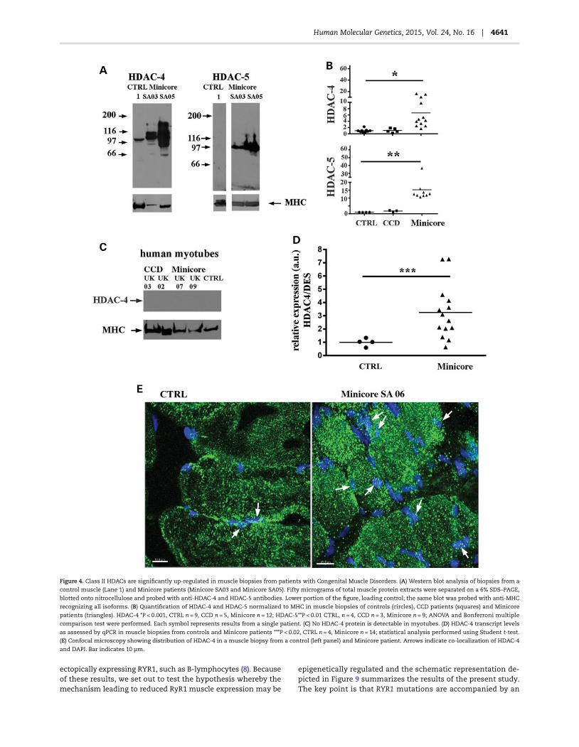
The levels of expression of HDAC-4 and HDAC-5 in muscle biop-sies from controls and patients were subsequently determinedfor the following reasons: (i) class II histone deacetylases(HDACs) can be recruited in association with DNA methylation,(ii) these enzymes repress transcription by deacetylating corehistones (17), (iii) theyaffectmyogenesis by binding to themuscletranscription factor mef2 (18), (iv) they are predominantly ex-pressed in those tissues expressingmef2, that is skeletal muscle,heart and brain (18) and (v) HDAC-4 is a target of miR-22 whosedown-regulation potentiates HDAC-4 expression (19). Figure 4Ashows a representative western blot of total muscle homogenatestained with anti-HDAC-4 and HDAC-5 antibodies; the bottomlane shows a loading control of the same blot stripped andprobedwith anti-myosin heavy chain (MHC) antibodies recogniz-ing allMHC isoforms. The control biopsyshows low levels of classII HDACs (Lane 1 Fig. 4A), whereas samples from theMinicore pa-tients contain abnormally high levels of HDAC-4 and HDAC-5.Figure 4B shows the relative content of HDAC-4 and HDAC-5normalized for MHC content in biopsies from all the available
Figure 1. Myotubes from Minicore patients harbouring recessive RYR1 mutations do not show alterations of the resting [Ca2+] nor decreased Ca2+ release after in vitro
stimulation. (A) Fura-2-loaded myotubes were imaged in Krebs Ringer solution containing 2 m Ca2+. No difference in the resting [Ca2+]i was observed between
controls (white bar), cells from patients with recessive mutations (grey bars) or a patient initially diagnosed as MmD carrying the heterozygous p.G297D RYR1
mutation but who also carries two compound heterozygous NEB mutations (black bar). Bars represent the mean (± SEM) fluorescence (340/380 nm) from the indicated
number of cells. (B) KCl-dependent peak Ca2+ release in Krebs Ringer containing 100 µ La3+. Each point represents the mean (±SEM) increase in fura-2 fluorescence
ratio (340/380 nm) of at least 10 myotubes. The data were analysed through Bolzmann equation using Origin 6.0. (C) Western blot analysis of total protein extracts
from muscle biopsies shows major decrease in RyR1 protein expression in Minicore patients. Desmin served as loading control.
4638 | Human Molecular Genetics, 2015, Vol. 24, No. 16

patients. Biopsies from patients with recessive compound het-erozygous RYR1 mutations, with a non-sense, intronic splicesite or a frameshift mutation in one allele and a missense muta-tion in the other allele show a 6- to 15-fold increase in class IIHDAC content, and the increase in HDAC-4 expression was alsodetectable at the transcriptional level (Fig. 4D). Interestingly,HDAC-4 was not detectable in any of the myotube cultures(Fig. 4C) supporting the observed lack of effect of the mutationson the ECC characteristics of myotubes from the Minicorepatients (Fig. 1). Figure 4E shows a photomicrograph taken byconfocal microscopy on a sample from Minicore SA06; as can beseen, though thevastmajorityofHDAC-4 isdistributed throughoutthe muscle fibre, the amount co-localizing with nuclei is higher inthe patient’s biopsy than in the control biopsy. Analysis of twoMinicore patients and two controls confirmed that the percentageof HDAC-4 co-localizingwith nuclei is∼10 times higher inmusclesfrom Minicore patients than that from controls (9.2 ± 3.8% versus1.1 ± 0.3%, respectively). We also compared HDAC-4 and HDAC-5up-regulation and RYR1 hypermethylation in the samples of thefour Minicore patients and found a positive correlation betweenhigh HDAC-4/5 levels and hypermethylation of the studied RYR1CpG-III gene sequence (Fig. 5A and B).
Effect of HDAC4/5 over-expression and RYR1 silencingin mouse muscle fibres
To demonstrate a causative link between RYR1 mutations and theabove-described epigenetic changes, wemanipulated gene expres-sion by creating transgenic intact adult mouse skeletal muscle flex-or digitorum brevis (FDB) fibres by either (i) over-expressing HDAC-4and HDAC-5 or (ii) knocking down RyR1 by siRNA silencing. Whencompared with acute transfection with an empty plasmid, acuteover-expression of HDAC-4 and HDAC-5 directly recapitulates theeffects observed in muscle biopsies from Minicore patients(Fig. 6). That is, acute over-expression of HDAC-4 and HDAC-5 inmouse FDB fibres decreases RYR1 transcript expression by ∼70%(Fig. 6) and RyR1 protein content by 75% (Supplementary Material,Fig. S3), down-regulates muscle-specific miRs and down-regulatesthe expression of myomesin-1, a muscle-specific gene whose ex-pression is regulated by the transcription factor mef2 (20,21)(Fig. 6). No changes were observed in mef2, miR-126, miR-221 andmiR-486 expression (Supplementary Material, Table S2). On theother hand, silencing RYR1 for 8 days with siRYR1 significantlyincreased HDAC-4 and HDAC-5 expression levels but did notchange the expression of muscle-specific miRs (Fig. 7).
Figure 2. Muscle-specific miR expression levels differ in biopsies from patients with dominant and recessive RYR1 mutations. Each symbol represents the mean relative
expression of the indicated miR from a single patient normalized to RNU44 content and to the muscle-specific housekeeping genes (DES/ACTN2). Control healthy
individuals, circles; CCD with dominant RYR1 mutations, squares; Minicore, triangles. Statistical analysis was performed using ANOVA and Bonferroni multiple
comparison test (95% confidence interval). miR-22 *P < 0.0005, CTRL n = 9, CCD n = 4, Minicore n = 15; miR-124 **P < 0.05, CTRL and CCD n = 5, Minicore n = 12;
miR-1*P < 0.0005, ***P < 0.009, CTRL n = 8, CCD n = 5, Minicore n = 16; miR133 ****P < 0.002, CTRL n = 10, CCD n = 4, Minicore n = 16; miR-206; no statistical significance change
between groups. CTRL n = 9, CCD n = 5, Minicore n = 16. Statistical analysis was performed using ANOVA and Bonferroni multiple comparison test (95% confidence interval).
Human Molecular Genetics, 2015, Vol. 24, No. 16 | 4639

Increasing class II HDAC expression does not affectmef2 expression
Having established that down-regulation of RYR1 causes an in-crease in HDAC-4/5 expression, we reasoned that this elevationcould lead to downstream effects onmef2, amaster trans-activa-tor of skeletal muscle gene expression (22). Mef2 is sequesteredby class II HDACs resulting in blockage of mef2-dependent genetranscription (23), and the RYR1 andmiR-1/miR-133 genes containintragenic mef2-dependent enhancer sequences that regulatetheir transcription in muscle (24,25). We therefore examined thepatient’s muscle biopsies for: (i) mef2 content, to ascertain thatthere was no compensatory up-regulation of its expressionowing to the increased expression of HDAC-4/5 and (ii) myomesina protein that is transcriptionally regulated by mef2 (20,21).Figure 8 shows that the transcript levels of MEF2A, MEF2C andMEF2D are not significantly different between controls and biop-sies from patients. On the other hand, myomesin-1 protein levelsnormalized to MHC are significantly decreased (by ∼50% in biop-sies isolated from patients with minicores (Fig. 8B and C).
HDAC-4/5 are also up-regulated in other congenitalmyopathies
To verify whether these observed effects (that is increased levelsof HDACs, decreased levels of RyR1 and decreased levels of mus-cle-specificmiRs) are specific for congenital myopathies owing torecessive RYR1 mutations or a more general response occurringin patients with other congenital myopathies, we analysed biop-sies from 11 patients with NM harbouringmutations in KBTBD13,
ACTA1 or NEB (NEM 6, NEM3 and NEM2, respectively). A similardecrease in muscle-specific miR-22, miR-133 and miR-1s wasobserved (Supplementary Material, Fig. S2). We also tested themuscle biopsies from the patients with NM for RyR1 contentand HDAC-4 and HDAC-5 expression. Surprisingly, RyR1 proteincontent was significantly reduced (control versus NEMwas 100 ±27.9% versus 0.03 ± 0.02%, P < 0.01, Student’s t-test), and HDAC-4and HDAC-5 protein levels were significantly increased (Supple-mentary Material, Fig. S2).
DiscussionHere, we identify a novel pathophysiological mechanism occur-ring in skeletal muscles of patients with congenital myopathieswherebyactivation of a cascade of events leads to the down-regu-lation of muscle-specific genes. We report that recessive com-pound heterozygous RYR1 mutations are accompanied by thefollowing changes in skeletal muscle: (i) hypermethylation ofthe RYR1 gene, (ii) a 6- to 15-fold increase in class II HDAC expres-sion and (iii) reduction in muscle-specific miRs. Our results re-present a major advancement in the field as to date the modeof action of recessive RYR1 mutations identified in patientswith MmD, CNM and CFTD has been elusive and the functionalcharacterization of cells harbouring such mutations has failedto yield a mechanism compatible with the disease phenotype(8,10,26). A regular finding in muscle biopsies of patients with re-cessive RYR1 mutations has been a reduced expression level ofRyR1 protein and transcript (2–4,26–28). This reduction appearsto be muscle-specific and has not been observed in other tissues
Figure 3. The RYR1 is hypermethylated, and DNA methyltransferases 1 and 2 are up-regulated in muscles fromMinicore patients. (A) Schematic representation showing
the location of the CpG region III within the RYR1 gene, the position of the CpG sites (indicated by arrowheads) and the location of the 5′ CCGG 3′HpaII/MspI site (arrowed);
the location of the internal control region lacking HpaII/MspI sites is also shown, aswell as the location of the PCR primers used to amplify the DNA. (B) Hypermethylation
of CpG region III of the RYR1. Each symbol represents the mean relativemethylation value from a patient (CTRL n = 4; Minicore n = 4). Experimental details are outlined in
Materials and Methods. (C) DNA methyltransferase 1 (DNMT1) and DNMT2 are significantly up-regulated in muscles of Minicore patients. Each symbol represents the
mean relative expression of DNMT1 (CTRL n = 5; Minicore n = 9) and DNMT2 (CTRL n = 4, Minicore n = 8) from a single patient normalized to the muscle-specific
housekeeping gene DES (*P < 0.02, **P < 0.025, Student’s t-test).
4640 | Human Molecular Genetics, 2015, Vol. 24, No. 16
ectopically expressing RYR1, such as B-lymphocytes (8). Becauseof these results, we set out to test the hypothesis whereby themechanism leading to reduced RyR1 muscle expression may be
epigenetically regulated and the schematic representation de-picted in Figure 9 summarizes the results of the present study.The key point is that RYR1 mutations are accompanied by an
Figure 4. Class II HDACs are significantly up-regulated in muscle biopsies from patients with Congenital Muscle Disorders. (A) Western blot analysis of biopsies from a
control muscle (Lane 1) and Minicore patients (Minicore SA03 and Minicore SA05). Fifty micrograms of total muscle protein extracts were separated on a 6% SDS–PAGE,
blotted onto nitrocellulose and probed with anti-HDAC-4 and HDAC-5 antibodies. Lower portion of the figure, loading control; the same blot was probed with anti-MHC
recognizing all isoforms. (B) Quantification of HDAC-4 and HDAC-5 normalized to MHC in muscle biopsies of controls (circles), CCD patients (squares) and Minicore
patients (triangles). HDAC-4 *P < 0.001, CTRL n = 9, CCD n = 5, Minicore n = 12; HDAC-5**P < 0.01 CTRL, n = 4, CCD n = 3, Minicore n = 9; ANOVA and Bonferroni multiple
comparison test were performed. Each symbol represents results from a single patient. (C) No HDAC-4 protein is detectable in myotubes. (D) HDAC-4 transcript levels
as assessed by qPCR in muscle biopsies from controls and Minicore patients ***P < 0.02, CTRL n = 4, Minicore n = 14; statistical analysis performed using Student t-test.
(E) Confocal microscopy showing distribution of HDAC-4 in a muscle biopsy from a control (left panel) and Minicore patient. Arrows indicate co-localization of HDAC-4
and DAPI. Bar indicates 10 µm.
Human Molecular Genetics, 2015, Vol. 24, No. 16 | 4641

increased expression of class II HDACs. We are aware that an in-crease inHDACexpression has been reported in other conditions,including denervation, muscle atrophy, ALS and Huntington’sdisease (29–34). However, we would like to point out that (i)such high levels of expression of class II HDACs have not been re-ported in any other human neuromuscular abnormalities inves-tigated so far; (ii) such an increase of HDAC is specific because thesignal to background ratio of our set of data is 6–16 times greaterthan that reported in other muscle disorders and (iii) over-expression of HDAC-4 and HDAC-5 in mouse FDB fibres resultsin the down-regulation of RYR1 and of muscle-specific miRs.High levels of HDACs not only lead to chromatin condensationthereby decreasing gene transcription (21,35,36), but also seques-ter mef2 (23,37–39). In this context, it should be pointed out thatthere is no compensatory up-regulation of mef2 in muscles fromthe patients, so that up-regulation of HDACs would lead to thedown-regulation of mef2-dependent proteins. That this is thecase is supported by the fact that RYR1, myomesin and muscle-specific miRs containing mef2-dependent binding domains(21,24,25) are significantly down-regulated in the muscles ofMinicore patients. Though few studies have focussed on the me-chanisms regulating RYR1 expression, the 5′ region of the humanand porcine RYR1 gene contains, aside a mef2-binding domain(24), consensus sequences for the transcription factor SP1, formuscle-specific promoter elements as well as for a number oftranscriptional activators (40). SP1 is a zinc finger transcriptionfactor that binds to CG-rich regions present in many promoters.
In fibroblasts, SP1 interacts with HDAC-2 leading to the transcrip-tional silencing of the human telomerase reverse transcriptase(hTERT) gene in normal somatic cells (41). Whether SP1 canalso interact with class II HDACs and whether this interactionis modified by CpG methylation also leading to repression ofRYR1 gene transcription remains to be investigated.
Interestingly, the 3′ UTR of HDAC-4 and that of HDAC-5 havebinding sites for miR-22/mir-124/miR-1/miR-206 and miR-206,respectively, and HDAC-4 is a target ofmiR-22 whose down-regu-lation potentiates its expression (19). Thus, it follows that a de-crease of miR-22, miR-124 and miR-1 activates a pathologicalloop leading to the further up-regulation of class II HDACs(Fig. 9). This mechanism is compatible with and gives mechanis-tic insight to two previous observations: (i) other muscle-specificgenes besides the RYR1 are down-regulated in patients with con-genital myopathies owing to RYR1mutations (7) and (ii) the RYR1was reported to be imprinted in some patients with MmD be-cause of epigenetic factors (42). The former observation is likelydue to the sequestration of mef2 by class II HDACs (37–39). Thelatter observations on the other hand can be explained by thefinding that the DNA methyltransferases DNMT1 and DNMT2are over-expressed in muscle biopsies of Minicore patients,bringing about RYR1hypermethylation. DNMT1 is amaintenancemethyltransferases preservingmethylation patterns but also hasde novo activity (43). DNMT2 on the other hand is thought to par-ticipate in the recognition of damaged DNA and mutation repair(44). In fact, the two observations are mechanistically linked ashypermethylation goes hand in hand with HDAC activation andgene down-regulation (23,24,35,36,45); furthermore, DNA dam-age can activate DNA-methylation via activation of DNMT1 (46),resulting in a pathological loop that will ultimately shut downgene transcription of mef2-dependent genes.
As to the role miRs in neuromuscular diseases, this is still un-clear: over-expression ofmiR-22 is sufficient to cause cardiomyo-cyte hypertrophy (47) and several miRs are up-regulated inmuscular dystrophies (48–50) but depending on the disease,some miRs may appear to be down-regulated (50). Interestingly,a recent study demonstrated that mice lacking miR-133 developan adult onset CNM in type-2 fibres, and this is accompanied byimpaired mitochondrial function, fast to slow myofiber conver-sion and disarrangement of triads (51), histopathologicalchanges very similar to those observed in recessive humanRYR1-related myopathies. Though in the paper the authors con-clude that this is principally due to the dysregulation of dyna-min-2, one of miR-133′s targets, the similarities between thephenotype of the miR-133a knockout mice and that of patientswith RYR1 mutations is striking and is indicative of a commonpathophysiological pathway.
The main point emerging from our studies is that epigeneticfactors are central culprits in recessive RYR1-linked myopathies.Our results also show that these factors are likely to also play amajor role in other congenital muscle diseases such as NM. Insupport of a role of epigenetics, moderate exercise has beenshown to improve the muscle function of some patients withcongenital myopathies (52,53) and there is increasing evidencethat physical activity influences DNA methylation in humans(53,54). Taken together our results suggest that a common patho-physiological mechanism is activated in skeletal muscles ofpatients with some congenital myopathies. The presence ofmutations in muscle-specific genes (in this case RYR1) activatesfactors that lead to the up-regulation of DNMs and of class IIHDACs, the master regulator of chromatin structure. Thoughthe primary mechanism causing HDAC up-regulation is at themoment unclear, our data provide the proof of concept that
Figure 5. Correlation between DNA methylation and HDAC-4/HDAC-5 expression
and up-regulation of DNA methyltransferases in muscles of patients with
Minicore. The data shown in this figures were obtained from biopsies from four
control individuals (empty squares) and from biopsies from four Minicore patients
(filled squares) (Minicore NL02, Minicore NL03; Minicore SA03, Minicore SA05).
(A) Correlation between HDAC-4 and RYR1 hypermethylation (correlation
coefficient r = 0.4695). (B) Correlation between HDAC-5 and RYR1 hypermethylation
(correlation coefficient r = 0.7925).
4642 | Human Molecular Genetics, 2015, Vol. 24, No. 16

DNM and HDAC are potential pharmacological targets to treat awide range of inherited neuromuscular conditions with differentgenetic backgrounds that as a common feature lead to a decreasein RyR1.
Materials and MethodsQuantitative PCR
Total RNA was extracted using Trizol (Life Technologies,#15596018). cDNAwas synthesized with the High Capacity cDNAsynthesis kit or Taqman microRNA Reverse Transcription kit(Applied Biosystems, #4366596). Transcript levels were quantifiedusing Syber-Green reagent on an Applied Biosystem platform(7500 fast real-time PCR system); levels of expression from tripli-cate replicas were averaged and normalized to the content of themuscle-specific gene desmin (DES). In the case of human samples,because of the limited amount of biological material, not all biop-sies could be investigated for all genes. The sequences of the pri-mers used for qPCR are listed in SupplementaryMaterial, Table S3.
MicroRNA determination
Quantification of selected miRs was performed using TaqManmaster mix no-UNG 2 (Life Technologies, # PN 4427788) and thefollowing miR assays (Life Technologies, # PN 4427975): miR-22,miR124a, miR-133a, miR-1, miR-206, miR-486-3p, miR-221 andmiR-126. Each reaction was performed in triplicate, and the re-sults from each muscle sample were analysed and averaged. Inhuman muscle biopsies, miR expression levels were normalizedto RNU44 and to the muscle-specific genes DES and Actinin2(ACTN2) that show similar Ct values in patients and healthy indi-viduals. In mouse FDBs, miR expression levels were normalizedto U6 snRNA. In the case of human samples, because of thelimited amount of biological material, not all biopsies could beinvestigated for all microRNAs.
DNA methylation
Total genomic DNAwas isolated using the GeneElutemammaliangenomic DNA Miniprep kit (Sigma Genosys). DNA methylation
Figure 6. In vivo over-expression of HDAC-4 and HDAC-5 causes down-regulation of RYR1 and of muscle-specific miRs. Each symbol shows the mean triplicate relative
expression value of the indicated transcript normalized to the indicated gene. Circles, control FDB fibres mock transfected with the empty pIRES2-dsRed2 plasmid;
squares, FDB fibres transfected with a plasmid encoding mouse HDAC-4 and HDAC-5. HDAC-4; *P < 0.0001, n = 6; HDAC-5 **P < 0.005, n = 6; RYR1 **P < 0.005, CTRL n = 4,
HDAC-4 and 5 n = 6; miR-22 and miR-1; ***P < 0.045, n = 6; miR-133 ****P < 0.035, n = 6; miR-206 ****P < 0.035, n = 5; Myomesin ****P < 0.035, n = 4; miR-124 was quantified and
no significance change was observed. Statistical analysis was performed using the Student’s t-test.
Human Molecular Genetics, 2015, Vol. 24, No. 16 | 4643

was assessed by PCR amplification of genomic DNA digested withthe methyl-sensitive HpaII and MspI restriction enzymes (55)(see schematic representation in Fig. 3A). Restriction enzymedigestion reactions were carried out overnight at 37°C, withHpaII or MspI (New England BioLabs), in final volume of 20 µl.The primers used for PCR amplification are listed in Supple-mentary Material, Table S3 as: Human RYR1 CG-rich (F and R)amplifying the CpG-III region of the RYR1 gene and Human
RYR1 Control (F and R), amplifying a control region of theRYR1 gene lacking HpaII/MspI sites (see Fig. 3A). To determinethe extent of DNA methylation, the ΔCt values were first ob-tained comparing the CpG-III and control PCRs of HpaII diges-tions, and then the ΔΔCt values were generated using asreference the samples exhibiting the higher ΔCt. Higher ΔΔCtvalues indicate higher extent of DNA methylation at the CpG-III RyR1 MspI/HpaII cleavage sites.
Figure 7. Down-regulation of RYR1 by siRNA leads to up-regulation of HDAC-4 and HDAC-5. Each symbol shows the mean triplicate value relative expression level of the
indicated transcript normalized to U6 snRNA (miR expression) or DES (RYR and HDAC expression), in fibres isolated from a single mouse. Circles, control (mock)
transfection with a scrambled siRNA; squares FDB transfected with siRYR RNA (see Materials and Methods for details). RYR1 *P < 0.0001, n = 7; HDAC-4 **P < 0.04, CTRL
n = 6, siRYR1 n = 7. HDAC-5; **P < 0.04, CTRL n = 6, siRYR1 n = 9. miR-22, miR-133, miR-124 and miR-1 show no statistical difference. Statistical analysis was performed
using the Student’s t-test.
Figure 8. Increased expression of HDAC-4/HDAC-5 leads to a decrease in the content of myomesin, without affectingmef2. (A) mef2A, mef2C andmef2D transcript levels
are similar in muscle biopsies of patients and controls (expression levels normalized to DES). (B) Representative western blot showing that the protein content of
myomesin is lower in patients with Minicore compared with healthy individuals (letters and numbers refer to patient no.; see Supplementary Material, Table S1);
(C). Quantification of myomesin protein content in muscle biopsies: controls, circles (n = 6); CCD, squares (n = 5); Minicore, triangles (n = 9). *P < 0.02. Statistical analysis
was performed using ANOVA and Bonferroni multiple comparison test (95% confidence interval).
4644 | Human Molecular Genetics, 2015, Vol. 24, No. 16

Electrophoresis and immunoblotting
Totalmuscleproteinswereextracted in10mHepespH7.0, 150m
NaCl, 1 m EDTA and anti-protease (Roche, # 11873580001). Pro-tein concentration was determined using Protein Assay Kit II(Bio-Rad Laboratories) using BSA as a standard. SDS–PAGE, pro-tein transfer on to nitrocellulose membranes and immunostain-ing were performed as described previously (2,8). The followingprimary antibodies were used:mouse anti-RyR1 (Ryanodine 1 Re-ceptor, Thermo Scientific, # MA3-925), mouse anti-MHC (MHC,Millipore, #05-716), Rabbit anti-HDAC-4 (Histone Deacetylase 4,Cell Signaling, #2072) and rabbit anti- HDAC-5 (Histone Deacety-lase 5, Abcam #1439), and rat anti-myomesin was a generousgift of Prof. Mathias Gautel, King’s College, London, UK (56). Sec-ondary peroxidase conjugateswere Protein G–peroxidase (Sigma,#P8170) and peroxidase-conjugated goat anti-mouse IgG (Sigma,#A2304). The immunopositive bandswere visualized by chemilu-minescence using the Super SignalWest Dura kit (Thermo Scien-tific). In order to perform statistical analysis, the intensity of theimmunopositive bands was determined using ImageJ/FIJI. Theintensity valueswere normalized to the intensity of the indicatedmuscle-specific housekeeping protein. The value (arbitrary units)obtained from the patient’s biopsies were divided by the meanvalue obtained from control biopsies and are expressed as 100%.
Mouse muscle fibre electroporation and isolation
The procedurewas as described byDiFranco et al. (57). Briefly, 8- to14-week-oldmicewere anaesthetizedusing isofluorane, and 7.5 µlof 2 mg/ml Hyaluronidase in RNase-free Tyroide’s Buffer (SigmaFine Chemicals, #H3506) was injected under the footpad. Themice were left 1 h under supervision, and subsequently, the fol-lowing constructs were injected into the footpad: pCMV6-HDAC4(Origene #MR211598) and pCMV6-HDAC5 (Origene # MC202550)whereas control mice (mock transfected) received 20 µg ofpIRES2-dsRed2 plasmid (Clonetech #632420). For siRNA silencingexperiments, 6 nmol of RNA either specific for the RYR1 (Ambion;siRNA RyR1-#4390771) or a scrambled siRNA sequence (Negativecontrol 2-#4390845) were used. siRNA-transfected FDBs were alsoinjected with lipofectamine RNAiMAX (Invitrogen, #13778-030).Ten minutes post-injection, FDBs were electroporated usingacupuncture needles placed parallel and perpendicular to thelong axis of the foot (with 1 cm distance), and twenty pulses(100v/cm, 20 ms duration and 1 Hz of frequency) were given. Six
to ten days post–transfection, the mice were sacrificed and FDBswere isolated by enzymatic dissociation at 37°C for 60 min inKrebs Ringer solution no Ca2+ (pH 7.4), containing 0.2% collage-nase I (Sigma Fine Chemicals, C-0130). Enzymatic digestion wasterminated by washing the muscle with Tyrode’s solution (pH7.4), and single fibres were isolated and total protein extractsprepared or RNAwas extracted and analysed by qPCR.
Ca2+ measurements
Primary skeletal muscle cultures and cell imaging were per-formed as previously described (58).
Confocal microscopy and immunofluorescence
Biopsies were embedded for pathological examination and slicedusing a cryostat (10 µm thickness). Cryosections were fixed withmethanol: acetone (1:1) for 30 min and then incubated in the fol-lowing solutions for 90 min at room temperature: blocking solu-tion (Roche, #115000694011), rabbit anti-HDAC-4 (Cell Signaling,#2072) andAlexa Fluor 647-conjugated anti-Rabbit IgG (Life Tech-nologies, #A21245). Nuclear staining was performed using DAPI(Invitrogen, #D21490), and slides were mounted with mountingmedium (Sigma, #1000-4) and sealed hermetically with 1.5-mm-thick coverslip. A Nikon A1R Confocal microscope wasused for 3D image acquisition with a 40× oil objective (N.A. = 1.3).Imageswere analysed using threshold co-localization function inImageJ2/FIJI program.
Compliance with ethical standards
All procedures performed in studies involving human partici-pants were in accordance with the ethical standards of the insti-tutional and/or national research committee and with the 1964Helsinki declaration and its later amendments or comparableethical standards. This study was approved by the Ethikkommis-sion beider Basel (permit No. EK64/12); all subjects gave writteninformed consent to carry out this work.
All applicable international, national and/or institutionalguidelines for the care and use of animals were followed. All pro-cedures performed in studies involving animals were in accord-ance with the ethical standards of the institution or practice atwhich the studies were conducted. Experiments on mouse mus-cles were approved by the local Cantonal Veterinary authorities(permit No. 2658).
Statistical analysis and graphical software
Statistical analysis was performed using the Student’s t-test;means were considered statistically significant when theP-value was < 0.05. When more than two groups were compared,analysis was performed using the ANOVA test followed by theBonferroni post hoc test using the statistical package included inGraphPad Prism 6.0 software. Origin 6 was used to generatedose–response curves. Images were assembled using AdobePhotoshop CS (version 8.0).
Supplementary MaterialSupplementary Material is available at HMG online.
Figure 9. Cartoon depicting how mutations in RYR1 lead to a decrease in
RyR1 content thereby leading to weak muscles. Mutations lead to DNA
hypermethylation and HDAC-4/HDAC-5 over-expression. This causes mef2
sequestration thereby inhibiting transcription of genes regulated by mef2,
including the RYR1 and muscle-specific miRs. A decrease in RyR1 would
severely affect muscle excitation–contraction coupling because this calcium
channel is a central player in this mechanism, releasing the calcium necessary
for muscle contraction from the sarcoplasmic reticulum.
Human Molecular Genetics, 2015, Vol. 24, No. 16 | 4645

AcknowledgementsWe thank Dr Joery Molenaar for contacting the patients, gather-ing their written permission and organizing the transport ofthe material from the Dutch patients.
Conflict of Interest statement. None declared.
FundingThisworkwas supported bya grant from the SwissNational ScienceFoundation (SNF No. 31003A-146198), by a grant from the Myotubu-larMyopathy (grant No. 12KCL 01-MT) and by a grant from the BaselNeuromuscular Association (NeRAB). O.R. was supported by a grantfrom the Botnar Stiftung. The support of the Department ofAnaesthesia Basel University Hospital and the technical supportof Anne-Sylvie Monnet are gratefully acknowledged.
References1. Maggi, L., Scoto, M., Cirak, S., Robb, S.A., Klein, A., Lillis, S.,
Cullup, T., Feng, L., Manzur, A.Y., Sewry, C.A. et al. (2013) Con-genital myopathies–clinical features and frequency of indi-vidual subtypes diagnosed over a 5-year period in theUnited Kingdom. Neuromuscul. Disord., 23, 195–205.
2. Zhou, H., Jungbluth, H., Sewry, C.A., Feng, L., Bertini, E., Bush-by, K., Straub, V., Roper, H., Rose, M.R., Brockington, M. et al.(2007) Molecular mechanisms and phenotypic variation inRYR1-related congenital myopathies. Brain, 130, 2024–2036.
3. Wilmhurst, J.M., Lillis, S., Zhou, H., Pillay, K., Henderson, H.,Kress, W., Müller, C.R., Ndondo, A., Cloke, V., Cullup, T. et al.(2010) RYR1mutations are a common cause of congenitalmy-opathies with central nuclei. Ann. Neurol., 68, 717–726.
4. Clarke, N.F., Waddell, L.B., Cooper, S.T., Perry, M., Smith, R.L.,Kornberg, A.J., Muntoni, F., Lillis, S., Straub, V., Bushby, K. et al.(2010) Recessivemutations in RYR1 are a commoncause of Con-genital Fiber Type disproportion. Hum. Mutat., 31, E1544–E1550
5. Franzini-Armstrong, C. and Protasi, F.G. (1997) Ryanodine re-ceptors of striated muscles: a complex channel capable ofmultiple interactions. Physiol. Rev., 77, 699–729.
6. Treves, S., Jungbluth, H., Muntoni, F. and Zorzato, F. (2008) Con-genital muscle disorders with cores: the ryanodine receptorcalcium channel paradigm. Curr. Opin. Pharmacol., 8, 319–326.
7. Zhou, H., Rokach, O., Feng, L., Munteanu, I., Mamchaoui, K.,Wilmshurst, J.M., Sewry, C., Manzur, A.Y., Pillay, K., Mouly, V.et al. (2013) RYR1 deficiency in congenital myopathies disruptsexcitation-contraction coupling. Hum. Mutat., 34, 986–996.
8. Attali, R., Aharoni, S., Treves, S., Rokach, O., Becker-Cohen,M.,Fellig, Y., Straussberg, R., Do, T., Daana, M., Mitrani-Rosen-baum, S. andNevo, Y. (2013) Variablemyopathic presentationin a single family with novel skeletal RYR1 mutation. PLoSOne, 8, e69296. doi:10.1371/journal. pone. 0069296.
9. Emery,A.E. (1991) Population frequenciesof inheritedneuromus-cular diseases—a world survey. Neuromuscul. Disord., 1, 19–29.
10. Zhou, H., Yamaguchi, N., Xu, L., Wang, Y., Sewry, C., Jung-bluth, H., Zorzato, F., Bertini, E., Muntoni, F., Meissner, G.and Treves, S. (2006) Characterization of recessive RYR1 mu-tations in core myopathies. Hum. Mol. Genet., 16, 2791–2803.
11. Chen, J.F., Mandel, E.M., Thomson, J.M., Wu, Q., Callis, T.E.,Hammond, S.M., Conlon, F.L. and Wang, D.Z. (2006) The roleof microRNA-1 and microRNA-133 in skeletal muscle prolif-eration and differentiation. Nat. Genet., 38, 228–233.
12. Kim, H.K., Lee, Y.S., Sivaprasad, U., Malhotra, A. and Dutta, A.(2006) Muscle-specific microRNA miR-206 promotes muscledifferentiation. J. Cell. Biol., 174, 677–687.
13. Small, E.M., O’Rourke, J.R., Moresi, V., Sutherland, L.B., McAn-ally, J., Gerard, R.D., Richardson, J.A. and Olson, E.N. (2010)Regulation of PI3-kinase/Akt signaling by muscle enrichedmicroRNA-486. Proc. Natl Acad. Sci. USA., 107, 4218–4223.
14. Wang, M., Zhao, C., Shi, H., Zhang, B., Zhang, L., Zhang, X.,Wang, S., Wu, X., Yang, T., Huang, F. et al. (2014) DeregulatedmicroRNAs in gastric cancer tissue-derived mesenchymalstem cells: novel biomarkers and a mechanism for gastriccancer. Br. J. Cancer., 110, 1199–1210.
15. Sun, X., Wang, Z.M., Song, Y., Tai, X.H., Ji, W.Y. and Gu, H.(2014) MicroRNA-126 modulates the tumor microenviron-ment by targeting calmodulin-regulated spectrin-associatedprotein 1 (Camsap1). Int. J. Oncol., 44, 1678–1684.
16. Jungbluth, H., Sewry, C. and Muntoni, F. (2011) Core myop-athies. Semin. Pediatr. Neurol., 18, 239–249.
17. Workmann, J.L. andKingston, R.E. (1998) Alteration of nucleo-some structure as amechanism of transcriptional regulation.Annu. Rev. Biochem., 67, 545–579.
18. Lu, J., McKinsey, T.A., Zhang, C.L. and Olson, E.N. (2000) Regu-lation of skeletal myogenesis by association of themef2 tran-scription factorwith class II Histone deacetylases.Mol. Cell., 6,233–244.
19. Zhang, J., Yang, Y., Yang, T., Liu, Y., Li, A., Fu, S.,Wu,M., Pan, Z.and Zhou, W. (2010) microRNA-22, downregulated in hepato-cellular carcinoma and correlatedwith prognosis, suppressescell proliferation and tumourigenicity. Br. J. Cancer, 103,1215–1220.
20. Potthoff, M.J., Arnold,M.A., McAnally, J., Richardson, J.A., Bas-sel-Duby, R. and Olson, E.N. (2007) Regulation of skeletalmuscle sarcomere integrity and postnatal muscle functionby Mef2c. Mol. Cell Biol., 27, 8143–8151.
21. Hinits, Y. and Hughes, S.M. (2007) Mef2s are required for thickfilament formation in nascent muscle fibres. Development,134, 2511–2519.
22. Naya, F.J. and Olson, E. (1999) MEF2: a transcriptional targetfor signaling pathways controlling skeletal muscle growthand differentiation. Curr. Opin. Cell Biol., 11, 683–688.
23. McKinsey, T.A., Zhang, C.L., Lu, J. and Olson, E.N. (2000)Signal-dependent nuclear export of a histone deacetylaseregulates muscle differentiation. Nature, 408, 106–111.
24. Schmoelzl, S., Leeb, T., Brinkmeier, H., Brem, G. and Brenig, B.(1996) Regulation of tissue-specific expression of the skeletalmuscle ryanodine receptor gene. J. Biol. Chem., 271, 4763–4769.
25. Liu, N., Williams, A.H., Kim, Y., McAnally, J., Bezprozvannaya,S., Sutherland, L.B., Richardson, J.A., Bassel-Duby, R. andOlson, E.N. (2007) An intragenicMEF2-dependent enhancer di-rects muscle-specific expression of microRNAs 1 and 133 Mef2and myomesin. Proc. Natl Acad. Sci. USA, 104, 20844–20849.
26. Zhou, H., Lillis, S., Loy, R.E., Ghassemi, F., Rose, M.R., Norwood,F., Mills, K., Al-Sarraj, S., Lane, R.J., Feng, L. et al. (2010) Multi-minicore disease and atypical periodic paralysis associatedwith novel mutations in the skeletal muscle ryanodine recep-tor (RYR1) gene. Neuromuscul. Disord., 20, 166–173.
27. Monnier, N., Marty, I., Faure, J., Castiglioni, C., Desnuelle, C., Sac-coni, S., Estournet, B., Ferreiro,A., Romero,N., Laquerriere, A. et al.(2008) Null mutations causing depletion of the type 1 ryanodinereceptor (RYR1) are commonly associated with recessive struc-tural congenitalmyopathieswith cores.Hum.Mutat., 29, 670–678.
28. Bevilacqua, J.A., Monnier, N., Bitoun, M., Eymard, B., Ferreiro,A., Monges, S., Lubieniecki, F., Taratuto, A.L., Laquerrière, A.,Claeys, K.G. et al. (2011) Recessive RYR1 mutations cause un-usual congenitalmyopathywith prominent nuclear internal-ization and large areas of myofibrillar disorganization.Neuropathol. Appl. Neurobiol., 37, 271–284.
4646 | Human Molecular Genetics, 2015, Vol. 24, No. 16

29. Bongers, K.S., Fox, D.K., Ebert, S.M., Kunkel, S.D., Dyle, M.C.,Bullard, S.A., Dierdorff, J.M. and Adams, C.M. (2013) Skeletalmuscle denervation causes skeletal muscle atrophy througha pathway that involves both Gadd45a and HDAC4.Am. J. Physiol. Endocrinol. Metab., 98, 4089–4096.
30. Bruneteau, G., Simonet, T., Bauché, S., Mandjee, N., Malfatti,E., Girard, E., Tanguy, M.L., Behin, A., Khiami, F., Sariali, E.et al. (2013) Muscle histone deacetylase 4 upregulation inamyotrophic lateral sclerosis: potential role in reinnervationability and disease progression. Brain, 136, 2359–2368.
31. Mielcarek, M., Landles, C., Weiss, A., Bradaia, A., Seredenina,T., Inuabasi, L., Osborne, G.F., Wadel, K., Touller, C., Butler, R.et al. (2013) HDAC4 reduction: a novel therapeutic strategy totarget cytoplasmic huntingtin and ameliorate neurodegen-eration. PLOS Biol., 11, e1001717.
32. Cohen, T.J., Waddell, D.S., Barrientos, T., Lu, Z., Feng, G., Cox,G.A., Bodine, S.C. and Yao, T.P. (2007) The histone deacetylaseHDAC4 connects neural activity to muscle transcriptional re-programming. J. Biol. Chem., 282, 33752–33759.
33. Consalvi, S., Saccone, V., Giordani, L., Minetti, G., Mozzetta, C.and Puri, P.L. (2011) Histone deacetylase inhibitors in thetreatment of muscular dystrophies: epigenetic drugs forgenetic diseases. Mol. Med., 17, 457–465.
34. Falkenberg, K.J. and Johnstone, R.W. (2014) Histone de-acetylases and their inhibitors in cancers, neurologicaldieases and immune disorders. Nature Rev. Drug. Discov., 13,673–691.
35. Wang, Z., Qin, G. and Zhao, T.C. (2014) HDAC4: mechanism ofregulation and biological functions. Epigenomics, 6, 139–150.
36. Baylin, S.B., Esteller, M., Rountree, M.R., Bachmanm, K.E.,Schuebelm, K. and Herman, J.G. (2001) Aberrant patterns ofDNAmethylation, chromatin formation and gene expressionin cancer. Hum. Mol. Genet., 10, 687–692.
37. Lu, J., McKinsey, T.A., Nicolm, R.L. and Olson, E.N. (2000) Sig-nal dependent activation of the MEF2 transcription factor bydissociation from histone deacytlases. Proc. Natl Acad. Sci.USA, 97, 4070–4075.
38. Miska, E.A., Karlsson, C., Langley, E., Nielesen, S.J., Pines, J.and Kouzaridesm, T. (1999) HDAC4 deacetylase associateswith and represses the MEF2 transcription factor MEF2 tran-scription factor. EMBO J., 18, 5099–5107.
39. Wang, A.H., Bertos, N.R., Vezmar, M., Pelletier, N., Crosato, M.,Heng, H.H., Th’ng, J., Han, J. and Yang, X.L. (1999) HDAC4, ahuman histone deacetylase related to yeast HDA1, is a tran-scriptional corepressor. Mol. Cell. Biol., 19, 7816–7827.
40. Phillips, M.S., Fujii, J., Khanna, V.K., DeLeon, S., Yokobata, K.,DeJong, P. and MacLennan, D.H. (1996) The structural organ-ization of the human skeletal muscle ryanodine receptor(RYR1) gene. Genomics, 34, 24–41.
41. Won, J., Yim, J. and Kim, T.K. (2002) Sp1 and Sp3 recruit his-tone deacetylase to repress transcription of human telomer-ase reverse transcriptase (hTERT) promoter in normalhuman somatic cells. J. Biol. Chem., 277, 38230–38238.
42. Zhou, H., Brockington, M., Jungbluth, H., Monk, D., Stanier, P.,Sewry, C.A., Moore, G.E. and Muntoni, F. (2006) Epigenetic al-lele silencing unveils recessive RYR1mutations in coremyop-athies. Am. J. Hum. Gen., 79, 859–868.
43. Denis, H., Ndlovu, M.N. and Fuks, F. (2011) Regulation ofmammalian DNA methyltransferases: a route to new me-chanisms. EMBO Reports, 12, 647–656.
44. Subramaniam, D., Thombre, R.T., Dhar, A. and Anant, S.(2014) DNAmethyltransferases: a novel target for preventionand therapy. Front. Oncol., 4. doi:10.3389/fonc.2014.00080.
45. Newell-Price, J., Clark, A.J. and King, P. (2000) DNA methyla-tion and silencing of gene expression. Trends. Endocrinol.Metab., 11, 142–148.
46. Cuozzo, C., Porcellini, A., Angrisano, T., Morano, A., Lee, B., DiPardo, A., Messina, S., Iuliano, R., Fusco, A., Santillo, M.R. et al.(2007) DNA damage, homology-directed repair, and DNAmethylation. PLOS Genet, 3, e110.
47. Huang,Z.P., Chen, J., Seok,H.Y., Zhang,Z., Kataoka,M.,Hu,X. andWang, D.Z. (2013) MicroRNA-22 regulates cardiac hypertrophyand remodeling in response to stress. Circ. Res., 112, 1234–1243.
48. Eisenberg, I., Eran, A., Nishinom, I., Moggio, M., Lamperti, C.,Amatom,A.A., Lidov, H.G., Kang, P.B., North, K.N., Mitrani-Ro-senbaum, S. et al. (2007) Distinctive patterns of microRNA ex-pression in primary muscle disorders. Proc. Natl Acad. Sci.USA, 104, 17016–17021.
49. Dmitriev, P., Stankevicins, L., Ansseau, E., Petrov, A., Barat, A.,Dessen, P., Robert, T., Turki, A., Lazar, V., Labourer, E. et al.(2013) Defective regulation of microRNA target genes inmyo-blasts from fascioscapulohumeral dystrophy patients. J. Biol.Chem., 288, 34989–35002.
50. Chen, J.F., Callis, T.E. and Wang, D.Z. (2009) microRNAs andmuscle disorders. J. Cell Sci., 122, 13–20.
51. Liu, N., Bezprozvannaya, S., Shelton, J.M., Frisard, M.I., Hulver,M.W., McMillan, R.P., Wu, Y., Voelker, K.A., Grange, R.W., Rich-ardson, J.A., Bassel-Duby, R. andOlson, E.N. (2011)Mice lackingmicroRNA 133a develop dynamin 2-dependent centronuclearmyopathy. J. Clin. Invest., 121, 3258–3268.
52. Vosin, S., Eynon, N., Yan, X. and Bishop, D.J. (2014) Exercisetraining and DNA methylation in humans. Acta. Physiol.,doi:10.1111/apha.12414.
53. Ling, C. and Rönn, T. (2014) Epigenetic adaptation to regularexercise in humans. Drug Discov. Today, 19, 1015–1018.
54. Barrès, R., Yan, J., Egan, B., Treebak, J.T., Rasmussen, M., Fritz,T., Caidahl, K., Krook, A., O’Gorman, D.J. and Zierath, J.R.(2012) Acute exercise remodels promoter methylation inhuman skeletal muscle. Cell Metabol., 15, 405–411.
55. Lee, W.H., Isaacs, W.B., Bova, G.S. and Nelson, W.G. (1997) CGislandmethylation changes near the GSTP1 gene in prostaticcarcinoma cells detected using the polymerase chain reac-tion: a new prostate cancer biomarker. Cancer Epidemiol. Bio-markers Prev., 6, 443–450.
56. Obermann, W.M.J., Gautel, M., Steiner, F., van der Ven, P.F.,Weber, K. and Fürst, D.O. (1996) The structure of thesarcomeric M band: localization of defined domains ofmyo-mesin, M-protein and the 250 kD carboxy-terminal regionof titin by immunoelectron microscopy. J. Cell Biol., 134,1441–1453.
57. DiFranco, M., Quinonez, M., Capote, J. and Vergara, J. (2009)DNA transfection of mammalian skeletal muscles usingin vivo electroporation. J. Vis. Exp., pii: 1520. doi:10.3791/1520.
58. Ducreux, S., Zorzato, F., Müller, C., Sewry, C., Muntoni, F.,Quinlivan, R., Restagno, G., Girard, T. and Treves, S. (2004) Ef-fect of ryanodine receptormutations on interleukin-6 releaseand intracellular calcium homeostasis in human myotubesfrom malignant hyperthermia-susceptible individuals andpatients affected by central core disease. J Biol. Chem., 279,43838–43846.
Human Molecular Genetics, 2015, Vol. 24, No. 16 | 4647