Embed Size (px)
Citation preview
Int J Clin Exp Pathol 2016;9(2):1285-1293www.ijcep.com /ISSN:1936-2625/IJCEP0019198
Original Article Enumeration of monocytes subsets using different gating methods by flow cytometry
Hua Wang1, Saili Wang1, Jun Ye2, Yong Yin1, Yupei Zhou1, Xiaolan Ma1, Yan Wang1
1Department of Obstetrics and Gynecology, Taizhou Hospital of Traditional Chinese Medicine, Taizhou, China; 2Department of Laboratory Medicine, Taizhou People’s Hospital, Taizhou, China
Received November 5, 2015; Accepted January 1, 2016; Epub February 1, 2016; Published February 15, 2016
Abstract: Objective: To compare the efficiency of different gating methods for the enumeration of monocytes sub-sets using flow cytometry. Methods: Forty-eight healthy individuals received physical examination in our hospital was randomly included in this study. Peripheral blood monocytes subsets were analyzed by flow cytometry with FSC-SSC, CD45 and CD14 gating methods. Besides, the effects of different storage times on the test results were analyzed. Results: Statistical difference was noticed in the enumeration of Mon1 and Mon3 obtained using three different gating methods (P<0.001). The detection results of Mon2 by gating with FSC-SSC showed no significant difference compared with those obtained by CD45 or CD14 gating (P>0.05). The percentage of Mon2 determined using CD45 gating showed remarkable difference compared with that of CD14 gating. Correlation test showed that the three gating methods for Mon1, Mon2, Mon3 detection showed a significant positive correlation (P<0.05). For the effects of storage duration on the test results, significant increase was noticed in the percentage of Mon2 determined by FSC-SSC gating at 8 h compared with that of CD14 gating at 1 h. Nevertheless, remarkable decrease was identified in the Mon3 by FSC-SSC gating at 8 h compared with that of CD14 gating at 1 h (P<0.05). Significant difference was observed in the Mon1, Mon2 and Mon3 proportion in SSClow cell populations at 8 h compared with those obtained at 4 h (P<0.05). For the samples stored at room temperature for 4 h, significant differences were identified in the Mon1 and Mon3 proportion in SSClow compared with those of the SSChi. For the samples stored at room temperature for 8 h, significant differences were observed in the Mon1, Mon2 and Mon3 proportion in SSClow compared with those of SSChi. Conclusion: FSC-SSC gating or CD45 gating was effective for the enumeration of Mon2. CD14 gat-ing is more suitable for the enumeration of Mon1. As the extension of storage duration may affect the analysis of monocytes subsets, clinical samples for analysis should be tested as timely as possible.
Keywords: Flow cytometry, monocytes, CD14, CD45
Introduction
Monocytes, one of the major sources of the macrophages and dendritic cells in tissue, play important roles in the regulation of inflamma-tion and inflammatory reactions [1, 2]. Cir- culating monocytes are divided into three sub-types according to the levels of CD14 and CD16 expression, including CD14++/CD16- (Mon1), CD14++/CD16+ (Mon2), and CD14+/CD16++ (Mon3) cells [3]. The expression of these sub-sets has been considered as an important eval-uation criteria for the pathogenesis in various inflammatory reactions.
To date, accumulating evidence reveals circu-lating monocyte subsets as surrogate cellular
biomarkers are closely related to the cardiovas-cular and cancer diseases. However, no well-acknowledged standards for the quantification of monocyte have been established [4-9], which precluded a routine monitoring and compara-tive interpretation of the clinical studies. In a previous study, Hristov et al [10] used flow cyto-metric protocol for enumeration of monocyte subsets in human blood, which proved the pos-sibility of such technique for the quantification of circulating monocytes. Ever since that study, rare studies have been carried out to investi-gate the efficiency of enumeration of monocyte subsets using flow cytometric protocols. In this study, different gating methods were used to analyze mononuclear cells in peripheral blood of healthy human. Besides, a compara-
Monocyte enumeration by flow cytometric gating
1286 Int J Clin Exp Pathol 2016;9(2):1285-1293
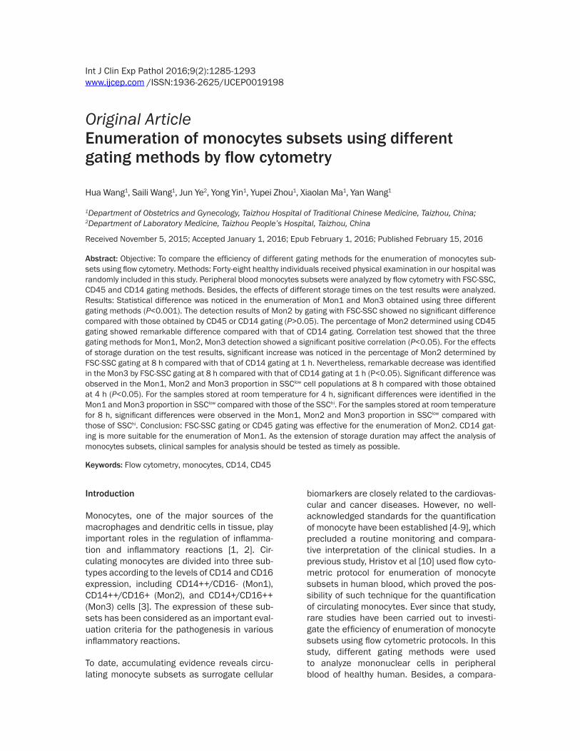
Figure 1. Analysis of different gating methods for the detection of monocytes subsets (A and B: FSC-SSC gating. C and D: CD45 gating. E and F: CD14 gating).
Monocyte enumeration by flow cytometric gating
1287 Int J Clin Exp Pathol 2016;9(2):1285-1293
resuspended in 100 μl PBS for the further analysis.
Flow cytometry
SSC was set as the y-coordinate, and FSC, CD45 and CD14 were set as the x-coordinate, respectively. Data of monocyte subsets were obtained on three types of charts (Figure 1), and 2000 mononuclear cells were obtained by CELL Quest software. On this basis, the pro-portion of the Mon1, Mon2 and Mon3 was analyzed.
Statistical analysis
Data were expressed as mean ± standard devi-ation. Student’s t-test or One-Way ANOVA test was used for the inter-group comparison. The potential correlation between two variants was analyzed using Pearson’s correlation coeffi-cient. All the statistical analysis in this study was performed using the Sigma Plot 12.0 soft-ware. P<0.05 was considered statistically significant.
Results
Comparison of different gating methods of monocytes subsets in the literatures
To date, few gating methods are available for the flow cytometric methods, including FSC-
tive study was performed to the results of dif-ferent monocytes subsets, based on which to identify the potential the factors affecting the test results.
Materials and methods
Subjects and sample collection
Forty-eight healthy subjects received physical examination in our hospital (male: 25, female: 23, mean age: 35.4 ± 8.3 years) were randomly included in this study. Written informed con-sent was obtained from each subject. The study protocols were approved by the Ethical Committee of Taizhou People’s Hospital. After inclusion, fasting peripheral venous blood (2 ml) was collected from each subject using EDTA-K2 as anticoagulant.
Staining of samples
The samples were divided into two groups in- cluding: Group A, which was added with CD45-PerCP (BD Bioscience Company, #347464) and CD14-FITC (BD Bioscience Company, #347493), PE Mouse Anti-Human CD16 (BD Bioscience Company, #347617); and Group B (isotype control), which was added with CD45-PerCP and Mouse IgG2a (BD Bioscience Company, #349051), as well as Mouse IgG1- PE (BD Bioscience Company, #349043). Sub- sequently, the mixture abovementioned was added with 50 μl blood, and was kept in the
dark conditions after vo- texing for 15 minutes. Afterwards, 2 ml hemo-lysin was added fol-lowed by keeping in the dark for 10 min after votexing. The mixture was centrifugated at 1,500 rpm for 5 min, and then the superna-tant was discarded. Af- ter washing with 2 ml PBS, the cell was finally
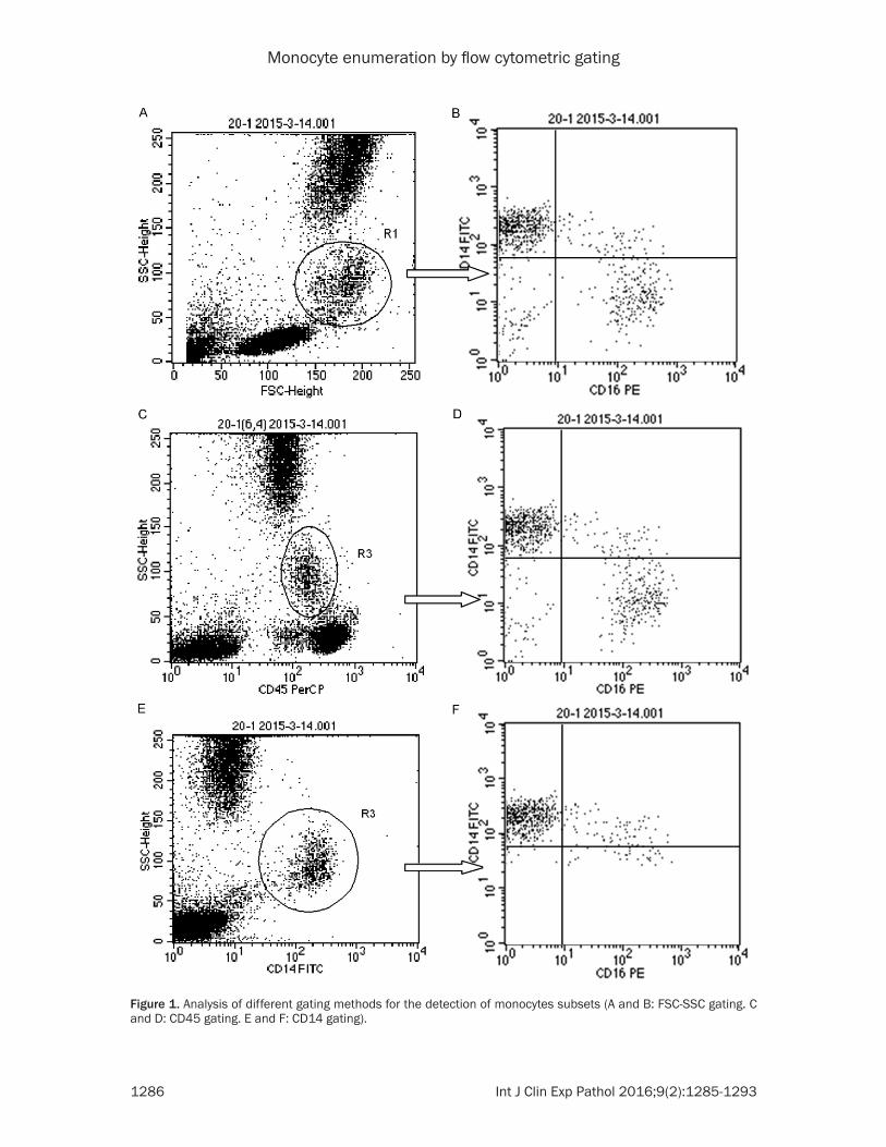
Table 1. Comparison of different gating methods of monocytes subsets in the literaturesAuthors Gating methods Mon1 (%) Mon2 (%) Mon3 (%)Hristov M [4] FSC-SSC 85.4 (83.2-88.1) 4.1 (2.5-4.9) 7.5 (6.4-8.3)Katarina E [5] FSC-SSC 67 (59-72) 7.1 (5.1-9.8) 3.5 (2.6-4.9)Krychtiuk KA [6] CD45 82.1 ± 6.7 5.6 ± 3.3 12.3 ± 5.9Chelombitko MA [7] CD14 gating 74.8 ± 7.6 12.8 ± 3.4Tiziano Tallone [8] HLA-DR gating 87.0 (78.5-95.9) 3.3 (1.1-7.5%) 5.8 (1.7-10.2)Van Craenenbroeck AH [9] FSC-SSC and CD86 88.09 ± 4.73 4.51 ± 2.05 7.39 ± 3.17
Table 2. Comparison of results by different gating methods of monocytes subsets
Mon1 (%) Mon2 (%) Mon3 (%)① FSC-SSC gating 69.92 ± 5.72 5.26 ± 3.29 12.22 ± 3.83② CD45 gating 80.10 ± 5.28 4.19 ± 2.76 7.83 ± 3.58③ CD14 gating 86.64 ± 4.66 6.62 ± 3.51 3.87 ± 2.10Comparison F=116.501 P<0.001 F=6.523 P=0.002 F=73.955 P<0.001①&② t=9.220 P<0.001 t=1.591 P=0.114 t=6.392 P<0.001①&③ t=15.146 P<0.001 t=2.012 P=0.090 t=12.156 P<0.001②&③ t=5.925 P<0.001 t=3.604 P=0.001 t=5.764 P<0.001
Monocyte enumeration by flow cytometric gating
1288 Int J Clin Exp Pathol 2016;9(2):1285-1293
SSC gating, CD45 gating and CD14 gating, respectively. As revealed in Table 1, large vari-ances were noticed in these methods.
Comparison of monocytes subsets percentage using different gating methods
Among the three gating methods used in this study, statistical difference was observed in the percentage of Mon1 and Mon3 (P<0.001, Table 2), respectively. The percentage of Mon2 determined using CD45 gating showed remark-able difference compared with that of CD14 gating. However, no significant difference was noticed in the percentage of Mon2 determined by FSC-SSC gating compared with those obtained using CD45 gating or CD14 gating (P>0.05).
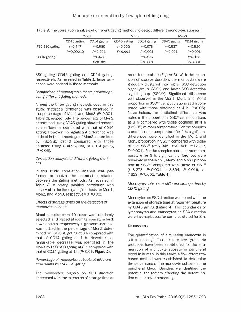
Correlation analysis of different gating meth-ods
In this study, correlation analysis was per-formed to analyze the potential correlation between the gating methods. As revealed in Table 3, a strong positive correlation was observed in the three gating methods for Mon1, Mon2, and Mon3, respectively (P<0.05).
Effects of storage times on the detection of monocytes subsets
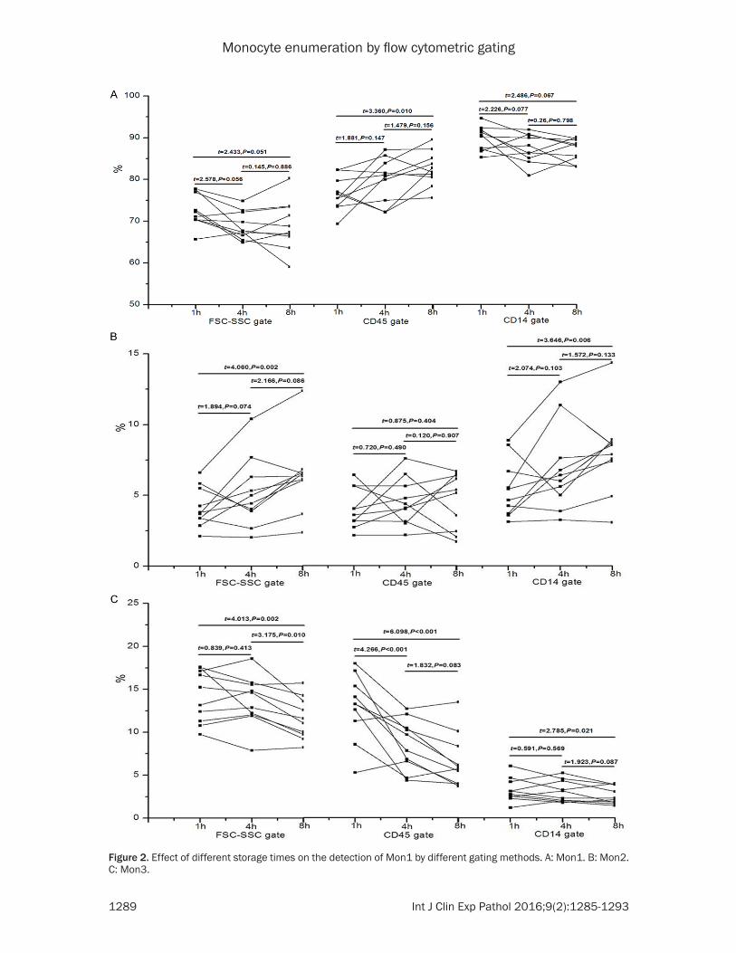
Blood samples from 10 cases were randomly selected, and placed at room temperature for 1 h, 4 h and 8 h, respectively. Significant increase was noticed in the percentage of Mon2 deter-mined by FSC-SSC gating at 8 h compared with that of CD14 gating at 1 h. Nevertheless, remarkable decrease was identified in the Mon3 by FSC-SSC gating at 8 h compared with that of CD14 gating at 1 h (P<0.05, Figure 2).
Percentage of monocytes subsets at different time points by FSC-SSC gating
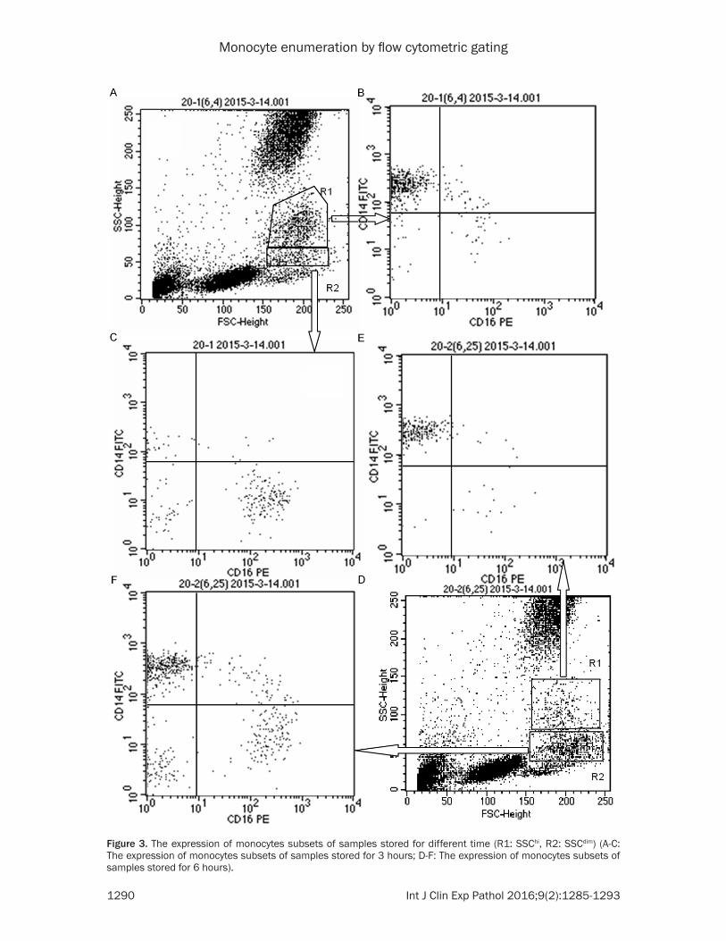
The monocytes’ signals on SSC direction decreased with the extension of storage time at
room temperature (Figure 3). With the exten-sion of storage duration, the monocytes were gradually clustered into higher SSC detection signal group (SSChi) and lower SSC detection signal group (SSClow). Significant difference was observed in the Mon1, Mon2 and Mon3 proportion in SSClow cell populations at 8 h com-pared with those obtained at 4 h (P<0.05). Nevertheless, no statistical difference was noted in the proportion in SSChi cell populations at 8 h compared with those obtained at 4 h (P>0.05) at room temperature. For the samples stored at room temperature for 4 h, significant differences were identified in the Mon1 and Mon3 proportion in SSClow compared with those of the SSChi (t=17.946, P<0.001; t=12.177, P<0.001). For the samples stored at room tem-perature for 8 h, significant differences were observed in the Mon1, Mon2 and Mon3 propor-tion in SSClow compared with those of SSChi (t=8.278, P<0.001; t=2.864, P=0.019; t= 7.323, P<0.001, Table 4).
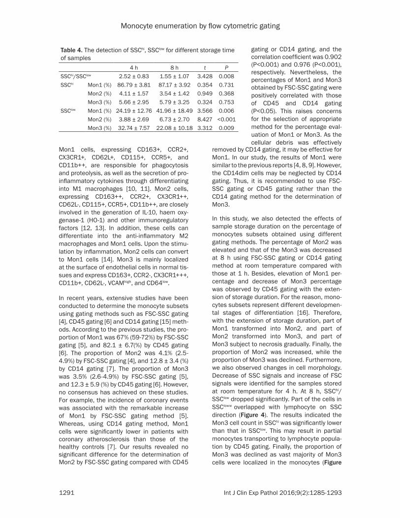
Monocytes subsets at different storage time by CD45 gating
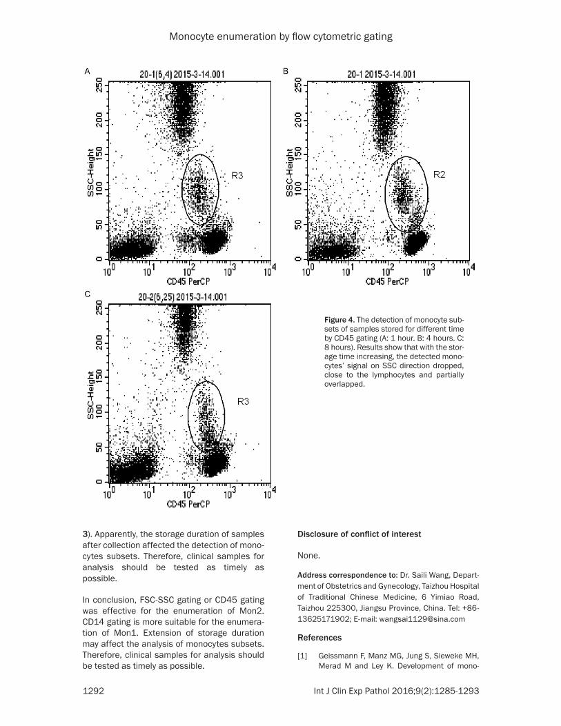
Monocytes on SSC direction weakened with the extension of storage time at room temperature by CD45 gating (Figure 4). The boundaries of lymphocytes and monocytes on SSC direction were inconspicuous for samples stored for 8 h.
Discussions
The quantification of circulating monocyte is still a challenge. To date, rare flow cytometric protocols have been established for the enu-meration of monocyte subsets in peripheral blood in human. In this study, a flow cytometry-based method was established to determine the percentage of the monocyte subsets in the peripheral blood. Besides, we identified the potential the factors affecting the determina-tion of monocyte percentage.
Table 3. The correlation analysis of different gating methods to detect different monocytes subsetsMon1 Mon2 Mon3
CD45 gating CD14 gating CD45 gating CD14 gating CD45 gating CD14 gatingFSC-SSC gating r=0.447 r=0.589 r=0.902 r=0.976 r=0.537 r=0.520
P=0.00210 P<0.001 P<0.001 P<0.001 P<0.001 P<0.001CD45 gating r=0.632 r=0.876 r=0.428
P<0.001 P<0.001 P<0.001
Monocyte enumeration by flow cytometric gating
1289 Int J Clin Exp Pathol 2016;9(2):1285-1293
Figure 2. Effect of different storage times on the detection of Mon1 by different gating methods. A: Mon1. B: Mon2. C: Mon3.
Monocyte enumeration by flow cytometric gating
1290 Int J Clin Exp Pathol 2016;9(2):1285-1293
Figure 3. The expression of monocytes subsets of samples stored for different time (R1: SSChi, R2: SSCdim) (A-C: The expression of monocytes subsets of samples stored for 3 hours; D-F: The expression of monocytes subsets of samples stored for 6 hours).
Monocyte enumeration by flow cytometric gating
1291 Int J Clin Exp Pathol 2016;9(2):1285-1293
removed by CD14 gating, it may be effective for Mon1. In our study, the results of Mon1 were similar to the previous reports [4, 8, 9]. However, the CD14dim cells may be neglected by CD14 gating. Thus, it is recommended to use FSC-SSC gating or CD45 gating rather than the CD14 gating method for the determination of Mon3.
In this study, we also detected the effects of sample storage duration on the percentage of monocytes subsets obtained using different gating methods. The percentage of Mon2 was elevated and that of the Mon3 was decreased at 8 h using FSC-SSC gating or CD14 gating method at room temperature compared with those at 1 h. Besides, elevation of Mon1 per-centage and decrease of Mon3 percentage was observed by CD45 gating with the exten-sion of storage duration. For the reason, mono-cytes subsets represent different developmen-tal stages of differentiation [16]. Therefore, with the extension of storage duration, part of Mon1 transformed into Mon2, and part of Mon2 transformed into Mon3, and part of Mon3 subject to necrosis gradually. Finally, the proportion of Mon2 was increased, while the proportion of Mon3 was declined. Furthermore, we also observed changes in cell morphology. Decrease of SSC signals and increase of FSC signals were identified for the samples stored at room temperature for 4 h. At 8 h, SSChi/SSClow dropped significantly. Part of the cells in SSCloww overlapped with lymphocyte on SSC direction (Figure 4). The results indicated the Mon3 cell count in SSChi was significantly lower than that in SSClow. This may result in partial monocytes transporting to lymphocyte popula-tion by CD45 gating. Finally, the proportion of Mon3 was declined as vast majority of Mon3 cells were localized in the monocytes (Figure
Mon1 cells, expressing CD163+, CCR2+, CX3CR1+, CD62L+, CD115+, CCR5+, and CD11b++, are responsible for phagocytosis and proteolysis, as well as the secretion of pro-inflammatory cytokines through differentiating into M1 macrophages [10, 11]. Mon2 cells, expressing CD163++, CCR2+, CX3CR1++, CD62L-, CD115+, CCR5+, CD11b++, are closely involved in the generation of IL-10, haem oxy-genase-1 (HO-1) and other immunoregulatory factors [12, 13]. In addition, these cells can differentiate into the anti-inflammatory M2 macrophages and Mon1 cells. Upon the stimu-lation by inflammation, Mon2 cells can convert to Mon1 cells [14]. Mon3 is mainly localized at the surface of endothelial cells in normal tis-sues and express CD163+, CCR2-, CX3CR1+++, CD11b+, CD62L-, VCAMhigh, and CD64low.
In recent years, extensive studies have been conducted to determine the monocyte subsets using gating methods such as FSC-SSC gating [4], CD45 gating [6] and CD14 gating [15] meth-ods. According to the previous studies, the pro-portion of Mon1 was 67% (59-72%) by FSC-SSC gating [5], and 82.1 ± 6.7(%) by CD45 gating [6]. The proportion of Mon2 was 4.1% (2.5-4.9%) by FSC-SSC gating [4], and 12.8 ± 3.4 (%) by CD14 gating [7]. The proportion of Mon3 was 3.5% (2.6-4.9%) by FSC-SSC gating [5], and 12.3 ± 5.9 (%) by CD45 gating [6]. However, no consensus has achieved on these studies. For example, the incidence of coronary events was associated with the remarkable increase of Mon1 by FSC-SSC gating method [5]. Whereas, using CD14 gating method, Mon1 cells were significantly lower in patients with coronary atherosclerosis than those of the healthy controls [7]. Our results revealed no significant difference for the determination of Mon2 by FSC-SSC gating compared with CD45
gating or CD14 gating, and the correlation coefficient was 0.902 (P<0.001) and 0.976 (P<0.001), respectively. Nevertheless, the percentages of Mon1 and Mon3 obtained by FSC-SSC gating were positively correlated with those of CD45 and CD14 gating (P<0.05). This raises concerns for the selection of appropriate method for the percentage eval-uation of Mon1 or Mon3. As the cellular debris was effectively
Table 4. The detection of SSChi, SSClow for different storage time of samples
4 h 8 h t PSSChi/SSClow 2.52 ± 0.83 1.55 ± 1.07 3.428 0.008SSChi Mon1 (%) 86.79 ± 3.81 87.17 ± 3.92 0.354 0.731
Mon2 (%) 4.11 ± 1.57 3.54 ± 1.42 0.949 0.368Mon3 (%) 5.66 ± 2.95 5.79 ± 3.25 0.324 0.753
SSClow Mon1 (%) 24.19 ± 12.76 41.96 ± 18.49 3.566 0.006Mon2 (%) 3.88 ± 2.69 6.73 ± 2.70 8.427 <0.001Mon3 (%) 32.74 ± 7.57 22.08 ± 10.18 3.312 0.009
Monocyte enumeration by flow cytometric gating
1292 Int J Clin Exp Pathol 2016;9(2):1285-1293
Disclosure of conflict of interest
None.
Address correspondence to: Dr. Saili Wang, Depart- ment of Obstetrics and Gynecology, Taizhou Hospital of Traditional Chinese Medicine, 6 Yimiao Road, Taizhou 225300, Jiangsu Province, China. Tel: +86-13625171902; E-mail: [email protected]
References
[1] Geissmann F, Manz MG, Jung S, Sieweke MH, Merad M and Ley K. Development of mono-
3). Apparently, the storage duration of samples after collection affected the detection of mono-cytes subsets. Therefore, clinical samples for analysis should be tested as timely as possible.
In conclusion, FSC-SSC gating or CD45 gating was effective for the enumeration of Mon2. CD14 gating is more suitable for the enumera-tion of Mon1. Extension of storage duration may affect the analysis of monocytes subsets. Therefore, clinical samples for analysis should be tested as timely as possible.
Figure 4. The detection of monocyte sub-sets of samples stored for different time by CD45 gating (A: 1 hour. B: 4 hours. C: 8 hours). Results show that with the stor-age time increasing, the detected mono-cytes’ signal on SSC direction dropped, close to the lymphocytes and partially overlapped.
Monocyte enumeration by flow cytometric gating
1293 Int J Clin Exp Pathol 2016;9(2):1285-1293
cytes, macrophages, and dendritic cells. Science 2010; 327: 656-661.
[2] Shantsila E, Wrigley B, Tapp L, Apostolakis S, Montoro-Garcia S, Drayson MT and Lip GY. Immunophenotypic characterization of human monocyte subsets: possible implications for cardiovascular disease pathophysiology. J Thromb Haemost 2011; 9: 1056-1066.
[3] Cros J, Cagnard N, Woollard K, Patey N, Zhang SY, Senechal B, Puel A, Biswas SK, Moshous D and Picard C. Human CD14 dim monocytes pa-trol and sense nucleic acids and viruses via TLR7 and TLR8 receptors. Immunity 2010; 33: 375-386.
[4] Hristov M, Schmitz S, Nauwelaers F and Weber C. A flow cytometric protocol for enumeration of endothelial progenitor cells and monocyte subsets in human blood. J Immunol Methods 2012; 381: 9-13.
[5] Berg KE, Ljungcrantz I, Andersson L, Bryngelsson C, Hedblad B, Fredrikson GN, Nilsson J and Björkbacka H. Elevated CD14++ CD16− monocytes predict cardiovascular events. Circ Cardiovasc Genet 2012; 5: 122-131.
[6] Krychtiuk KA, Kastl SP, Pfaffenberger S, Pongratz T, Hofbauer SL, Wonnerth A, Katsaros KM, Goliasch G, Gaspar L and Huber K. Small high-density lipoprotein is associated with monocyte subsets in stable coronary artery disease. Atherosclerosis 2014; 237: 589-596.
[7] Chelombitko MA, Shishkina VS, Ilyinskaya OP, Kaminnyi AI, Pavlunina TO, Samovilova NN, Gracheva EV, Tararak EM and Prokazova NV. A Cytofluorometric Study of Membrane Rafts in Human Monocyte Subsets in Atherosclerosis. Acta Naturae 2014; 6: 80.
[8] Tallone T, Turconi G, Soldati G, Pedrazzini G, Moccetti T and Vassalli G. Heterogeneity of hu-man monocytes: an optimized four-color flow cytometry protocol for analysis of monocyte subsets. J Cardiovasc Transl Res 2011; 4: 211-219.
[9] Van Craenenbroeck AH, Van Ackeren K, Hoymans VY, Roeykens J, Verpooten GA, Vrints CJ, Couttenye MM and Van Craenenbroeck EM. Acute Exercise-Induced Response of Monocyte Subtypes in Chronic Heart and Renal Failure. Mediators Inflamm 2014; 2014: 216534.
[10] Hristov M and Weber C. Differential role of monocyte subsets in atherosclerosis. Thromb Haemost 2011; 106: 757.
[11] Nahrendorf M, Pittet MJ and Swirski FK. Monocytes: protagonists of infarct inflamma-tion and repair after myocardial infarction. Circulation 2010; 121: 2437-2445.
[12] Skrzeczyńska-Moncznik J, Bzowska M, Grage-Griebenow E, Zembala M and Pryjma J. Peripheral Blood CD14high CD16+ Monocytes are Main Producers of IL-10. Scand J Immunol 2008; 67: 152-159.
[13] Mizuno K, Toma T, Tsukiji H, Okamoto H, Yamazaki H, Ohta K, Kasahara Y, Koizumi S and Yachie A. Selective expansion of CD16highCCR2-subpopulation of circulating monocytes with preferential production of haem oxygenase (HO)-1 in response to acute inflammation. Clin Exp Immunol 2005; 142: 461-470.
[14] Stout RD, Jiang C, Matta B, Tietzel I, Watkins SK and Suttles J. Macrophages sequentially change their functional phenotype in response to changes in microenvironmental influences. J Immunol 2005; 175: 342-349.
[15] Lu W, Zhang Z, Fu C and Ma G. Intermediate Monocytes Lead to Enhanced Myocardial Remodelling in STEMI Patients With Diabetes. Int Heart J 2015; 56: 22-28.
[16] Auffray C, Sieweke MH and Geissmann F. Blood monocytes: development, heterogene-ity, and relationship with dendritic cells. Ann Rev Immunol 2009; 27: 669-692.