Embed Size (px)
Citation preview
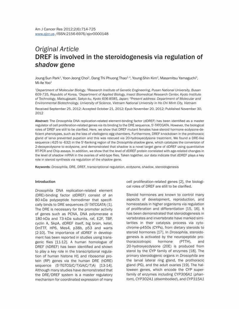
Am J Cancer Res 2012;2(6):714-725 www.ajcr.us /ISSN:2156-6976/ajcr0000148
Original ArticleDREF is involved in the steroidogenesis via regulation of shadow gene
Joung-Sun Park1, Yoon-Jeong Choi1, Dang Thi Phuong Thao3, 4, Young-Shin Kim2, Masamitsu Yamaguchi3, Mi-Ae Yoo1
1Department of Molecular Biology, 2Research Institute of Genetic Engineering, Pusan National University, Busan 609-735, Republic of Korea, 3Department of Applied Biology, Insect Biomedical Research Center, Kyoto Institute of Technology, Matsugasaki, Sakyo-ku, Kyoto 606-8585, Japan; 4Present address: Department of Molecular and Environmental Biotechnology, University of Science, Vietnam National University in Ho Chi Minh City, Vietnam
Received September 25, 2012; Accepted October 21, 2012; Epub November 20, 2012; Published November 30, 2012
Abstract: The Drosophila DNA replication-related element-binding factor (dDREF) has been identified as a master regulator of cell proliferation-related genes via its binding to the DRE sequence, 5′-TATCGATA. However, the biological roles of DREF are still to be clarified. Here, we show that DREF mutant females have steroid hormone ecdysone-de-ficient phenotypes, such as the loss of vitellogenic egg chambers. Furthermore, DREF knockdown in the prothoracic gland of larva prevented pupation and this was rescued via 20-hydroxyecdysone treatment. We found a DRE-like sequence (-625 to -632) in the 5′-flanking region of the Drosophila shadow gene, which catalyzes the conversion of 2-deoxyecdysone to ecdysone, and demonstrated that shadow is a novel target gene of dDREF using quantitative RT-PCR and Chip assays. In addition, we show that the level of dDREF protein correlated with age-related changes in the level of shadow mRNA in the ovaries of wild-type flies. Taken together, our data indicate that dDREF plays a key role in steroid synthesis via regulation of the shadow gene.
Keywords: Drosophila, DRE, DREF, transcriptional regulation, ecdysone, shadow, steroidogenesis
Introduction
Drosophila DNA replication-related element (DRE)-binding factor (dDREF) consist of an 80-kDa polypeptide homodimer that specifi-cally binds to DRE sequences (5′-TATCGATA) [1]. The DRE is necessary for the promoter activity of genes such as PCNA, DNA polymerase α 180-kDa and 73-kDa subunits, raf, E2F, TBP, cyclin A, SkpA, dDREF itself, big brain, ketel, DmTTF, HP6, Mes4, p38b, p53 and warts [2-10]. The importance of dDREF in develop-ment has been reported in studies using trans-genic flies [11-12]. A human homologue of DREF (hDREF) has been identified and shown to play a key role in the transcriptional regula-tion of human histone H1 and ribosomal pro-tein (RP) genes via the human DRE (hDRE) sequence (5′-TGTCG(C/T)GA(C/T)A) [13-14]. Although many studies have demonstrated that the DRE/DREF system is a master regulatory mechanism for coordinated expression of many
cell proliferation-related genes [2], the biologi-cal roles of DREF are still to be clarified.
Steroid hormones are known to control many aspects of development, reproduction, and homeostasis in higher organisms via regulation of proliferation and differentiation [15, 16]. It has been demonstrated that steroidogenesis in vertebrates and invertebrate have marked simi-larities in their catalysis process via cyto-chrome-p450s (CYPs), from dietary steroids to steroid hormones [17]. In Drosophila, steroido-genesis is activated by the neuropeptide pro-thoracicotropic hormone (PTTH), and 20-hydroxyecdysone (20E) is produced from sterol by the CYP family of enzymes [18]. The primary steroidogenic organs in Drosophila are the larval lateral ring gland, the prothoracic gland (PG), and the adult ovaries [19]. The hal-loween genes, which encode the CYP super-family of enzymes including CYP306A1 (phan-tom), CYP302A1 (disembodied), and CYP315A1
Regulation of steroidogenesis by DREF
715 Am J Cancer Res 2012;2(6):714-725
(shadow), catalyze three sequential hydroxyl-ations yielding ecdysone from dietary choles-terol [20]. Ecdysone is then converted into the principal molting hormone, 20-hydroxyecdy-sone (20E), by the P450 enzyme (CYP314A1; shade) in peripheral target tissues [21]. Steroid hormones such as 20E trigger the differentia-tion and morphogenesis of the imaginal discs, giveing rise to adult tissues, and the pro-grammed cell death of larval cells [22]. Each hormone pulse leads to the induction of three early response genes, BR-C, E74, and E75, each of which encode multiple forms of related transcription factors [23-25]. Although steroid hormones play an essential role during an organism’s lifespan, little is known about the regulatory mechanisms of steroidogenesis.
In this study, we show that adult females het-erozygous for DrefKG09294 and larvae carrying DREF knockdown in the PG, induced hormone-deficient phenotypes and provided evidence that dDREF plays a role in the growth of the pri-mary steroidogenic organs and in steroidogen-esis via the regulation of a steroidogenesis-related gene shadow. Our results suggest that the conservation of DREF in human indicates a broad role for DREF proteins during steroidogenesis.
Results
DREF mutant has steroid hormone ecdysone-deficient phenotypes
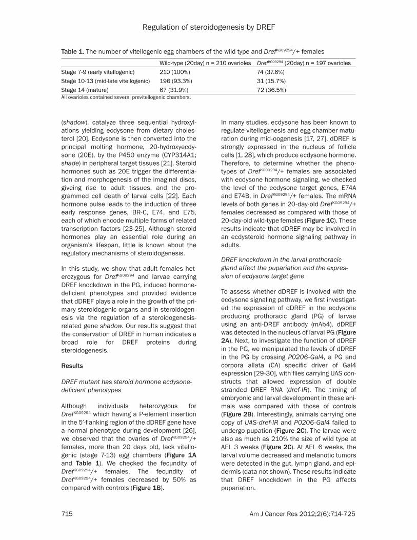
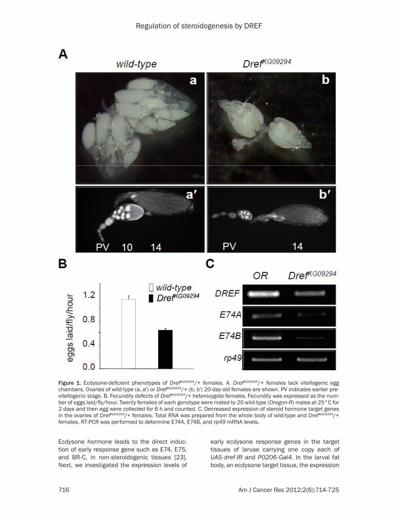
Although individuals heterozygous for DrefKG09294 which having a P-element insertion in the 5′-flanking region of the dDREF gene have a normal phenotype during development [26], we observed that the ovaries of DrefKG09294/+ females, more than 20 days old, lack vitello-genic (stage 7-13) egg chambers (Figure 1A and Table 1). We checked the fecundity of DrefKG09294/+ females. The fecundity of DrefKG09294/+ females decreased by 50% as compared with controls (Figure 1B).
In many studies, ecdysone has been known to regulate vitellogenesis and egg chamber matu-ration during mid-oogenesis [17, 27]. dDREF is strongly expressed in the nucleus of follicle cells [1, 28], which produce ecdysone hormone. Therefore, to determine whether the pheno-types of DrefKG09294/+ females are associated with ecdysone hormone signaling, we checked the level of the ecdysone target genes, E74A and E74B, in DrefKG09294/+ females. The mRNA levels of both genes in 20-day-old DrefKG09294/+ females decreased as compared with those of 20-day-old wild-type females (Figure 1C). These results indicate that dDREF may be involved in an ecdysteroid hormone signaling pathway in adults.
DREF knockdown in the larval prothoracic gland affect the pupariation and the expres-sion of ecdysone target gene
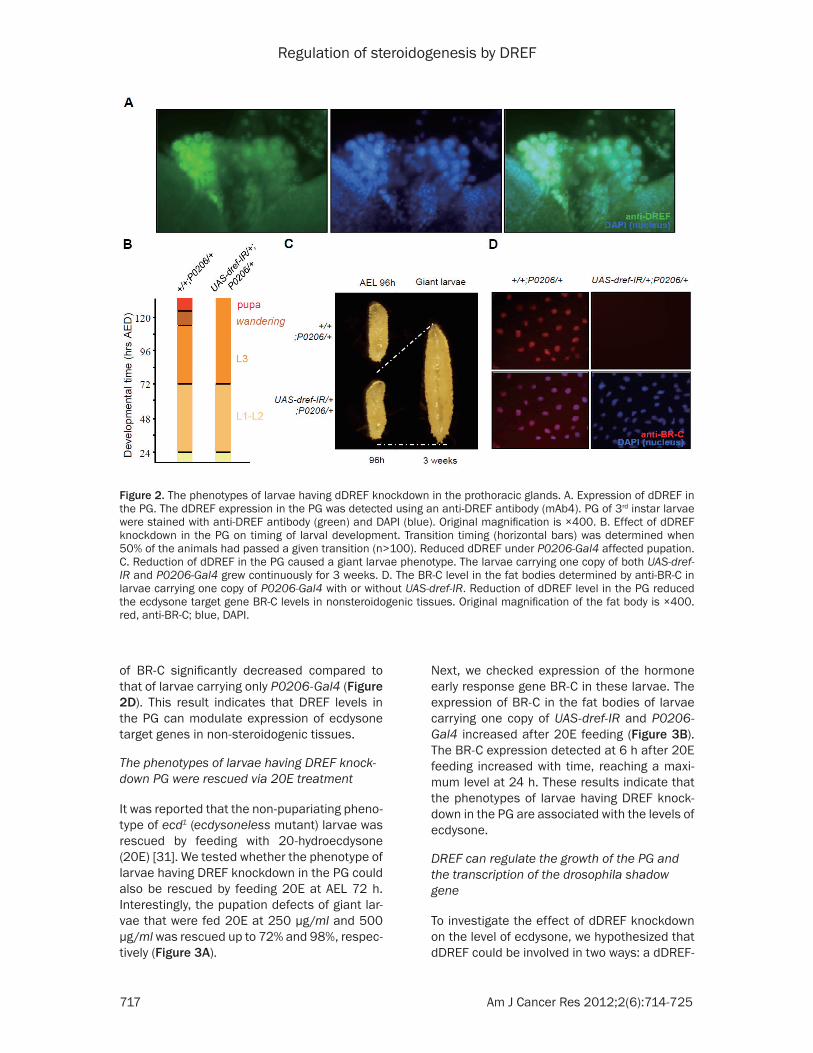
To assess whether dDREF is involved with the ecdysone signaling pathway, we first investigat-ed the expression of dDREF in the ecdysone producing prothoracic gland (PG) of larvae using an anti-DREF antibody (mAb4). dDREF was detected in the nucleus of larval PG (Figure 2A). Next, to investigate the function of dDREF in the PG, we manipulated the levels of dDREF in the PG by crossing P0206-Gal4, a PG and corpora allata (CA) specific driver of Gal4 expression [29-30], with flies carrying UAS con-structs that allowed expression of double stranded DREF RNA (dref-IR). The timing of embryonic and larval development in these ani-mals was compared with those of controls (Figure 2B). Interestingly, animals carrying one copy of UAS-dref-IR and P0206-Gal4 failed to undergo pupation (Figure 2C). The larvae were also as much as 210% the size of wild type at AEL 3 weeks (Figure 2C). At AEL 6 weeks, the larval volume decreased and melanotic tumors were detected in the gut, lymph gland, and epi-dermis (data not shown). These results indicate that DREF knockdown in the PG affects pupariation.
Table 1. The number of vitellogenic egg chambers of the wild type and DrefKG09294/+ females
Wild-type (20day) n = 210 ovarioles DrefKG09294 (20day) n = 197 ovariolesStage 7-9 (early vitellogenic) 210 (100%) 74 (37.6%)Stage 10-13 (mid-late vitellogenic) 196 (93.3%) 31 (15.7%)Stage 14 (mature) 67 (31.9%) 72 (36.5%)All ovarioles contained several previtellogenic chambers.
Regulation of steroidogenesis by DREF
716 Am J Cancer Res 2012;2(6):714-725
Ecdysone hormone leads to the direct induc-tion of early response gene such as E74, E75, and BR-C, in non-steroidogenic tissues [23]. Next, we investigated the expression levels of
early ecdysone response genes in the target tissues of larvae carrying one copy each of UAS-dref-IR and P0206-Gal4. In the larval fat body, an ecdysone target tissue, the expression
Figure 1. Ecdysone-deficient phenotypes of DrefKG09294/+ females. A. DrefKG09294/+ females lack vitellogenic egg chambers. Ovaries of wild-type (a, a′) or DrefKG09294/+ (b, b′) 20-day-old females are shown. PV indicates earlier pre-vitellogenic stage. B. Fecundity defects of DrefKG09294/+ heterozygote females. Fecundity was expressed as the num-ber of eggs laid/fly/hour. Twenty females of each genotype were mated to 20 wild-type (Oregon-R) males at 25°C for 2 days and then egg were collected for 6 h and counted. C. Decreased expression of steroid hormone target genes in the ovaries of DrefKG09294/+ females. Total RNA was prepared from the whole body of wild-type and DrefKG09294/+ females. RT-PCR was performed to determine E74A, E74B, and rp49 mRNA levels.
Regulation of steroidogenesis by DREF
717 Am J Cancer Res 2012;2(6):714-725
of BR-C significantly decreased compared to that of larvae carrying only P0206-Gal4 (Figure 2D). This result indicates that DREF levels in the PG can modulate expression of ecdysone target genes in non-steroidogenic tissues.
The phenotypes of larvae having DREF knock-down PG were rescued via 20E treatment
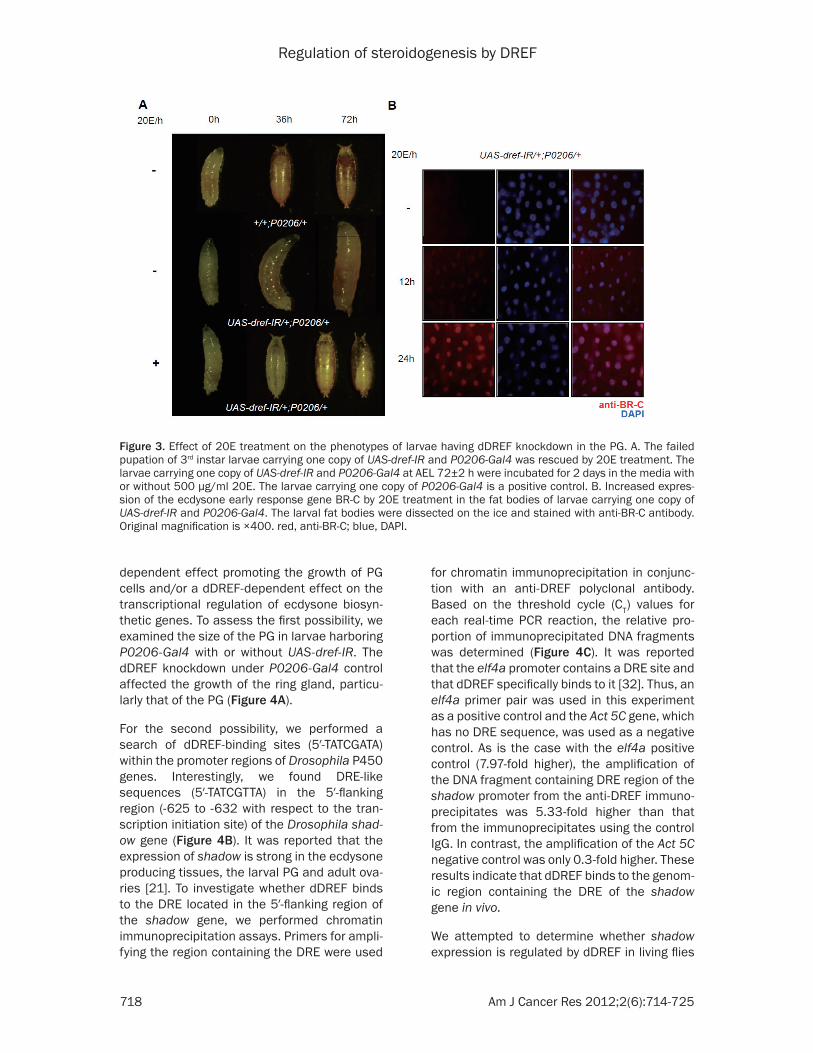
It was reported that the non-pupariating pheno-type of ecd1 (ecdysoneless mutant) larvae was rescued by feeding with 20-hydroecdysone (20E) [31]. We tested whether the phenotype of larvae having DREF knockdown in the PG could also be rescued by feeding 20E at AEL 72 h. Interestingly, the pupation defects of giant lar-vae that were fed 20E at 250 µg/ml and 500 µg/ml was rescued up to 72% and 98%, respec-tively (Figure 3A).
Next, we checked expression of the hormone early response gene BR-C in these larvae. The expression of BR-C in the fat bodies of larvae carrying one copy of UAS-dref-IR and P0206-Gal4 increased after 20E feeding (Figure 3B). The BR-C expression detected at 6 h after 20E feeding increased with time, reaching a maxi-mum level at 24 h. These results indicate that the phenotypes of larvae having DREF knock-down in the PG are associated with the levels of ecdysone.
DREF can regulate the growth of the PG and the transcription of the drosophila shadow gene
To investigate the effect of dDREF knockdown on the level of ecdysone, we hypothesized that dDREF could be involved in two ways: a dDREF-
Figure 2. The phenotypes of larvae having dDREF knockdown in the prothoracic glands. A. Expression of dDREF in the PG. The dDREF expression in the PG was detected using an anti-DREF antibody (mAb4). PG of 3rd instar larvae were stained with anti-DREF antibody (green) and DAPI (blue). Original magnification is ×400. B. Effect of dDREF knockdown in the PG on timing of larval development. Transition timing (horizontal bars) was determined when 50% of the animals had passed a given transition (n>100). Reduced dDREF under P0206-Gal4 affected pupation. C. Reduction of dDREF in the PG caused a giant larvae phenotype. The larvae carrying one copy of both UAS-dref-IR and P0206-Gal4 grew continuously for 3 weeks. D. The BR-C level in the fat bodies determined by anti-BR-C in larvae carrying one copy of P0206-Gal4 with or without UAS-dref-IR. Reduction of dDREF level in the PG reduced the ecdysone target gene BR-C levels in nonsteroidogenic tissues. Original magnification of the fat body is ×400. red, anti-BR-C; blue, DAPI.
Regulation of steroidogenesis by DREF
718 Am J Cancer Res 2012;2(6):714-725
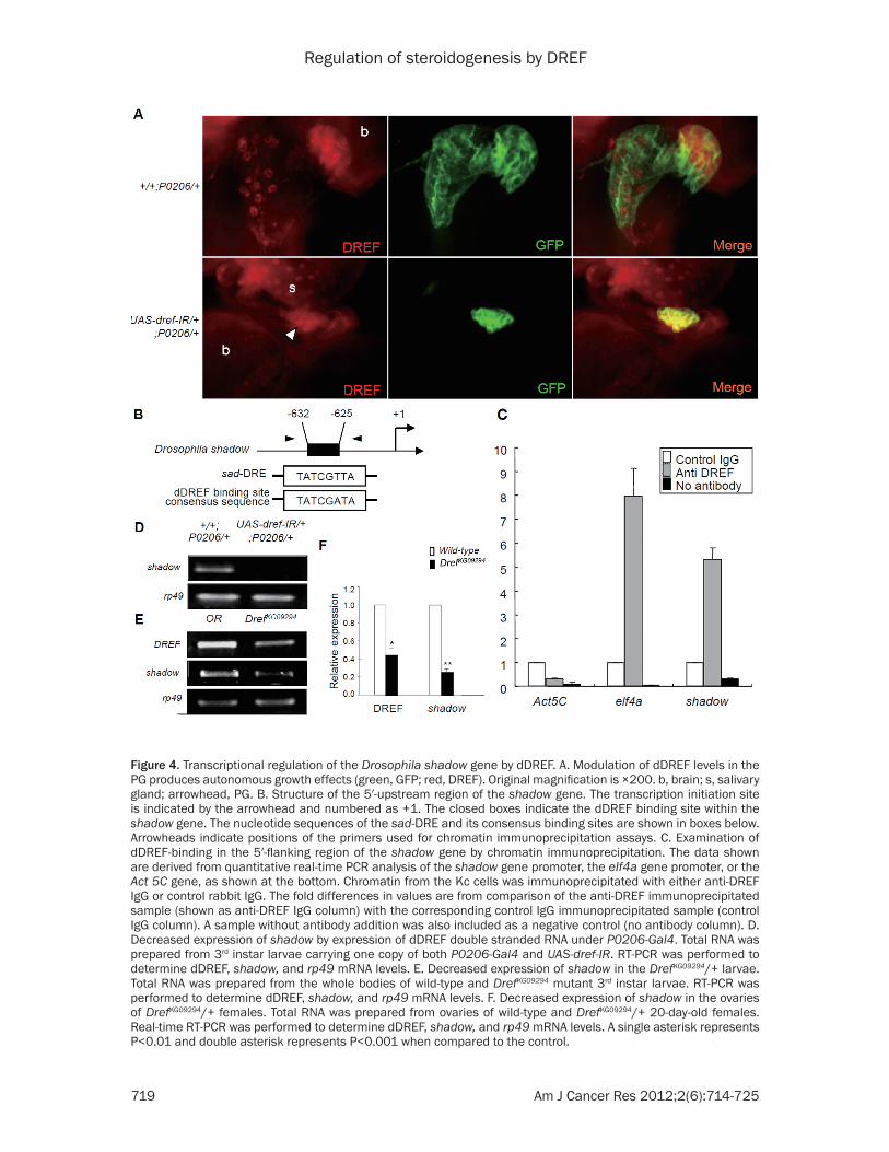
dependent effect promoting the growth of PG cells and/or a dDREF-dependent effect on the transcriptional regulation of ecdysone biosyn-thetic genes. To assess the first possibility, we examined the size of the PG in larvae harboring P0206-Gal4 with or without UAS-dref-IR. The dDREF knockdown under P0206-Gal4 control affected the growth of the ring gland, particu-larly that of the PG (Figure 4A).
For the second possibility, we performed a search of dDREF-binding sites (5′-TATCGATA) within the promoter regions of Drosophila P450 genes. Interestingly, we found DRE-like sequences (5′-TATCGTTA) in the 5′-flanking region (-625 to -632 with respect to the tran-scription initiation site) of the Drosophila shad-ow gene (Figure 4B). It was reported that the expression of shadow is strong in the ecdysone producing tissues, the larval PG and adult ova-ries [21]. To investigate whether dDREF binds to the DRE located in the 5′-flanking region of the shadow gene, we performed chromatin immunoprecipitation assays. Primers for ampli-fying the region containing the DRE were used
for chromatin immunoprecipitation in conjunc-tion with an anti-DREF polyclonal antibody. Based on the threshold cycle (CT) values for each real-time PCR reaction, the relative pro-portion of immunoprecipitated DNA fragments was determined (Figure 4C). It was reported that the elf4a promoter contains a DRE site and that dDREF specifically binds to it [32]. Thus, an elf4a primer pair was used in this experiment as a positive control and the Act 5C gene, which has no DRE sequence, was used as a negative control. As is the case with the elf4a positive control (7.97-fold higher), the amplification of the DNA fragment containing DRE region of the shadow promoter from the anti-DREF immuno-precipitates was 5.33-fold higher than that from the immunoprecipitates using the control IgG. In contrast, the amplification of the Act 5C negative control was only 0.3-fold higher. These results indicate that dDREF binds to the genom-ic region containing the DRE of the shadow gene in vivo.
We attempted to determine whether shadow expression is regulated by dDREF in living flies
Figure 3. Effect of 20E treatment on the phenotypes of larvae having dDREF knockdown in the PG. A. The failed pupation of 3rd instar larvae carrying one copy of UAS-dref-IR and P0206-Gal4 was rescued by 20E treatment. The larvae carrying one copy of UAS-dref-IR and P0206-Gal4 at AEL 72±2 h were incubated for 2 days in the media with or without 500 µg/ml 20E. The larvae carrying one copy of P0206-Gal4 is a positive control. B. Increased expres-sion of the ecdysone early response gene BR-C by 20E treatment in the fat bodies of larvae carrying one copy of UAS-dref-IR and P0206-Gal4. The larval fat bodies were dissected on the ice and stained with anti-BR-C antibody. Original magnification is ×400. red, anti-BR-C; blue, DAPI.
Regulation of steroidogenesis by DREF
719 Am J Cancer Res 2012;2(6):714-725
Figure 4. Transcriptional regulation of the Drosophila shadow gene by dDREF. A. Modulation of dDREF levels in the PG produces autonomous growth effects (green, GFP; red, DREF). Original magnification is ×200. b, brain; s, salivary gland; arrowhead, PG. B. Structure of the 5′-upstream region of the shadow gene. The transcription initiation site is indicated by the arrowhead and numbered as +1. The closed boxes indicate the dDREF binding site within the shadow gene. The nucleotide sequences of the sad-DRE and its consensus binding sites are shown in boxes below. Arrowheads indicate positions of the primers used for chromatin immunoprecipitation assays. C. Examination of dDREF-binding in the 5′-flanking region of the shadow gene by chromatin immunoprecipitation. The data shown are derived from quantitative real-time PCR analysis of the shadow gene promoter, the eIf4a gene promoter, or the Act 5C gene, as shown at the bottom. Chromatin from the Kc cells was immunoprecipitated with either anti-DREF IgG or control rabbit IgG. The fold differences in values are from comparison of the anti-DREF immunoprecipitated sample (shown as anti-DREF IgG column) with the corresponding control IgG immunoprecipitated sample (control IgG column). A sample without antibody addition was also included as a negative control (no antibody column). D. Decreased expression of shadow by expression of dDREF double stranded RNA under P0206-Gal4. Total RNA was prepared from 3rd instar larvae carrying one copy of both P0206-Gal4 and UAS-dref-IR. RT-PCR was performed to determine dDREF, shadow, and rp49 mRNA levels. E. Decreased expression of shadow in the DrefKG09294/+ larvae. Total RNA was prepared from the whole bodies of wild-type and DrefKG09294 mutant 3rd instar larvae. RT-PCR was performed to determine dDREF, shadow, and rp49 mRNA levels. F. Decreased expression of shadow in the ovaries of DrefKG09294/+ females. Total RNA was prepared from ovaries of wild-type and DrefKG09294/+ 20-day-old females. Real-time RT-PCR was performed to determine dDREF, shadow, and rp49 mRNA levels. A single asterisk represents P<0.01 and double asterisk represents P<0.001 when compared to the control.
Regulation of steroidogenesis by DREF
720 Am J Cancer Res 2012;2(6):714-725
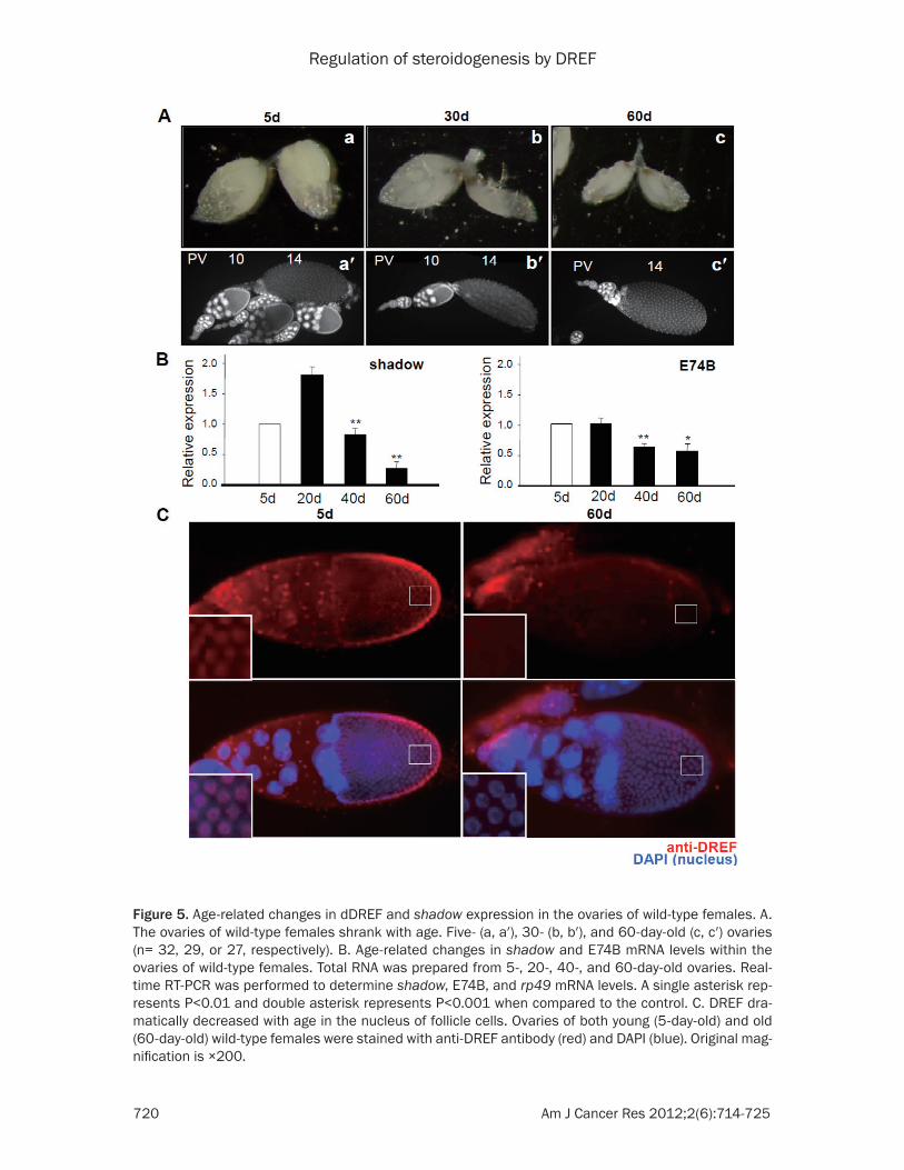
Figure 5. Age-related changes in dDREF and shadow expression in the ovaries of wild-type females. A. The ovaries of wild-type females shrank with age. Five- (a, a′), 30- (b, b′), and 60-day-old (c, c′) ovaries (n= 32, 29, or 27, respectively). B. Age-related changes in shadow and E74B mRNA levels within the ovaries of wild-type females. Total RNA was prepared from 5-, 20-, 40-, and 60-day-old ovaries. Real-time RT-PCR was performed to determine shadow, E74B, and rp49 mRNA levels. A single asterisk rep-resents P<0.01 and double asterisk represents P<0.001 when compared to the control. C. DREF dra-matically decreased with age in the nucleus of follicle cells. Ovaries of both young (5-day-old) and old (60-day-old) wild-type females were stained with anti-DREF antibody (red) and DAPI (blue). Original mag-nification is ×200.
Regulation of steroidogenesis by DREF
721 Am J Cancer Res 2012;2(6):714-725
using the GAL4-UAS system. In RT-PCR experi-ments, the level of shadow mRNA in the whole larvae having UAS-dref-IR and P0206-Gal4 was lower than those observed in the controls (Figure 4D). We also assessed the level of shadow mRNA in DrefKG09294/+ larvae. The level of shadow mRNA in the 3rd instar larvae of the DrefKG09294/+ heterozygous was reduced in comparison with that of wild type (Figure 4E). To confirm the RT-PCR data, shadow mRNA lev-els in the ovaries of the DrefKG09294/+ females were examined by real-time RT-PCR. The expression level was significantly reduced in comparison with that of wild type (Figure 4F). These results indicate that dDREF positively regulates the expression of shadow. Taken together, we conclude that dDREF can regulate ecdysteroidogenesis via regulation of PG growth and transcriptional regulation of the Drosophila shadow gene.
dDREF is involved in age-related decrease in steroidogenesis
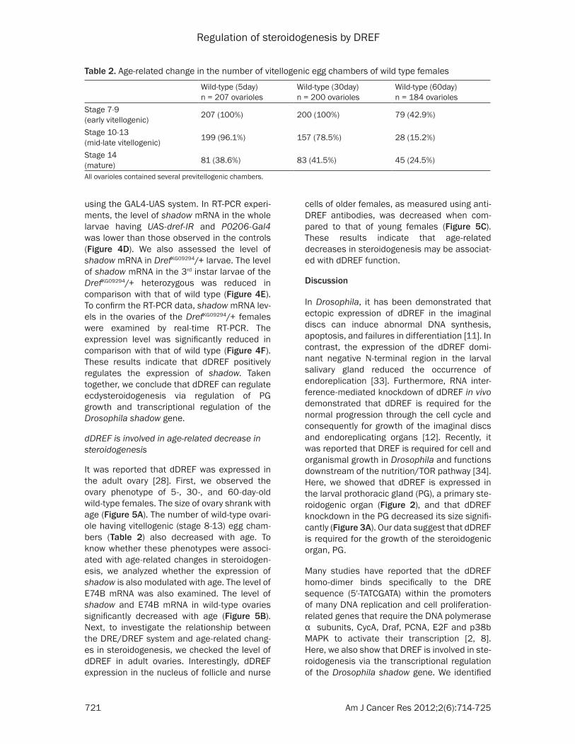
It was reported that dDREF was expressed in the adult ovary [28]. First, we observed the ovary phenotype of 5-, 30-, and 60-day-old wild-type females. The size of ovary shrank with age (Figure 5A). The number of wild-type ovari-ole having vitellogenic (stage 8-13) egg cham-bers (Table 2) also decreased with age. To know whether these phenotypes were associ-ated with age-related changes in steroidogen-esis, we analyzed whether the expression of shadow is also modulated with age. The level of E74B mRNA was also examined. The level of shadow and E74B mRNA in wild-type ovaries significantly decreased with age (Figure 5B). Next, to investigate the relationship between the DRE/DREF system and age-related chang-es in steroidogenesis, we checked the level of dDREF in adult ovaries. Interestingly, dDREF expression in the nucleus of follicle and nurse
cells of older females, as measured using anti-DREF antibodies, was decreased when com-pared to that of young females (Figure 5C). These results indicate that age-related decreases in steroidogenesis may be associat-ed with dDREF function.
Discussion
In Drosophila, it has been demonstrated that ectopic expression of dDREF in the imaginal discs can induce abnormal DNA synthesis, apoptosis, and failures in differentiation [11]. In contrast, the expression of the dDREF domi-nant negative N-terminal region in the larval salivary gland reduced the occurrence of endoreplication [33]. Furthermore, RNA inter-ference-mediated knockdown of dDREF in vivo demonstrated that dDREF is required for the normal progression through the cell cycle and consequently for growth of the imaginal discs and endoreplicating organs [12]. Recently, it was reported that DREF is required for cell and organismal growth in Drosophila and functions downstream of the nutrition/TOR pathway [34]. Here, we showed that dDREF is expressed in the larval prothoracic gland (PG), a primary ste-roidogenic organ (Figure 2), and that dDREF knockdown in the PG decreased its size signifi-cantly (Figure 3A). Our data suggest that dDREF is required for the growth of the steroidogenic organ, PG.
Many studies have reported that the dDREF homo-dimer binds specifically to the DRE sequence (5′-TATCGATA) within the promoters of many DNA replication and cell proliferation-related genes that require the DNA polymerase α subunits, CycA, Draf, PCNA, E2F and p38b MAPK to activate their transcription [2, 8]. Here, we also show that DREF is involved in ste-roidogenesis via the transcriptional regulation of the Drosophila shadow gene. We identified
Table 2. Age-related change in the number of vitellogenic egg chambers of wild type females
Wild-type (5day)n = 207 ovarioles
Wild-type (30day)n = 200 ovarioles
Wild-type (60day)n = 184 ovarioles
Stage 7-9(early vitellogenic) 207 (100%) 200 (100%) 79 (42.9%)
Stage 10-13(mid-late vitellogenic) 199 (96.1%) 157 (78.5%) 28 (15.2%)
Stage 14(mature) 81 (38.6%) 83 (41.5%) 45 (24.5%)
All ovarioles contained several previtellogenic chambers.
Regulation of steroidogenesis by DREF
722 Am J Cancer Res 2012;2(6):714-725
the DRE-like sequence (5′-TATCGTTA) in the region between -625 and -632 with respect to the transcription initiation site of the shadow gene. Using a chip assay and quantitative RT-PCR, we demonstrated that dDREF binds to the genomic region containing DRE in the shad-ow gene promoter and regulate the levels of shadow mRNA. Therefore, our data indicate that shadow is a novel target of dDREF. It was demonstrated that hDREF is an important tran-scription factor that plays a role in cell cycle-dependent regulation of RP genes and histone H1 [13, 35]. Interestingly, we found an 8 out of 10 nucleotides match within the hDRE-like sequence (5′-TGTCGCCCA) in the region between -524 and -533 with respect to the transcription initiation site of the human CYP19A1 gene. This raises the question of whether the human CYP19 gene is also regu-lated by hDREF. Whether hDREF is involved in the human steroidogenesis would be an inter-esting avenue of research.
In humans, many reports have shown that abnormal levels of steroid hormones cause diverse diseases including hormone-related cancer, osteoporosis, and neurodegenerative diseases [36-38]. It was reported that an estro-gen deficient state is characterized by acceler-ated aging [39]. Here, we demonstrated that the number of ovariole having vitellogenic egg chambers and the expression of shadow and E74B mRNAs at the adult stage both decreased with age. These data indicate an age-related decrease of steroidogenesis in Drosophila. We found that the ovaries of 20-day-old DrefKG09294/+ females demonstrated pheno-types similar to those of 60-day-old wild-type females. Furthermore, we found that the dDREF levels within the follicle cell nucleus of 60-day-old flies were markedly lower than that of 5-day-old flies. Our data suggest that dDREF may also be involved an age-related decrease in ste-roidogenesis and that DrefKG09294 would be an excellent model for studying steroid hormone function in adults. Recently, it was reported that hDREF is associated with importin β1 dur-ing nuclear transport via the hATC domain [35]. It was also reported that the protein levels of nucleocytoplasmic transport factors (NCT) con-taining importin β1 were reduced with age [40]. Interestingly, we found that Drosophila DREF has an ATC sequence on its 3′-carboxyl-termi-nal region (data not shown). Further studies
investigating the molecular mechanisms by which dDREF levels in the nucleus of follicle cells are modulated with age are necessary.
Experimental procedures
Fly stocks
Fly stocks were cultured at 25°C on standard media. The Oregon-R flies were used as the wild-type strain. The P0206-Gal4 fly stock was kindly provided by Drs W. Janning and L. Pierre. Oregon-R and DrefKG09294 stocks used in this study were obtained from the Bloomington Drosophila stock center.
Immunohistochemistry
Immunohistology was performed as described previously [41]. We used the following primary antibodies: mouse anti-DREF (mAb4) 1:200 and mouse anti-BR-C 1:100 [Developmental Studies Hybridoma Bank (DSHB), Iowa City, IA, USA]. We used the following secondary antibod-ies: mouse-Cy3 (Jackson ImmunoResearch, West Grove, PA, USA) 1:400. DAPI (Molecular Probes, Eugene, OR, USA) was used at 1 µg/ml. We used Vectashield Mounting Medium (Vector Labs, Burlingame, CA, USA) for microscopic observation. Fluorescent samples were exam-ined on a Karl Zeiss AxioPlan2 fluorescent microscope (Carl Zeiss Inc., Jena, Germany). Images were processed and assembled in Photoshop CS.
Fecundity analysis
Egg laying rate analysis was performed as described [42]. Fecundity was expressed as the number of eggs laid/fly/hour. Twenty 5-day-old wild-type or mutant virgin females were crossed to 20 wild-type males for 2 days and their eggs collected on a molasses agar plate and count-ed at 24 h intervals. This experiments were conducted at 25°C.
Quantification of vitellogenic egg chambers
Wild-type and heterozygous DrefKG09294 females were mated within 1 day of eclosion to Oregon-R males. Number of vitellogenic egg chambers was counted as described previously [43]. Ovaries were dissected, fixed in 4% paraformal-dehyde for 30 min, stained in 1 µg/ml DAPI (Molecular Probes) for 20 min, washed three times with PBS, and mounted in Vectashield
Regulation of steroidogenesis by DREF
723 Am J Cancer Res 2012;2(6):714-725
statin A), and lysed in 2 ml of SDS lysis buffer (Upstate). Lysates were sonicated to break the DNA into fragments of less than 1 kb and cen-trifuged at 15,300 x g for 10 min at 4˚C. The sonicated cell supernatants were diluted 10 fold in chip dilution buffer (Upstate Biotechnology Inc., Lake Placid, NY, USA) and pre-cleared with 80 µl of salmon sperm DNA/Protein G agarose-50% slurry for 30 min at 4°C. After a brief centrifugation, supernatants were incubated with 1 µg of the normal rabbit-IgG antibody (Sigma) or 1 µg of anti-DREF poly-clonal antibody for 16 h at 4°C. Salmon sperm DNA/Protein G agarose-50% slurry was added and incubated for 1 h at 4°C. After washing, immunoprecipitated DNA was eluted with buf-fer containing 1% SDS and 0.1 M NaHCO3. The protein-DNA crosslinks were then reversed by heating at 65°C for 4 h. After deproteinization with Proteinase K, DNA was recovered by phe-nol-chloroform extraction and ethanol precipi-tation. The immunoprecipitated DNA fragments were then detected by quantitative real-time PCR using SYBR Green I (TaKaRa Dalian Biotechnology Co., Ltd. Dalian, China) and the Applied Biosystems 7500 Real Time PCR sys-tem. The ΔCT value of each sample was calcu-lated by subtracting the CT value of the input sample from the CT value obtained of the immu-noprecipitated samples. Fold differences of each sample, relative to the control using non-immune IgG, were then calculated by raising 2 to the ΔΔCT power. The ΔΔCT was calculated by subtracting the ΔCT value of the sample immu-noprecipitated with control IgG.
Oligonucleotide primers for Chip assay: eif4ad-re1p: 5′-CTTTACCATACACACTGCGAAG; eif4aan-tidre1p: 5′-CAAAAGAGGCTCCATCTTGCAAAAG; actin5CP: 5′-CTCCATCATGAAGTGTGATGTG; actin5CantiP: 5′-CGTACTCCTGCTTGGACGTC; shadow Fn : 5′-GCGATTGCATGGGCTTAAAATCAGA GTCGAAAAAGG; shadow Rn: 5′-CTGTAAATAATG TTATCTATTGAATGCGTTTTTGG.
Address correspondence to: Mi-Ae Yoo, Department of Molecular Biology, College of Natural Science, Pusan National University, Busan 609-735, Republic of Korea. Tel: +82-51-510-2278; Fax: +82-51-513-9258; E-mail: [email protected]. Joung-Sun Park, Department of Molecular Biology, College of Natural Science, Pusan National University, Busan 609-735, Republic of Korea. Tel: +82-51-510-3362; Fax: +82-51-513-9258; E-mail: [email protected]
Mounting Medium (Vector Labs). The number of ovarioles containing early vitellogenic (stage 7-9), mid-late vitellogenic (stage 10-13), or mature (stage 14) egg chambers were counted.
20E feeding
Hormone feeding experiment was performed as described previously [31]. Two hundred early third instar larvae (AEL 72±2 h) of each geno-type were placed in vials with a sucrose-yeast medium containing 20-hydroxyecdysone (20E) (Sigma-Aldrich, St. Louis, MO, USA) at concen-trations of 250 or 500 µg/ml. This experiment was conducted at 25°C on dark conditions.
Quantitative RT-PCR
Real-time RT-PCR and RT-PCR were performed as described previously [32]. Total RNA from larvae was isolated with Trizol Reagent (Molecular Research Center Inc., Cincinnati, OH, USA) according to the manufacturer’s pro-tocol and cDNAs were synthesized with M-MLV-RT (reverse transcriptase) (Promega, Madison, WI, USA).
Oligonucleotide primers for real-time RT-PCR: dDREF sense: 5́ -ACACCTATCTGCGAGAGAAAAAT ATAC; dDREF antisense: 5́ -CAAAGAACTTCAATG TTTCGTACAG; shadow Up2: 5́ -CGGAGAGTGG TGAAATACGA; shadow Do1: 5́ -TACGCTGTCAAC GGGCATCT; Oligonucleotide primers for RT-PCR: E74A sense: 5́ -TCCGAGAGCAACTTCGAGAT; E74A antisense: 5́ -TTGATCAAATCGCCACAGAG; E74B sense: 5́ -ATGTGTCCAGCTCCAGCTCT; E74B antisense: 5́ -TAGTGACTCGGGGACTTTGG.
Oligonucleotide primers of ribosomal protein 49 (rp49) were described previously [44]. The real-time RT-PCR products were analyzed using OpticMonitor3.
Chip assay
Chromatin immunoprecipitation was performed using a Chip Assay kit as recommended by the manufacturer (Upstate Biotechnology Inc., Lake Placid, NY, USA) . Approximately 2 x 10⁷ Kc cells were fixed in 1% formaldehyde at 37°C for 10 min and then quenched in 125 mM glycine for 5 min at 25°C. Cells were collected, washed twice in PBS containing protease inhibitors (1 mM PMSF, 1 µg/ml aprotinin and 1 µg/ml pep-
Regulation of steroidogenesis by DREF
724 Am J Cancer Res 2012;2(6):714-725
References
[1] Hirose F, Yamaguchi M, Kuroda K, Omori A, Hachiya T, Ikeda M, Nishimoto Y, Matsukage A. Isolation and characterization of cDNA for DREF, a promoter-activating factor for Dro-sophila DNA replication-related genes. J Biol Chem 1996; 271: 3930-3937.
[2] Matsukage A, Hirose F, Yoo MA, Yamaguchi M. The DRE/DREF transcriptional regulatory sys-tem: a master key for cell proliferation. Biochim Biophys Acta 2008; 1779: 81-89.
[3] Park JS, Kim SR, Park SY, Yang DJ, Lee SH, Choi YJ, Bae MK, Yamaguchi M, Kim YS, Yoo MA. Big brain, a Drosophila homologue of mammalian aquaporin, is regulated by the DRE/DREF system. Biochim Biophys Acta 2008; 1779:789-796.
[4] Villanyi Z, Papp B, Szikora S, Boros I, Szabad J. The DRE motif is a key component in the ex-pression regulation of the importin-beta en-coding Ketel gene in Drosophila. Mech Dev 2008; 125: 822-831.
[5] Suyari O, Ida H, Yoshioka Y, Kato Y, Hashimoto R, Yamaguchi M. Identification of the Drosoph-ila Mes4 gene as a novel target of the tran-scription factor DREF. Exp Cell Res 2009; 315: 1403-1414.
[6] Fernández-Moreno MA, Bruni F, Adán C, Sierra RH, Polosa PL, Cantatore P, Garesse R, Roberti M. The Drosophila nuclear factor DREF posi-tively regulates the expression of the mito-chondrial transcription termination factor Dm-TTF. Biochem J 2009; 418: 453-462.
[7] Ida H, Suzusho N, Suyari O, Yoshida H, Ohno K, Hirose F, Itoh M, Yamaguchi M. Genetic screen-ing for modifiers of the DREF pathway in Dro-sophila melanogaster: identification and char-acterization of HP6 as a novel target of DREF. Nucleic Acids Res 2009; 37: 1423-1437.
[8] Park JS, Kim YS, Kim JG, Lee SH, Park SY, Ya-maguchi M, Yoo MA. Regulation of the Dro-sophila p38b gene by transcription factor DREF in the adult midgut. Biochim Biophys Acta 2010; 1799: 510-519.
[9] Trong-Tue N, Thao DT, Yamaguchi M. Role of DREF in transcriptional regulation of the Dro-sophila p53 gene. Oncogene 2010; 29: 2060-2069.
[10] Fujiwara S, Ida H, Yoshioka Y, Yoshida H, Yama-guchi M. The warts gene as a novel target of the Drosophila DRE/DREF transcription path-way. Am J Cancer Res 2012; 2: 36-44.
[11] Hirose F, Ohshima N, Shiraki M, Inoue YH, Ta-guchi O, Nishi Y, Matsukage A, Yamaguchi M. Ectopic expression of DREF induces DNA syn-thesis, apoptosis, and unusual morphogenesis in the Drosophila eye imaginal disc: possible
interaction with Polycomb and trithorax group proteins. Mol Cell Biol 2001; 21: 7231-7242.
[12] Hyun J, Jasper H, Bohmann D. DREF is re-quired for efficient growth and cell cycle pro-gression in Drosophila imaginal discs. Mol Cell Biol 2005; 25: 5590-5598.
[13] Ohshima N, Takahashi M, Hirose F. Identifica-tion of a human homologue of the DREF tran-scription factor with a potential role in regula-tion of the histone H1 gene. J Biol Chem 2003; 78: 22928-22938.
[14] Yamashita D, Sano Y, Adachi Y, Okamoto Y, Osada H, Takahashi T, Yamaguchi T, Osumi T, Hirose F. hDREF regulates cell proliferation and expression of ribosomal protein genes. Mol Cell Biol 2007; 27: 2003-2013.
[15] Sanderson JT. The steroid hormone biosynthe-sis pathway as a target for endocrine-disrupt-ing chemicals. Toxicol Sci 2006; 94: 3-21.
[16] Maturana MA, Irigoyen MC, Spritzer PM. Meno-pause, estrogens, and endothelial dysfunction: current concepts. Clinics 2007; 62: 77-86.
[17] Wurgler FE. Polysubstrate monooxygenases in Drosophila, mammals and man. Basic Life Sci 1985; 34: 157-182.
[18] Caldwell PE, Walkiewicz M, Stern M. Ras activ-ity in the Drosophila prothoracic gland regu-lates body size and developmental rate via ec-dysone release. Curr Biol 2005; 15: 1785-1795.
[19] Dai JD, Gilbert LI. Metamorphosis of the cor-pus allatum and degeneration of the protho-racic glands during the larval-pupal-adult transformation of Drosophila melanogaster: a cytophysiological analysis of the ring gland. Dev Biol 1991; 144: 309-326.
[20] Rewitz KF, O’Connor MB, Gilbert LI. Molecular evolution of the insect Halloween family of cy-tochrome P450s: phylogeny, gene organization and functional conservation. Insect Biochem Mol Biol 2007; 37: 741-753.
[21] Warren JT, Petryk A, Marques G, Jarcho M, Parvy JP, Dauphin-Villemant C, O’Connor MB, Gilbert LI. Molecular and biochemical charac-terization of two P450 enzymes in the ecdys-teroidogenic pathway of Drosophila melano-gaster. Proc Natl Acad Sci U S A 2002; 99: 11043-11048.
[22] Baehrecke EH. Ecdysone signaling cascade and regulation of Drosophila metamorphosis. Arch Insect Biochem Physiol 1996; 33: 231-244.
[23] Burtis KC, Thummel CS, Jones CW, Karim FD, Hogness DS. The Drosophila 74EF early puff contains E74, a complex ecdysone-inducible gene that encodes two ets-related proteins. Cell 1990; 61: 85-99.
[24] DiBello PR, Withers DA, Bayer CA, Fristrom JW, Guild GM. The Drosophila Broad-Complex en-
Regulation of steroidogenesis by DREF
725 Am J Cancer Res 2012;2(6):714-725
codes a family of related proteins containing zinc fingers. Genetics 1991; 129: 385-397.
[25] Segraves WA, Hogness DS. The E75 ecdysone-inducible gene responsible for the 75B early puff in Drosophila encodes two new members of the steroid receptor superfamily. Genes Dev 1990; 4: 204-219.
[26] Yoshida H, Kwon E, Hirose F, Otsuki K, Yamada M, Yamaguchi M. DREF is required for EGFR signalling during Drosophila wing vein develop-ment. Genes Cells 2004; 9: 935-944.
[27] Buszczak M, Freeman MR, Carlson JR, Bender M, Cooley L, Segraves WA. Ecdysone response genes govern egg chamber development dur-ing mid-oogenesis in Drosophila. Development 1999; 126: 4581-4589.
[28] Thao DT, Seto H, Yamaguchi M. Drosophila Myc is required for normal DREF gene expression. Exp Cell Res 2008; 314: 184-192.
[29] Janning W. FlyView, a Drosophila image data-base, and other Drosophila databases. Semin Cell Dev Biol 1997; 8: 469-475.
[30] Colombani J, Bianchini L, Layalle S, Pondeville E, Dauphin-Villemant C, Antoniewski C, Carre C, Noselli S, Leopold P. Antagonistic actions of ecdysone and insulins determine final size in Drosophila. Science 2005; 310: 667-670.
[31] Gaziova I, Bonnette PC, Henrich VC, Jindra M. Cell-autonomous roles of the ecdysoneless gene in Drosophila development and oogene-sis. Development 2004; 131: 2715-2725.
[32] Ida H, Yoshida H, Nakamura K, Yamaguchi M. Identification of the Drosophila eIF4A gene as a target of the DREF transcription factor. Exp Cell Res 2007; 313: 4208-4220.
[33] Hirose F, Yamaguchi M, Matsukage A. Targeted expression of the DNA binding domain of DRE-binding factor, a Drosophila transcription fac-tor, attenuates DNA replication of the salivary gland and eye imaginal disc. Mol Cell Biol 1999; 19: 6020-6028.
[34] Killip LE, Grewal SS. DREF is required for cell and organismal growth in Drosophila and func-tions downstream of the nutrition/TOR path-way. Dev Biol 2012; (http://dx.doi.org/10.1016/j.ydbio.2012.08.020).
[35] Yamashita D, Komori H, Higuchi Y, Yamaguchi T, Osumi T, Hirose F. Human DNA replication-related element binding factor (hDREF) self-association via hATC domain is necessary for its nuclear accumulation and DNA binding. J Biol Chem 2007; 282: 7563-7575.
[36] Ko YJ, Balk SP. Targeting steroid hormone re-ceptor pathways in the treatment of hormone dependent cancers. Curr Pharm Biotechnol 2004; 5: 459-470.
[37] McCormick RK. Osteoporosis: integrating bio-markers and other diagnostic correlates into the management of bone fragility. Altern Med Rev 2007; 12: 113-145.
[38] Huang X, Warren JT, Buchanan J, Gilbert LI, Scott MP. Drosophila Niemann-Pick type C-2 genes control sterol homeostasis and steroid biosynthesis: a model of human neurodegen-erative disease. Development 2007; 134: 3733-3742.
[39] Savonenko AV, Markowska AL. The cognitive effects of ovariectomy and estrogen replace-ment are modulated by aging. Neuroscience 2003; 119: 821-830.
[40] Pujol G, Soderqvist H, Radu A. Age-associated reduction of nuclear protein import in human fibroblasts. Biochem Biophys Res Commun 2002; 294: 354-358.
[41] Yoshida H, Inoue YH, Hirose F, Sakaguchi K, Matsukage A, Yamaguchi M. Over-expression of DREF in the Drosophila wing imaginal disc induces apoptosis and a notching wing pheno-type. Genes Cells 2001; 6: 877-886.
[42] Dodson GS, Guarnieri DJ, Simon MA. Src64 is required for ovarian ring canal morphogenesis during Drosophila oogenesis. Development 1998; 125: 2883-2892.
[43] Carney GE, Bender M. The Drosophila ecdy-sone receptor (EcR) gene is required mater-nally for normal oogenesis. Genetics 2000; 154: 1203-1211.
[44] Park SY, Kim YS, Yang DJ, Yoo MA. Transcrip-tional regulation of the Drosophila catalase gene by the DRE/DREF system. Nucleic Acids Res 2004; 32: 1318-1324.