Embed Size (px)
Citation preview
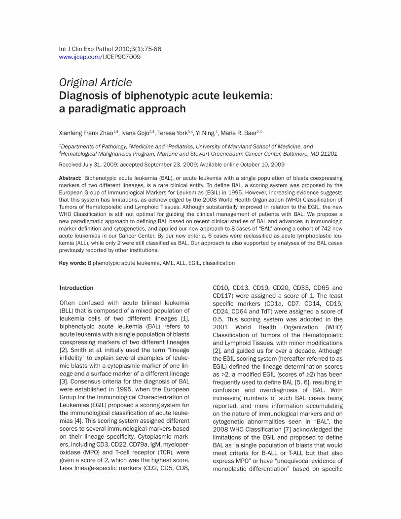
Int J Clin Exp Pathol 2010;3(1):75-86 www.ijcep.com/IJCEP907009
Original ArticleDiagnosis of biphenotypic acute leukemia: a paradigmatic approach
Xianfeng Frank Zhao1,4, Ivana Gojo2,4, Teresa York3,4, Yi Ning,1, Maria R. Baer2,4
1Departments of Pathology, 2Medicine and 3Pediatrics, University of Maryland School of Medicine, and 4Hematological Malignancies Program, Marlene and Stewart Greenebaum Cancer Center, Baltimore, MD 21201
Received July 31, 2009; accepted September 23, 2009; Available online October 10, 2009
Abstract: Biphenotypic acute leukemia (BAL), or acute leukemia with a single population of blasts coexpressing markers of two different lineages, is a rare clinical entity. To define BAL, a scoring system was proposed by the European Group of Immunological Markers for Leukemias (EGIL) in 1995. However, increasing evidence suggests that this system has limitations, as acknowledged by the 2008 World Health Organization (WHO) Classification of Tumors of Hematopoietic and Lymphoid Tissues. Although substantially improved in relation to the EGIL, the new WHO Classification is still not optimal for guiding the clinical management of patients with BAL. We propose a new paradigmatic approach to defining BAL based on recent clinical studies of BAL and advances in immunologic marker definition and cytogenetics, and applied our new approach to 8 cases of “BAL” among a cohort of 742 new acute leukemias in our Cancer Center. By our new criteria, 6 cases were reclassified as acute lymphoblastic leu-kemia (ALL), while only 2 were still classified as BAL. Our approach is also supported by analyses of the BAL cases previously reported by other institutions.
Key words: Biphenotypic acute leukemia, AML, ALL, EGIL, classification
Introduction
Often confused with acute bilineal leukemia (BLL) that is composed of a mixed population of leukemia cells of two different lineages [1], biphenotypic acute leukemia (BAL) refers to acute leukemia with a single population of blasts coexpressing markers of two different lineages [2]. Smith et al. initially used the term “lineage infidelity” to explain several examples of leuke-mic blasts with a cytoplasmic marker of one lin-eage and a surface marker of a different lineage [3]. Consensus criteria for the diagnosis of BAL were established in 1995, when the European Group for the Immunological Characterization of Leukemias (EGIL) proposed a scoring system for the immunological classification of acute leuke-mias [4]. This scoring system assigned different scores to several immunological markers based on their lineage specificity. Cytoplasmic mark-ers, including CD3, CD22, CD79a, IgM, myeloper-oxidase (MPO) and T-cell receptor (TCR), were given a score of 2, which was the highest score. Less lineage-specific markers (CD2, CD5, CD8,
CD10, CD13, CD19, CD20, CD33, CD65 and CD117) were assigned a score of 1. The least specific markers (CD1a, CD7, CD14, CD15, CD24, CD64 and TdT) were assigned a score of 0.5. This scoring system was adopted in the 2001 World Health Organization (WHO) Classification of Tumors of the Hematopoietic and Lymphoid Tissues, with minor modifications [2], and guided us for over a decade. Although the EGIL scoring system (hereafter referred to as EGIL) defined the lineage determination scores as >2, a modified EGIL (scores of >2) has been frequently used to define BAL [5, 6], resulting in confusion and overdiagnosis of BAL. With increasing numbers of such BAL cases being reported, and more information accumulating on the nature of immunological markers and on cytogenetic abnormalities seen in “BAL”, the 2008 WHO Classification [7] acknowledged the limitations of the EGIL and proposed to define BAL as “a single population of blasts that would meet criteria for B-ALL or T-ALL but that also express MPO” or have “unequivocal evidence of monoblastic differentiation” based on specific
Paradigmatic diagnosis of biphenotypic acute leukemia
76 Int J Clin Exp Pathol 2010;3(1):75-86
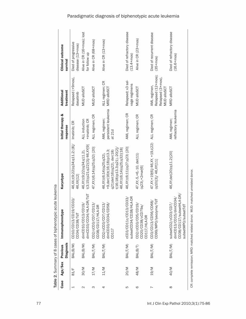
(M:F = 7:1), ages 11, 17, 19, 20, 30, 40, 48 and 81 years (median 25 years). Leukemia cells in most of the cases had scant basophilic cyto-plasm, high nuclear/cytoplasmic ratio, fine or slightly clumped chromatin and inconspicuous nucleoli, and resembled lymphoblasts. These blasts often show substantial variation in size (Figure 1). Two cases expressed both myeloid and B-cell (B/M) markers and harbored t(9;22)(q34;q11.2). One of these two cases had an addi-tional t(3;15)(p21;q22). Two cases expressed both myeloid and T-cell (T/M) markers and had t(6;14)(q25;q32) [8]. An additional T/M leukemia had t(6;11)(q27;q23). One case expressed both B- and T-cell markers and had a complex karyo-type including 25, +6, -10 and del(11q23); molecular studies revealed a clonal TCR rear-rangement and polyclonal VDJ recombination. The last two cases were additional T/M cases with +19, i(22)(q10) and with del(20)(q11.2).
Treatment and clinical outcome of these “BAL” cases are also summarized in Table 2. Five patients were treated with acute lymphoblastic leukemia (ALL) regimens and three with acute myeloid leukemia (AML) regimens. Six patients underwent allogeneic hematopoietic stem cell transplantation (alloSCT). In our small series, several patients had favorable outcomes with ALL regimens, which is consistent with the find-ings of two recent large series [5, 6].
Discussion
BAL is a rare clinical and pathological entity that is listed as a “rare disease” by the Office of Rare
requirements (Table 1). However, the new WHO Classification did not address the clinical signifi-cance of the proposed new definition of BAL, nor the implications for treatment.
Based on recent advances in immunology, genetics and clinical studies, here we discuss the limitations of the EGIL scoring system and propose a paradigmatic approach for defining BAL. We analyzed cases of BAL diagnosed at our Cancer Center from 2000 to 2007 and reclassified them according to our new approach, and discuss the potential implica-tions for clinical decision-making.
Materials and methods
With Institutional Review Board approval, we retrospectively reviewed consecutive acute leu-kemias newly diagnosed at our Cancer Center from 2000 to 2007. Cases that were previously diagnosed as “BAL” were identified. The mor-phology, immunophenotype, cytogenetic find-ings, treatment and clinical outcome of these cases were analyzed.
Results
Among 742 consecutive acute leukemias, 8 cases were previously diagnosed as “BAL”. Four cases (cases 4, 6, 7, 8) fulfilled the EGIL criteria for BAL, but the other 4 cases only met the modi-fied EGIL criteria (score >2) [5] and were also included in this study. These cases, representing an incidence of 1.1%, are summarized in Table 2. The BAL patients include 7 males and 1 female
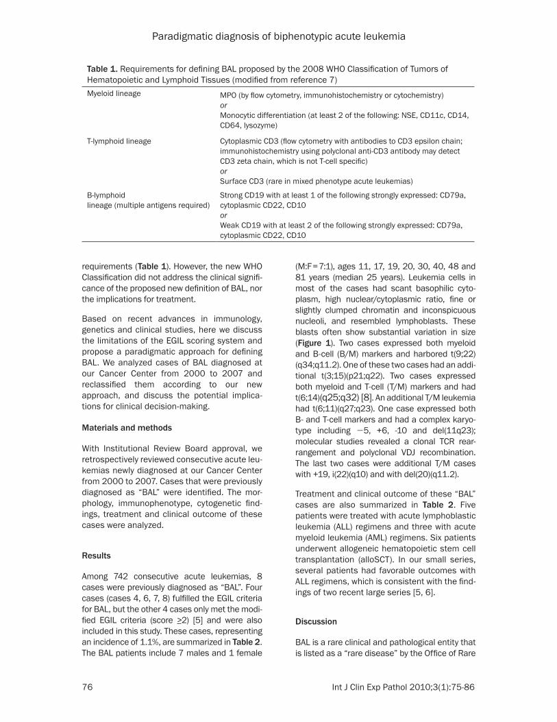
Table 1. Requirements for defining BAL proposed by the 2008 WHO Classification of Tumors of Hematopoietic and Lymphoid Tissues (modified from reference 7)Myeloid lineage MPO (by flow cytometry, immunohistochemistry or cytochemistry)
orMonocytic differentiation (at least 2 of the following: NSE, CD11c, CD14, CD64, lysozyme)
T-lymphoid lineage Cytoplasmic CD3 (flow cytometry with antibodies to CD3 epsilon chain; immunohistochemistry using polyclonal anti-CD3 antibody may detect CD3 zeta chain, which is not T-cell specific)orSurface CD3 (rare in mixed phenotype acute leukemias)
B-lymphoid lineage (multiple antigens required)
Strong CD19 with at least 1 of the following strongly expressed: CD79a, cytoplasmic CD22, CD10orWeak CD19 with at least 2 of the following strongly expressed: CD79a, cytoplasmic CD22, CD10
Paradigmatic diagnosis of biphenotypic acute leukemia
77 Int J Clin Exp Pathol 2010;3(1):75-86
Tabl
e 2.
Sum
mar
y of
8 c
ases
of b
iphe
noty
pic
acut
e le
ukem
ia
Case
Age/
Sex
Prev
ious
D
iagn
osis
Imm
unop
heno
type
Kary
otyp
eIn
itial
ther
apy
&
resp
onse
Addi
tiona
l tr
eatm
ent
Clin
ical
out
com
e
surv
ival
181
/FBA
L(B/
M)
CD10
/CD
13/C
D19
/CD
33/
CD34
/CD
38/T
dT46
,XX,
t(9;2
2)(q
34;q
11.2
) [8]
/ 46
,XX[
12]
Imat
inib
; CR
Rela
psed
(+9m
os),
dasa
tinib
Die
d of
pro
gres
sive
di
seas
e (1
3+m
os)
230
/MBA
L(B/
M)
dim
CD10
/CD
13/C
D19
/ di
mCD
33/C
D34
/HLA
-DR/
TdT
46,X
Y,t(9
;22)
(q34
;q11
.2),
t(3
;15)
(p21
;q22
)[3]/
46,X
Y[9]
ALL
indu
ctio
n +i
mat
inib
; CR
MUD
allo
SCT
Aliv
e in
CR
(16+
mos
); lo
st
for f
ollo
w u
p3
17/M
BAL(
T/M
)CD
2/cC
D3/
CD7/
CD13
/CD
38/C
D11
7/H
LA-D
R47
,XY,
t(6;1
4)(q
25;q
32) [
20]
ALL
regi
men
; CR
MUD
allo
SCT
Aliv
e in
CR
(68+
mos
)
411
/MBA
L(T/
M)
CD2/
cCD
3/CD
7/CD
13/
dim
CD33
/CD
34/C
D58
/CD
117
46,X
Y,t(6
;14)
(q25
;q32
), +9
,der
(16)
t(16;
18)(p
13.3
; q2
1)de
l(16)
(q22
), de
r(18)
t(1
6;18
)(p13
.3;q
21),-
20[2
]/
46,X
Y,t(6
;14)
(q25
;q32
)[18]
AML
regi
men
; pe
rsis
tent
leuk
emia
at
21d
ALL
regi
men
; CR
MRD
allo
SCT
Aliv
e in
CR
(13+
mos
)
520
/MBA
L(T/
M)
cCD
3/CD
11c/
CD13
/CD
33/
subs
etCD
34/C
D38
/HLA
-DR
46,X
Y,t(6
;11)
(q27
;q23
) [20
]AM
L re
gim
en; C
R Re
laps
ed; x
3 sa
l-va
ge re
gim
ens
Die
d of
refra
ctor
y di
seas
e (1
3.4+
mos
)6
48/M
BAL(
B/T)
CD2/
cCD
3/CD
5/CD
19/
CD20
/CD
38/c
CD79
a/
CD11
7/H
LA-D
R
47,X
Y,-5
,+6,
-10,
del
(11)
(q23
),+2m
ar[6
]AL
L re
gim
en; C
RM
UD a
lloSC
T A
live
in C
R (2
3+m
os)
719
/MBA
L(T/
M)
CD3/
CD13
/CD
56/C
D68
/CD
99/M
PO/l
ysoz
yme/
TdT
47,X
Y,+1
9[6]
/46,
XY, +
19,i(
22)
(q10
)[3]/
46,
XY[1
1]AL
L re
gim
en; C
RAM
L re
gim
en,
Rela
psed
(12+
mos
); Re
laps
ed (3
+mos
), M
UD a
lloSC
T
Die
d of
recu
rren
t dis
ease
(3
5+m
os)
840
/MBA
L(T/
M)
subs
etCD
2/cC
D3/
CD7/
dim
CD13
/CD
15/d
imCD
34/
CD38
/CD
117/
subs
etH
LA-D
R/
subs
etM
PO/s
ubse
tTdT
46,X
Y,de
l(20)
(q11
.2)[2
0]AM
L re
gim
en;
refra
ctor
y le
ukem
iaM
RD a
lloSC
TD
ied
of re
fract
ory
dise
ase
(16.
6+m
os)
CR: c
ompl
ete
rem
issi
on; M
RD: m
atch
ed re
late
d do
nor;
MUD
: mat
ched
unr
elat
ed d
onor
.
Paradigmatic diagnosis of biphenotypic acute leukemia
78 Int J Clin Exp Pathol 2010;3(1):75-86
non-covalently associated with surface immu-noglobulin, thus forming the B-cell antigen receptor complex, which initiates the B-cell anti-gen receptor signal transduction pathway [11]. Although CD79a was previously considered to be specific for B-cell lineage [12], there have been increasing numbers of reports of its aber-rant expression in both acute myeloid and acute T-cell leukemias [13-15].
CD19 is the most commonly used marker to define B cells. The new WHO Classification uses CD19, together with at least one antigen among CD10, cytoplasmic CD22, and CD79a, to define B-cell lineage. However, CD19 expression is seen in approximately one third of AML with t(8;21)(q22;q22), and serves to predict the presence of this cytogenetic abnormality in AML [16]. Those leukemias also express CD79a [17]. Thus, if CD19 and CD79a are employed as B-cell lineage-specific markers, some AML cases will be diagnosed as BAL [17].
The human B-lymphocyte-restricted antigen CD22, also known as sialic acid binding immuno-globulin-like lectin 2 (SIGLEC-2) [18], is expressed early in pro-B cells as a cytoplasmic protein and later in pre-B cells as a surface protein. Therefore, the presence of CD22 is a specific marker for precursor B-cells. Compared to CD79a, CD22 is considered more reliable in acute leukemia lin-eage determination [19, 20]. Although CD22 has also been detected in basophils [21], due to the distinct morphology of acute basophilic leuke-mia, it is less a problem to differentiate B lym-phoblasts from basophilic blasts. Furthermore, certain clones of CD22 monoclonal antibody (B3 and 4KB128) can detect B lymphoblasts, but not basophilic blasts [21].
Among the three high-scoring markers, cytoplas-mic IgM may be the most specific, though less sensitive, for B cells. To our knowledge, IgM has never been detected in myeloid or T-cells.
In contrast, the other B-cell markers are less specific. Although known as common acute lym-phoblastic leukemia antigen (CALLA) [22], CD10 is also expressed on the surface of granulocytes [23] as well as on several malignant lymphomas [24]. CD20 expression is variable in B-ALL. CD24 is expressed in B cells as well as in myeloid cells [25]. Terminal deoxynucleotidyl transferase (TdT) is an early lymphoid marker that is shared by precursor T and precursor B cells.
Diseases (ORD) of the National Institutes of Health (NIH) [9]. An accurate definition of BAL has significance in guiding treatment decisions. The 2008 WHO Classification uses more restrictive criteria than the EGIL to define BAL [7]. However, ambiguity still exists and a guide-line for clinicians is still lacking. We propose a paradigmatic approach to defining BAL based on recent data on the the nature of the immunologi-cal markers that are the basis of defining BAL, the cytogenetic data, and clinical studies.
Nature of immunological markers
B-lymphoid markers
In the EGIL, the highest scoring markers for B-lymphoid lineage are cytoplasmic CD79a, cytoplasmic CD22 and cytoplasmic IgM. CD79a, also known as immunoglobulin-associated α (Igα), is encoded by the mb-1 gene [10]. With a similar structure to the CD3γ chain, CD79a is
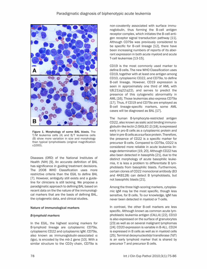
Figure 1. Morphology of some BAL blasts. The T/M leukemia cells (A) and B/T leukemia cells (B) show more variation in size and morphology than typical lymphoblasts (original magnification x1000).
Paradigmatic diagnosis of biphenotypic acute leukemia
79 Int J Clin Exp Pathol 2010;3(1):75-86
cells; 2) CD3 and TCR for T cells; and 3) MPO for myeloid/monocytic cells.
Limitations of EGIL
Since the publication of the EGIL in 1995 [4], approximately 250 reports of BAL have been published in the English literature and EGIL crite-ria have been used for the diagnosis of BAL in up to 20 papers. Based on those studies, we have identified the following limitations of the EGIL:
(1) The EGIL did not define lineage-specific markers: Although the EGIL gave the high-est score to some lineage-specific markers (such as CD3, CD22 and MPO), these mark-ers score only slightly higher than the lin-eage-associated markers (such as CD7, CD13, CD19, CD20, and CD33), thus lead-ing to overdiagnosis of BAL.
(2) The high score given to CD79a needs to be changed: The EGIL recognized the specific-ity of CD79a for B cells, but ignored its fre-quent aberrant expression in myeloid and T cells, leading to more frequent diagnosis of B/myeloid and B/T BAL.
(3) The EGIL ignored cytogenetic data: The EGIL is based on immunological markers, and omitted cytogenetic data. Because of this, even well-defined AML may be misdi-agnosed as BAL.
(4) The EGIL does not optimally guide treat-ment decisions: Overdiagnosis of BAL cre-ates uncertainty for clinicians. Due to the lack of standard regimens for BAL, hema-tologists/oncologists may choose to treat their patients with regimens for either AML [48] or ALL [5], or both [17]. Better lineage definition will provide clinicians better guidelines for choosing therapeutic regi-mens for patients, since the treatment of AML is quite different from that of ALL.
Proposed redefinition of BAL
The above limitations of the EGIL result in over-diagnosis of BAL. Therefore, it is necessary to better define acute leukemias with expression of multi-lineage markers. We propose that cyto-genetic abnormalities should be considered the most important factors in classifying acute
T-lymphoid markers
CD3 and T-cell receptor (TCR) are both parts of the TCR complex [26]. CD3 itself is a protein complex composed of four distinct chains (CD3γ, CD3d and two CD3ε), that associate with TCR and the z-chain to generate an activation signal in T cells [27]. Many studies have shown that CD3 and TCR are the most specific mark-ers for T cells [28, 29]. To date, aberrant expres-sion of CD3 has been extremely rare in other lineages.
Conversely, expression of CD1a, CD2, CD5, CD7 and CD8 has been identified in various other lin-eages. CD1a is expressed in a subset of precur-sor T cells as well as in Langerhans cells [30]. CD2, CD5 and CD7 are frequently expressed in myeloid leukemias [31-33]. CD8 is often co-ex-pressed with CD4 in precursor T cells. CD10 and TdT expression is shared with precursor B cells (see above) and these two markers can also be expressed in AML.
Myeloid/monocytic markers
Thirty-three years after the French-American-British (FAB) classification was published [34], myeloperoxidase (MPO) remains the most spe-cific marker of myeloid differentiation [7, 35]. In AML, MPO is usually associated with expres-sion of other myeloid markers, such as CD13, CD33 and CD117. Even in AML-M0, there is evi-dence of MPO expression in the blasts by either flow cytometry or electron microscopy [35, 36]. However, aberrant MPO expression has been detected in ALL and even lymphomas by flow cytometry, immunohistochemistry and electron microscopy [37-41]. MPO expression is most commonly detected in B-ALL with t(9;22)(q34;q11) [40]. Fortunately, MPO activity is not detectable by cytochemical stain in cases of ALL [39, 40], suggesting that cytochemistry is still the most discriminating assay for differenti-ating AML from ALL.
In contrast, co-expression of CD13 and CD33 is particularly associated with B-ALL with cytoge-netic abnormalities [42-44]. CD14, CD15 and CD64 are very rarely expressed in the absence of CD13 and CD33. CD65 is less well studied. CD117 expression has been identified in T-cells, B-cells and mast cells [44-47].
In summary, the reliable lineage-specific immu-nologic markers include: 1) IgM and CD22 for B
Paradigmatic diagnosis of biphenotypic acute leukemia
80 Int J Clin Exp Pathol 2010;3(1):75-86
even if immunological markers of other lin-eages are present. For example, even with CD79a expression, acute leukemia with t(8;21)(q22;q22) should still be classified as AML with t(8;21)(q22;q22) [7]. Therefore, the immunological markers only apply when the well-defined recurrent cytogenetic abnormalities are not present.
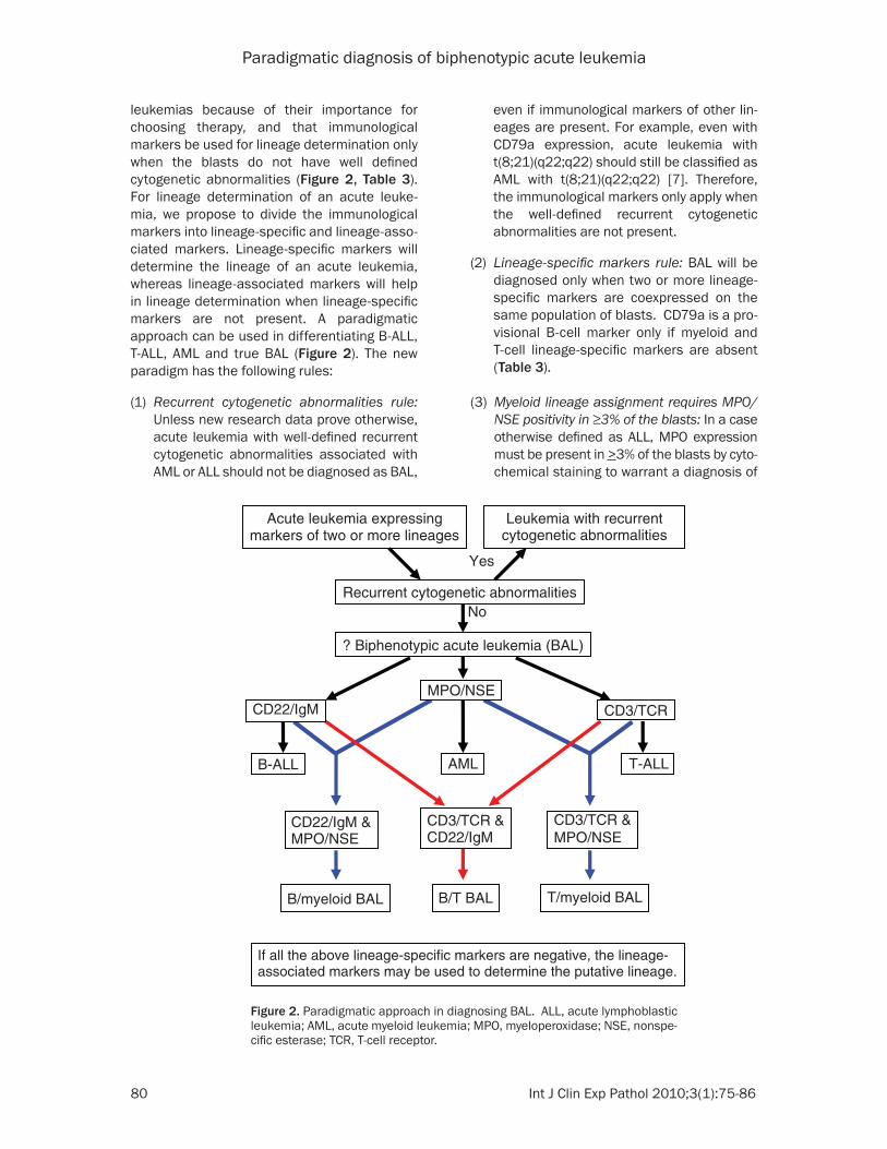
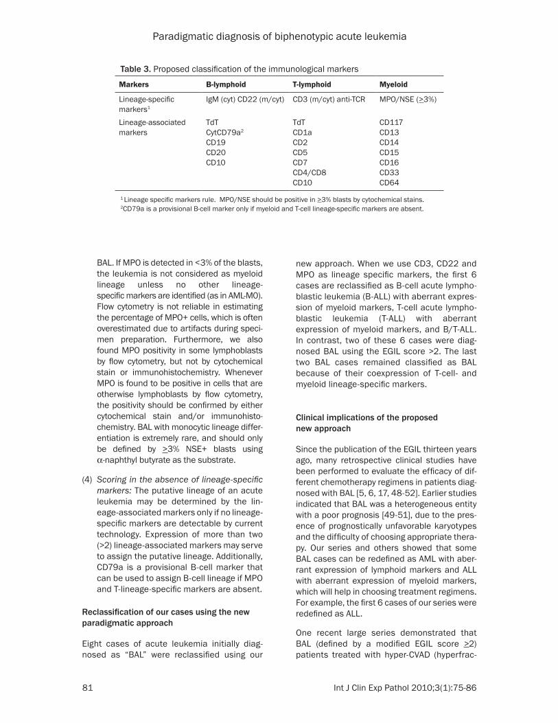
(2) Lineage-specific markers rule: BAL will be diagnosed only when two or more lineage-specific markers are coexpressed on the same population of blasts. CD79a is a pro-visional B-cell marker only if myeloid and T-cell lineage-specific markers are absent (Table 3).
(3) Myeloid lineage assignment requires MPO/NSE positivity in ≥3% of the blasts: In a case otherwise defined as ALL, MPO expression must be present in >3% of the blasts by cyto-chemical staining to warrant a diagnosis of
leukemias because of their importance for choosing therapy, and that immunological markers be used for lineage determination only when the blasts do not have well defined cytogenetic abnormalities (Figure 2, Table 3). For lineage determination of an acute leuke-mia, we propose to divide the immunological markers into lineage-specific and lineage-asso-ciated markers. Lineage-specific markers will determine the lineage of an acute leukemia, whereas lineage-associated markers will help in lineage determination when lineage-specific markers are not present. A paradigmatic approach can be used in differentiating B-ALL, T-ALL, AML and true BAL (Figure 2). The new paradigm has the following rules:
(1) Recurrent cytogenetic abnormalities rule: Unless new research data prove otherwise, acute leukemia with well-defined recurrent cytogenetic abnormalities associated with AML or ALL should not be diagnosed as BAL,
Yes
No
CD3/TCR
B-ALL T-ALLAML
CD3/TCR & MPO/NSE
B/myeloid BAL
CD22/IgM &MPO/NSE
T/myeloid BAL B/T BAL
CD3/TCR & CD22/IgM
If all the above lineage-specific markers are negative, the lineage-associated markers may be used to determine the putative lineage.
CD22/IgMMPO/NSE
Recurrent cytogenetic abnormalities
? Biphenotypic acute leukemia (BAL)
Acute leukemia expressing markers of two or more lineages
Leukemia with recurrentcytogenetic abnormalities
Figure 2. Paradigmatic approach in diagnosing BAL. ALL, acute lymphoblastic leukemia; AML, acute myeloid leukemia; MPO, myeloperoxidase; NSE, nonspe-cific esterase; TCR, T-cell receptor.
Paradigmatic diagnosis of biphenotypic acute leukemia
81 Int J Clin Exp Pathol 2010;3(1):75-86
new approach. When we use CD3, CD22 and MPO as lineage specific markers, the first 6 cases are reclassified as B-cell acute lympho-blastic leukemia (B-ALL) with aberrant expres-sion of myeloid markers, T-cell acute lympho-blastic leukemia (T-ALL) with aberrant expression of myeloid markers, and B/T-ALL. In contrast, two of these 6 cases were diag-nosed BAL using the EGIL score >2. The last two BAL cases remained classified as BAL because of their coexpression of T-cell- and myeloid lineage-specific markers.
Clinical implications of the proposed new approach
Since the publication of the EGIL thirteen years ago, many retrospective clinical studies have been performed to evaluate the efficacy of dif-ferent chemotherapy regimens in patients diag-nosed with BAL [5, 6, 17, 48-52]. Earlier studies indicated that BAL was a heterogeneous entity with a poor prognosis [49-51], due to the pres-ence of prognostically unfavorable karyotypes and the difficulty of choosing appropriate thera-py. Our series and others showed that some BAL cases can be redefined as AML with aber-rant expression of lymphoid markers and ALL with aberrant expression of myeloid markers, which will help in choosing treatment regimens. For example, the first 6 cases of our series were redefined as ALL.
One recent large series demonstrated that BAL (defined by a modified EGIL score >2) patients treated with hyper-CVAD (hyperfrac-
BAL. If MPO is detected in <3% of the blasts, the leukemia is not considered as myeloid lineage unless no other lineage- specific markers are identified (as in AML-M0). Flow cytometry is not reliable in estimating the percentage of MPO+ cells, which is often overestimated due to artifacts during speci-men preparation. Furthermore, we also found MPO positivity in some lymphoblasts by flow cytometry, but not by cyto chemical stain or immunohistochemistry. Whenever MPO is found to be positive in cells that are otherwise lymphoblasts by flow cytometry, the positivity should be confirmed by either cytochemical stain and/or immunohisto-chemistry. BAL with monocytic lineage differ-entiation is extremely rare, and should only be defined by >3% NSE+ blasts using α-naphthyl butyrate as the substrate.
(4) Scoring in the absence of lineage-specific markers: The putative lineage of an acute leukemia may be determined by the lin-eage-associated markers only if no lineage-specific markers are detectable by current technology. Expression of more than two (>2) lineage-associated markers may serve to assign the putative lineage. Additionally, CD79a is a provisional B-cell marker that can be used to assign B-cell lineage if MPO and T-lineage-specific markers are absent.
Reclassification of our cases using the new paradigmatic approach
Eight cases of acute leukemia initially diag-nosed as “BAL” were reclassified using our
Table 3. Proposed classification of the immunological markers
Markers B-lymphoid T-lymphoid Myeloid
Lineage-specific markers1
IgM (cyt) CD22 (m/cyt) CD3 (m/cyt) anti-TCR MPO/NSE (>3%)
Lineage-associated markers
TdT CytCD79a2
CD19CD20CD10
TdTCD1aCD2CD5CD7CD4/CD8CD10
CD117CD13CD14CD15CD16CD33CD64
1 Lineage specific markers rule. MPO/NSE should be positive in >3% blasts by cytochemical stains. 2CD79a is a provisional B-cell marker only if myeloid and T-cell lineage-specific markers are absent.
Paradigmatic diagnosis of biphenotypic acute leukemia
82 Int J Clin Exp Pathol 2010;3(1):75-86
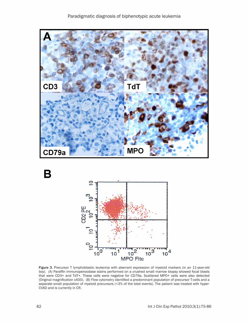
Figure 3. Precursor T lymphoblastic leukemia with aberrant expression of myeloid markers (in an 11-year-old boy). (A) Paraffin immunoperoxidase stains performed on a crushed small marrow biopsy showed focal blasts that were CD3+ and TdT+. These cells were negative for CD79a. Scattered MPO+ cells were also detected (Original magnification x400). (B) Flow cytometry identified a predominant population of precursor T-cells and a separate small population of myeloid precursors (<3% of the total events). The patient was treated with hyper-CVAD and is currently in CR.
Paradigmatic diagnosis of biphenotypic acute leukemia
83 Int J Clin Exp Pathol 2010;3(1):75-86
Possible diagnostic pitfalls
Even with the new approach, BAL could still be overdiagnosed due to inappropriate interpretation of immunological studies. For example, misinterpretation of MPO staining could lead to diagnosis of “BAL.” In one of the cases presented (Case 4), small numbers of MPO+ cells mixed together with CD3+/TdT+ cells might have suggested that the leukemia was also of myeloid lineage (Figure 3A), even though the marrow was packed with lympho-blasts and flow cytometry indicated a separate population of MPO+ cells (Figure 3B). However, if these myeloid precursors morphologically resembled myeloblasts, the leukemia might be misdiagnosed as BAL or BLL.
Conclusion
BAL is a rare clinical entity. With our proposed paradigmatic approach, BAL will be even rarer (~0.3%). The evolving definition of BAL reflects our increasing knowledge and understanding of this rare type of leukemia. With our proposed paradigmatic approach, acute leukemia will be better defined and better managed by the clini-cians. Future larger series may be required to further validate this approach.
Acknowledgments
This work was supported by a Seed Fund of the Department of Pathology, University of Maryland School of Medicine, Baltimore, MD (XFZ). We would like to thank Ms. Lenore Lawrence for help with flow cytometry.
Please address correspondence to: X. Frank Zhao, MD PhD, Department of Pathology, University of Maryland School of Medicine, 10 S. Pine Street, MSTF 711B, Baltimore, MD 21201, Tel: (410)- 328-5555, Fax: (410)-328-5508, E-mail: [email protected]
References
[1] Weir EG, Ali Ansari-Lari M, Batista DA, Griffin CA, Fuller S, Smith BD, Borowitz MJ. Acute bi-Acute bi-lineal leukemia: a rare disease with poor out-come. Leukemia 2007; 21: 2264-2270.
[2] Brunning RD, Head D, Matutes E, et al. In: Jaffe ES, Harris NL, Stein H, Vardiman JW (Eds):
tionated cyclophosphamide, vincristine, doxo-rubicin and dexamethasone) had a higher CR rate (78%) than patients treated with AML reg-imens (57%) [5]. Although data on some cyto-plasmic lineage-specific markers such as CD3, CD79a, IgM or TCR were not given in the paper, based on low MPO (MPO = 0 in 10 cases, MPO < 3 in 15 cases), high CD22 (CD22 > 50 in 9 cases, not available in 12 cases) and TdT (TdT > 50 in 21 cases) expression in most of the 31 cases of BAL [5] , those cases were actually ALL rather than BAL when using the new approach. Thus it is not surprising that most of those patients responded favorably to hyper-CVAD therapy for ALL [53]. We also ana-lyzed a recently reported large series of pedi-atric BAL [6]. Based on the new definition, cases 5, 6, and 24 should be reclassified as ALL with aberrant expression of myeloid mark-ers. Two of the 3 redefined ALL cases respond-ed favorably to ALL therapy and gained CR, whereas one died of toxicity [6]. One recent multicenter study indicated that T/myeloid BAL had a much worse prognosis than other BAL [48]. However some of those T/myeloid BAL might be reclassified as T-ALL with aber-rant expression of myeloid markers by using the new approach, and this group of patients was treated with AML regimens and did not respond [48]. A single-institution study defined a group of “biphenotypic AML” in mostly young patients with better outcome than other AML, without mentioning their treatment reg imen (52). Using the new approach, most of those patients would be reclassified as ALL with aberrant expression of myeloid markers. Another study reported a series of BAL with recurrent t(8;21)(q22;q22) and CD79a expres-sion [17]. Since expression of CD79a is usually driven by the aberrant expression of PAX5 in myeloblasts [54, 55], based on this new approach, these cases should be diagnosed as AML with t(8;21)(q22;q22), rather than BAL. The patients in that series responded favorably to concurrent AML and ALL chemo-therapy [17], but they might have responded equally well to AML therapy alone. One earlier study showed that the use of combined lym-phoid and myeloid regimens for induction in BAL patients resulted in a high incidence of early death [50], suggesting that accurate lin-eage determination might decrease patient mortality.
Paradigmatic diagnosis of biphenotypic acute leukemia
84 Int J Clin Exp Pathol 2010;3(1):75-86
ambiguity. Cancer Genet Cytogenet 2005; 163: 62-67.
[14] Cruse JM, Lewis RE, Pierce S, Lam J, Tadros Y. Aberrant expression of CD7, CD56, and CD79a antigens in acute myeloid leukemias. Exp Mol Pathol 2005; 79: 39-41.
[15] Lai R, Juco J, Lee SF, Nahirniak S, Etches WS. Flow cytometric detection of CD79a expression in T-cell acute lymphoblastic leukemias. Am J Clin Pathol 2000; 113: 823-830.
[16] Kita K, Nakase K, Miwa H, Masuya M, Nishii K, Morita N, Takakura N,
Otsuji A, Shirakawa S, Ueda T, Nasu K, Kyo T, Dohy H, Kamada N. Phenotypical characteris-tics of acute myelocytic leukemia associated with the t(8;21)(q22;q22) chromosomal ab-normality: frequent expression of immature B-cell antigen CD19 together with stem cell anti-gen CD34. Blood 1992; 80: 470-477.
[17] He G, Wu D, Sun A, Xue Y, Jin Z, Qiu H, Tang X, Miao M, Fu Z, Ma X, Wang X, Chen Z, Ruan C. B-Lymphoid and myeloid lineages biphenotypic acute leukemia with t(8;21)(q22;q22). Int J Hematol 2008; 87: 132-136.
[18] Brinkman-Van der Linden ECM, Sjoberg ER, Juneja LR, Crocker PR, Varki N, Varki A. Loss of N-glycolylneuraminic acid in human evolution: implications for sialic acid recognition by si-glecs. J Biol Chem 2000; 275: 8633-8640.
[19] Janossy G, Coustan-Smith E, Campana D. The reliability of cytoplasmic CD3 and CD22 anti-gen expression in the immunodiagnosis of acute leukemia: a study of 500 cases. Leuke-mia 1989; 3: 170-181.
[20] Paredes-Aguilera R, Romero-Guzman L, Lopez-Santiago N, Burbano-Ceron L, Camacho-Del Monte O, Nieto-Martinez S. Flow cytometric analysis of cell-surface and intracellular anti-gens in the diagnosis of acute leukemia. Am J Hematol 2001; 68: 69-74.
[21] Toba K, Hanawa H, Fuse I, Sakaue M, Wa-tanabe K, Uesugi Y, Higuchi W, Takahashi M, Aizawa Y. Difference in CD22 molecules in hu-man B cells and basophils. Exp Hematol 2002; 30: 205-211.
[22] Brown G, Hogg N, Greaves M. Candidate leukaemia-specific antigen in man. Nature 1975; 258: 454-456.
[23] Braun MP, Martin PJ, Ledbetter JA, Hansen JA. Granulocytes and cultured human fibroblasts express common acute lymphoblastic leukemia-associated antigens. Blood 1983; 61: 718-725.
[24] Ritz J, Nadler LM, Bhan AK, Notis-McConarty J, Pesando JM, Schlossman SF. Expression of common acute lymphoblastic leukemia anti-gen (CALLA) by lymphomas of B-cell and T-cell lineage. Blood 1981; 58: 648-652.
[25] Raife TJ, Lager DJ, Kemp JD, Dick FR. Expres-sion of CD24 (BA-1) predicts monocytic lineage
World Health Organization Classification of Tumors: Pathology and Genetics of Tumors of Haematopoietic and Lymphoid Tissues. Lyon: IARC Press; 2001. p106-107.
[3] Smith LJ, Curtis JE, Messner HA, Senn JS, Furth-mayr H, McCulloch EA. Lineage infi delity in acu-Lineage infidelity in acu-te leukemia. Blood 1983; 61: 1138-1145.
[4] Bene MC, Castoldi G, Knapp W, Ludwig WD, Matutes E, Orfao A, van’t Veer MB. Proposals for the immunological classification of acute leukemias. European Group for the Immuno-logical Characterization of Leukemias (EGIL). Leukemia 1995; 9: 1783-1786.
[5] Aribi A, Bueso-Ramos C, Estey E, Estrov Z, O’Brien S, Giles F, Faderl S, Thomas D, Kebria-ei P, Garcia-Manero G, Pierce S, Cortes J, Kan-tarjian H, Ravandi F. Biphenotypic acute leu-Biphenotypic acute leu-kaemia: a case series. Br J Haematol 2007; 138: 213-216.
[6] Rubnitz JE, Onciu M, Pounds S, Shurtleff S, Cao X, Raimondi SC, Behm FG, Campana D, Razzouk BI, Ribeiro RC, Downing JR, Pui CH. Acute mixed lineage leukemia in children: The experience of St. Jude Children’s Research Hospital. Blood 2009; 113: 5083-5089.
[7] Borowitz MJ, Bene M-C, Harris NL, Porwit A, Matutes E. Acute leukemias of ambiguous lin-eage. In: Swerdlow SH, Campo E, Harris NL, Jaffe ES, Pileri SA, Stein H, Thiele J, Vardiman JW (Eds.): WHO Classification of Tumors of Haematopoietic and Lymphoid Tissues. IARC: Lyon 2008. p149-155.
[8] Georgy M, Yonescu R, Griffin CA, Batista DA. Acute mixed lineage leukemia and a t(6;14)(q25;q32) in two adults. Cancer Genet Cyto-genet 2008; 185: 28-31.
[9] Acute biphenotypic leukemia, Genetic and Rare Diseases Information Center (GARD), NIH. Available from: http://rarediseases. info.nih.gov/GARD/Disease.aspx?PageID= 4&diseaseID=8638
[10] Sakaguchi N, Kashiwamura, S, Kimoto M, Thalmann P, Melchers F. B lymphocyte lineage-restricted expression of mb-1, a gene with CD3-like structural properties. EMBO J 1988; 7: 3457–3464.
[11] Sanchez M, Misulovin Z, Burkhardt AL, Mahajan S, Costa T, Franke R, Bolen JB, Nussenzweig M. Signal transduction by immunoglobulin is me-diated through Ig alpha and Ig beta. J Exp Med 1993; 178: 1049-1055.
[12] Buccheri V, Mihaljević B, Matutes E, Dyer MJ, Mason DY, Catovsky D.
mb-1: a new marker for B-lineage lymphoblas-tic leukemia. Blood 1993; 82: 853-857.
[13] Kozlov I, Beason K, Yu C, Hughson M. CD79a expression in acute myeloid leukemia t(8;21) and the importance of cytogenetics in the diag-nosis of leukemias with immunophenotypic
Paradigmatic diagnosis of biphenotypic acute leukemia
85 Int J Clin Exp Pathol 2010;3(1):75-86
mia-lymphoma cell lines. Blood 1993; 82: 1599-1607.
[38] Conlin PA, Orden MB, Hough TR, Morgan DL. Myeloperoxidase-positive intravascular large B-cell lymphoma. Arch Pathol Lab Med 2001; 125: 948-950.
[39] Nakase K, Sartor M, Bradstock K. Detection of myeloperoxidase by flow cytometry in acute leukemia. Cytometry 1998; 34: 198-202.
[40] Arber DA, Snyder DS, Fine M, Dagis A, Niland J, Slovak ML. Myeloperoxidase immunoreactivity in adult acute lymphoblastic leukemia. Am J Clin Pathol 2001; 116: 25-33.
[41] Kantarjian HM, Hirsch-Ginsberg C, Yee G, Huh Y, Freireich EJ, Stass S. Mixed-lineage leuke-mia revisited: acute lymphocytic leukemia with myeloperoxidase-positive blasts by electron microscopy. Blood 1990; 76: 808-813.
[42] Nakase K, Kita K, Shiku H, Tanaka I, Nasu K, Dohy H, Kyo T, Tsutani H, Kamada N. Myeloid antigen, CD13, CD14, and/or CD33 expres-sion is restricted to certain lymphoid neo-plasms. Am J Clin Pathol 1996; 105: 761-768.
[43] Baruchel A, Cayuela JM, Ballerini P, Landman-Parker J, Cezard V, Firat H, Haddad E, Auclerc MF, Valensi F, Cayre YE, Macintyre EA, Sigaux F. The majority of myeloid-antigen-positive (My+) childhood B-cell precursor acute lymphoblastic leukaemias express TEL-AML1 fusion tran-scripts. Br J Haematol 1997; 99: 101-106.
[44] Abdelhaleem M. Frequent but nonrandom ex-pression of myeloid markers on de novo child-hood acute lymphoblastic leukemia. Exp Mol Pathol 2007; 83: 138-141.
[45] Godfrey DI, Zlotnik A, Suda T. Phenotypic and functional characterization of c-kit expression during intrathymic T-cell development. J Immu-nol 1992; 149: 2281-2285.
[46] Suggs JL, Cruse JM, Lewis RE. Aberrant myeloid marker expression in precursor B-cell and T-cell leukemias. Exp Mol Pathol 2007; 83: 471-473.
[47] Arber DA, Tamayo R, Weiss LM. Paraffin section detection of the c-kit gene product (CD117) in human tissues: value in the diagnosis of mast cell disorders. Hum Pathol 1998; 29: 498-504.
[48] Lee JH, Min YH, Chung CW, Kim BK, Yoon HJ, Jo DY, Shin HJ, Bang SM, Won JH, Zang DY, Kim HJ, Chi HS, Lee KH, Cheong JW, Kim JS, Kim SH, Park S, Park SY, Chung JS, Lee JH, Park CJ; Korean Society of Hematology AML/MDS Working Party. Prognostic implications of the immunophenotype in biphenotypic acute leukemia. Leuk Lymphoma 2008; 49: 700-709.
[49] Legrand O, Perrot JY, Simonin G, Baudard M, Cadiou M, Blanc C, Ramond S, Viguié F, Marie JP, Zittoun R. Adult biphenotypic acute leukae-Adult biphenotypic acute leukae-mia: an entity with poor prognosis which is related to unfavourable cytogenetics and
in acute myeloid leukemia. Am J Clin Pathol 1994; 101: 296-299.
[26] Abbas AK, Lichtman AH. Cellular and Molecu-lar Immunology (5th Ed.): Saunders, Philadel-phia, 2003.
[27] Minguet S, Schamel WW. A permissive geom-etry model for TCR-CD3 activation. Trends Bio-Trends Bio-chem Sci 2008; 33: 51-57.
[28] Urbano-Ispizua A, Matutes E, Villamor N, Ribe-ra JM, Feliu E, Montserrat E, Grañena A, Vives-Corrons JL, Rozman C. Clinical significance of the presence of myeloid associated antigens in acute lymphoblastic leukaemia. Br J Haema-Br J Haema-tol 1990; 75: 202-207.
[29] Inukai T, Saito M, Mori T, Nishino K, Abe T, Ki-noshita A, Suzuki T, Kurosawa Y, Okazaki T, Su-gita K, et al. Analysis of cytoplasmic and sur-Analysis of cytoplasmic and sur-face antigens in childhood T-cell acute lymphoblastic leukaemias: clinical relevance of cytoplasmic TCR beta chain expression. Br J Haematol 1994; 87: 273-281.
[30] Krenács L, Tiszalvicz L, Krenács T, Boumsell L. Immunohistochemical detection of CD1A anti-gen in formalin-fixed and paraffin-embedded tissue sections with monoclonal antibody 010. J Pathol 1993; 171: 99-104.
[31] Drexler HG, Thiel E, Ludwig WD. Acute myeloid leukemias expressing lymphoid-associated an-tigens: diagnostic incidence and prognostic significance. Leukemia 1993; 7: 489-498.
[32] Launder TM, Bray RA, Stempora L, Chenggis ML, Farhi DC. Lymphoid-associated antigen ex-pression by acute myeloid leukemia. Am J Clin Pathol 1996; 106: 185-191.
[33] Khalidi HS, Medeiros LJ, Chang KL, Brynes RK, Slovak ML, Arber DA. The immunophenotype of adult acute myeloid leukemia: high fre-quency of lymphoid antigen expression and comparison of immunophenotype, French-American-British classification, and karyotypic abnormalities. Am J Clin Pathol 1998; 109: 211-220.
[34] Bennett JM, Catovsky D, Daniel MT, Flandrin G, Galton DA, Gralnick HR, Sultan C. Proposals for the classification of the acute leukaemias. French-American-British (FAB) co-operative group. Br J Haematol 1976; 33: 451-458.
[35] Imamura N. Sensitive detection technique of myeloperoxidase precursor protein by flow cy-tometry with monoclonal antibodies. Am J He-matol 1998; 58: 241-243.
[36] Hewson JW, Bradstock KF, Kerr A, Rose RG. Characterizing ”difficult” acute leukemias. A combined electron microscopic and immu-nological marker study. Pathology 1986; 18: 99-110.
[37] Hu ZB, Ma W, Uphoff CC, Metge K, Gignac SM, Drexler HG. Myeloperoxidase: expression and modulation in a large panel of human leuke-
Paradigmatic diagnosis of biphenotypic acute leukemia
86 Int J Clin Exp Pathol 2010;3(1):75-86
Freireich EJ. Long-term follow-up results of hyperfractionated cyclophosphamide, vincris-tine, doxorubicin, and dexamethasone (Hyper-CVAD), a dose-intensive regimen, in adult acute lymphocytic leukemia. Cancer 2004; 101: 2788-2801.
[54] Tiacci E, Pileri S, Orleth A, Pacini R, Tabarrini A, Frenguelli F, Liso A, Diverio D, Lo-Coco F, Falini B. PAX5 expression in acute leukemias: higher B-lineage specificity than CD79a and selective association with t(8;21)-acute myel-ogenous leukemia. Cancer Res 2004; 64: 7399–7404.
[55] Anderson K, Rusterholz C, Månsson R, Jensen CT, Bacos K, Zandi S, Sasaki Y, Nerlov C, Sig-vardsson M, Jacobsen SE. Ectopic expression of PAX5 promotes maintenance of bipheno-typic myeloid progenitors coexpressing myeloid and B-cell lineage-associated genes. Blood 2007; 109: 3697-3705.
P-glycoprotein over-expression. Br J Haematol 1998; 100: 147-155.
[50] Killick S, Matutes E, Powles RL, Hamblin M, Swansbury J, Treleaven JG, Zomas A, Atra A, Catovsky D. Outcome of biphenotypic acute leukemia. Haematologica 1999; 84: 699-706.
[51] Rubio MT, Dhedin N, Boucheix C, Bourhis JH, Reman O, Boiron JM, Gallo JH, Lhéritier V, Thomas X, Fière D, Vernant JP. Adult T-biphenotypic acute leukaemia: clinical and biological fea-tures and outcome. Br J Haematol 2003; 123: 842-849.
[52] Lee MY, Tan TD, Feng AC. Clinicopathologic analysis of acute myeloid leukemia in a single institution: biphenotypic acute myeloid leuke-mia may not be an aggressive subtype. J Chin Med Assoc 2007; 70: 269-273.
[53] Kantarjian H, Thomas D, O’Brien S, Cortes J, Giles F, Jeha S, Bueso-Ramos CE, Pierce S, Shan J, Koller C, Beran M, Keating M,