Embed Size (px)
Citation preview

Cyanidin-3-O-b-Glucoside and Protocatechuic Acid ExertInsulin-Like Effects by Upregulating PPARg Activity inHuman Omental AdipocytesBeatrice Scazzocchio,
1Rosaria Varì,
1Carmelina Filesi,
1Massimo D’Archivio,
1Carmela Santangelo,
1
Claudio Giovannini,1Annunziata Iacovelli,
2Gianfranco Silecchia,
3Giovanni Li Volti,
4,5
Fabio Galvano,4and Roberta Masella
1
OBJECTIVE—Insulin resistance (IR) represents an independentrisk factor for metabolic, cardiovascular, and neoplastic disor-ders. Preventing/attenuating IR is a major objective to be reachedto preserve population health. Because many insulin-sensitizingdrugs have shown unwanted side effects, active harmless com-pounds are sought after. Dietary anthocyanins have been demon-strated to ameliorate hyperglycemia and insulin sensitivity. Thisstudy aimed at investigating whether cyanidin-3-O-b-glucoside(C3G) and its metabolite protocatechuic acid (PCA) might havea role in glucose transport activation in human omental adipocytesand 3T3-L1 cells.
RESEARCH DESIGN AND METHODS—In cells treated with50 mmol/L C3G and 100 mmol/L PCA, [3H]-2-deoxyglucose uptake,GLUT4 translocation by immunoblotting, adiponectin secretion,and peroxisome proliferator–activated receptor-g (PPARg) acti-vation by enzyme-linked immunosorbent assay kits were evalu-ated. Parallel experiments were carried out in murine adipocyte3T3-L1. To define the role of PPARg in modulating polyphenoleffects, small interfering RNA technique and PPARg antagonistwere used to inhibit transcription factor activity.
RESULTS—C3G and PCA increased adipocyte glucose uptake(P , 0.05) and GLUT4 membrane translocation (P , 0.01). Sig-nificant increases (P, 0.05) in nuclear PPARg activity, as well asin adiponectin and GLUT4 expressions (P , 0.01), were alsoshown. It is interesting that PPARg inhibition counteracted thepolyphenol-induced adiponectin and GLUT4 upregulations, sug-gesting a direct involvement of PPARg in this process.
CONCLUSIONS—Our study provides evidence that C3G andPCA might exert insulin-like activities by PPARg activation, evi-dencing a causal relationship between this transcription factorand adiponectin and GLUT4 upregulation. Dietary polyphenolscould be included in the preventive/therapeutic armory againstpathological conditions associated with IR. Diabetes 60:2234–2244, 2011
The prevalence of type 2 diabetes is estimated toreach .300 million cases by year 2030 (1). Met-abolic syndrome, which is often a precursor todiabetes and cardiovascular diseases, is charac-
terized by insulin resistance (IR), increased fasting glucose,decreased HDL, hypertension, and obesity (specifically,visceral obesity) (2). Furthermore, metabolic syndrome aswell as atherosclerosis, type 2 diabetes, and obesity is as-sociated with increased circulating oxidized LDL (oxLDL)(3–6). In cultured cells, oxLDLs have been demonstratedto lower insulin sensitivity (7) and to impair the insulin-dependent GLUT4-mediated uptake of glucose (8–10).
Many studies have shown that adipocytes play an impor-tant role in the development of obesity-associated patholo-gies and IR, mostly by synthesizing and secreting biologicallyactive molecules called adipocytokines (11), such as adipo-nectin, which is able to improve insulin sensitivity of targetcells (12). Serum levels of adiponectin protein correlate withsystemic insulin sensitivity (13) and are decreased in insulin-resistant, diabetic, and obese subjects (14).
Adiponectin is regulated by peroxisome proliferator–activated receptor-g (PPARg) (15). PPARg is a ligand-activated nuclear hormone receptor that controls glucoseand lipid metabolism (16,17), as well as the transcription ofproteins involved in glucose and fatty acid cellular uptake.For these reasons, it represents a main target for antidiabeticdrugs, such as thiazolidinediones (TZDs) (18). In additionto their insulin-sensitizing effects, TZDs have a number ofside effects, such as promoting adipogenesis, causing bodyweight gain, and increasing risk for bone fracture and car-diovascular diseases. Hence, ligands for PPARg that do notprocure these unwanted side effects are being sought.
Recently, great interest has arisen regarding evidencethat the consumption of a diet rich in vegetables and fruitcan exert beneficial healthy effects, likely because of thehigh content in fiber, mineral salts, vitamins, and poly-phenols (19–22). Among polyphenols, anthocyanins (ACNs)are flavonoids of great nutritional interest because theirdaily intake (180–250 mg/day) is much higher than that ofother polyphenols (23). ACNs are absorbed in animals andhumans (24–26) and rapidly metabolized, ultimately leadingto the formation of phenolic acids and aldehydes (27). Inparticular, at physiological pH (such as in the bloodstream),ACNs easily convert to protocatechuic acid (PCA), which isalso abundantly formed and absorbed in the large intestineafter microbial metabolization (28). Owing to the potentialbenefits for human health (29), many studies have focusedon cyanidin-3-O-b-glucoside (C3G), the best known and
From the 1Department of Veterinary Public Health and Food Safety, NationalInstitute of Health, Rome, Italy; 2Fabia Mater Hospital, Rome, Italy; the 3De-partment of Surgery P. Stefanini, University of Rome La Sapienza, Rome,Italy; the 4Department of Biological Chemistry, Medical Chemistry, and Mo-lecular Biology, University of Catania, Catania, Italy; and the 5Department ofCardiac Surgery, IRCSS, S. Donato Hospital, Milan, Italy.
Corresponding author: Roberta Masella, [email protected] 15 October 2010 and accepted 15 June 2011.DOI: 10.2337/db10-1461� 2011 by the American Diabetes Association. Readers may use this article as
long as the work is properly cited, the use is educational and not for profit,and the work is not altered. See http://creativecommons.org/licenses/by-nc-nd/3.0/ for details.
2234 DIABETES, VOL. 60, SEPTEMBER 2011 diabetes.diabetesjournals.org
ORIGINAL ARTICLE
most investigated ACN, highlighting its potential activities infree radical scavenging and prevention of oxLDL genera-tion, exerting beneficial effects on cardiovascular diseases,obesity, and inflammation (30–32).
Some compelling studies have reported that ACNs im-prove insulin sensitivity and glucose uptake in diabeticrats (33) and effectively upregulate the signaling pathwayof PPARg in mouse peritoneal macrophages (34), stronglysuggesting that they could be successfully used as insulin-sensitizing agents. However, the molecular mechanism ofaction and the effectiveness of C3G and PCA in exertingprotective effects against IR are still poorly understood.
Finally, most of the research on adipocytes has beenconducted on the murine cell line 3T3-L1, which is con-sidered a suitable model for studying the pathophysiologyof adipocytes and, in particular, for assessing the responseto insulin (10). Conversely, to the best of our knowledge,only a few studies have been specifically carried out onhuman omental adipocytes.
This study investigated the effects of C3G and PCA onglucose uptake machinery in adipocytes by evaluating theirability to reverse the oxLDL-induced impairment of adipo-cyte response to insulin and the molecular events un-derlying their effects. Specifically, we demonstrated thatC3G and PCA enhanced glucose uptake and GLUT4 trans-location in both insulin-stimulated human omental adipo-cytes and 3T3-L1 cells. Notably, the polyphenols elicited thesame response in the absence of insulin, showing insulin-like activity; specifically, they upregulated PPARg activityand the expression and secretion of its target gene adipo-nectin. These findings support the hypothesis that C3G, andits main metabolite PCA, might play a role in the therapeuticarmory against disease states associated with IR, such astype 2 diabetes and obesity.
RESEARCH DESIGN AND METHODS
Plasma LDL isolation and oxidation. LDLs (1.019–1.063 g/mL) were isolatedby density gradient ultracentrifugation from fresh pooled plasma of healthy vol-unteers as described elsewhere (35). The protein content was measured by Lowrymethod (8). Native LDLs (nLDLs) were oxidized as previously described (10).Isolation of human omental adipocytes. Human omental adipocytes werecollected from anesthetized individuals undergoing abdominal surgery or lap-aroscopy for benign conditions (i.e., gallbladder disease without icterus,umbilical hernia, or uterine fibromatosis) (11 females and 9 males, age 50–70years, BMI 20.0–26.9, waist circumference#107 for males and#92 for females).Exclusion criteria were steroidal and nonsteroidal anti-inflammatory thera-pies, hormonal substitutive or contraceptive therapy, hormonal therapy forany thyroid dysfunctions, drug or alcohol abuse, diabetes, chronic renal fail-ure, cancer, pregnancy, and mental disability.
The omental sampling was performed at the same site (great omentum) withthe patient in anti-Trendelenburg position (25° head up) with the surgeonstanding between the legs. The biopsies were obtained by monopolar electro-cautery or harmonic scalpel. The standard sampling was considered 2 3 2 cm,avoiding bleeding and other possible contamination. The omentum was col-lected into an endobag and extracted through the umbilical trocar to avoidcrash and contamination. The study protocol has been approved by the ethicscommittee of the La Sapienza University. All the subjects were volunteers andgave their informed consent according to the Italian law on this matter (Leg-islative Decree of the Italian Ministry of Health, 25 January 2001, published inthe Official Gazette of 3 April 2001).
From 5 to 10 grams of omental biopsies were microdissected, rinsed severaltimes in 0.9% NaCl, and digested with 5 mL of Krebs-Ringer solution (0.12 mol/LNaCl, 4.7 mol/L KCl, 2.5 mmol/L CaCl2, 1.2 mmol/L MgSO4, and 1.2 mmol/LKH2PO4) containing 20 mmol/L HEPES pH 7.4, 3.5% fatty acid–free BSA, 200nmol/L adenosine, 2 mmol/L glucose, and collagenase (type 1) for 1 h (1 mg/gadipose tissue) at 37°C in shaking water bath (36). After collagenase digestion, theadipocytes were separated from tissue debris by filtering through sterile nylonmesh (250 mm). Cells were then washed three times with Krebs-Ringer solutioncontaining 20 mmol/L HEPES, pH 7.4, 1% fatty acid–free BSA, 200 nmol/L aden-osine, and 2 mmol/L glucose and resuspended in 199-medium containing 1% fatty
acid–free BSA and 25 mmol/L HEPES. Floating fraction of isolated omental adi-pocytes from different individuals was used for the experiments described below.3T3-L1 preadipocyte differentiation. 3T3-L1 preadipocytes (American TypeCulture Collection) were induced to differentiate according to Masella et al. (8).The cells were used for the experiments on day 14, when .90% of cells pre-sented the adipocyte phenotype (8).Treatment of adipocytes with oxLDL. Different oxLDL concentrations (25–200 mg protein/L) were used to test both oxLDL cytotoxicity and their effects onglucose uptake to determine the best concentration. The 100 mg/L concentra-tion was chosen because it effectively reduces the glucose uptake by 50% inhuman and murine adipocytes without showing any sign of cytotoxicity, asassessed by Neutral Red assay, or affecting the morphology or the metabolismof adipocytes, as determined by the expression of leptin and adipocyte protein 2(aP2) and by the incorporation of [3H]Uridine, which were both comparable tothe controls (data not shown). Under all the experimental conditions describedbelow, adipocytes, untreated and treated with nLDL (100 mg/L), were used ascontrols. Because we obtained wholly overlapping results, we report data foruntreated cells only.Treatment of adipocytes with C3G and PCA. Adipocytes were incubatedwith C3G (Polyphenols Laboratories AS, Sandnes, Norway) or PCA (Sigma-Aldrich, St. Louis, MO) at concentrations of 50 and 100 mmol/L, respectively, forhuman omental adipocytes and 10 and 100 mmol/L, respectively, for 3T3-L1 18h before the addition of nLDL or oxLDL for 4 or 18 h. To define the experi-mental conditions, we carried out preliminary trials, incubating the cells withdifferent concentrations of the polyphenols (1–150 mmol/L) for different timesbefore oxLDL addition and determining the percentage of glucose internalizedin the cells after insulin stimulation. On the basis of the data obtained (notshown), the time and the lowest concentration of the two polyphenols able toprovide a 50% recovery of glucose uptake in oxLDL-treated cells were used inall the experiments. To define the effect of the polyphenols on PPARg, weassessed the mRNA expression and activity of the transcription factor in cellsincubated for 2 or 18 h with C3G or PCA. In the experiments intended toevaluate the specific involvement of PPARg in the activation of its targetgenes, the cells were treated with 10 mmol/L GW9662, a PPARg antagonist, 30min before the treatment with polyphenols.Glucose uptake assay. Glucose transport was measured as described else-where (37). Briefly, human and 3T3-L1 adipocytes, plated in low-glucoseDulbecco’s modified Eagle’s medium (1,000 mg/L D-(+)-glucose), were serumstarved for 18 h and stimulated with 20 nmol/L insulin for 15 min. [3H]-2-DG(2-deoxyglucose) (1 mCi/well) was added to the cells, and 45 min was allowedfor its uptake by the cells. The reaction was stopped by ice-cold PBS in 3T3-L1cells and by rapid centrifugation at 8,000 rpm for 5 min through 300 mL cushionof silicon oil in human omental adipocytes. The total incorporated radioactivitywas determined in a liquid scintillation counter. The results were corrected foraspecific absorption (37). Aspecific absorption was always ,10% of total up-take. Results were normalized for protein content.Protein determination by immunoblotting analysis. To evaluate GLUT4 inplasma membranes (PM) and in low-density microsome (LDM) fractionsenriched in the intracellular GLUT4 storage vesicles (38), the cells werefractionated according to McKeel and Jarett (39). Whole-cell extracts were pre-pared from cells as previously described (8). Nuclear protein extracts wereprepared by the Nuclear/Cytosol Fractionation Kit (Medical & Biological Labo-ratories, Watertown, LA) according to the manufacturer’s instructions. Immu-noblotting analyses were carried out using specific antibodies for PPARgand GLUT4 (Santa Cruz Biotechnology, Santa Cruz, CA). Blots were treatedwith appropriate secondary antibodies conjugated with horseradish peroxi-dase (Santa Cruz Biotechnology) followed by enhanced chemiluminescencedetection (Amersham Bio-sciences, Buckinghamshire, U.K.). Equal loadingof proteins was verified by immunoblotting with a goat anti–glyceraldehyde-3-phosphate dehydrogenase (GAPDH) antibody. Densitometric analysis wasperformed using a molecular imager FX (Bio-Rad, Hercules, CA).mRNA determination by quantitative real-time PCR. Total RNA wasisolated with TRIZOL reagent (Invitrogen-Life Technologies, Carlsbad, CA) asreported elsewhere (8). Quantitative real-time PCR (RTq-PCR) was carried outwith gene-specific TaqMan MGB probes and primers (Applied Biosystems,Carlsbad, CA) in an ABI 7700 sequence detector (Applied Biosystems).PPARg, GLUT4, adiponectin, and endogenous controls TATA-box bindingprotein (TBP) and GAPDH were purchased from Applied Biosystems as pre-designed assays. All gene expression assays have a FAM reporter dye at 59 endof TaqMan MGB probe and a nonfluorescent quencher at 39 end of the probe.
Expression of PPARg, GLUT4, and adiponectin genes were determined asthe amount of individual mRNA relative to mRNA for TBP (murine adipocytes)or GAPDH (human adipocytes) using the comparative CT method described inthe ABI 7700 sequence detection system, user bulletin number two.Assessment of PPARg activity. PPARg activity was determined in nuclearextracts with the TransAM ELISA Kit (Active Motif Europe, Rixensart, Belgium)according to the manufacturer’s instructions.
B. SCAZZOCCHIO AND ASSOCIATES
diabetes.diabetesjournals.org DIABETES, VOL. 60, SEPTEMBER 2011 2235
Evaluation of adiponectin secretion. The release of adiponectin wasevaluated in the culture media by ELISA Kit (R&D Systems Inc., Minneapolis,MN) according to the manufacturer’s instructions.PPARg silencing by small interfering RNA. PPARg expression was in-hibited with specific small interfering RNA (siRNA) reagents (mouse PPARgsiGENOME SMARTpool siRNA; Dharmacon, Lafayette, CO) as previously de-scribed (8). Scrambled nontargeting siRNA was used as negative control. At se-lected time points after transfection, mRNA and protein were extracted to assessPPARg, GLUT4, and adiponectin mRNA expressions and GLUT4 protein in PM.Statistical analysis. The results are expressed as means 6 SEM of at leastfour independent experiments performed in duplicate. In the human studies,depending on the amount of adipocytes isolated from each subject, each ex-periment was performed in at least 4 different individuals (men-to-women 1:1)randomly chosen from the 20 recruited subjects. Comparisons between twogroups were carried out by Student t test. ANOVA followed by Student-Newman-Keuls multiple comparison test were used when .2 groups werecompared. Differences were considered significant when P , 0.05.
RESULTS
Impairment of glucose uptake in human omental adi-pocytes by oxLDL. We have previously demonstrated thatoxLDLs are able to affect cell sensitivity toward insulin,
inhibiting glucose uptake by interfering with the cell re-sponse to insulin in 3T3-L1 adipocytes (10). Similar resultswere obtained in insulin-stimulated human omental adipo-cytes treated with oxLDL (100 mg/L). Indeed, these cellsshowed a strong decrease (240%) in glucose uptake anda concomitant reduction of GLUT4 protein (260%) in thePMs after oxLDL treatment (Fig. 1A and B).Effects of PCA and C3G on glucose transport andGLUT4 translocation. In the present article, we evaluatedwhether polyphenols, specifically PCA and C3G, couldcounteract the detrimental effects induced by oxLDL inadipocyte cell models. To this end, glucose uptake wasevaluated in oxLDL-treated adipocytes, previously incubatedwith each polyphenol for 18 h, as described in RESEARCH
DESIGN AND METHODS. Our results evidenced that both thepolyphenols were able to counteract the drop in glucoseuptake of human as well as murine insulin-treated adipo-cytes (Fig. 1A and C, respectively). In addition, they re-versed the impairment of GLUT4 translocation (Fig. 1Band D) induced by oxLDL.
FIG. 1. Effects of C3G and PCA on the impairment of glucose uptake by oxLDL in human and 3T3-L1 adipocytes. Human and murine adipocyteswere serum starved in low-glucose medium for 18 h, incubated with 50 mmol/L and 10 mmol/L C3G, respectively, or with 100 mmol/L PCAfor a further 18 h, and then treated with 100 mg/L oxLDL for 4 h. The rate of glucose uptake was determined after the addition of [
3H]-2-DG
(2-deoxyglucose) in cells with or without 20 nmol/L insulin stimulation. Glucose uptake in human (A) and murine (C) adipocytes was expressed asradioactivity per minute per milligram of cell proteins (DPM/min/mg cell proteins). Data are means 6 SEM of four independent experiments. PMfractions from human (B) and 3T3-L1 (D) adipocytes, isolated as described in RESEARCH DESIGN AND METHODS, were resolved by SDS-PAGE and an-alyzed using antibodies against GLUT4. Results were normalized to GAPDH protein content. Representative blots are shown. ANOVA, P < 0.0001;post hoc test, *P< 0.001 compared with unstimulated control cells, #P< 0.05 compared with insulin-stimulated cells, and §P< 0.05 compared withoxLDL-treated cells stimulated with insulin.
ANTHOCYANINS UPREGULATE PPARg ACTIVITY
2236 DIABETES, VOL. 60, SEPTEMBER 2011 diabetes.diabetesjournals.org
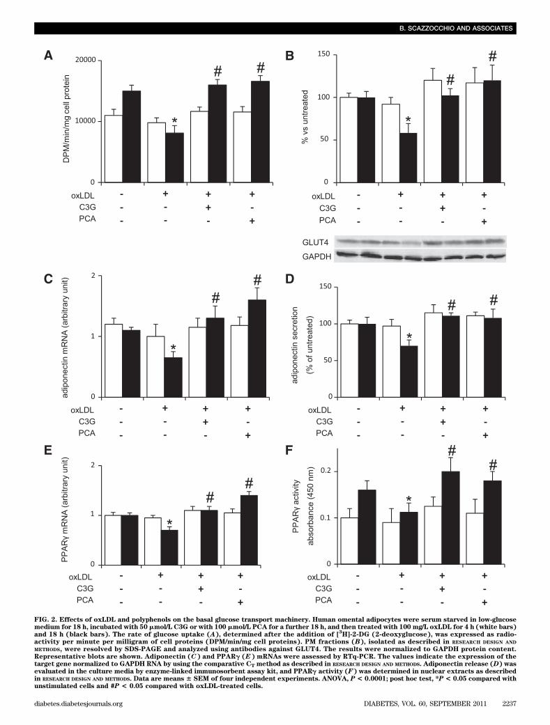
FIG. 2. Effects of oxLDL and polyphenols on the basal glucose transport machinery. Human omental adipocytes were serum starved in low-glucosemedium for 18 h, incubated with 50 mmol/L C3G or with 100 mmol/L PCA for a further 18 h, and then treated with 100 mg/L oxLDL for 4 h (white bars)and 18 h (black bars). The rate of glucose uptake (A), determined after the addition of [
3H]-2-DG (2-deoxyglucose), was expressed as radio-
activity per minute per milligram of cell proteins (DPM/min/mg cell proteins). PM fractions (B), isolated as described in RESEARCH DESIGN AND
METHODS, were resolved by SDS-PAGE and analyzed using antibodies against GLUT4. The results were normalized to GAPDH protein content.Representative blots are shown. Adiponectin (C) and PPARg (E) mRNAs were assessed by RTq-PCR. The values indicate the expression of thetarget gene normalized to GAPDH RNA by using the comparative CT method as described in RESEARCH DESIGN AND METHODS. Adiponectin release (D) wasevaluated in the culture media by enzyme-linked immunosorbent assay kit, and PPARg activity (F) was determined in nuclear extracts as describedin RESEARCH DESIGN AND METHODS. Data are means 6 SEM of four independent experiments. ANOVA, P < 0.0001; post hoc test, *P < 0.05 compared withunstimulated cells and #P < 0.05 compared with oxLDL-treated cells.
B. SCAZZOCCHIO AND ASSOCIATES
diabetes.diabetesjournals.org DIABETES, VOL. 60, SEPTEMBER 2011 2237
Notably, the levels of both glucose uptake and GLUT4translocation were increased in insulin-stimulated adi-pocytes treated with polyphenols with respect to thosetreated with insulin alone (up to 40% at 50 mmol/L and 100mmol/L of C3G and PCA, respectively). This increase mightbe the result of an enhancement of cell sensibility to in-sulin as well as to an additive effect (i.e., the polyphenolscould improve glucose uptake and GLUT4 translocationby activating other factors than those specifically involvedin insulin pathways). However, we have no evidence tosupport any hypothesis.
To better define the possible effects of oxLDL on thebasal uptake of glucose, we determined glucose uptake andGLUT4 translocation in unstimulated human omental adi-pocytes after 4 and 18 h of incubation with oxLDL alone andin the presence of polyphenols. The glucose transport ma-chinery appeared not to be affected by 4-h oxLDL treatment,whereas 18-h treatment caused the lowering of glucoseuptake and GLUT4 translocation. Both the polyphenolswere able to counteract such decrease (Fig. 2A and B).
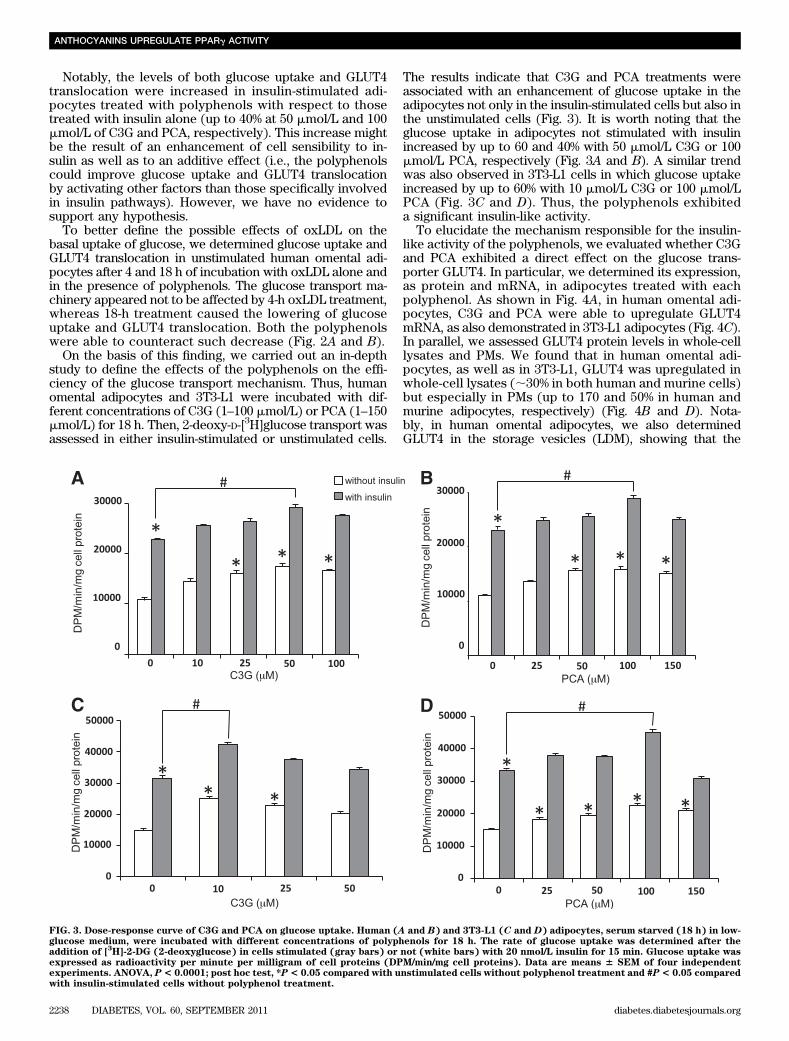
On the basis of this finding, we carried out an in-depthstudy to define the effects of the polyphenols on the effi-ciency of the glucose transport mechanism. Thus, humanomental adipocytes and 3T3-L1 were incubated with dif-ferent concentrations of C3G (1–100 mmol/L) or PCA (1–150mmol/L) for 18 h. Then, 2-deoxy-D-[3H]glucose transport wasassessed in either insulin-stimulated or unstimulated cells.
The results indicate that C3G and PCA treatments wereassociated with an enhancement of glucose uptake in theadipocytes not only in the insulin-stimulated cells but also inthe unstimulated cells (Fig. 3). It is worth noting that theglucose uptake in adipocytes not stimulated with insulinincreased by up to 60 and 40% with 50 mmol/L C3G or 100mmol/L PCA, respectively (Fig. 3A and B). A similar trendwas also observed in 3T3-L1 cells in which glucose uptakeincreased by up to 60% with 10 mmol/L C3G or 100 mmol/LPCA (Fig. 3C and D). Thus, the polyphenols exhibiteda significant insulin-like activity.
To elucidate the mechanism responsible for the insulin-like activity of the polyphenols, we evaluated whether C3Gand PCA exhibited a direct effect on the glucose trans-porter GLUT4. In particular, we determined its expression,as protein and mRNA, in adipocytes treated with eachpolyphenol. As shown in Fig. 4A, in human omental adi-pocytes, C3G and PCA were able to upregulate GLUT4mRNA, as also demonstrated in 3T3-L1 adipocytes (Fig. 4C).In parallel, we assessed GLUT4 protein levels in whole-celllysates and PMs. We found that in human omental adi-pocytes, as well as in 3T3-L1, GLUT4 was upregulated inwhole-cell lysates (;30% in both human and murine cells)but especially in PMs (up to 170 and 50% in human andmurine adipocytes, respectively) (Fig. 4B and D). Nota-bly, in human omental adipocytes, we also determinedGLUT4 in the storage vesicles (LDM), showing that the
FIG. 3. Dose-response curve of C3G and PCA on glucose uptake. Human (A and B) and 3T3-L1 (C and D) adipocytes, serum starved (18 h) in low-glucose medium, were incubated with different concentrations of polyphenols for 18 h. The rate of glucose uptake was determined after theaddition of [
3H]-2-DG (2-deoxyglucose) in cells stimulated (gray bars) or not (white bars) with 20 nmol/L insulin for 15 min. Glucose uptake was
expressed as radioactivity per minute per milligram of cell proteins (DPM/min/mg cell proteins). Data are means 6 SEM of four independentexperiments. ANOVA, P< 0.0001; post hoc test, *P< 0.05 compared with unstimulated cells without polyphenol treatment and #P< 0.05 comparedwith insulin-stimulated cells without polyphenol treatment.
ANTHOCYANINS UPREGULATE PPARg ACTIVITY
2238 DIABETES, VOL. 60, SEPTEMBER 2011 diabetes.diabetesjournals.org

upregulation of GLUT4 in the PMs was accompanied bya significant decrease in the LDM fractions (Fig. 4B).
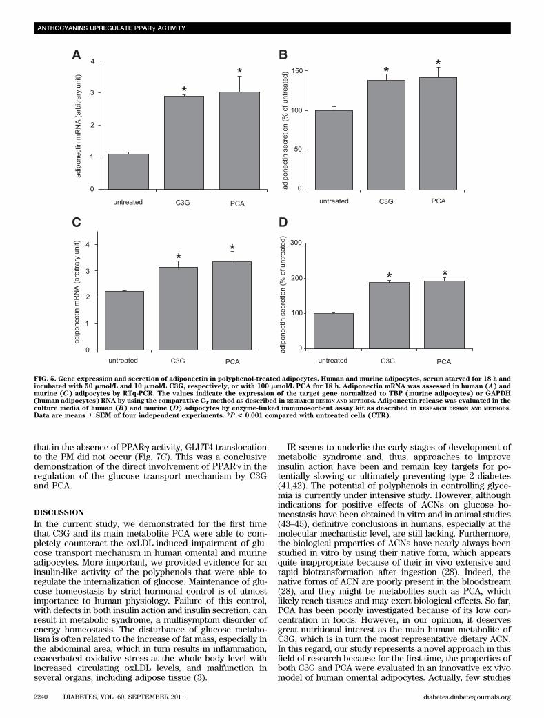
These findings indicated that polyphenols increased glu-cose uptake by significantly inducing GLUT4 expressionand mostly, GLUT4 translocation.PCA and C3G mediate induction of adiponectin geneexpression. Adiponectin has been shown to have someinsulin-sensitizing properties (13,40) and to be decreasedin serum of insulin-resistant, diabetic, and obese subjects(14). Thus, we hypothesized that oxLDL could affect adi-ponectin production/secretion and that the polyphenolscould counteract this effect. To verify our hypothesis, hu-man omental adipocytes were incubated for 4 and 18 h withoxLDL alone or in the presence of the phenolic compounds.After 4 h of oxLDL treatment, both in presence or absenceof polyphenols, the levels of adiponectin expression andsecretion were not substantially changed (Fig. 2C and D).On the contrary, at 18 h, oxLDL reduced adiponectin mRNAlevels by 50% (Fig. 2C) and adiponectin secretion by 30%(Fig. 2D), whereas both polyphenols prevented such reduc-tions. Worthy of note is the finding that polyphenols wereable to upregulate both mRNA expression and secretion ofadiponectin by themselves (Fig. 5A and B).
A similar increase in adiponectin expression and secre-tion was also demonstrated in 3T3-L1 cells after polyphe-nol treatment (Fig. 5C and D).PCA and C3G induce PPARg expression and activity.To identify a possible mechanism responsible for the dif-ferent modulation of GLUT4 and adiponectin observed inoxLDL- or polyphenol-treated adipocytes, we assessed
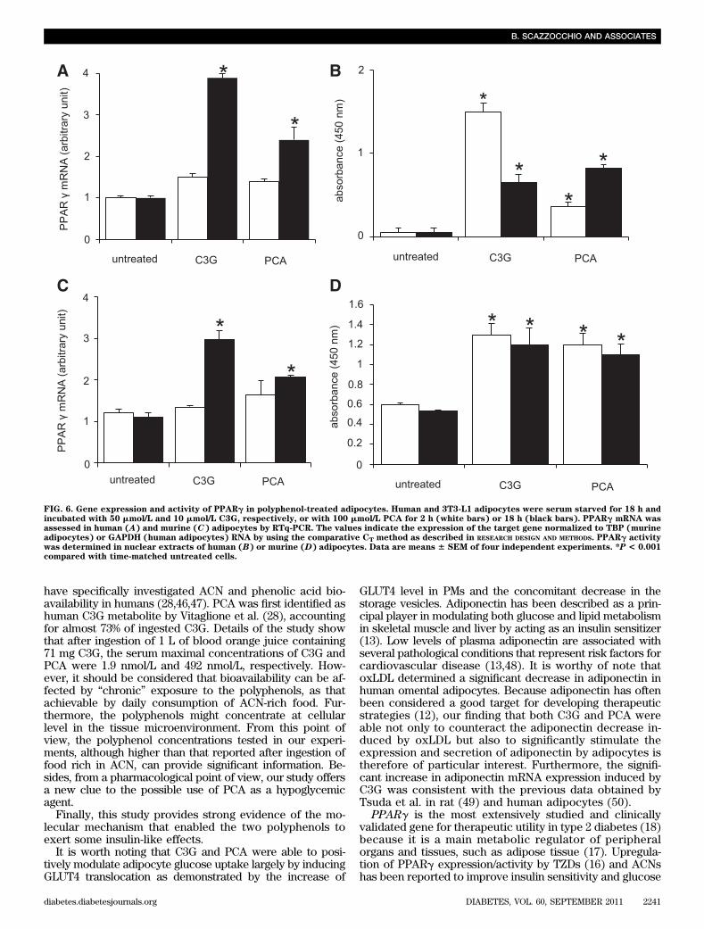
whether PPARg, the master regulator of mature adipocytegenes (15), could be involved.PPARg mRNA expression and activity. For this pur-pose, we carried out experiments to determine PPARgmRNA after treatment with oxLDL and/or polyphenols.RTq-PCR analysis showed a significant decrease in PPARgmRNA expression (P, 0.001) in human omental adipocyteswithin 18 h of treatment with oxLDL that was counteractedby polyphenols (Fig. 2E). Furthermore, we determinedthe activation status of PPARg in nuclear extracts of cellstreated with oxLDL and/or polyphenols, further demon-strating that oxLDL negatively affected PPARg activa-tion, whereas C3G and PCA were able to counteract theoxLDL-induced detrimental action (Fig. 2F). It is interestingthat the polyphenols elicited per se both the rise of PPARggene transcription (Fig. 6A) and an early and prolonged in-crease in PPARg activity (Fig. 6B). In 3T3-L1 cells, C3G andPCA also upregulated PPARg gene expression and activity(Fig. 6C and D).PPARg silencing. To further demonstrate the involve-ment of PPARg in the insulin-like activity exerted by C3Gand PCA, we silenced PPARg expression in 3T3-L1 by usingthe small interfering mRNA technique and the PPARg an-tagonist GW9662. In the transfected cells, as well as in thePPARg inhibitor–treated cells, we found that the signifi-cant upregulation of adiponectin and GLUT4 mRNAs in-duced by polyphenols were counteracted (Fig. 7A and B),providing additional evidence for the causal relationshipbetween PPARg activation and polyphenol-induced up-regulation of adiponectin and GLUT4. We also demonstrated
FIG. 4. Gene and protein expressions of GLUT4 in polyphenol-treated adipocytes. Human and 3T3-L1 adipocytes were serum starved for 18 h andincubated with 50 mmol/L and 10 mmol/L C3G (striped bars), respectively, or with 100 mmol/L PCA (black bars) for 2 or 18 h. GLUT4 mRNA wasassessed in human (A) and murine (C) adipocytes by RTq-PCR. The values indicate the expression of target gene normalized to TBP (murineadipocytes) or GAPDH (human adipocytes) RNA by using the comparative CT method as described in RESEARCH DESIGN AND METHODS. GLUT4 proteinexpression was determined at 18 h of polyphenol treatment in 1) whole-cell lysates (Total), PMs, and storage vesicles (LDM) prepared from humanadipocytes (B), and 2) whole-cell lysates (Total) and PMs from 3T3-L1 cells (D). Results were normalized to GAPDH protein content. Data areexpressed as means 6 SEM of four independent experiments. *P < 0.05 compared with matched control cells (CTR, white bars). Representativeblots are shown.
B. SCAZZOCCHIO AND ASSOCIATES
diabetes.diabetesjournals.org DIABETES, VOL. 60, SEPTEMBER 2011 2239
that in the absence of PPARg activity, GLUT4 translocationto the PM did not occur (Fig. 7C). This was a conclusivedemonstration of the direct involvement of PPARg in theregulation of the glucose transport mechanism by C3Gand PCA.
DISCUSSION
In the current study, we demonstrated for the first timethat C3G and its main metabolite PCA were able to com-pletely counteract the oxLDL-induced impairment of glu-cose transport mechanism in human omental and murineadipocytes. More important, we provided evidence for aninsulin-like activity of the polyphenols that were able toregulate the internalization of glucose. Maintenance of glu-cose homeostasis by strict hormonal control is of utmostimportance to human physiology. Failure of this control,with defects in both insulin action and insulin secretion, canresult in metabolic syndrome, a multisymptom disorder ofenergy homeostasis. The disturbance of glucose metabo-lism is often related to the increase of fat mass, especially inthe abdominal area, which in turn results in inflammation,exacerbated oxidative stress at the whole body level withincreased circulating oxLDL levels, and malfunction inseveral organs, including adipose tissue (3).
IR seems to underlie the early stages of development ofmetabolic syndrome and, thus, approaches to improveinsulin action have been and remain key targets for po-tentially slowing or ultimately preventing type 2 diabetes(41,42). The potential of polyphenols in controlling glyce-mia is currently under intensive study. However, althoughindications for positive effects of ACNs on glucose ho-meostasis have been obtained in vitro and in animal studies(43–45), definitive conclusions in humans, especially at themolecular mechanistic level, are still lacking. Furthermore,the biological properties of ACNs have nearly always beenstudied in vitro by using their native form, which appearsquite inappropriate because of their in vivo extensive andrapid biotransformation after ingestion (28). Indeed, thenative forms of ACN are poorly present in the bloodstream(28), and they might be metabolites such as PCA, whichlikely reach tissues and may exert biological effects. So far,PCA has been poorly investigated because of its low con-centration in foods. However, in our opinion, it deservesgreat nutritional interest as the main human metabolite ofC3G, which is in turn the most representative dietary ACN.In this regard, our study represents a novel approach in thisfield of research because for the first time, the properties ofboth C3G and PCA were evaluated in an innovative ex vivomodel of human omental adipocytes. Actually, few studies
FIG. 5. Gene expression and secretion of adiponectin in polyphenol-treated adipocytes. Human and murine adipocytes, serum starved for 18 h andincubated with 50 mmol/L and 10 mmol/L C3G, respectively, or with 100 mmol/L PCA for 18 h. Adiponectin mRNA was assessed in human (A) andmurine (C) adipocytes by RTq-PCR. The values indicate the expression of the target gene normalized to TBP (murine adipocytes) or GAPDH(human adipocytes) RNA by using the comparative CT method as described in RESEARCH DESIGN AND METHODS. Adiponectin release was evaluated in theculture media of human (B) and murine (D) adipocytes by enzyme-linked immunosorbent assay kit as described in RESEARCH DESIGN AND METHODS.Data are means 6 SEM of four independent experiments. *P < 0.001 compared with untreated cells (CTR).
ANTHOCYANINS UPREGULATE PPARg ACTIVITY
2240 DIABETES, VOL. 60, SEPTEMBER 2011 diabetes.diabetesjournals.org
have specifically investigated ACN and phenolic acid bio-availability in humans (28,46,47). PCA was first identified ashuman C3G metabolite by Vitaglione et al. (28), accountingfor almost 73% of ingested C3G. Details of the study showthat after ingestion of 1 L of blood orange juice containing71 mg C3G, the serum maximal concentrations of C3G andPCA were 1.9 nmol/L and 492 nmol/L, respectively. How-ever, it should be considered that bioavailability can be af-fected by “chronic” exposure to the polyphenols, as thatachievable by daily consumption of ACN-rich food. Fur-thermore, the polyphenols might concentrate at cellularlevel in the tissue microenvironment. From this point ofview, the polyphenol concentrations tested in our experi-ments, although higher than that reported after ingestion offood rich in ACN, can provide significant information. Be-sides, from a pharmacological point of view, our study offersa new clue to the possible use of PCA as a hypoglycemicagent.
Finally, this study provides strong evidence of the mo-lecular mechanism that enabled the two polyphenols toexert some insulin-like effects.
It is worth noting that C3G and PCA were able to posi-tively modulate adipocyte glucose uptake largely by inducingGLUT4 translocation as demonstrated by the increase of
GLUT4 level in PMs and the concomitant decrease in thestorage vesicles. Adiponectin has been described as a prin-cipal player in modulating both glucose and lipid metabolismin skeletal muscle and liver by acting as an insulin sensitizer(13). Low levels of plasma adiponectin are associated withseveral pathological conditions that represent risk factors forcardiovascular disease (13,48). It is worthy of note thatoxLDL determined a significant decrease in adiponectin inhuman omental adipocytes. Because adiponectin has oftenbeen considered a good target for developing therapeuticstrategies (12), our finding that both C3G and PCA wereable not only to counteract the adiponectin decrease in-duced by oxLDL but also to significantly stimulate theexpression and secretion of adiponectin by adipocytes istherefore of particular interest. Furthermore, the signifi-cant increase in adiponectin mRNA expression induced byC3G was consistent with the previous data obtained byTsuda et al. in rat (49) and human adipocytes (50).
PPARg is the most extensively studied and clinicallyvalidated gene for therapeutic utility in type 2 diabetes (18)because it is a main metabolic regulator of peripheralorgans and tissues, such as adipose tissue (17). Upregula-tion of PPARg expression/activity by TZDs (16) and ACNshas been reported to improve insulin sensitivity and glucose
FIG. 6. Gene expression and activity of PPARg in polyphenol-treated adipocytes. Human and 3T3-L1 adipocytes were serum starved for 18 h andincubated with 50 mmol/L and 10 mmol/L C3G, respectively, or with 100 mmol/L PCA for 2 h (white bars) or 18 h (black bars). PPARg mRNA wasassessed in human (A) and murine (C) adipocytes by RTq-PCR. The values indicate the expression of the target gene normalized to TBP (murineadipocytes) or GAPDH (human adipocytes) RNA by using the comparative CT method as described in RESEARCH DESIGN AND METHODS. PPARg activitywas determined in nuclear extracts of human (B) or murine (D) adipocytes. Data are means 6 SEM of four independent experiments. *P < 0.001compared with time-matched untreated cells.
B. SCAZZOCCHIO AND ASSOCIATES
diabetes.diabetesjournals.org DIABETES, VOL. 60, SEPTEMBER 2011 2241
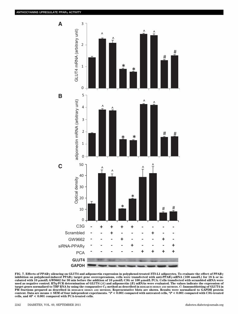
FIG. 7. Effects of PPARg silencing on GLUT4 and adiponectin expression in polyphenol-treated 3T3-L1 adipocytes. To evaluate the effect of PPARginhibition on polyphenol-induced PPARg target gene overexpressions, cells were transfected with anti-PPARg-siRNA (100 nmol/L) for 18 h or in-cubated with 10 mmol/L GW9662 for 30 min before the addition of 10 mmol/L C3G or 100 mmol/L PCA. Cells transfected with scrambled siRNA wereused as negative control. RTq-PCR determination of GLUT4 (A) and adiponectin (B) mRNAs were evaluated. The values indicate the expression oftarget genes normalized to TBP RNA by using the comparative CT method as described in RESEARCH DESIGN AND METHODS. C: Immunoblotting of GLUT4 inPM fractions prepared as described in RESEARCH DESIGN AND METHODS. Representative blots are shown. Results were normalized to GAPDH proteincontent. Data are means6 SEM of four independent experiments. ^P< 0.001 compared with untreated cells, *P< 0.001 compared with C3G-treatedcells, and #P < 0.001 compared with PCA-treated cells.
ANTHOCYANINS UPREGULATE PPARg ACTIVITY
2242 DIABETES, VOL. 60, SEPTEMBER 2011 diabetes.diabetesjournals.org
uptake in human adipocytes (50) and in animal models ofdiabetes (33).
We hypothesized that oxLDL and polyphenols couldaffect GLUT4 and adiponectin expressions by differentlymodulating PPARg. Our results allowed us to stronglysupport this hypothesis. Indeed, oxLDL significantly re-duced the expression and activity of PPARg in humanomental adipocytes, as already reported in 3T3-L1 (10),whereas the polyphenols were able to counteract such de-crease. It is interesting that in untreated cells, both C3G andPCA significantly increased the expression of PPARg geneand especially its activity with respect to basal values.PPARg activity remained higher during the entire experi-mental period, likely through the promotion of its binding tothe oligonucleotide at its consensus binding site.
Finally, our data strongly suggest that PPARg playsa key role in the activation of its target genes by C3G andPCA. In fact, the silencing of PPARg overrode the increasein GLUT4 and adiponectin and the translocation of GLUT4on the PM induced by the two polyphenols.
In conclusion, we demonstrated for the first time thatC3G and PCA exert insulin-like activity in human omentaladipocytes. The increase in glucose uptake was associ-ated with enhanced GLUT4 translocation and adiponectinsecretion, which were probably caused by the increasedactivity of PPARg induced by the polyphenols. We alsoconfirmed that the 3T3-L1 cell line represents a suitablemodel for the study of human adipocyte biology becausethey showed the same response to polyphenol treatmentas human adipocytes.
Altogether, our data provide new evidence on the bi-ological activity of C3G and PCA, supporting a possibleuse of these polyphenols as dietary bioactive compoundsagainst the IR condition linked to the occurrence of met-abolic syndrome.
ACKNOWLEDGMENTS
This study was partially supported by the ProvinciaRegionale di Catania through the Antioxidant Properties ofSicilian Pigmented Oranges Project.
No potential conflicts of interest relevant to this articlewere reported.
B.S. provided research data, contributed to discussion,and wrote the manuscript. R.V. contributed to researchdata and discussion. C.F. contributed to research data.M.D., C.S., and C.G. contributed to discussion and re-viewed the manuscript. A.I. and G.S. provided the humanbiopsies and reviewed the manuscript. G.L.V. and F.G. con-tributed to discussion and reviewed the manuscript. R.M.contributed to discussion and wrote the manuscript.
The authors wish to thank Professor Gabriella Girelli,Director of Centro Trasfusionale, University of Rome LaSapienza, for providing human plasma and AntoniettaPucciarmati, Department of Veterinary Public Health andFood Safety, Italian National Institute of Health, for technicalassistance.
REFERENCES
1. Wild S, Roglic G, Green A, Sicree R, King H. Global prevalence of diabetes:estimates for the year 2000 and projections for 2030. Diabetes Care 2004;27:1047–1053
2. Eckel RH, Grundy SM, Zimmet PZ. The metabolic syndrome. Lancet 2005;365:1415–1428
3. Holvoet P, Lee DH, Steffes M, Gross M, Jacobs DR Jr. Association betweencirculating oxidized low-density lipoprotein and incidence of the metabolicsyndrome. JAMA 2008;299:2287–2293
4. Tsuzura S, Ikeda Y, Suehiro T, et al. Correlation of plasma oxidized low-density lipoprotein levels to vascular complications and human serumparaoxonase in patients with type 2 diabetes. Metabolism 2004;53:297–302
5. Holvoet P, Vanhaecke J, Janssens S, Van de Werf F, Collen D. Oxidized LDLand malondialdehyde-modified LDL in patients with acute coronary syn-dromes and stable coronary artery disease. Circulation 1998;98:1487–1494
6. Weinbrenner T, Schröder H, Escurriol V, et al. Circulating oxidized LDL isassociated with increased waist circumference independent of body massindex in men and women. Am J Clin Nutr 2006;83:30–35; quiz 181–182
7. Mazière C, Morlière P, Santus R, et al. Inhibition of insulin signaling byoxidized low density lipoprotein. Protective effect of the antioxidant vi-tamin E. Atherosclerosis 2004;175:23–30
8. Masella R, Varì R, D’Archivio M, et al. Oxidised LDL modulate adipo-genesis in 3T3-L1 preadipocytes by affecting the balance between cellproliferation and differentiation. FEBS Lett 2006;580:2421–2429
9. D’Archivio M, Scazzocchio B, Filesi C, et al. Oxidised LDL up-regulateCD36 expression by the Nrf2 pathway in 3T3-L1 preadipocytes. FEBS Lett2008;582:2291–2298
10. Scazzocchio B, Varì R, D’Archivio M, et al. Oxidized LDL impair adipocyteresponse to insulin by activating serine/threonine kinases. J Lipid Res2009;50:832–845
11. Scherer PE. Adipose tissue: from lipid storage compartment to endocrineorgan. Diabetes 2006;55:1537–1545
12. Yamauchi T, Kamon J, Waki H, et al. The fat-derived hormone adiponectinreverses insulin resistance associated with both lipoatrophy and obesity.Nat Med 2001;7:941–946
13. Berg AH, Combs TP, Scherer PE. ACRP30/adiponectin: an adipokine regu-lating glucose and lipid metabolism. Trends Endocrinol Metab 2002;13:84–89
14. Lindsay RS, Funahashi T, Hanson RL, et al. Adiponectin and developmentof type 2 diabetes in the Pima Indian population. Lancet 2002;360:57–58
15. Iwaki M, Matsuda M, Maeda N, et al. Induction of adiponectin, a fat-derivedantidiabetic and antiatherogenic factor, by nuclear receptors. Diabetes2003;52:1655–1663
16. Spiegelman BM. PPAR-gamma: adipogenic regulator and thiazolidinedionereceptor. Diabetes 1998;47:507–514
17. Badman MK, Flier JS. The adipocyte as an active participant in energybalance and metabolism. Gastroenterology 2007;132:2103–2115
18. Miyazaki Y, Mahankali A, Wajcberg E, Bajaj M, Mandarino LJ, DeFronzo RA.Effect of pioglitazone on circulating adipocytokine levels and insulin sensi-tivity in type 2 diabetic patients. J Clin Endocrinol Metab 2004;89:4312–4319
19. Biesalski HK. Diabetes preventive components in the Mediterranean diet.Eur J Nutr 2004;43(Suppl. 1):I/26–30
20. Lindström J, Ilanne-Parikka P, Peltonen M, et al.; Finnish Diabetes Pre-vention Study Group. Sustained reduction in the incidence of type 2 di-abetes by lifestyle intervention: follow-up of the Finnish Diabetes PreventionStudy. Lancet 2006;368:1673–1679
21. Tuomilehto J, Lindström J, Eriksson JG, et al.; Finnish Diabetes PreventionStudy Group. Prevention of type 2 diabetes mellitus by changes in lifestyleamong subjects with impaired glucose tolerance. N Engl J Med 2001;344:1343–1350
22. Saura-Calixto F, Goñi I. Definition of the Mediterranean diet based onbioactive compounds. Crit Rev Food Sci Nutr 2009;49:145–152
23. Hertog MG, Hollman PC, Katan MB, Kromhout D. Intake of potentiallyanticarcinogenic flavonoids and their determinants in adults in the Netherlands.Nutr Cancer 1993;20:21–29
24. Kay CD, Mazza G, Holub BJ, Wang J. Anthocyanin metabolites in humanurine and serum. Br J Nutr 2004;91:933–942
25. Kay CD, Mazza GJ, Holub BJ. Anthocyanins exist in the circulation pri-marily as metabolites in adult men. J Nutr 2005;135:2582–2588
26. Mazza G, Kay CD. Bioactivity, absorption, and metabolism of anthocya-nins. In Recent Advances in Polyphenols Research. Vol. 1. Lattanzio V,Daayf F, Eds. Oxford, Blackwell, 2008, p. 228–262
27. Tsuda T, Horio F, Osawa T. Absorption and metabolism of cyanidin 3-O-beta-D-glucoside in rats. FEBS Lett 1999;449:179–182
28. Vitaglione P, Donnarumma G, Napolitano A, et al. Protocatechuic acid is themajor human metabolite of cyanidin-glucosides. J Nutr 2007;137:2043–2048
29. Cao G, Muccitelli HU, Sánchez-Moreno C, Prior RL. Anthocyanins areabsorbed in glycated forms in elderly women: a pharmacokinetic study.Am J Clin Nutr 2001;73:920–926
30. Galvano F, La Fauci L, Lazzarino G, et al. Cyanidins: metabolism and bi-ological properties. J Nutr Biochem 2004;15:2–11
31. Serraino I, Dugo L, Dugo P, et al. Protective effects of cyanidin-3-O-glucoside from blackberry extract against peroxynitrite-induced endothelialdysfunction and vascular failure. Life Sci 2003;73:1097–1114
32. Tsuda T, Horio F, Uchida K, Aoki H, Osawa T. Dietary cyanidin 3-O-beta-D-glucoside-rich purple corn color prevents obesity and ameliorates hyper-glycemia in mice. J Nutr 2003;133:2125–2130
B. SCAZZOCCHIO AND ASSOCIATES
diabetes.diabetesjournals.org DIABETES, VOL. 60, SEPTEMBER 2011 2243
33. Seymour EM, Lewis SK, Urcuyo-Llanes DE, et al. Regular tart cherry intakealters abdominal adiposity, adipose gene transcription, and inflammation inobesity-prone rats fed a high fat diet. J Med Food 2009;12:935–942
34. Xia M, Hou M, Zhu H, et al. Anthocyanins induce cholesterol efflux frommouse peritoneal macrophages: the role of the peroxisome proliferator-activated receptor gamma-liver X receptor alpha-ABCA1 pathway. J BiolChem 2005;280:36792–36801
35. Masella R, Varì R, D’Archivio M, et al. Extra virgin olive oil biophenolsinhibit cell-mediated oxidation of LDL by increasing the mRNA tran-scription of glutathione-related enzymes. J Nutr 2004;134:785–791
36. Kristensen K, Pedersen SB, Richelsen B. Regulation of leptin by steroid hor-mones in rat adipose tissue. Biochem Biophys Res Commun 1999;259:624–630
37. Tanti JF, Cormont M, Grémeaux T, Le Marchand-Brustel Y. Assays ofglucose entry, glucose transporter amount, and translocation. MethodsMol Biol 2001;155:157–165
38. Piper RC, Hess LJ, James DE. Differential sorting of two glucose transportersexpressed in insulin-sensitive cells. Am J Physiol 1991;260:C570–C580
39. McKeel DW, Jarett L. Preparation and characterization of a plasmamembrane fraction from isolated fat cells. J Cell Biol 1970;44:417–432
40. Arai Y, Kojima T, Takayama M, Hirose N. The metabolic syndrome, IGF-1,and insulin action. Mol Cell Endocrinol 2009;299:124–128
41. Cusi K, Maezono K, Osman A, et al. Insulin resistance differentially affectsthe PI 3-kinase- and MAP kinase-mediated signaling in human muscle.J Clin Invest 2000;105:311–320
42. Folli F, Saad MJ, Backer JM, Kahn CR. Regulation of phosphatidylinositol3-kinase activity in liver and muscle of animal models of insulin-resistantand insulin-deficient diabetes mellitus. J Clin Invest 1993;92:1787–1794
43. Adisakwattana S, Charoenlertkul P, Yibchok-Anun S. alpha-Glucosidaseinhibitory activity of cyanidin-3-galactoside and synergistic effect withacarbose. J Enzyme Inhib Med Chem 2009;24:65–69
44. Takikawa M, Inoue S, Horio F, Tsuda T. Dietary anthocyanin-rich bil-berry extract ameliorates hyperglycemia and insulin sensitivity via ac-tivation of AMP-activated protein kinase in diabetic mice. J Nutr 2010;140: 527–533
45. Nizamutdinova IT, Jin YC, Chung JI, et al. The anti-diabetic effect of an-thocyanins in streptozotocin-induced diabetic rats through glucose trans-porter 4 regulation and prevention of insulin resistance and pancreaticapoptosis. Mol Nutr Food Res 2009;53:1419–1429
46. Mazza G, Kay CD, Cottrell T, Holub BJ. Absorption of anthocyanins fromblueberries and serum antioxidant status in human subjects. J Agric FoodChem 2002;50:7731–7737
47. Koli R, Erlund I, Jula A, Marniemi J, Mattila P, Alfthan G. Bioavailability ofvarious polyphenols from a diet containing moderate amounts of berries.J Agric Food Chem 2010;58:3927–3932
48. Liang KW, Lee WJ, Lee WL, Ting CT, Sheu WH. Decreased ratio of high-molecular-weight to total adiponectin is associated with angiographiccoronary atherosclerosis severity but not restenosis. Clin Chim Acta 2009;405:114–118
49. Tsuda T, Ueno Y, Aoki H, et al. Anthocyanin enhances adipocytokine se-cretion and adipocyte-specific gene expression in isolated rat adipocytes.Biochem Biophys Res Commun 2004;316:149–157
50. Tsuda T, Ueno Y, Yoshikawa T, Kojo H, Osawa T. Microarray profiling ofgene expression in human adipocytes in response to anthocyanins. Bio-chem Pharmacol 2006;71:1184–1197
ANTHOCYANINS UPREGULATE PPARg ACTIVITY
2244 DIABETES, VOL. 60, SEPTEMBER 2011 diabetes.diabetesjournals.org


![[6,8,10,3 ,5 13C ]cyanidin-3-glucoside, for use in oral ... · PDF file1 A gram scale synthesis of a multi-13C-labelled anthocyanin, [6,8,10,3 ,5 -13C 5]cyanidin-3-glucoside, for use](https://img.pdfslide.us/doc/110x75/5a9bf0f27f8b9a9c5b8e48c7/68103-5-13c-cyanidin-3-glucoside-for-use-in-oral-a-gram-scale-synthesis.jpg)