Embed Size (px)
Citation preview
Int J Clin Exp Med 2017;10(12):16037-16048www.ijcem.com /ISSN:1940-5901/IJCEM0065622
Original ArticleCoptis chinensis inhibits growth and metastasis and induces cell apoptosis in non-small cell lung cancer cells
Lulu Ni1*, Jiangan Li2*, Weixing Zhang1, Zhiyi Zhou3, Hui Liu4, Jianhui Tian3, Haizhou Liu5,6, Hongli Ren1
1Institute of Science, Technology and Humanities, Shanghai University of Traditional Chinese Medicine, Shanghai, China; 2Wuxi No.2 People’s Hospital, The Affiliated Hospital of Nanjing Medical University, Wuxi, China; 3Depart-ment of Oncology, Longhua Hospital, Shanghai University of Traditional Chinese Medicine, Shanghai, China; 4Department of Oncology, Shuguang Hospital, Shanghai University of Traditional Chinese Medicine, Shanghai, China; 5Department of Medicine, Division of Hematology-Oncology, University of Pittsburgh, Pittsburgh, Pennsylva-nia, United States of America; 6Cancer Therapeutics Program, University of Pittsburgh Cancer Institute, University of Pittsburgh, Pittsburgh, Pennsylvania, United States of America. *Equal contributors and co-first authors.
Received September 9, 2017; Accepted October 18, 2017; Epub December 15, 2017; Published December 30, 2017
Abstract: Objective: Coptis chinensis (COP), also known as Huang Lian, is a common herb used in Traditional Chi-nese Medicine. Little is known about its biological functions and mechanisms in cancer cells. This study aimed to evaluate the anti-tumor effects and biological mechanisms of COP in non-small cell lung cancer (NSCLC) cells. Methods: The quality of COP was determined by high performance liquid chromatography (HPLC). A549 and H1299 human NSCLC cells were used to explore the effect of COP in vitro. Effect of COP on cell proliferation, metastasis and apoptosis was measured by the MTT, cell invasion and Annexin V-conjugated FITC assay, respectively. Effect of COP on genes expression in NSCLC cells was determined by PrimeView Human Gene Expression Array. The in vivo anti-tumor effect of COP was evaluated using the tumor xenograft model. Results: The results showed that the compo-nents of the three batches of COP granules are identical. COP exhibited remarkable cytotoxic activities against the A549 and H1299 cells by inhibiting cell proliferation and clone formation. We also found that COP inhibited NSCLC cell migration and invasion ability. In addition, COP induced cell apoptosis and regulated the gene expression of cell growth, death, replication and repair of NSCLC cells. Moreover, experimental results on C57 mice showed that orally administration of COP could inhibit tumor growth without obvious toxicity. Conclusions: These results indicate that COP inhibits the growth and metastasis and induces cell apoptosis in NSCLC cells, suggesting that COP is a potential anti-tumor candidate in NSCLC.
Keywords: Non-small cell lung cancer, coptis chinensis, metastasis, apoptosis
Introduction
Lung cancer has become the most common cause of cancer-related mortality in the world population [1]. Non-small cell lung cancer (NS- CLC) accounting for 80%, is the most common pathological pattern of lung cancer [2]. In re- cent years, although the diagnosis and treat-ment of lung cancer have been greatly im- proved, the survival rate of lung cancer still remains low [3]. Chemotherapy and radiothe- rapy remain as the main therapeutic methods for NSCLC [4, 5], but the efficacy are less than satisfaction. Natural products have been pr- oved as an effective source of anticancer
agents. It is up to 30-40% of the antitumor agents are derived from natural plant source [6]. The investigation of medicinal plants con-tinues to hold promise for the prevention and treatment of cancer.
The dried rhizome of coptis chinensis (COP), also known as Huang Lian, is a popular herb used in Traditional Chinese Medicine (TCM). Due to its relaxant, pyretic, anti-diabetic, anti- viral and antibacterial activity, COP has been used in the treatment of various diseases, such as carbuncles, cardiovascular diseases, ulcers, gastro enteric disorders, dysentery and diabe-tes [7]. In recent years, a number of studies
Coptis chinensis inhibits NSCLC tumor growth
16038 Int J Clin Exp Med 2017;10(12):16037-16048
have reported the pharmacological properties of COP, including anti-oxidant, anti-inflammato-ry activity and neuroprotective effect [8, 9]. In addition, Ye et al. reported that the aqueous extract of COP can protect against carbon tet-rachloride induced liver injuries [10]. Moreover, it was indicated that COP can activate MOLT-4 cell to Th1 cell and activate the Mitogen-activated protein kinase (MAPKs) signaling pa- thways [11]. Recent study revealed that com- ponent of COP extract possesses anticancer activities, as indicated by its ability to sup- press cell growth and induce cell apoptosis in cancer cells [12]. All above studies indicate that COP displays positive effect on various diseases and shows potent anti-tumor activi-ties. However, COP’s role as an anti-cancer agent has not been fully established, and the anti-tumor effect of COP on non-small cell lung cancer cell still remains unclear.
Therefore, to investigate the potential anti-tumor effect of the COP on NSCLC, we first examined the quality of different batches of COP granules by HPLC. Then we examined the role of COP on the cell proliferation, metastas- is and apoptosis in human A549 and H1299 cells. We also explored the effect of COP on gene expression of A549 cells. We further investigated the inhibitory action on tumor growth using tumor xenograft mice.
Materials and methods
Experiment reagents
COP granules were purchased from Tianjiang Pharmaceutical Ltd. (Jiangyin, China). The gran-ules were dissolved in phosphate buffer saline (1 × PBS) at 80°C for 30 minutes, centrifuged at 1500 rpm for 5 minutes and then passed through 0.22 μm filter. Epiberberine (catalog number: 140621) and coptisine (catalog num-ber: 140423) were purchased from Shengli Biological Technology Company (Sichuan Pro- vince, Chengdu, China) and acted as controls for COP granules. Palmatine chloride (catalog number: 110723-201510) and berberine hy- drochloride (catalog number: 110713-201212) were purchased from National Institutes for Food and Drug Control and also served as controls for COP granules. RPMI1640 and Du- lbecco’s modified Eagle medium (DMEM) for cell culture were purchased from Gibco (Grand Irsland, NY, USA). Fetal bovine serum (FBS) was
obtained from HyClone (South Logan, UT, USA) and trypsin was bought from Gibco (Grand Irsland, NY, USA). 3-(4,5-Dimethylthiazol-2-yl)-2,5-diphenyltetrazolium bromide (MTT) was purchased from Sigma (St. Louis, MO, USA). Matrigel was purchased from BD Bioscienc- es (San Jose, CA, USA). Propidium iodide (PI) and FITC-Annexin V were both purchased from BD Biosciences (San Jose, CA, USA). Other chemical agents were purchased from Guoyao Chemical Reagent Co., Ltd.
HPLC analysis of COP granules
The quality of COP granule was determined by high performance liquid chromatography (HP- LC). Four components of COP including epiber-berine, coptisine, palmatine chloride and ber-berine hydrochloride were prepared in metha-nol, respectively and served as the internal standard control. Different batches of COP were prepared in methanol-hydrochloric acid (100:1). Following filtration, 10 μl of standard control and COP solution were injected onto C18 column (kromasil, 250 mm × 4.6 mm, 5 μm). The mobile phase was composed of ace- tonitrile and 0.05 mol/L potassium dihydrogen phosphate solution (50:50), pH 4.0. The detec-tion wavelength was set at 345 nm. The con-tent of the main components of COP were cal-culated by comparing their peak areas to tho- se of standard controls.
Cell culture
Lewis lung carcinoma (LLC) cells and human NSCLC cell lines A549 and H1299, were pur-chased from Shanghai Cell Bank of Chinese Academy of Sciences (Shanghai, China). Cells were cultured with RPMI-1640 medium (Gibco, Life Technologies, Grand Island, NY, USA) sup-plemented with 10% fetal bovine serum (In- vitrogen), penicillin (100 U/mL) and streptomy-cin sulfate (100 μg/mL) at 37°C in a humidi- fied incubator with 5% CO2.
MTT assay
The effect of COP on cell proliferation was determined by MTT assay as previously de- scribed [13]. Briefly, A549 or H1299 cells (3 × 10³ cells/well) were seeded in 96-well plates and allowed to attach overnight. COP was ad- ded to wells at various concentrations (0.16, 0.31, 0.62, 1.2, 2.5, and 5 mg/ml). After incu-

Coptis chinensis inhibits NSCLC tumor growth
16039 Int J Clin Exp Med 2017;10(12):16037-16048
Clone formation assay
A549 and H1299 cells were plated in 6-well plates at a density of 500 cells/well. On the following day, cells were treated with COP (0.08, 0.16, 0.31 and 0.62 mg/ml) for 24 h. After an additional 10 days, colonies were fix- ed with crystal violet before counting colonies > 50 cells.
Wound-healing assay
A549 or H1299 cells (6 × 10⁵ cells per well) were seeded on 6-well plates. Sixteen hours later, the cells were wounded with a sterile 200 μl pipette tip to remove cells and were treated with COP (A549 cells: 0.16 and 0.31 mg/ml; H1299 cells: 0.08 and 0.16 mg/ml) for 24 h. The progress of migration was pho- tographed (after identification of each wound-ed zone) in six regions, immediately and 24 hours after wounding.
Cell invasion assay
To determine the effect of COP on NSCLC cell invasion, transwell chamber with 24 wells was used and 40 μl matrigel (BD Biosciences) was inserted in each chamber. NSCLC cells were treated with various concentrations of COP (A549 cells: 0.16 and 0.31 mg/ml; H1299 cells: 0.08 and 0.16 mg/ml) for 24 h. Then 2 × 10⁴ A549 or H1299 cells in 200 ml medium with 5% FBS were seeded into each chamber. Culture medium (750 μl) with 20% FBS was injected into lower chamber and maintained in 37°C, 5% CO2 incubator. After 24 h, cells without invasion were erased by cotton balls. The invading cells were fixed with 95% ethanol and stained with 1% crystal violet. Photogra- phs of the stained cells were taken with micro-scope (× 10) and 10 visions were selected randomly.
Detection of apoptosis
Annexin V-conjugated FITC was used to deter-mine the effect of COP on apoptosis of NSCLC cells. A549 and H1299 cells were incubated with various concentrations of COP (A549 ce- lls: 0.16, 0.31 and 0.62 mg/ml; H1299 cells: 0.08, 0.16 and 0.31 mg/ml) for 24 h. Cells were harvested, stained with Annexin V and PI, and analyzed on facscalibur cytometer (BD Bio- sciences).
Figure 1. HPLC profile of three batches of COP gran-ules. A. The chemical fingerprint of COP granules batch #2 was measured by HPLC analysis. a: blank; b: standard control; c: batch #2 COP granules. B. The chemical fingerprint of COP granules batch #1, #2 and #3. d: batch #1; e: batch #2; f: batch #3.
bation for 24, 48 and 72 h, respectively, 10 µL of the MTT solution was added to each well at a concentration of 5 mg/mL and incubated for 4 h. The culture medium was aspirated and 150 µL of Dimethyl sulfoxide (DMSO) was added to each well. Optical density (OD) value was determined at 570 nm. Cell viability was calculated using the following formula: cell via-bility (%) = OD570 nm in cells treated with COP/OD570 nm in control cells × 100%. The IC50 value is defined as the concentration of drug requir- ed to inhibit cell growth by 50% when com- pared to control cells. All assays were per-formed in triplicate with at least 3 indepen- dent experiments.
Coptis chinensis inhibits NSCLC tumor growth
16040 Int J Clin Exp Med 2017;10(12):16037-16048
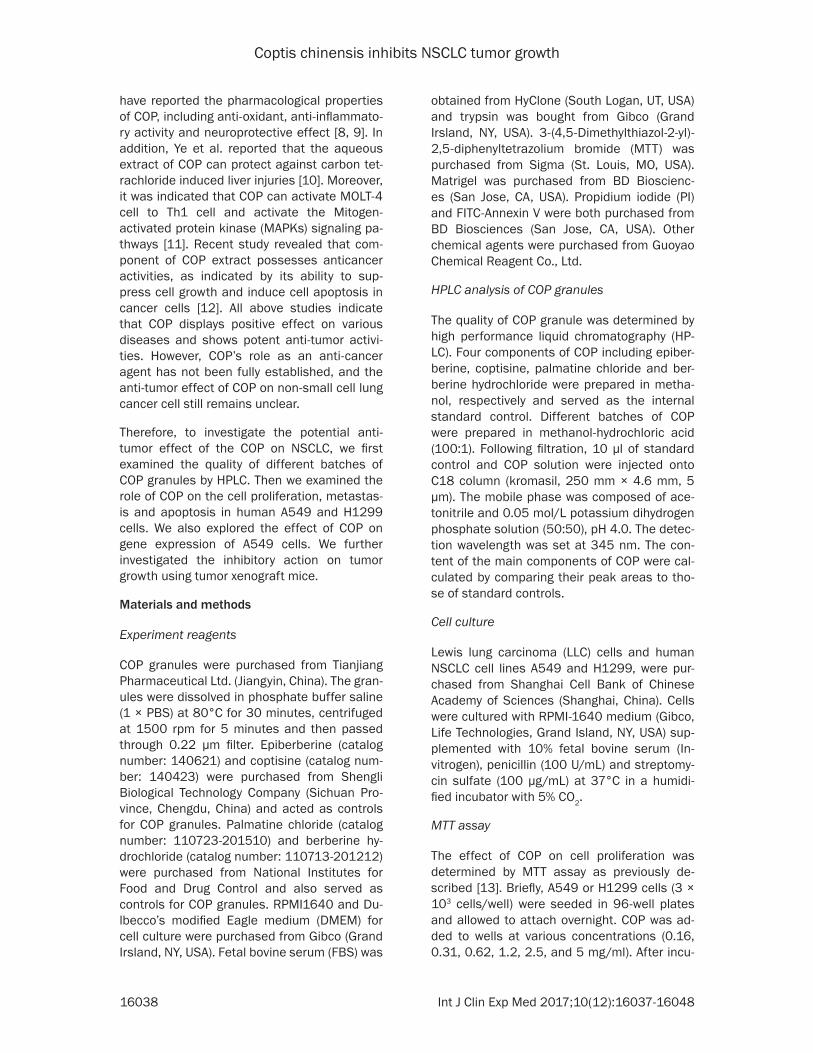
Table 1. Main components of three different batches of COP granules
No. Epiberberine (%)
Coptisine (%)
Palmatine chloride
(%)
Berberine hydrochloride
(%)#1 1.58 2.23 2.15 8.1#2 1.56 2.18 2.14 8.06#3 1.58 2.2 2.15 8.1
PrimeView human gene expression array
PrimeView Human Gene Expression Array was used to determine the effect of COP on genes expression of NSCLC cells. A549 cells were treated with COP (0.31 mg/ml) for 24 h. Total RNA was extracted with Trizol reagent (In- vitrogen, Life Technologies). Gene chip was hybridized and then scanned with Affymetrix Scanner 3000.
Antitumor activity in vivo
Animal experiments involving in this study were conducted in accordance with the internation-ally accepted principles for laboratory animal use and care. The study was approved by the Committee on the Ethics of Animal Experi- ments of Shanghai University of Traditional Chinese Medicine. Female C57 mice (5 weeks) were purchased from SLRC Shanghai and fed in a standard feeding atmosphere in Shanghai University of Traditional Chinese Medicine. LLC cells (3 × 10⁶ cells per mouse) were inoculat- ed subcutaneously in C57 mice at the right axillary region, after three days, mice were ran-domly assigned to three different treatment groups (5 mice per group): (A) vehicle control (water); (B) COP 100 mg/kg; (C) COP 200 mg/kg. The mice were administered drug orally once daily for 3 weeks. Tumor size was mea-sured on two axes with the aid of Vernier cali-pers and tumor volume (mm3) was calculated using the formula: 1/2 (a × b2), where a is the longest and b is the shortest axis. Mice were euthanized at the end of the study and/or when tumor size exceeded 2,000 mm3.
Statistical analysis
Statistical software SPSS version 13.0 was used for analysis. All data are presented as mean ± SD unless otherwise indicated. The data was analyzed by Student t test or a one-way analysis of variance (ANOVA), followed by
pairwise multiple comparisons to determine any difference between groups Values of P < 0.05 were considered statistically significant.
Results
Batch to batch comparison chemical finger-prints of COP granules
Three different batches of COP granules were determined by HPLC. Four main components of COP including epiberberine, coptisine, pal-matine chloride and berberine hydrochloride serve as the standard control. As shown in Figure 1A, epiberberine, coptisine, palmatine chloride and berberine hydrochloride are de- tectable in COP granules. We also found that the magnitude, number, and retention time of the peaks were highly similar among the three batches of COP (Figure 1B). In addition, the standard linear curve for epiberberine, copti-sine, palmatine chloride and berberine hydro-chloride was Y = 3074.2X-2.7816 (X: 0.017-2.18 μg, R = 1), Y = 3476.6X + 10.254 (X: 0.0163-2.088 μg, R = 0.9999), Y = 4025.4X-1.8929 (X: 0.0157-2.004 μg, R = 1), Y = 3895.4X-2.9426 (X: 0.0197-2.52 μg, R = 1), respectively. The content (μg) of control solu-tion was recorded as X and the peak area (mAu) of chromatographic peak was Y. The content of four standard controls in each batch of COP sample was calculated as follows: epiberber-ine, #1: 1.58%, #2: 1.56%, #3: 1.58%; copti-sine, #1: 2.23%, #2: 2.18%, #3: 2.20%; palma-tine chloride, #1: 2.15%, #2: 2.14%, #3: 2.15%; berberine hydrochloride, #1: 8.1%, #2: 8.06%, #3: 8.1% (Table 1). These findings suggest that the three batches of COP granules are identical.
COP inhibits cell proliferation of NSCLC cells
To study the effect of COP on cell proliferation of NSCLC cells, A549 and H1299 cells were exposed to various concentrations of COP for 24, 48 and 72 h, respectively, and cell viability was analyzed by the MTT assay. As shown in Figure 2A and 2B, COP (0.16 mg/ml) signifi-cantly inhibited cell growth of A549 and H12- 99 cells. We also observed that COP treatment resulted in cell growth inhibition in a dose- and time- dependent manner (Figure 2C and 2D). A549 and H1299 cells were incubated in the presence of COP for 48 h, the IC50 values were 0.29 mg/ml and 0.23 mg/ml, respective-

Coptis chinensis inhibits NSCLC tumor growth
16041 Int J Clin Exp Med 2017;10(12):16037-16048
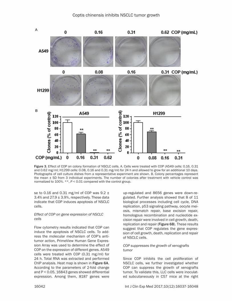
ly. In addition to the MTT assay, colony forma-tion assays were performed on A549 and H1299 cells. We found that low dose of COP (0.16 mg/ml for A549 cells and 0.08 mg/ml for H1299 cells) significantly decreased grow- th of NSCLC cells in colony formation assays (Figure 3A and 3B). These observations indi-cated that COP inhibits cell proliferation of NSCLC cells in vitro.
COP suppresses migratory and invasive ability in NSCLC cells
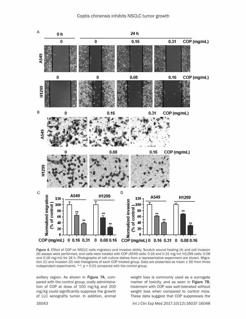
Given that COP inhibits growth of NSCLC ce- lls, we postulated that COP could affect the migratory and invasive ability of NSCLC cells. To further explore the effect of COP on NSC- LC metastasis, using wound healing assay, we determined changes in cell migration after 24 h of incubation with COP. Compared with the vehicle control treated cells, COP treated A549 and H1299 cells both showed significantly de- creased migratory ability (Figure 4A and 4C).
We also tested the effect of COP in NSCLC in- vasion using a transwell chamber. The results further confirmed that COP inhibits NSCLC ce- lls invasion ability (Figure 4B and 4D). Taken together, these in vitro results suggest that COP inhibits NSCLC cell migration and invasion ability.
COP induces apoptosis of NSCLC cells
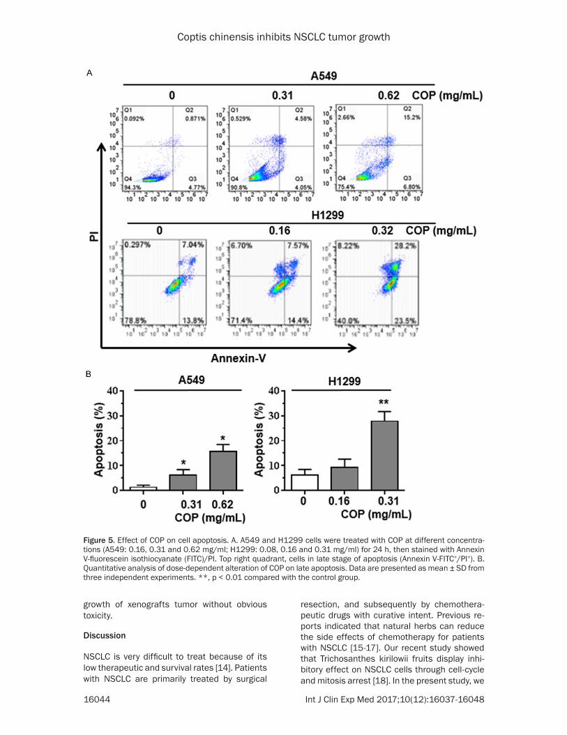
We further investigated the effect of COP on NSCLC cell apoptosis. A549 and H1299 cells were treated with COP at different concen- trations for 24 h, then stained with Annexin V-fluorescein isothiocyanate (FITC)/PI. As sh- own in Figure 5A and 5B, treatment of A549 and H1299 cells with COP induced a dose-dependent increase in the number of late ap- optotic cells. The relative percentage of A549 cells in late apoptosis in response to 0.31 and 0.62 mg/ml of COP was 6.2 ± 2.2% and 15.6 ± 2.9%, respectively, while the relative percent-age of H1299 cells in late apoptosis in respon-
Figure 2. Effect of COP on proliferation of NSCLC cells. A549 (A) and H1299 (B) cells were treated with COP (0.15 mg/ml) for 24 h, respectively. Photographs of cell morphology from a representative experiment are shown. A549 (C) and H1299 (D) cells were treated with COP (0.16, 0.3, 0.6, 1.2, 2.5, and 5 mg/ml) for 24, 48 and 72 h, respec-tively, and cell viability was analyzed by the MTT assay. All values represent the mean ± SD from 3 independent experiments.
Coptis chinensis inhibits NSCLC tumor growth
16042 Int J Clin Exp Med 2017;10(12):16037-16048
se to 0.16 and 0.31 mg/ml of COP was 9.2 ± 3.4% and 27.9 ± 3.9%, respectively. These data indicate that COP induces apoptosis of NSCLC cells.
Effect of COP on gene expression of NSCLC cells
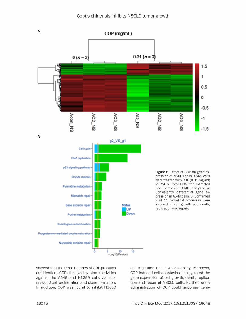
Flow cytometry results indicated that COP can induce the apoptosis of NSCLC cells. To add- ress the molecular mechanism of COP’s anti-tumor action, PrimeView Human Gene Expres- sion Array was used to determine the effect of COP on the expression of different genes. A549 cells were treated with COP (0.31 mg/ml) for 24 h. Total RNA was extracted and performed ChIP analysis. Heat map is shown in Figure 6A. According to the parameters of 2-fold change and P < 0.05, 16843 genes showed differential expression. Among them, 8187 genes were
up-regulated and 8656 genes were down-re- gulated. Further analysis showed that 8 of 11 biological processes including cell cycle, DNA replication, p53 signaling pathway, oocyte mei-osis, mismatch repair, base excision repair, homologous recombination and nucleotide ex- cision repair were involved in cell growth, death, replication and repair (Figure 6B). These results suggest that COP regulates the gene expres-sion of cell growth, death, replication and repair of NSCLC cells.
COP suppresses the growth of xenografts tumor
Since COP inhibits the cell proliferation of NSCLC cells, we further investigated whether COP can suppress the growth of xenografts tumor. To validate this, LLC cells were inoculat-ed subcutaneously in C57 mice at the right
Figure 3. Effect of COP on colony formation of NSCLC cells. A. Cells were treated with COP (A549 cells: 0.16, 0.31 and 0.62 mg/ml; H1299 cells: 0.08, 0.16 and 0.31 mg/ml) for 24 h and allowed to grow for an additional 10 days. Photographs of cell culture dishes from a representative experiment are shown. B. Colony percentages represent the mean ± SD from 3 individual experiments. The number of colonies after treatment with vehicle control was normalized to 100%. **, P < 0.01 compared with the control group.
Coptis chinensis inhibits NSCLC tumor growth
16043 Int J Clin Exp Med 2017;10(12):16037-16048
axillary region. As shown in Figure 7A, com-pared with the control group, orally administra-tion of COP at dose of 100 mg/kg and 200 mg/kg could significantly suppress the growth of LLC xenografts tumor. In addition, animal
weight loss is commonly used as a surrogate marker of toxicity, and as seen in Figure 7B, treatment with COP was well-tolerated without weight loss when compared to control mice. These data suggest that COP suppresses the
Figure 4. Effect of COP on NSCLC cells migratory and invasive ability. Scratch wound healing (A) and cell invasion (B) assays were performed, and cells were treated with COP (A549 cells: 0.16 and 0.31 mg/ml; H1299 cells: 0.08 and 0.16 mg/ml) for 24 h. Photographs of cell culture dishes from a representative experiment are shown. Migra-tion (C) and invasion (D) rate histograms of each COP treated group. Data are presented as mean ± SD from three independent experiments. **, p < 0.01 compared with the control group.
Coptis chinensis inhibits NSCLC tumor growth
16044 Int J Clin Exp Med 2017;10(12):16037-16048
growth of xenografts tumor without obvious toxicity.
Discussion
NSCLC is very difficult to treat because of its low therapeutic and survival rates [14]. Patients with NSCLC are primarily treated by surgical
resection, and subsequently by chemothera-peutic drugs with curative intent. Previous re- ports indicated that natural herbs can reduce the side effects of chemotherapy for patients with NSCLC [15-17]. Our recent study showed that Trichosanthes kirilowii fruits display inhi- bitory effect on NSCLC cells through cell-cycle and mitosis arrest [18]. In the present study, we
Figure 5. Effect of COP on cell apoptosis. A. A549 and H1299 cells were treated with COP at different concentra-tions (A549: 0.16, 0.31 and 0.62 mg/ml; H1299: 0.08, 0.16 and 0.31 mg/ml) for 24 h, then stained with Annexin V-fluorescein isothiocyanate (FITC)/PI. Top right quadrant, cells in late stage of apoptosis (Annexin V-FITC+/PI+). B. Quantitative analysis of dose-dependent alteration of COP on late apoptosis. Data are presented as mean ± SD from three independent experiments. **, p < 0.01 compared with the control group.
Coptis chinensis inhibits NSCLC tumor growth
16045 Int J Clin Exp Med 2017;10(12):16037-16048
showed that the three batches of COP granules are identical. COP displayed cytotoxic activities against the A549 and H1299 cells via sup-pressing cell proliferation and clone formation. In addition, COP was found to inhibit NSCLC
cell migration and invasion ability. Moreover, COP induced cell apoptosis and regulated the gene expression of cell growth, death, replica-tion and repair of NSCLC cells. Further, orally administration of COP could suppress xeno-
Figure 6. Effect of COP on gene ex-pression of NSCLC cells. A549 cells were treated with COP (0.31 mg/ml) for 24 h. Total RNA was extracted and performed ChIP analysis. A. Consistently differential gene ex-pression in A549 cells. B. Confirmed 8 of 11 biological processes were involved in cell growth and death, replication and repair.

Coptis chinensis inhibits NSCLC tumor growth
16046 Int J Clin Exp Med 2017;10(12):16037-16048
grafts tumor growth without obvious toxicity. These results suggested that COP may be a novel potential anti-tumor candidate in the NSCLC cells related lung cancer.
Given the batch to batch consistency of natu- ral herb extracts is fundamental for basic re- search and clinical studies, at the beginning of our study, three different batches of COP gran-ules were compared by HPLC. We found that four of main components including epiberber-ine, coptisine, palmatine chloride and berber-ine hydrochloride are detectable, and the con-tent of them are highly similar in COP granules. These results indicate that the components of the three batches of COP granules are identi-cal. It has been identified that the extract of COP contains several bioactivity components [19] and berberine has been recognized as the dominant one [20]. Interestingly, previous stud-ies reported that purified berberine was less effective than the whole COP extract [21, 22]. These reports indicated that it is unusual for a single component to be isolated from an herbal medicine with optimal biological activity. Our previous study also provided evidence that an extract is more effective in vivo than a single isolated component [13]. In this regard, it seems better to develop the whole herbal me- dicine extract rather than its single dominant component for cancer treatment. COP has vari-ous pharmacological activities and caused high interests on its anti-cancer activity.
It has been shown that COP extract inhibits tumor progression via suppressing cell proli- feration, inducing cell death and arresting cell cycle [23]. In this study, we found that COP dis-played cytotoxic activities against the NSCLC cells (A549 and H1299) by inhibiting cell prolif-eration and clone formation. Previous reports demonstrated the effects of COP in suppress-ing cancer cell invasion, which in turn inhibits cancer metastasis [23]. In agreement with this published data, we also observed that COP inhibits the migration and invasion ability of A549 and H1299 cells. The anti-tumor actions of some natural products have been proved to be the result of their ability to induce cell apop-tosis [24]. Recently, it has been reported that COP and its main active component, berbe- rine, could initiate human cancer cell apopto-sis, causing to death of tumor cells. Treatment of COP extract and berberine could induce the intrinsic pathway of apoptosis [25]. In the pres-ent study, flow cytometric analysis showed th- at exposure to COP resulted in a significant increase in the percentage of apoptosis cells, suggesting that COP initiates apoptosis in A549 and H1299 cells. It is now well recog-nized that understanding of molecular effect (i.e. gene expression) of an herb is very impor-tant for assessing its efficacy and safety [22]. A clearer understanding as to how COP regu-lates the cellular pathway would provide impor-tant insights into the potential mechanisms for the development of anti-cancer herbs with im- proved selectivity. Microarray technology is qui-
Figure 7. Antitumor activity of COP in C57 mice bearing LLC tumors. LLC cells (3 × 10⁶ cells per mouse) were in-oculated subcutaneously in C57 mice at the right axillary region, after three days, mice were randomly assigned to three different treatment groups (5 mice per group): vehicle control (water); COP 100 mg/kg and COP 200 mg/kg. The mice were administered drug orally once daily for 3 weeks. Tumor volume (A) and mice body weight (B) were recorded at different time. Data in the graphs represent the mean ± SD (n = 5 mice per group).
Coptis chinensis inhibits NSCLC tumor growth
16047 Int J Clin Exp Med 2017;10(12):16037-16048
te useful in this regard. Our human gene ex- pression array data clearly showed that COP regulated the gene expression of cell growth, death, replication and repair of NSCLC cells. These results may help identifying novel thera-peutic effect of COP. Our strategy used in this study could serve as a framework to study medicinal herbs. Taken together, our results suggest that there are potentially multiple me- chanisms by which COP is able to inhibit the growth of NSCLC cancer cells.
Our in vivo data further showed that COP (100 mg/kg and 200 mg/kg) treatment significantly reduced tumor volume. In addition, we observ- ed that the COP treatment did not affect the body weight of mice. This indicates that COP is a low toxicity herb. COP has been widely used in China for several thousand years for the treat-ment of infectious conditions. COP has been shown to be safe for human consumption [26]. This advantage plus the accumulating eviden- ce of its anti-tumor effects make COP a promis-ing candidate for being an anti-cancer agent. This study may shed light on future direction of studies featuring COP as a novel anti-tumor agent, which should be further proven in future animal and clinical studies.
In conclusion, our studies demonstrated that COP displayed anti-tumor activity both in vitro and in vivo. COP may represent a novel thera-peutic herb for NSCLC treatment. Further stud-ies are needed to elucidate the whole signaling pathway of the anti-tumor action of COP and to determine the role of metabolism as it relates to the in vivo activity of COP.
Acknowledgements
This work was supported by the Natural Scien- ce Foundation of China (No#. 81173224 and 81373621).
Disclosure of conflict of interest
None.
Address correspondence to: Dr. Hongli Ren, Institute of Science, Technology and Humanities, Shanghai University of Traditional Chinese Medicine, Shanghai, China. Tel: +86-021-51322139; E-mail: [email protected]; Dr. Haizhou Liu, Department of Medicine, University of Pittsburgh Cancer Institute, University of Pittsburgh, 5117 Centre Ave, Pitts-
burgh, PA 15213, United States of America. Tel: +1-412-628-3289; E-mail: [email protected]
References
[1] Broodman I, Lindemans J, van Sten J, Bischoff R and Luider T. Serum protein markers for the early detection of lung cancer: a focus on auto-antibodies. J Proteome Res 2017; 16: 3-13.
[2] Pearce A, Bradley C, Hanly P, O’Neill C, Thomas AA, Molcho M and Sharp L. Projecting produc-tivity losses for cancer-related mortality 2011-2030. BMC Cancer 2016; 16: 804.
[3] Siegel R, Ward E, Brawley O and Jemal A. Can-cer statistics, 2011: the impact of eliminating socioeconomic and racial disparities on pre-mature cancer deaths. CA Cancer J Clin 2011; 61: 212-236.
[4] Billiet C, Peeters S, Decaluwe H, Vansteenkiste J, Mebis J and Ruysscher D. Postoperative ra-diotherapy for lung cancer: is it worth the con-troversy? Cancer Treat Rev 2016; 51: 10-18.
[5] Koyi H, Hillerdal G, Kolbeck KG, Brodin D, Liv P and Branden E. Non-small cell lung cancer (NSCLC) in octogenarians in clinical practice. Anticancer Res 2016; 36: 5397-5402.
[6] Newman DJ, Cragg GM and Snader KM. Natu-ral products as sources of new drugs over the period 1981-2002. J Nat Prod 2003; 66: 1022-1037.
[7] Feng YB, Luo WQ and Zhu SQ. [Explore new clinical application of Huanglian and corre-sponding compound prescriptions from their traditional use]. Zhongguo Zhong Yao Za Zhi 2008; 33: 1221-1225.
[8] Friedemann T, Ying Y, Wang W, Kramer ER, Schumacher U, Fei J and Schroder S. Neuro-protective effect of coptis chinensis in MPP [Formula: see text] and MPTP-induced parkin-son’s disease models. Am J Chin Med 2016; 44: 907-925.
[9] Kwon OJ, Kim MY, Shin SH, Lee AR, Lee JY, Seo BI, Shin MR, Choi HG, Kim JA, Min BS, Kim GN, Noh JS, Rhee MH and Roh SS. Antioxidant and anti-inflammatory effects of Rhei rhizoma and coptidis rhizoma mixture on reflux esophagitis in rats. Evid Based Complement Alternat Med 2016; 2016: 2052180.
[10] Ye X, Feng Y, Tong Y, Ng KM, Tsao S, Lau GK, Sze C, Zhang Y, Tang J, Shen J and Kobayashi S. Hepatoprotective effects of Coptidis rhi-zoma aqueous extract on carbon tetrachloride-induced acute liver hepatotoxicity in rats. J Eth-nopharmacol 2009; 124: 130-136.
[11] Kim E, Ahn S, Rhee HI and Lee DC. Coptis chi-nensis Franch. Extract up-regulate type I help-er T-cell cytokine through MAPK activation in MOLT-4 T cell. J Ethnopharmacol 2016; 189: 126-131.

Coptis chinensis inhibits NSCLC tumor growth
16048 Int J Clin Exp Med 2017;10(12):16037-16048
[12] Chuang TY, Wu HL, Min J, Diamond M, Azziz R and Chen YH. Berberine regulates the protein expression of multiple tumorigenesis-related genes in hepatocellular carcinoma cell lines. Cancer Cell Int 2017; 17: 59.
[13] Liu H, Schmitz JC, Wei J, Cao S, Beumer JH, Strychor S, Cheng L, Liu M, Wang C, Wu N, Zhao X, Zhang Y, Liao J, Chu E and Lin X. Clove extract inhibits tumor growth and promotes cell cycle arrest and apoptosis. Oncol Res 2014; 21: 247-259.
[14] Delbaldo C, Michiels S, Syz N, Soria JC, Le Che-valier T and Pignon JP. Benefits of adding a drug to a single-agent or a 2-agent chemother-apy regimen in advanced non-small-cell lung cancer: a meta-analysis. JAMA 2004; 292: 470-484.
[15] Cassileth BR, Rizvi N, Deng G, Yeung KS, Vick-ers A, Guillen S, Woo D, Coleton M and Kris MG. Safety and pharmacokinetic trial of docetaxel plus an astragalus-based herbal for-mula for non-small cell lung cancer patients. Cancer Chemother Pharmacol 2009; 65: 67-71.
[16] Lee J, Chae J, Lee S, Kim K, Eo W, Kim S, Choi W and Cheon SH. The efficacy and safety of standardized allergen-removed Rhus vernici-flua extract as maintenance therapy after first-line chemotherapy in patients with advanced non-small cell lung cancer. Am J Chin Med 2013; 41: 773-787.
[17] McCulloch M, See C, Shu XJ, Broffman M, Kramer A, Fan WY, Gao J, Lieb W, Shieh K and Colford JM Jr. Astragalus-based Chinese herbs and platinum-based chemotherapy for ad-vanced non-small-cell lung cancer: meta-anal-ysis of randomized trials. J Clin Oncol 2006; 24: 419-430.
[18] Ni L, Zhu X, Gong C, Luo Y, Wang L, Zhou W, Zhu S and Li Y. Trichosanthes kirilowii fruits in-hibit non-small cell lung cancer cell growth through mitotic cell-cycle arrest. Am J Chin Med 2015; 43: 349-364.
[19] Fang XP, Wang TZ, Zhang H, Shuai H, Li D and Xie CK. [Quantitative determination of 5 alka-loids in plants of coptis from China]. Zhongguo Zhong Yao Za Zhi 1989; 14: 33-35, 63.
[20] Li XK, Motwani M, Tong W, Bornmann W and Schwartz GK. Huanglian, a Chinese herbal ex-tract, inhibits cell growth by suppressing the expression of cyclin B1 and inhibiting CDC2 kinase activity in human cancer cells. Mol Pharmacol 2000; 58: 1287-1293.
[21] Iizuka N, Oka M, Yamamoto K, Tangoku A, Mi-yamoto K, Miyamoto T, Uchimura S, Hamamo-to Y and Okita K. Identification of common or distinct genes related to antitumor activities of a medicinal herb and its major component by oligonucleotide microarray. Int J Cancer 2003; 107: 666-672.
[22] Kang JX, Liu J, Wang J, He C and Li FP. The ex-tract of huanglian, a medicinal herb, induces cell growth arrest and apoptosis by upregula-tion of interferon-beta and TNF-alpha in hu-man breast cancer cells. Carcinogenesis 2005; 26: 1934-1939.
[23] Wang N, Tan HY, Li L, Yuen MF and Feng Y. Ber-berine and Coptidis Rhizoma as potential anti-cancer agents: recent updates and future per-spectives. J Ethnopharmacol 2015; 176: 35-48.
[24] Fulda S. Modulation of apoptosis by natural products for cancer therapy. Planta Med 2010; 76: 1075-1079.
[25] Eom KS, Kim HJ, So HS, Park R and Kim TY. Berberine-induced apoptosis in human glio-blastoma T98G cells is mediated by endoplas-mic reticulum stress accompanying reactive oxygen species and mitochondrial dysfunction. Biol Pharm Bull 2010; 33: 1644-1649.
[26] Qin CL, Liu JY and Cheng ZM. [Pharmacological studies on the effects of huanglian decoction on experimental gastric lesions in rats and an-tiemetic in pigeons]. Zhongguo Zhong Yao Za Zhi 1994; 19: 427-430, 448.