Embed Size (px)
Citation preview

30
Original article
Comparative protein profile studies and in silicostructural/functional analysis of HMGR (ApHMGR)
in Andrographis paniculata (Burm.f.) Wall. ex NeesByreddi Bhavani Venkata Bindu, Mote Srinath, Aayeti Shailaja and Charu Chandra Giri
Centre for Plant Molecular Biology (CPMB), Osmania University, Hyderabad-500007, Telangana State, India
Received May 7, 2017: Revised June 5, 2017: Accepted June 10, 2017: Published online June 30, 2017
Abstract
Andrographis paniculata (Burm.f.) Wall. ex Nees (Ap) contains andrographolide, i.e., synthesizedvia cytosolic MVA and plastidial MEP pathways using IPP and DMAPP as precursor molecules.HMGR catalyzes the first committed and rate-limiting step in MVA pathway. Limited informationis available on structural and functional aspects of key enzyme HMGR. In a preliminary study, thein vivo leaves and light/dark grown callus was used for protein profile analysis. The callus growthand andrographolide content through HPLC analysis was carried out. SDS-PAGE total proteinprofile of in vivo leaves, light, and dark grown callus was investigated. A high intensity band of62.3 KD protein indicating its proximity with HMGR enzyme was observed in leaves and lightcallus compared to dark grown callus. On this background, the present communication reportsbioinformatics analysis of HMGR enzyme to decipher its functional and structural properties. Theisoelectric point (pI), molecular weight and hydrophobicity were calculated using Expasy tools.The secondary (2D) and tertiary (3D) structures were predicted and validated using PROCHECKSAVES algorithm. Our finding on the allowed percentage (88.2%) of amino acid residues in theRamachandran plot indicated that the simulated 3D structure was reliable. Phylogenetic analysisof A. paniculata HMGR (ApHMGR) with different plants and other organisms including humansrevealed close relationships and common lineage. The protein-protein interaction studies usingSTRING 10 tool revealed a close association of HMGR and nine other proteins in MVA and MEPpathways. The present analyzed data will give an insight on the structural and function attributescorresponding to this important HMGR protein. The molecular and biochemical understanding ofHMGR protein will be advantageous for andrographolide yield enhancement through metabolicengineering.
Key words: Andrographis paniculata (Burm.f.) Wall. ex Nees, ApHMGR, 3D structures, proteinprofile, phylogenetic analysis, protein-protein interactions
1. Introduction
Andrographis paniculata of family Acanthaceae, popularly knownas king of bitters, is widely cultivated in Southern and South easternAsia (Sareer et al., 2014). A. paniculata contains main bioactivecompounds such as andrographolide (AD), neo-andrographolide(NAD), and 14-deoxy-11, 12-didehydroandrographolide (DAD)(Subramanian et al., 2012; Latha et al., 2017). These compoundsare produced mainly by two distinct biosynthetic pathways, i.e.,mevalonate (MVA) and methylerythritol 4-phosphate (MEP). MVApathway occurs in cytosol whereas MEP pathway takes place inplastid (Figure 1). Until date, these two pathways have not beenstudied thoroughly and very less information is available on thekey genes and enzymes of these pathways (Srivastava and Akhila,2010). Recently, as per Lipko and Swiezewska (2016), MVA is one
Copyright @ 2017 Ukaaz Publications. All rights reserved.Email: [email protected]; Website: www.ukaazpublications.com
Author for correspondence: Dr. Charu Chandra GiriProfessor, Centre for Plant Molecular Biology (CPMB) , OsmaniaUniversity, Hyderabad-500007, Telangana State, IndiaE-mail: [email protected].: +91-040-27098087
of the major biosynthetic routes associated with terpenoidsynthesis, which provides the key precursors such as isopentenylpyrophosphate (IPP) and dimethylallyl pyrophosphate (DMAPP).A. paniculata has several medicinal properties such as antidiarrhoeal,anti-inflammatory, hepatoprotective, anticancer, antidepressive andanti-HIV (Niranjan et al., 2010; Suriyo et al., 2017).
3-hydroxy-3-methylglutaryl-coenzyme A reductase (HMGR)enzyme is found to be coded and regulated by multigene families inplants (Guo et al., 2015). The presence of several copies of thesegenes in plants attributes its relationship with many physiologicalprocesses, thus depicts its biological significance and importance.However, the number of structurally characterized proteins in plantsis small and limited compared to number of known proteinsequences. The 3D structure of proteins is stable and conservedthan primary structure (amino acid sequence). Homology modellinghas proven as the method of choice to generate reliable 3D model ofa protein from its amino acid sequence (Prasada et al., 2010).Currently, in silico methods and tools have provided numerousadvantages over time consuming wet-lab experiments oncharacterization of proteins (Proctor et al., 2016; Singh andSrivastava, 2016). The MVA is synthesized from HMG-CoA by the
Annals of Phytomedicine 6(1): 30-44, 2017DOI: 10.21276/ap.2017.6.1.5; Print ISSN : 2278-9839 and Online ISSN : 2393-9885 6(1):30-44 (2017)
Ann. Phytomed.,

31
regulation of key rate limiting enzyme HMGR. In addition, HMGRhas also shown to be a fundamental key enzyme of terpenoidbiosynthesis in A. paniculata (Jha et al., 2011).
Further, in our laboratory we have a comprehensive researchprogramme on the geographical distribution of A. paniculata andrelated species, haplotyping, screening and yield enhancement ofbioactive compounds using biotechnological and molecularapproaches (Neeraja et al., 2015; Arolla et al., 2015; Parlapally etal., 2015; Zaheer and Giri, 2015; Neeraja et al., 2016; Zaheer andGiri, 2017). Plant tissue culture is an alternative tool for productionof bioactive compounds and callus culture system generally usedfor detection of biologically active molecules in cultured plant tissuein vitro (Dias et al., 2016; Giri and Zaheer, 2016; Abd El-Aal et al.,2016). Meagre information is available on gene and enzymes involvedin diterpene lactone biosynthesis with particular reference to HMGR
in non-model plant A. paniculata. HMGR being a key regulatoryrate-limiting enzyme in MVA pathway, the study on ApHMGR gainssignificance. Therefore, there is a need to understand and elucidatestructural/functional properties of HMGR. HPLC analysis of in vivoleaves, callus grown in dark/light and andrographolide content wasestimated to elucidate possible correlation of the enzyme andspecific secondary metabolite synthesis. The protein profiling ofin vivo leaves and callus using sodium dodecyl sulfatepolyacrylamide gel electrophoresis (SDS-PAGE) was carried out tostudy proteins related to enzymes in andrographolide biosyntheticpathway. In silico analysis of HMGR protein is an attempt in thisdirection to generate further information. The presentcommunication reports protein profile analysis and in detail insilico study on the structural, functional, phylogenic analysis andprotein-protein interaction of ApHMGR.
MVA PathwayMEP Pathway
Acetyl Co-A Aceto acetyl Co-A
HMG Co-A
Mevalonate
MVAP
MVAPP
IPP
Pyruvate Glyceraldehyde 3-P
DOXP
MEP
CDPME
CDPMEP
MEcPP
HMBPP
IPP DMAPPDMAPPIPPI
PMD
PMK
MVK
HMGR*
HMGS
HDS
MCS
CMK
MCT
DXR
DXS
HDR
IPPI
Monoterpenes
Sesquiterpenes
Diterpenes
PolyterpenesTriterpenes
HMG Co A MevalonateHMGR*
Cytosol Plastid
PLANT CELLAndrographis paniculata
Figure 1: Net house grown Andrographis paniculata plant and MVA/MEP pathways showing key HMGR enzyme in A. paniculata.
2. Materials and Methods
2.1 Plant tissue culture, HPLC and protein profile analysis
2.1.1 Collection of seeds and establishment of A. paniculata plants in vivo and in vitro
The seeds of A.paniculata were sown on soil in net house conditionsat Centre for Plant Molecular Biology (CPMB), OsmaniaUniversity, Hyderabad, India. The plants were established by regularsupplement of Hoagland nutrient solution (Hoagland and Arnon,
1938) twice a day. Seeds were collected from these establishedplants and used for different experiments in the laboratory. Thegreen pods were surface sterilized with 0.1% (w/v) mercuric chloridefor 10 min, followed by washing with sterile distilled water 5 timeswith five minutes for each wash. The surface sterilized pods wereinoculated aseptically on MS (Murashige and Skoog, 1962) mediumwithout plant growth regulators (PGRs) and 2.0 mg/l GA3supplemented media for germination and establishment of asepticcultures. Seeds were incubated in dark for one week with subsequenttransfer to light conditions and all the cultures were maintained at25 ± 2ºC.

32
2.1.2 Induction of callus and growth study
The callus was induced, established using cotyledon and hypocotylexplants on MS media containing 1.0 mg/1 2, 4-dichlorophenoxyacetic acid (2, 4-D) and 0.5 mg/1 Kinetin (Kn). The callus inducedand maintained both in dark/light conditions with same combinationof MS medium. These cultures were maintained by sub-culturing ata regular interval of four weeks. The growth study of callus cultureswere performed by taking fresh weight (FW) and dry weight (DW)of cultures every week until the sub-culturing time (up to 4 weeks).The fresh weight was taken, samples were kept for drying in anincubator for 72 h. at 600C and dry weight data was scored.
2.1.3 Statistical analysis
All the data on germination of seeds and callus growth in A.paniculata was recorded for analysis after four weeks of culture.Statistical analysis was carried out to calculate the mean and thestandard error.
2.1.4 Extraction of andrographolide from leaf, callus andHPLC analysis
Samples of leaf and callus were oven-dried and ground using mortarand pestle. Dry powdered material of leaf and callus samplesweighing 50 mg were transferred to vials. About 5.0 mL of HPLC-grade methanol was added to each sample and incubated for 24 h. atambient temperature. Final extract was ultra-sonicated using anultrasonic cleaning bath (Spectra Lab., Model UCB 30, India) for 30min. The final extract was first filtered using Whatman filter paperNo. 41 subsequent to sonication. The filtrate was further passedthrough 0.45 µm disposable hydrophilic PVDF membrane filters(Millex-HV, Millipore, Ireland) before its injection into HPLC systemfor analysis. The HPLC analysis was performed as per the protocolreported earlier from our laboratory (Zaheer and Giri, 2015).
2.1.5 Protein isolation and SDS-PAGE
Total protein was isolated from in vivo leaf, dark and light growncallus using acetone precipitation method as per Talei et al. (2014).The quantitative analysis was performed by Bradford method(Bradford, 1976). The spectrophotometric analysis of protein wasdone at 595 nm. Bovine serum albumin (BSA) was used as a standardprotein for all the experiments. The qualitative analysis wasperformed using SDS-PAGE (Laemmli, 1970). The protein sampleswere run on 5% stacking gel and 12% separating gel at 90 volts percm. The staining was done using 0.25% (w/v) coomassie brilliantblue G-250 in glacial acetic acid, methanol and water at 1:2:2 ratio.The bands were observed on image viewer and data was collected.
2.2 In silico study
2.2.1 Primary sequence collection and analysis of biochemicalproperties
ApHMGR sequence with accession number AAL28015.2 was obtainedfrom NCBI and biochemical properties were analyzed. Estimationof pI and molecular weight was carried out using Expasy toolswebsite: Compute pI/Mw tool (Kyte and Doolittle, 1982).Hydropathy-plot analysis was performed using the ProtScale tool,available from the Expasy online server.
2.2.2 Prediction of secondary, tertiary (3D) structure andvalidation
The secondary structure of protein was predicted using CFSSPprediction server (Chou and Fasman, 1974; Dor et al., 2016).Analysis of protein sequence was carried out to predict the alphahelix, extended beta sheet and random coil structures contributionat each position based on 17 amino acid sequence window. SWISS-MODEL server was used for protein 3D structure prediction (Kieferet al., 2009). Structure databases and protein sequence was identifiedfor homology modelling. Energy levels and protein stabilizationwas taken into account to optimise model structure. Visualizationof 3D structure was carried out using WebLab Viewer Lite 4.PROCHECK analysis was performed for the evaluation of 3D modelstereo chemical qualities. 3D structure stereo chemical reliabilitywas analyzed using Ramchandran plot (Laskowski et al., 1996).All 20 amino acids were analyzed considering most allowed anddisallowed regions.
2.2.3 BLAST analysis and multiple sequence alignment (MSA)
BLASTp (Basic Local Alignment Search Tool- protein) wasperformed to compare the ApHMGR protein sequence with otherplant non-redundant protein data available in the NCBI. To find outthe percentage of similarity and conserved regions between theApHMGR, Homo sapiens (HsHMGR) and Mus musculus(MmHMGR)multiple sequence alignment (CLUSTAL W2) wasperformed. The default parameters were considered for running theprogramme and analysing the data.
2.2.4 Motifs and domain analysis
The conserved motifs were analyzed among different plants of 30species which are highly similar with ApHMGR and other organismslike HsHMGR and MmHMGR using the program of multiple Em formotif elicitation (MEME; version 4.10.2). The parameters of MEMEanalysis were applied as follows: minimum width for each Motif:six; maximum width for each motif: fifty; maximum number ofmotifs to find is three and the numbers of repetitions are zero orone per sequence (Bailey et al., 2006). TMHMM server V.2.0 (CBS;Denmark) guided by hidden Markov model (HMM) was used toanalyze trans-membrane helices in the ApHMGR as per Krogh et al.(2001). The elucidation of ApHMGR structural orientation wasperformed using Phyre2 (Kelley et al., 2015).
2.2.5 Analysis of ligand binding site, phylogenetic and protein-protein interaction studies
The evaluation of predicted 3D structure for ligand binding wascarried out using bioinformatics tool for site prediction (Wass et al.,2010). The identification of homologous structures with boundligands from structural library was assisted by predicted 3Dstructure. The elucidation of a ligand-binding site was accomplishedby superimposing ligands onto predicted protein structure.Neighbor-Joining method was used to analyze the evolutionarytrend. The optimal tree was depicted with the sum of branch lengthvalue (1.80027160). The clustering of associated taxa together visibleas percentage of replicate trees was performed using bootstrap test(1000 replicates) and is being portrayed beside the branches. Thetree is drawn to scale, with branch lengths (next to the branches) inthe same units as those of the evolutionary distances used to inferthe phylogenetic tree. The evolutionary distances were computedusing the Poisson correction method and are in the units of the

33
number of amino acid substitutions per site. The analysis involved30 amino acid sequences. All positions containing gaps and missingdata were eliminated. There were 66 positions in the final dataset.Evolutionary analyzes were conducted in MEGA6 (Saitou et al.,1987; Kumar and Gadagkar, 2000; Tamura et al., 2013). STRING 10was used to find out the protein-protein interactions. Arabidopsisthaliana was used as reference sequence to draw protein-proteininteractions (Szklarczyk et al., 2015).
3. Results and Discussion
3.1 Plant tissue culture, HPLC and protein profile analysis
3.1.1 Establishment of A. paniculata seedlings from pods invitro
Germination of A. paniculata seeds was observed after three daysof inoculation. These germinated seedlings were used as explantsfor induction of callus. The highest germination frequency(46.5 ± 5.5) was obtained on MS medium without hormone whencompared to 37 ± 4.84 MS media supplemented with GA3(2.0 mg/l) and is depicted in Figure 2.
Figure 2:Comparative study for germination frequency of seeds inMSO and MS+GA3.
3.1.2 Induction of callus, growth and HPLC analysis
The highest percentage of callus induction (100 ± 0) was observedfrom cotyledon explants and initiation within 4-5 days on MSmedium containing 1.0 mg/1 2, 4-D and 0.5 mg/1 Kn. The callusinduction frequency of 97±0 was observed within 5-6 days inhypocotyl explants on same callus induction medium (Figure 3,Figures 4 A, B). Callus cultures are generally used for detection ofsecondary metabolites in vitro and their further exploitation forproduction (Giri and Zaheer, 2016; Elenaet et al., 2016). The growthstudy of callus cultures up to four weeks revealed that the freshand dry weights of callus cultures gradually increased (Figure 5).
The HPLC analysis of callus revealed that the andrographolidecontent was increased up to four weeks. There was increase inandrographolide content with increase in callus age. The
andrographolide content of leaves was up to 1.667% DW followingHPLC analysis. The andrographolide content of callus cultures wasless compared to in vivo leaves (25 fold). The light callusandrographolide content was 1.46 fold higher than dark callus (Figure6, Figures 7 A, B). The study on andrographolide content of callusin both light and dark is reported first time in A. paniculata. Thisplant contains two different pathways, i.e., MEP and MVA for thesynthesis of andrographolides. The MEP pathway takes place inplastid and MVA takes place in cytosol. Light has a direct influenceon plant development, morphogenic potential and synthesis ofactive compounds in medicinal herbs (Fazal et al., 2016). Likewiselight may perhaps have some influence in enhancing theandrographolide content. The molecular analysis of light callus canprovide an insight into study the MEP pathway in detail.
Figure 3 : Callus induction from cotyledon and hypocotyl explants.
(A)

34
(B)Figure 4 : (A) Callus induced in dark and (B) Callus induced in light
Figure 5 : Growth study of callus cultures in light and dark
Figure 6 : HPLC analysis of callus cultures (Light and Dark)
(A)
(B)
Figure 7 : (A) HPLC analysis of Dark callus and (B) HPLC analysisof Light callus.
3.1.3 Protein profile analysis
The Bradford estimation determines that the total protein contentin leaves of A.paniculata is 0.588±0.22. In callus cultures, it wasobserved that the total protein content in both light and dark inducedcallus was almost same, i.e., 0.251±0.152 and 0.256±0.10,respectively. The qualitative analysis by SDS-PAGE showed thatleaves are having 22 bands, though the protein content is almostsame in light and dark callus, the number of bands varied (Figure 8).In SDS-PAGE protein profile analysis, 13 bands were observedwith callus grown in light compared to 8 bands in callus grown indark (Figure 9). The banding pattern was almost similar in light anddark callus samples. The extra bands found in light induced calluswere also coinciding with in vivo leaf samples which are absent indark callus. The intensity of bands indicated abundance of proteins

35
influenced by presence of light in both callus and in vivo leaves.The protein band intensity was higher in leaves followed by lightcallus and low in dark callus. A high intensity band of 62.3 KDprotein indicating its proximity with HMGR enzyme was found inin vivo leaves and light callus compared to callus grown in dark.The similar studies were also reported in Artemisia vulgaris (Kumaret al., 2009), Glycine max L. Merr (Radhakrishnan and Ranjitha,2009), and Plumbago zeylanica L. (Rout et al., 2010). Protein andenzyme analysis of stem in Tinospora cordifolia was also reported(Sharma and Batra, 2015). Some other medicinal plant species weretoo examined for their protein profile in vivo and in vitro, viz.,Bacopa monnieri (Mohapatra and Rath, 2005).
Figure 8 : Qualitative analysis was performed using SDS-PAGE DC-Dark callus; LC-Light callus; PM-Protein marker; 62.3KD protein similar to HMGR.
Figure 9 : Qualitative protein profile analysis showing presence andabsence of bands with specific molecular weight in leaf,Light callus (LC) and dark callus (DC). The gaps betweenthe bars in LC and DC indicates the absence of specificbands in SDS-PAGE profile.Lane 10 indicated with starrepresents 62.3 KD protein similar to HMGR
3.2 In silico study
3.2.1 ApHMGR primary sequence and analysis of biochemicalproperties
The collected primary structures of ApHMGR with Accessionnumber AAL28015.2 comprise 595 amino acid residues. It indicatedthe gene size, which is approximately 1.7 kb (1785 base pairs).Elucidation of protein properties such as solubility and mobilityare based on theoretical pI and being used to develop buffer systemor salt bridge in expression studies (Dewald et al., 2015). TheApHMGR protein has theoretical pI value of 5.75 and molecularweight of 63268.59 daltons (~63 KD). The theoretical molecularweight obtained from the present study will be helpful in isoenzymestudies and their purification. Generally, hydropathy plots areused for visualization of hydrophobicity of a peptide sequencelength (Fung et al., 2010). The hydrophobicity plot for ApHMGRshowed the percentage of buried amino acid residues inside proteincore. The highest percentage is of Val (12.9%), which is mostlyhydrophobic, with least of 0.5% in both Arg and Lys, which aremostly hydrophilic (Table 1). It indicated that the ApHMGR N-terminus is highly hydrophobic with two highest hydrophobicpeaks (Figure 10). Generally, scales of hydrophobicity were usedto envisage the genetic code preservation. In an earlier study, thehydrophobicity profiles of the N-terminus showed twohydrophobic sequences in Arabidopsis thaliana (Uozumi et al.,1997). This was similar to our finding where we identified residues,which were long enough to cover the membrane bilayer in ApHMGR.
Table 1: Percentage of hydrophobicity along each amino acid ofApHMGR protein*
S.No. Amino Percentage S.No. Amino Percentageacid (%) acid (%)
1. Val 12.9 1 1 Asp 2.902. Gly 11.8 1 2 Pro 2.703. Leu 11.7 1 3 Tyr 2.604. Ala 11.2 1 4 T rp 2.205. Ile 8.6 15. His 2.006. Ser 8.0 16. Met 1.907. Phe 5.1 17. Glu 1.808 T hr 4.90 18. Gln 1.609 Cys 4.10 19. Arg 0.5
1 0 Asn 2.90 20. Lys 0.5* Data presented as per Hydropathy-plot analysis using the ProtScale
tool
Figure 10 : The hydrophobicity plot of ApHMGR protein showingpeaks for each amino acid.

36
3.2.2 Prediction of secondary, tertiary (3D) structure andvalidation
The secondary structure analysis showed that 455 residues possiblefor the -helix (H); extended sheet (E) 355 residues and turns (T)66 residues. Hence, ApHMGR consists of 76.5% helices, 59.7%extended sheets and 11.1% turns (Figure 11). - helices and extendedsheets are the most abundant structural elements of ApHMGRpenetrating through most part of the secondary structure. On theother hand, beta turns and random coils were found to be distributedintermittently in trans-membrane protein. The multiple sequencealignment by simultaneous assessment of many homologoussequences, the evolutionary conservation of secondary structurescan be exploited. This can be achieved by calculating the netsecondary structure propensity of an aligned column of aminoacids. Development of related homologous protein three-dimensional structure requires homology or comparativemodelling through the involvement of an atomic-resolution proteinmodel from its primary structure. In general, structures are moreconserved compared to sequences of homologous proteins.However, it has been found that, the sequence identity below 20%can have different structure and it will be functionally different(Xiang et al., 2006). Predicted 3D structures of proteins are of greatimportance while planning wet lab biological experiments in theabsence of experimental 3D structures (Bhagavathi et al., 2014).
Figure 11 : Secondary structure of ApHMGR showing 76.5% helices,59.7% extended sheets and 11.1% turns.
The tertiary structure prediction using homology-modelingprogramme SWISS-MODEL revealed that, the template havingmaximum identity, the lowest e-value was selected and three modelswere created. The model having lowest energy was considered forfurther study and searched against the Uni-prot database. Fortytemplates found to match the target sequence out of 250-plantprotein database. This database list then further filtered and 18templates were finally selected. It has given three most suitablepredicted 3D models with co-factors such as 2 CoA and 6 NADP+
binding onto it. Model-I, II has 56.97% and Model-III 55.61%sequence identity, respectively. Model-I was visualized by WebLabViewer Lite 4 depicting 3D view (Figure 12).The ApHMGR is ahomo-tetramer and the interaction between the enzyme and co-factor was non-covalent.
Figure 12 : The 3D structure of ApHMGR established by homology-based modelling. The 3D structure shown as Schematicmodel by WebLab viewer Lite 4.
The quality of predicted 3D structure and its stereochemistryanalysis by Ramchandran plot showed that its structure has goodchain of 88.2% acceptable stereochemistry with least disallowedregion 0.6% (Figure 13, Table 2). The Ramchandran plot for 20different amino acids is shown in Figure 14. The result obtainedwas in credence with the 3D structure of Centella asiatica (84.9%)HMGR (Kalita et al., 2015), Withania somnifera (80.1%) HMGR(Sanchita et al., 2014), Cyanobium sp. (Siqueira et al., 2016), Musaparadisiaca (Hemmati, 2016). Recently, similar kind of studies isalso reported in A. paniculata (Alexander et al., 2015) andCryptococcus laurentii strain RY1 (Sarkar et al., 2017).
Figure 13 : Ramchandran plot analysis of ApHMGR Red (core);Dark yellow (allowed); Light yellow (generouslyallowed); Triangles (Glycine).
Table 2 : Validation of modelled structure of ApHMGR using varioustools with respect to templates*
Tools Parameter ApHMGR
Procheck Most favoured region (A, B, I in residues) 88.2%Saves
Additional allowed region (a, b, i, p) 10.1%
Generously allowed region (~a,~b, ~i, ~p) 1.1%
Disallowed region (empty white spaces) 0.6%
* Data presented for validation as per Procheck Saves
3.2.3 BLAST analysis and multiple sequence alignment (MSA)
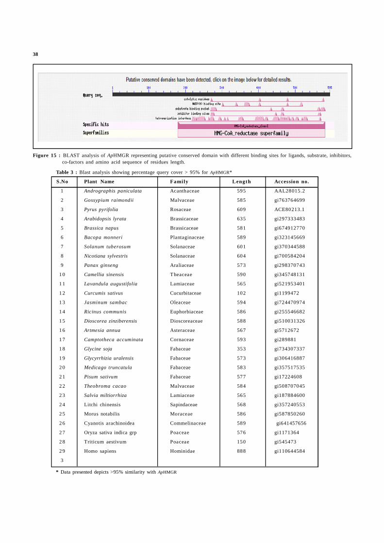
Blast analysis of ApHMGR showed similarity with 420 plant species.Identification of conserved domain was based on plants with querycover greater than 95% (Figure 15, Table 3). It contains 402 aminoacid residues. 97 tetramerization sites, 9 inhibitor binding sites, 18substrate binding sites, 20 NADPH binding sites and 4 catalyticresidues.
The MSA (CLUSTAL W2) analysis of ApHMGR showed 43.03%homology with Homo sapiens and 42.86% homology with Musmusculus HMGR (Figure 16). It indicated that the H. sapiens HMGRwas matching with ApHMGR. But the H. sapiens HMGR is veryless identical (15.3%) compared to Oryza sativa (Os HMGR) inmonocot (Darabi et al., 2012).

37
Figure 14: Ramachandran plot of ApHMGR. The plot shows separate Ramachandran plots for each of the 20 different amino acid types.Numbers of amino acid residues are shown in brackets. Those in unfavourable conformations (Z score<-3.00) are labelled. Greenshading on the background of yellow shows favourable conformations as obtained from an analysis of 163 structures at resolution2.0A or better.
38
Figure 15 : BLAST analysis of ApHMGR representing putative conserved domain with different binding sites for ligands, substrate, inhibitors,co-factors and amino acid sequence of residues length.
Table 3 : Blast analysis showing percentage query cover > 95% for ApHMGR*
S.No Plant Name Fami ly Length Accession no.
1 Andrographis paniculata Acanthaceae 595 AAL28015.2
2 Gossypium raimondii Malvaceae 585 gi763764699
3 Pyrus pyrifolia Rosaceae 609 ACE80213.1
4 Arabidopsis lyrata Brassicaceae 635 gi297333483
5 Brassica napus Brassicaceae 581 gi674912770
6 Bacopa monneri Plantaginaceae 589 gi323145669
7 Solanum tuberosum Solanaceae 601 gi370344588
8 Nicotiana sylvestris Solanaceae 604 gi700584204
9 Panax ginseng Araliaceae 573 gi298370743
1 0 Camellia sinensis Theaceae 590 gi345748131
1 1 Lavandula augustifolia Lamiaceae 565 gi521953401
1 2 Curcumis sativus Cucurbitaceae 102 gi1199472
1 3 Jasminum sambac Oleaceae 594 gi724470974
1 4 Ricinus communis Euphorbiaceae 586 gi255546682
1 5 Dioscorea zinziberensis Dioscoreaceae 588 gi510031326
1 6 Artmesia annua Asteraceae 567 gi5712672
1 7 Camptotheca accuminata Cornaceae 593 gi289881
1 8 Glycine soja Fabaceae 353 gi734307337
1 9 Glycyrrhizia uralensis Fabaceae 573 gi306416887
2 0 Medicago truncatula Fabaceae 583 gi357517535
2 1 Pisum sativum Fabaceae 577 gi17224608
2 2 Theobroma cacao Malvaceae 584 gi508707045
2 3 Salvia miltiorrhiza Lamiaceae 565 gi187884600
2 4 Litchi chinensis Sapindaceae 568 gi357240553
2 5 Morus notabilis Moraceae 586 gi587850260
2 6 Cyanotis arachinoidea Commelinaceae 589 gi641457656
2 7 Oryza sativa indica grp Poaceae 576 gi1171364
2 8 Triticum aestivum Poaceae 150 gi545473
2 9 Homo sapiens Hominidae 888 gi110644584
3
* Data presented depicts >95% similarity with ApHMGR

39
3.2.4 Motifs and domain analysis
A mathematical model is helpful in deriving motif sequences inbiology and is similar to a defined group of organisms. In our study,three conserved motifs were found through MEME tool and aredepicted in Figure 17, Figure 18 and Table 4. Domain prediction ishelpful to find out the evolutionary relationship. Further, domainalignments convey potential information in simpler manner (Fonget al., 2008). The domain structure analysis of ApHMGR usingTMHMM server showed two transmembrane domains at 36 (lys)-58 (lys) and 78 (Ile)-100 (val) positions (Figure 19). Similar resultwas also found with OsHMGR (Darabi et al., 2012).
PHYRE2 analysis revealed that ApHMGR contains threetransmembrane regions, namely S1 (39-57), S2 (95-70) and S3 (553-568). The N-terminal is present towards extracellular side and theC-terminal is present towards cytoplasmic side and was connectedthrough linker sequences (Figure 20). The C-terminal portion of theprotein, which included the catalytic domain, was highly conserved(74 to 98%) across all plant species as well as animal and yeastHMGRs with 65% identity. The linker region is highly divergent inboth size and sequence among all HMGRs in plants and animals.The most striking difference is in the size of the N-terminal domainin plants, which contains the putative membrane-spanning region.This is highly conserved among plant species, which may be areflection of functional attributes (Saur et al., 2015).
Figure16: Multiple sequence alignment (MSA) of amino acid sequences in ApHMGR, HsHMGR and MmHMGR using CLUSTAL W2 programme.
Figure 17 : Common motifs among the different plants and other species.

40
Motif-1 Motif-2
Motif-3
Figure 18: All motifs and their sequences for HMGR proteins in selected plants. The MEME motifs are shown as different-coloured boxes.
(colour figure online generated) and amino acid residues of motif-1, 2 and 3.

41
Table 4: Motifs sequence showing width, e-value and similarity for HMGR*
Motif Width Sequence e-value Simi lar ity
1 2 3
1 3 5 F(Y)P(SF)D(E)MD(EQH)V(I)I(LT)G(SA)I(VL) SG(W)N(K)Y 1.1e-896 - 0.13 0.12(F)CS (T)DKKP(SA)A(TS)AV(I) NWIEGRGKS(T)VVC(F)E(QG)
2 4 1 G(L)G(V)F(YD)N(L)A(M)HA(M)S(AL)N(SH)I(L)VS(T)AI(V)Y 4.6e-958 0.13 - 0.13(F)I(L)AT(C)GQDP(A)AQNV(I)E (G)SSH(QN)CI(L)T(A)M(L)M(LF)EA(PR)V(ISD)N(G)D(PQNKGE)G(TD)
3 5 0 F(S)N(DTK)K(RSQ)S(T)S(I)RFA(G)R(K)LQ(SGRKTN)I(VL) 1.1e-117 0.12 0.13 -Q(KH) C(PVT)A(TS)I(VML)AGK(R)NL(AV)YI(MLVT)R(G)F(YL)C(STRQVN) C(S)S(RG)T(S)G(V)DA(V)MGMNMV(I)SKGV(TA)Q(E)N(K)V(A) L(MI)D(ESL)F(YK)L
*Data presented as per MEME; version 4.10.2
Figure 19 : Graphical representation of transmembrane domain pre-diction in ApHMGR using TMHMM server.
Figure 20 : Structural representation of transmembrane helices inApHMGR using PHYRE2 online tool.
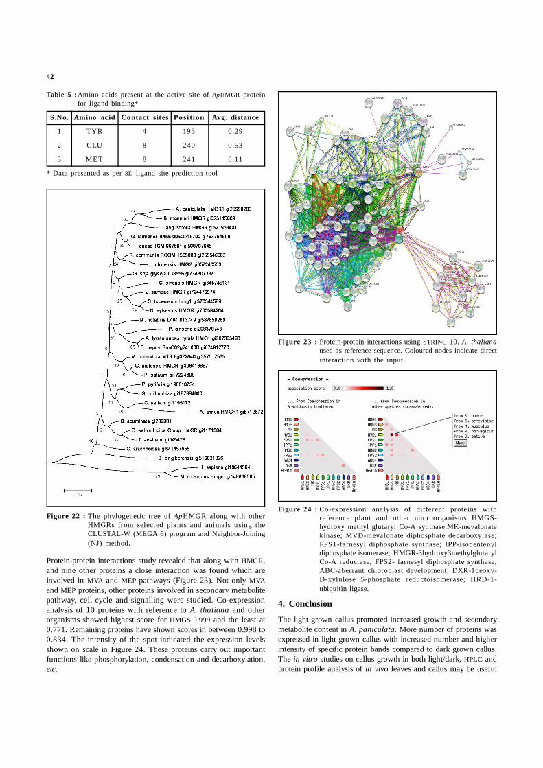
3.2.5 Analysis of ligand binding site, phylogenetic and protein-protein interaction studies
Ligand a small signal-triggering molecule binds to proteins by feebleinteractions. Drug design, drug side effects and elucidation of proteinfunction is facilitated by protein-ligand binding site predictionstudies (Krivák et al., 2015). The ligand binding site of ApHMGRcontains amino acids Tyr (193), Glu (240) and Met (241) at theactive site (Figure 21, Table 5). Two molecules of CoA and sixmolecules of ADP were found as ligands for ApHMGR. These bindto enzyme non-covalently and provide energy to process theenzymatic reaction. The phylogenetic analysis usually comparesthe sequences of proteins and finds out identities among differentorganisms. Phylogenetic tree was constructed by amino acid
sequences of ApHMGR involving 28-plant species with highersimilarity (query cover > 95%). Further, other organisms like H.sapiens and Mus musculus was also analyzed to find out theevolutionary relationship among different organisms. The resultshowed that HMGRs are derived from common ancestor gene ofplants and animals. However, plants are categorized into monocotsand dicots. Phylogenetic analysis revealed that both are closelyrelated evolutionarily with bootstrap 21. In animals, it is againdiverged with bootstrap 99. ApHMGR was closely related to Bacopamonneri HMGR with bootstrap 33 (Figure 22). Further, in ourstudy the monocot group included O. sativa, Triticum aestivum,C arachinoidea and D. zinziberensis. In this group, dicot plantC. accuminata also present with bootstrap 21 indicated closerelationship with monocots. The similar studies were carried out inthe starch-binding domain family CBM41 (Janecek et al., 2017).The biosynthesis of isoprenoids via MVA and MEP pathways iswell studied in plants compared to other organisms. However, theregulation of ApHMGR a key enzyme in MVA pathway still needsattention. Recently, the revised data of Ap chloroplast genome(150,249 bp) provided information regarding MEP pathway genesin chloroplast and is a positive endeavour in this direction (Ding etal., 2015). Likewise the complete chloroplast genome was annotatedin Scrophularia dentate (Mehrotra et al., 2016)
Figure 21: Ligand (green on the background of yellow) binding toApHMGR showing active sites with amino acids TYR, METand GLU at 193, 241 and 240 positions.
42
Table 5 :Amino acids present at the active site of ApHMGR proteinfor ligand binding*
S.No. Amino acid Contact sites Po si tion Avg. distance
1 TYR 4 193 0.29
2 GLU 8 240 0.53
3 MET 8 241 0.11
* Data presented as per 3D ligand site prediction tool
Figure 22 : The phylogenetic tree of ApHMGR along with otherHMGRs from selected plants and animals using theCLUSTAL-W (MEGA 6) program and Neighbor-Joining(NJ) method.
Protein-protein interactions study revealed that along with HMGR,and nine other proteins a close interaction was found which areinvolved in MVA and MEP pathways (Figure 23). Not only MVAand MEP proteins, other proteins involved in secondary metabolitepathway, cell cycle and signalling were studied. Co-expressionanalysis of 10 proteins with reference to A. thaliana and otherorganisms showed highest score for HMGS 0.999 and the least at0.771. Remaining proteins have shown scores in between 0.998 to0.834. The intensity of the spot indicated the expression levelsshown on scale in Figure 24. These proteins carry out importantfunctions like phosphorylation, condensation and decarboxylation,etc.
Figure 23 : Protein-protein interactions using STRING 10. A. thalianaused as reference sequence. Coloured nodes indicate directinteraction with the input.
Figure 24 : Co-expression analysis of different proteins withreference plant and other microorganisms HMGS-hydroxy methyl glutaryl Co-A synthase;MK-mevalonatekinase; MVD-mevalonate diphosphate decarboxylase;FPS1-farnesyl diphosphate synthase; IPP-isopentenyldiphosphate isomerase; HMGR-3hydroxy3methylglutarylCo-A reductase; FPS2- farnesyl diphosphate synthase;ABC-aberrant chloroplast development; DXR-1deoxy-D-xylulose 5-phosphate reductoisomerase; HRD-1-ubiquitin ligase.
4. Conclusion
The light grown callus promoted increased growth and secondarymetabolite content in A. paniculata. More number of proteins wasexpressed in light grown callus with increased number and higherintensity of specific protein bands compared to dark grown callus.The in vitro studies on callus growth in both light/dark, HPLC andprotein profile analysis of in vivo leaves and callus may be useful

43
to understand the possible role of light (MEP pathway inchloroplast) on andrographolide synthesis in A. paniculata. Thisstudy may help to find the genes and proteins responsible for theproduction of bioactive compounds such as andrographolide.
ApHMGR structural analysis in the present study will be useful infunctional prediction, regulation and location of specific proteinsacross organisms. Functional aspects of ApHMGR can find out theligand binding sites and active site information, for their use indocking studies. The phylogenetic analysis revealed the genestructure and protein architecture of all plant HMGRs wereconsiderably conserved. This is the first report describing thestructural, physicochemical and functional information of ApHMGR,which will provide a good foundation and can be exploited forprimer designing and isolation of genes from this important medicinalplant. The protein-protein interactions will be helpful to understandcell-cell interactions for the elucidation of biological mechanisms atthe molecular level and drug-target relationships. These findingsmay also lead to the possibilities of metabolic engineering ofisoprenoids, for increased synthesis of diterpene lactoneandrographolide there by overcoming metabolic bottlenecks.
Acknowledgements
Authors would like to thank OU-UGC-CPEPA programme sponsoredby University Grants Commission (UGC), New Delhi for financialsupport. Ms. B. B. V. Bindu, Mr. M. Srinath and Ms. A. Shailajathank UGC, New Delhi for Research Fellowships.
Conflict of interest
We declare that we have no conflict of interest.
References
Abd El-Aal, S.M.; Rabie, K.A.E. and Manaf, H.H. (2016). The effect of UV-Con secondary metabolites production of Echinacea purpureaculture in vitro. J. Biol. Chem. Environ. Sci., 2:465-483.
Alexander, R.A.; Sankari, S.; Ramaraj, K. and Rajaram, S.K. (2016). In silicoanalysis and molecular interactions studies of selectedphytoconstituents from Andrographis paniculata as potentialinhibitors of monoamine oxidase b. Asian J. Pharm. Clinical Res.,2:239-242.
Arolla, R.G.; Cherukupalli, N.; Rao, K.V. and Reddy, V.D. (2015). DNA barcodingand haplotyping in different species of Andrographis. Biochem.Syst. Ecol., 62:91-97.
Bailey, T.L.; Williams, N.; Misleh, C. and Li, W.W. (2006). MEME: Discoveringand analyzing DNA and protein sequence motifs. Nucleic AcidsRes., 34:369-373. MEME; version 4.10.2) at website (http://meme.nbcr.net/meme/meme.html)
Bhagavathi, S.; Prakash, A. and Gulshan, W. (2014). An insight to virtualligand screening methods for structure-based drug design andmethods to predict protein structure and function in lung cancer:approaches and progress. J. Crit. Rev., 1:10-24.
Bradford, M.M. (1976). A rapid and sensitive method for the quantitationof microgram quantities of protein utilizing the principle of protein-dye binding. Anal. Biochem., 72: 248-254.
Chou, P.Y. and Fasman, G.D. (1974). Prediction of proteininformation. Biochem., 13:222-245. CFSSP prediction server(http://cib.cf.ocha.ac.jp/bitool/MIX/)
Darabi, M.; Izadi-Darbandi, A.; Masoudi N.A. and Nemat, Z.G. (2012).Bioinformatics study of the 3-hydroxy-3-methylglotaryl-coenzyme A reductase (HMGR) gene in Gramineae. Mol. Biol.Rep., 39:8925-35.
Daryush, T.; Alireza, V.; Mohd Y.R. and Mahmood, M. (2014). Proteomic analysisof the salt-responsive leaf and root proteins in the anticancerplant, Andrographis paniculata nees. PLoS One., 9:1-10.
Dewald, I.; Isakin, O.; Schubert, J.; Kraus, T. and Chanana, M. (2015). Proteinidentity and environmental parameters determine the finalphysicochemical properties of protein-coated metal nanoparticles.J. Phys. ChemC., 119:25482-25492.
Dias, M.I.; Sousa, M.J.; Alves, R.C. and Ferreira, I.C. (2016). Exploring planttissue culture to improve the production of phenolic compounds:A review. Indust. Crops Prod., 82:9-22.
Ding, P.; Shao, Y.; Li, Q.; Gao, J. Zhang, R.; Lai, X. and Zhang, H. (2015). Thecomplete chloroplast genome sequence of the medicinal plantAndrographis paniculata. Mitochond. DNA, 4:1-2.
Dor, O.; Zhou, Y. and Zhou. (2006). Achieving 80% tenfold cross-validatedaccuracy for secondary structure prediction by large-scaletraining. Proteins, 66:838-845.
Elena, A.; Günter and Oxana V.P. (2016). Calcium pectinate gel beads obtainedfrom callus cultures pectins as promising systems for colon-targeteddrug delivery. Carbohydr. Polym., 147:490-499.
Fazal, H.; Abbasi, B.H.; Ahmad, N.; Ali, M. and Ali, S. (2016). Sucrose inducedosmotic stress and photoperiod regimes enhanced the biomass andproduction of antioxidant secondary metabolites in shake-flasksuspension cultures of Prunella vulgaris L. Plant Cell Tiss. OrganCult., 3:573-581.
Fong, J.H. and Marchler, B.A. (2008). Protein subfamily assignment usingthe Conserved Domain Database. BMC. Res. Notes, 1:114-120.
Fung, P.K.; Krushkal, J and Weathers, P.J. (2010). Computational analysis ofthe evolution of 1 deoxy d xylulose 5 phosphate reductoisomerase,an important enzyme in plant terpene biosynthesis. Chem.Biodivers., 7:1098-1110.
Giri, C.C. and Zaheer, M. (2016). Chemical elicitors versus secondarymetabolite production in vitro using plant cell, tissue and organcultures: Recent trends and a sky eye view appraisal. Plant CellTiss. Organ Cult., 126:1-18.
Guo, D.; Yi, H.Y.; Li, H.L.; Liu, C.; Yang, Z.P. and Peng, S.Q. (2015). Molecularcharacterization of HbCZF1, a Hevea brasiliensis CCCH-type zincfinger protein that regulates hmg1. Plant Cell Tiss. Organ Cult.,34:1569-1578.
Hemmati, S. (2016). Predicting the functionality of major intrinsicproteins: An in silico analysis in Musa. Trends Phram. Sci., 2:139-150.
Hoagland, D.R. and Arnon, D.I. (1938) The water culture method for growingplants without soil. California Agri. Exp. Station Circul., 347: 32
Janecek, S.; Majzlová, K.; Svensson, B. and MacGregor, E.A. (2017). The starch-binding domain family CBM41-an in silico analysis of evolutionaryrelationships Proteins (Online First).
Jha, Z.; Sharam, S.N. and Sharma, D.K. (2011). Differential expression of 3-hydroxy- 3-methylglutaryl-coenzyme A reductase of Andrographispaniculata in andrographolide accumulation. J. Chem. Pharm. Res.,3:499-504.
Kalita, R.; Patar, L.; Shasany, A.K.; Modi, M.K. and Sen, P. (2015). Molecularcloning, characterization and expression analysis of 3-hydroxy-3-methylglutaryl coenzyme A reductase gene from Centella asiaticaL. Mol. Biol. Rep., 42:1431-1439.
Kelley, L.A.; Mezulis, S.; Yates, C.M.; Wass, M.N. and Sternberg, M.J. (2015). ThePhyre2 web portal for protein modeling, prediction and analysis.Nat. Protoc., 10:845-58. Phyre2 An online tool (http://www.sbg.bio.ic.ac.uk/phyre2/html/page.cgi?id=index
Kiefer, F.; Arnold, K.; Künzli, M.; Bordoli, L. and Schwede, T. (2009). The SWISS-MODEL repository and associated resources. Nucleic Acids Res.,37:387-392. SWISS-MODEL server (http://swissmodel.expasy.org/interactive/BYrhz7/models/)

44
Krivák, R. and Hoksza, D. (2015). Improving protein-ligand binding siteprediction accuracy by classification of inner pocket points usinglocal features. J. Cheminformatics, 1:1-25.
Krogh, A.; Larsson, B; Von Heijne, G. and Sonnhammer, E.L. (2001). Predictingtransmembrane protein topology with a hidden Markov model:Application to complete genomes. J. Mol. Biol., 3:567-580.
Kumar, P.S. and Kumari R.B.D. (2009). In vitro and in vivo identification ofvariation in protein expression in Artemisia vulgaris L. Adv. Biol.Res., 5-6:63-70.
Kumar, S. and Gadagkar, S.R. (2000). Efficiency of the neighbor-joiningmethod in reconstructing deep and shallow evolutionaryrelationships in large phylogenies. J. Mol. Evol., 51:544-553.
Kyte, J. and Doolittle, R.F. (1982). A simple method for displaying thehydropathic character of a protein. J. Mol. Biol., 1:105-132.Expasy tools website (http://web.expasy.org/cgi-bin/compute_pi/pi_tool), (http://web.expasy.org/protscale/).
Laemmli, U.K. (1970). Cleavage of structural proteins during assemblyof the head of bacteriophage T4. Nature, 227:680-685.
Laskowski, R.A.; Rullmannn, J.A.; MacArthur, M.W.; Kaptein, R. and Thornton,J.M. (1996). AQUA and PROCHECK-NMR: programs for checking thequality of protein structures solved by NMR. J. Biomol. NMR, 4:477-486. PROCHECK (http://services.mbi.ucla.edu/SAVES/)
Latha, B.C.; Ahalya,.S.; Naidu, P.D.; Mounica, K. and Kumar, A.R. (2017).Phytochemical evaluation of Andrographis paniculata , Cassiaangustifolia and Eclipta alba. Ind. J. Res. Pharm. Biotechnol.,5:160-163
Lipko, A. and Swiezewska, E. (2016). Isoprenoid generating systems in plants-a handy toolbox how to assess contribution of the mevalonate andmethylerythritol phosphate pathways to the biosynthetic process.Prog. Lipid Res., 63:70-92
Mehrotra, P.; Ramakrishnan, G.; Dhandapani, G.; Srinivasan, N. and Madanan,M.G. (2016). The Complete Chloroplast Genome of Ye-Xing-Ba(Scrophularia dentata; Scrophulariaceae), an Alpine Tibetan HerbMol. Biosyst., 13:883-891
Mohapatra, H.P. and Rath, S.P. (2005) In vitro studies of Bacopa monnieri-an important medicinal plant with reference to its biochemicalvariations. Ind. J. Exp. Biol., 4:373-376.
Murashige, T. and Skoog, F. (1962). A revised medium for rapid growth andbioassays with tobacco tissue cultures. Physiol. Plant., 15:473-497.
Neeraja, C.; Krishna, P.H.; Reddy, C.S.; Giri, C.C.; Rao, K.V. and Reddy, V.D.(2015). Distribution of Andrographis species in different districtsof Andhra Pradesh. Proc. Natl. Acad. Sci. India Sect. B. Biol. Sci.,85:601-606.
Neeraja, C.; Mayur, D.; Mittapelli, S.R.; Rao, K.V. and Reddy, V.D. (2016). Denovo assembly of leaf transcriptome in the medicinal plantAndrographis paniculata. Front. Plant Sci., 7:1-13.
Niranjan, A.; Tewari, S.K. and Lehri, A. (2010). Biological activities ofKalmegh (Andrographis paniculata Nees) and its active principles:A review. Ind. J. Natl. Prod. Resources, 2:125-135.
Uozumi, N.; Nakamura, T.; Schroeder, J.I. and Muto, S. (1997). Determinationof transmembrane topology of an inward-rectifying potassiumchannel from Arabidopsis thaliana based on functional expressionin Escherichia coli. Proc. Natl. Acad. Sci., 95:9773-9778.
Parlapally, S.; Cherukupalli, N.; Bhumireddy, S.R.; Sripadi, P.; Anisetti, R.; Giri,C.C. and Reddy, V.D. (2015). Chemical profiling and anti-psoriaticactivity of methanolic extract of Andrographis nallamalayanaJ.L. Ellies. Nat. Prod. Res., 30:1256-1261.
Prasada, R.K.; Sharmaa, R. and Prajapati, G.L. (2010). Homology modelingand evaluation of human TEK tyrosine kinase using SWISS-MODELworkspace. J. Chem. Pharm. Res., 2:440-451.
Proctor, E.A. and Dokholyan, N.V. (2016). Applications of discrete moleculardynamics in biology and medicine. Curr. Opin. Struct. Biol., 37:9-13.
Radhakrishnan, R. and Ranjitha, K.B.D. (2009). Changes in protein contentin micropropagated and conventional soybean plants (Glycinemax (L.) Merr.). World J. Agric. Sci., 2:186-189.
Rout, J.R.; Kanungo, S.; Das, R. and Sahoo, S.L. (2010). In vivo proteinprofiling and catalase activity of Plumbago zeylanica L. Nat. Sci.,1:87-90.
Saitou, N. and Nei, M. (1987). The neighbor-joining method: a new methodfor reconstructing phylogenetic trees. Mol. Biol. Evol., 4:406-425.
Sanchita, S.S. and Sharma, A. (2014). Bioinformatics approaches forstructural and functional analysis of proteins in secondarymetabolism in Withania somnifera. Mol. Biol. Rep., 41:7323-7330.
Sareer O, Ahmad S, Umar S (2014). Andrographis paniculata: A criticalappraisal of extraction, isolation and quantification of androgra-pholide and other active constituents. Nat. Prod. Res., 23:2081-2101.
Sarkar, S.; Gupta, S.; Chakraborty, W.; Senapati, S. and Gachhui, R. (2017).Homology modeling, molecular docking and molecular dynamicsstudies of the catalytic domain of chitin deacetylase fromCryptococcus laurentii strain RY1. Int. J. Biol. Macromol. (OnlineFirst).
Saur, I.M.L.; Conlan, B.F and Rathjen, J.P. (2015). The N-Terminal domainof the tomato immune protein Prf contains multiple homotypicand Pto kinase interaction sites. J. Biol. Chem., 290:11258-11267.
Sharma, A. and Batra, A. (2015). In vivo and in vitro protein profiling inTinospora cordifolia-an antidiabetic plant species. Anal. Int. DailyJ. Species, 51:88-96.
Singh, S. and Srivastava, A.K. (2016). In silico and wet lab study revealedcadmium is the potent inhibitor of HupL in Anabaena sp. PC C7120. Arch. Microbiol., 198:27-34.
Siqueira, A.S.; Lima, A.R.J.; DallAgnol, L.T.; de Azevedo, J.S.N.; VianezmJ.L.D.S.G. and Gonçalves, E.C. (2016). Comparative modeling andmolecular dynamics suggest high carboxylase activity of theCyanobium sp. CACIAM14 RbcL protein. J. Mol. Model., 3:1-8.
Srivastava, N. and Akhila, A. (2010). Biosynthesis of andrographolide inAndrographis paniculata . Phytochem., 71:1298-1304.
Subramanian, R.; Asmawi, M.Z. and Sadikun, A. (2012). A bitter plant with asweet future? A comprehensive review of an oriental medicinalplant: Andrographis paniculata . Phytochem. Rev., 11:39-75.
Suriyo, T.; Pholphana, N.; Ungtrakul, T.; Rangkadilok, N.; Panomvana, D.; Thiantanawat, A.; Pongpun, W. And Satayavivad, J. (2017). ClinicalParameters following multiple oral dose administration of astandardized Andrographis paniculata capsule in healthy Thaisubjects. Planta Med. (Online first).
Szklarczyk, D.; Franceschini, A.; Wyder, S.; Forslund, K.; Heller, D.; Huerta-Cepas, J.; Simonovic, M.; Roth, A.; Santos, A.; Tsafou, K.P.; Kuhn, M.; Bork, P.; Jensen, L.J. and von Mering, C. (2015). STRING v10: protein-protein interaction networks, integrated over the tree of life.Nucleic Acids Res., 43:447-452.
Tamura, K.; Stecher, G.; Peterson, D.; Filipski, A. and Kumar, S. (2013). MEGA6:Molecular Evolutionary Genetics Analysis Version 6.0. Mol. Biol.Evol., 30:2725-2729.
Wass, M.N.; Kelley, L.A. and Sternberg, M.J. (2010). 3DLigandSite: predictingligand-binding sites using similar structures. Nucleic AcidsRes., 38:469-73. Three dimensional (3D) ligand site (http://www.sbg.bio.ic.ac.uk/3dligandsite/)
Xiang, Z. (2006) Advances in homology protein structure modeling. Curr.Protein Pept. Sci., 7:217-227.
Zaheer, M. and Giri, C.C. (2015). Multiple shoot induction and jasmonicversus salicylic acid driven elicitation for enhanced andrographolideproduction in Andrographis paniculata . Plant Cell Tiss. OrganCult., 122:553-563.
Zaheer, M. and Giri, C.C (2017) Enhanced diterpene lactone (androgra-pholide) production from elicited adventitious root cultures ofAndrographis paniculata. Res. Chem. Intermed., 43:2433-2444 .


![Nanomagnetic double-charged diazoniabicyclo[2.2.2]octane](https://img.pdfslide.us/doc/110x75/61d93679997f9e51ce0e3333/nanomagnetic-double-charged-diazoniabicyclo222octane-.jpg)