Embed Size (px)
Citation preview
EXCLI Journal 2016;15:772-780 – ISSN 1611-2156 Received: October 16, 2016, accepted: November 04, 2016, published: December 06, 2016
772
Original article:
BROMELAIN CAPPED GOLD NANOPARTICLES AS THE NOVEL DRUG DELIVERY CARRIERS TO AGGRANDIZE EFFECT OF THE
ANTIBIOTIC LEVOFLOXACIN Paramdeep Bagga1, Tarique Mahmood Ansari1, Hefazat Hussain Siddiqui1, Asad Syed3, Ali H. Bahkali3, Md. Azizur Rahman1, Mohd. Sajid Khan2* 1 Department of Pharmacy, Integral University, Lucknow, Uttar Pradesh 226026 (India) 2 Nanotechnology Lab, Department of Biosciences, Integral University, Lucknow,
Uttar Pradesh 226026 (India) 3 Botany and Microbiology Department, Faculty of Science, King Saud University, Riyadh,
Saudi Arabia * Corresponding author: Mohd. Sajid Khan, Nanotechnology Lab, Department of Biosciences,
Integral University, Lucknow, Uttar Pradesh 226026 (India), [email protected] http://dx.doi.org/10.17179/excli2016-710
This is an Open Access article distributed under the terms of the Creative Commons Attribution License (http://creativecommons.org/licenses/by/4.0/).
ABSTRACT
To develop bromelain capped gold nanoparticles (BRN capped Au-NPs) as the effective drug delivery carriers of the antibiotic levofloxacin (LvN) and evaluate antibacterial potential of its bioconjugated form compared to pure LvN. BRN capped Au-NPs were synthesized by in vitro method and bioconjugated to LvN using 1-ethyl-3-(3-dimethylamino-propyl)-carbodiimide as activator to form Au-BRN-LvN-NPs. These were characterized for mean particle size by dynamic light scattering analysis, zeta potential by Zetasizer nanosystem analysis and transmission electron microscopy (TEM) on carbon coated TEM copper grids by TEM respectively. Drug load-ing efficiency of LvN was calculated using UV-visible spectroscopy by standard curve of pure LvN. Antibacte-rial efficacy of Au-BRN-LvN-NPs and pure LvN was determined by evaluating minimum inhibitory concentra-tion (MIC) against Staphylococcus aureus and Eschereschia coli. Two peaks were observed in Au-BRN-LvN-NPs spectrum one at 307 nm and other at 526 nm while one peak in BRN capped Au-NPs at 522 nm during UV spectroscopy suggesting red shift. The drug loading efficiency of LvN was found to be 84.8 ± 2.41 %. The diam-eter of Au-BRN-LvN-NPs and BRN capped Au-NPs were found to be (58.65 ± 2 nm, 38.11 ± 2 nm), zeta poten-tial (-9.01 mV, -13.8 mV) and surface morphology (~13.2 nm, 11.4 nm) respectively. The MICs against S. aure-us and E. coli were found to be (0.128 µg/mL, 1.10 µg/mL) for Au-BRN-LvN-NPs and (0.547 µg/mL, 1.96 µg/mL) for pure LvN. The results suggested that BRN capped Au-NPs can be used as effective drug delivery carriers of the antibiotic LvN. The Au-BRN-LvN-NPs exhibited enhanced antibacterial activity compared to pure LvN alone. (Graphical abstract see Figure 1) Keywords: Antibacterial activity, bromelain, gold nanoparticles, levofloxacin, novel drug delivery carriers
Figure 1: Graphical abstract
EXCLI Journal 2016;15:772-780 – ISSN 1611-2156 Received: October 16, 2016, accepted: November 04, 2016, published: December 06, 2016
773
INTRODUCTION
The metallic nanoparticles derived from noble metals such as gold has attracted the researchers all over the world regarding their escalating use in delivery of drugs (Emerich and Thanos, 2006). These particles build up new physico-chemical properties which are absent in individual molecules. Specially, the gold nanoparticles are being targeted nowa-days by the researchers since it not only en-compasses the advantages of “nano-sizing” but it also includes surface functionalization necessary to augment stability and enable conjugation with biomolecules (Couto et al., 2016). Surface functionalization of the gold nanoparticles has opened new frontiers for delivery of drugs such as anticancer and the antimicrobial drugs (Dhar et al., 2008, Selva-raj and Alagar, 2007). Bovine serum albumin capped gold nanoparticles of amino-glycosidic antibiotics and the acridine deriv-atives bioconjugated on citrate stabilized gold nanoparticles have been used as the ef-fective drug delivery vehicles (Rastogi et al., 2012, Mitra et al, 2014). Thus, these exciting approaches have motivated the search for novel molecules for the synthesis of gold na-noparticles.
Levofloxacin (LvN) which is chemically (S)-9-fluoro-2,3-dihydro-3-methyl-10-(4-me-thylpiperazin-1-yl)-7-oxo-7H-pyrido[1,2,3-de]-1,4-benzoxazine-6-carboxylic acid, is a broad spectrum fluoroquinolone antibiotic active against both Gram positive and Gram negative bacteria functioning by inhibiting the type II topoisomerase enzymes namely DNA gyrase and topoisomerase IV (Drlica and Zhao, 1997). It is recommended as a first-line treatment option by ‘Infectious Disease Society of America’ for catheter as-sociated urinary tract infections in adults and in prosthetic joint infection in combination with rifampicin (Hooton et al., 2010; Osmon et al., 2013).
Bromelain (BRN) is a proteolytic en-zyme derived from the plant Ananas como-sus belonging to family Bromeliaceae (Taussig and Batkin, 1988). It has shown an-algesic, anti-inflammatory, anti-arthritis, an-
titumuor, anthelminthic, antimicrobial and several other pharmacological activities (Leipner et al, 2001; Beez et al, 2007; Ro-wan et al., 1990; Praveen et al., 2014). It was found to be less toxic having mutagenic po-tential (Castell et al., 1997; Chobotova et al., 2010). Hence, the present study was aimed to develop bromelain capped gold nanoparti-cles as the effective drug delivery carrier of the antibiotic levofloxacin and to evaluate the antibacterial potential of its bioconjugat-ed form as compared to pure LvN alone.
MATERIALS AND METHODS
Chemicals and reagents All the chemicals and solvents used in
the present investigation were of analytical grade. Levofloxacin was procured from Yar-row Chem Products, Mumbai (India) and bromelain from Merck Chemicals Darm-stadt, Germany.
Synthesis of gold nanoparticles
Gold nanoparticles (Au-NPs) were syn-thesized by in vitro method. The 3 μl of 1.0 mM H[AuCl4] prepared in 50 mM phos-phate buffer was taken in 3 mL of freshly prepared bromelain (0.33 mg/mL) and incu-bated at the temperature of 40 °C for 48 h in an incubator. The reaction mixture without bromelain was used as a control. To ensure the formation of nanoparticles, the samples were removed from the reaction mixture at regular intervals and analyzed by UV-visible spectroscopy. On completion of the reaction, Au-NPs were collected by centrifugation at 30000 g for 30 min, washed twice with Mil-liQ water and the excess bromelain was re-moved by treating with 50 % ethanol (v/v) (Khan et al., 2015a).
Bioconjugation of gold nanoparticles to levofloxacin
Synthesized Au-NPs were bioconjugated to LvN by using 1-ethyl-3-(3-dimethyl-amino-propyl)-carbodiimide (EDC) as the activator (Timkovich, 1977). The 5 mM EDC was added to the 5 ml reaction mixture containing 250 µg of LvN, 250 µg of Au-
EXCLI Journal 2016;15:772-780 – ISSN 1611-2156 Received: October 16, 2016, accepted: November 04, 2016, published: December 06, 2016
774
NPs and 50 mM MES/HEPES buffer (HiMedia laboratories, India) in aliquots within 3 h at 30 °C for the coupling process. The bioconjugates so formed were separated from unconjugated Au-NPs by passing the reaction mixture through Biogel P-30 gel fil-tration column pre-equilibrated with 20 mM HEPES buffer (pH 6.0) containing 150 mM sodium chloride. The fractions were scanned between 200-900 nm and subsequently pooled. The pooled samples were then dia-lyzed against distilled water and used for fur-ther characterization (Khan et al., 2015b). The samples were removed at regular inter-vals and analyzed in UV-visible spectrosco-py (Shimadzu dual-beam spectrophotometer, model UV-1601 PC, Japan) at a resolution of 1 nm to confirm the formation of ‘BRN capped Au-NPs’ and ‘levofloxacin conjugat-ed with BRN capped Au-NPs’(Au-BRN-LvN-NPs).
Characterization of synthesized gold nano-particles and bioconjugated gold nanoparti-cles
Measurement of mean particle size and zeta potential of ‘BRN capped Au-NPs’ and Au-BRN-LvN-NPs
The samples of ‘BRN capped Au-NPs’ and Au-BRN-LvN-NPs were sonicated sepa-rately for 1 min and then, taken in a DTS0112-low volume disposable sizing cu-vette of 1.5 mL for the measurement of mean particle size with a dynamic light scattering (DLS) particle size analyzer (Zetasizer Nano-ZS, model ZEN3600, Malvern Instru-ment Ltd, Malvern, UK) (Akhtar et al., 2014). Zeta potential values were also as-sessed to check the stability of the gold na-noparticles with a Malvern Zetasizer Nanosystem (Worcestershire, UK).
Transmission electron microscopy of ‘BRN capped Au-NPs’ and Au-BRN-LvN-NPs
Transmission electron microscopy (TEM) was analyzed by drop drying the so-lution of biosynthesized ‘BRN capped Au-NPs’ and Au-BRN-LvN-NPs on carbon coated TEM copper grids followed by meas-
urements on TEM (FEI Company, Tecnaiä G2 Spirit BioTWIN) at an accelerating volt-age of 80 kV.
Fourier transform infrared spectroscopy of ‘BRN capped Au-NPs’ and Au-BRN-LvN-NPs
The Fourier transform infrared (FTIR) spectra of ‘BRN capped Au-NPs’ and Au-BRN-LvN-NPs were recorded using Perkin-Elmer Spectrum Two FT-IR (Perkin Elmer Inc., Tres Cantos, Madrid) equipped with a Universal attenuated total reflectance sam-pling device and scanned at room tempera-ture in transmission mode over the wave number range of 4000-650 cm-1 at a resolu-tion of 4 cm-1.
Drug loading efficiency of levofloxacin Drug loading efficiency of LvN was cal-
culated using UV-visible spectroscopy at the wavelength of 307 nm/526 nm where the standard curve of the pure drug LvN was es-tablished and the amount of unbound LvN was calculated from it. The actual amount of bioconjugated LvN was calculated by sub-tracting unbound LvN from the total amount of LvN added. The amount of bioconjugated LvN was calculated using the equation [ % bioconjugation = (amount of drug conjugat-ed / total drug added) × 100].
Evaluation of antibacterial efficacy of bio-conjugated levofloxacin, Au-BRN-LvN-NPs over pure levofloxacin alone
The antibacterial efficacy of bioconju-gated LvN (Au-BRN-LvN-NPs) was deter-mined by evaluating the minimum inhibitory concentration (MIC) of Au-BRN-LvN-NPs and pure LvN against Gram positive bacteria Staphylococcus aureus (NCIM No. 2079) and Gram negative bacteria Eschereschia coli (NCIM No. 2065) (Amsterdam, 1996). The bacterial strains in mid logarithmic phase were harvested by centrifugation, washed with 10 mM sodium phosphate buff-er (PB) of pH 7.4, and diluted to 2×10⁵ col-ony forming units per ml (CFU/mL) in PB containing 0.03 % Luria-Bertani (LB) broth. Au-BRN-LvN-NPs were serially diluted in
EXCLI Journal 2016;15:772-780 – ISSN 1611-2156 Received: October 16, 2016, accepted: November 04, 2016, published: December 06, 2016
775
50 µL of LB medium in 96-well microtitre plates so as to achieve the desired concentra-tions with bacterial inoculums (5×10⁴ CFU/well) and were incubated at 37 °C overnight. The MIC was taken as the lowest Au-BRN-LvN-NPs concentration at which growth was inhibited (Khan et al., 2008). For agar plate count method (Stenger et al., 1998), 25 µL aliquot of bacteria (1×10⁵ CFU/mL) was incubated with 25 µL of sam-ples at 37 °C for 2 h (Hamamoto et al., 2002). The mixtures were 10-fold serially di-luted in PB, plated on LB agar and then in-cubated overnight at 37 °C. Bacterial colo-nies were enumerated following the day. Af-ter the determination of the MIC from the microtitre plate wells with no visible growth, the samples were removed for serial subcul-tivation (2 µL) into microtitre plates contain-ing 100 µL of broth/well and further incu-bated for 24 h in order to determine the bac-tericidal activity measurement (MBC), the lowest concentration at which there is no vis-ible bacterial growth indicating 99.5 % kill-ing of the original inoculums. The absorb-ance of each well was measured at a wave-length of 620 nm by microtitre plate reader (Bio-Rad laboratories Inc., India) and com-pared with a control. Autoclaved water and ‘BRN capped Au-NPs’ were used as a nega-tive control for each experiment. The proce-
dure was repeated for the pure levofloxacin alone too.
RESULTS
Bioconjugation of gold nanoparticles to levofloxacin and drug loading efficiency of levofloxacin
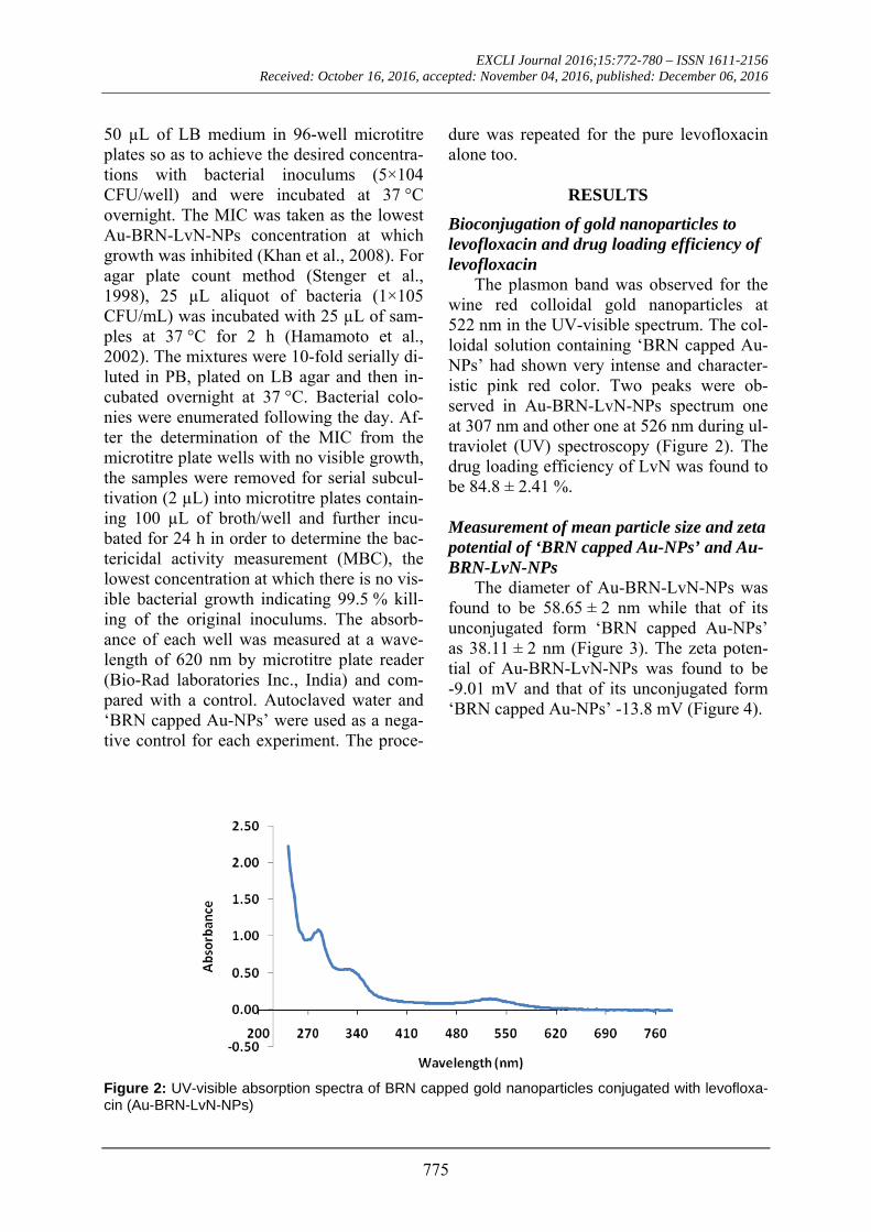
The plasmon band was observed for the wine red colloidal gold nanoparticles at 522 nm in the UV-visible spectrum. The col-loidal solution containing ‘BRN capped Au-NPs’ had shown very intense and character-istic pink red color. Two peaks were ob-served in Au-BRN-LvN-NPs spectrum one at 307 nm and other one at 526 nm during ul-traviolet (UV) spectroscopy (Figure 2). The drug loading efficiency of LvN was found to be 84.8 ± 2.41 %.
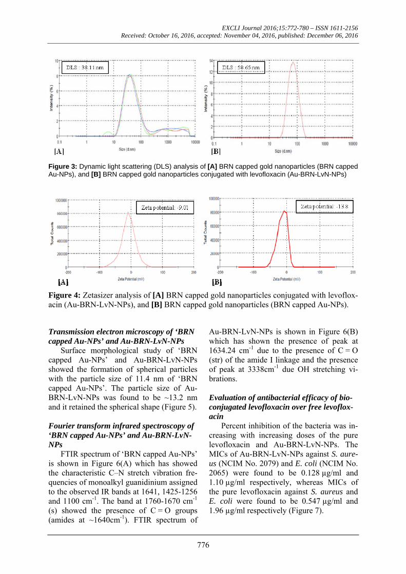
Measurement of mean particle size and zeta potential of ‘BRN capped Au-NPs’ and Au-BRN-LvN-NPs
The diameter of Au-BRN-LvN-NPs was found to be 58.65 ± 2 nm while that of its unconjugated form ‘BRN capped Au-NPs’ as 38.11 ± 2 nm (Figure 3). The zeta poten-tial of Au-BRN-LvN-NPs was found to be -9.01 mV and that of its unconjugated form ‘BRN capped Au-NPs’ -13.8 mV (Figure 4).
Figure 2: UV-visible absorption spectra of BRN capped gold nanoparticles conjugated with levofloxa-cin (Au-BRN-LvN-NPs)
EXCLI Journal 2016;15:772-780 – ISSN 1611-2156 Received: October 16, 2016, accepted: November 04, 2016, published: December 06, 2016
776
Figure 3: Dynamic light scattering (DLS) analysis of [A] BRN capped gold nanoparticles (BRN capped Au-NPs), and [B] BRN capped gold nanoparticles conjugated with levofloxacin (Au-BRN-LvN-NPs)
Figure 4: Zetasizer analysis of [A] BRN capped gold nanoparticles conjugated with levoflox-acin (Au-BRN-LvN-NPs), and [B] BRN capped gold nanoparticles (BRN capped Au-NPs).
Transmission electron microscopy of ‘BRN capped Au-NPs’ and Au-BRN-LvN-NPs
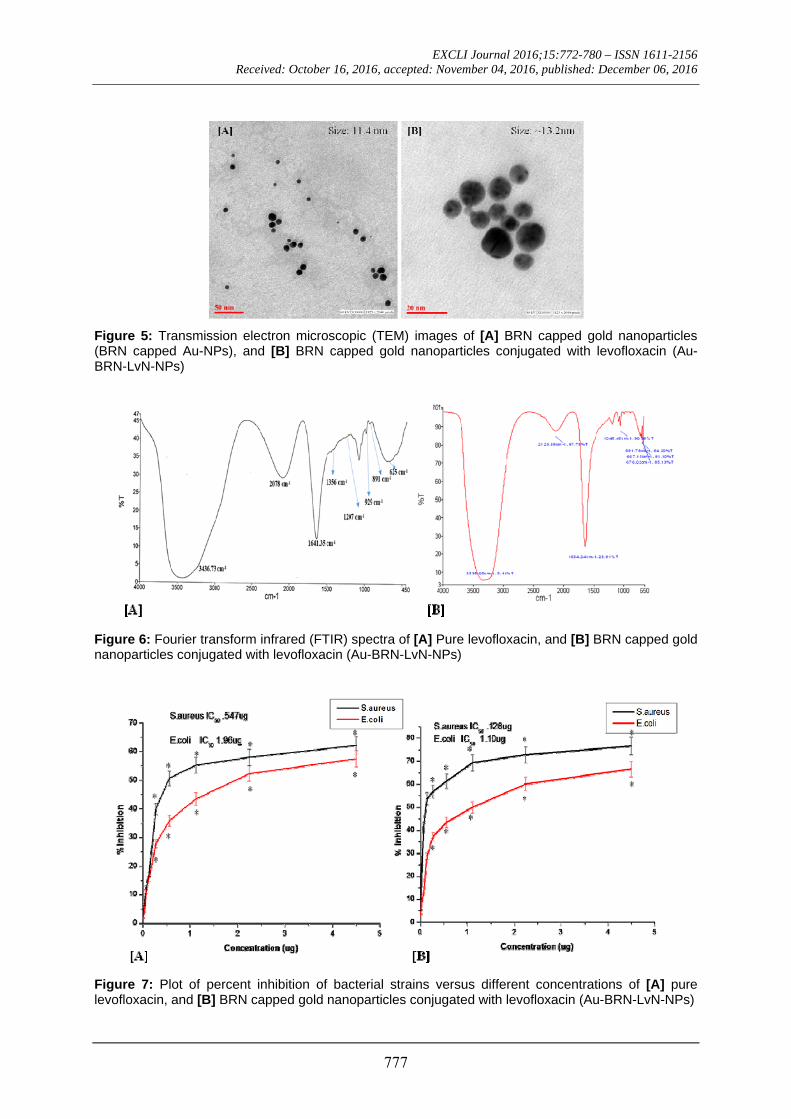
Surface morphological study of ‘BRN capped Au-NPs’ and Au-BRN-LvN-NPs showed the formation of spherical particles with the particle size of 11.4 nm of ‘BRN capped Au-NPs’. The particle size of Au-BRN-LvN-NPs was found to be ~13.2 nm and it retained the spherical shape (Figure 5).
Fourier transform infrared spectroscopy of ‘BRN capped Au-NPs’ and Au-BRN-LvN-NPs
FTIR spectrum of ‘BRN capped Au-NPs’ is shown in Figure 6(A) which has showed the characteristic C–N stretch vibration fre-quencies of monoalkyl guanidinium assigned to the observed IR bands at 1641, 1425-1256 and 1100 cm-1. The band at 1760-1670 cm-1 (s) showed the presence of C = O groups (amides at ~1640cm-1). FTIR spectrum of
Au-BRN-LvN-NPs is shown in Figure 6(B) which has shown the presence of peak at 1634.24 cm-1 due to the presence of C = O (str) of the amide I linkage and the presence of peak at 3338cm-1 due OH stretching vi-brations.
Evaluation of antibacterial efficacy of bio-conjugated levofloxacin over free levoflox-acin
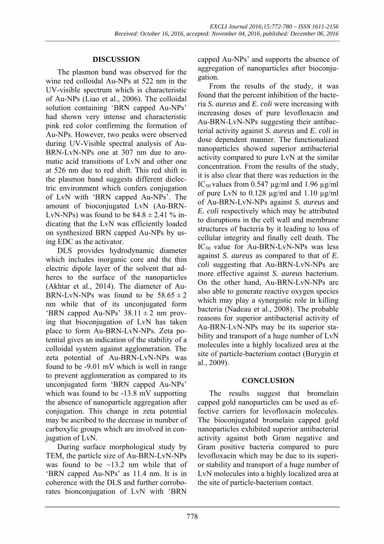
Percent inhibition of the bacteria was in-creasing with increasing doses of the pure levofloxacin and Au-BRN-LvN-NPs. The MICs of Au-BRN-LvN-NPs against S. aure-us (NCIM No. 2079) and E. coli (NCIM No. 2065) were found to be 0.128 µg/ml and 1.10 µg/ml respectively, whereas MICs of the pure levofloxacin against S. aureus and E. coli were found to be 0.547 µg/ml and 1.96 µg/ml respectively (Figure 7).
EXCLI Journal 2016;15:772-780 – ISSN 1611-2156 Received: October 16, 2016, accepted: November 04, 2016, published: December 06, 2016
777
Figure 5: Transmission electron microscopic (TEM) images of [A] BRN capped gold nanoparticles (BRN capped Au-NPs), and [B] BRN capped gold nanoparticles conjugated with levofloxacin (Au-BRN-LvN-NPs)
Figure 6: Fourier transform infrared (FTIR) spectra of [A] Pure levofloxacin, and [B] BRN capped gold nanoparticles conjugated with levofloxacin (Au-BRN-LvN-NPs)
Figure 7: Plot of percent inhibition of bacterial strains versus different concentrations of [A] pure levofloxacin, and [B] BRN capped gold nanoparticles conjugated with levofloxacin (Au-BRN-LvN-NPs)
EXCLI Journal 2016;15:772-780 – ISSN 1611-2156 Received: October 16, 2016, accepted: November 04, 2016, published: December 06, 2016
778
DISCUSSION
The plasmon band was observed for the wine red colloidal Au-NPs at 522 nm in the UV-visible spectrum which is characteristic of Au-NPs (Liao et al., 2006). The colloidal solution containing ‘BRN capped Au-NPs’ had shown very intense and characteristic pink red color confirming the formation of Au-NPs. However, two peaks were observed during UV-Visible spectral analysis of Au-BRN-LvN-NPs one at 307 nm due to aro-matic acid transitions of LvN and other one at 526 nm due to red shift. This red shift in the plasmon band suggests different dielec-tric environment which confers conjugation of LvN with ‘BRN capped Au-NPs’. The amount of bioconjugated LvN (Au-BRN-LvN-NPs) was found to be 84.8 ± 2.41 % in-dicating that the LvN was efficiently loaded on synthesized BRN capped Au-NPs by us-ing EDC as the activator.
DLS provides hydrodynamic diameter which includes inorganic core and the thin electric dipole layer of the solvent that ad-heres to the surface of the nanoparticles (Akhtar et al., 2014). The diameter of Au-BRN-LvN-NPs was found to be 58.65 ± 2 nm while that of its unconjugated form ‘BRN capped Au-NPs’ 38.11 ± 2 nm prov-ing that bioconjugation of LvN has taken place to form Au-BRN-LvN-NPs. Zeta po-tential gives an indication of the stability of a colloidal system against agglomeration. The zeta potential of Au-BRN-LvN-NPs was found to be -9.01 mV which is well in range to prevent agglomeration as compared to its unconjugated form ‘BRN capped Au-NPs’ which was found to be -13.8 mV supporting the absence of nanoparticle aggregation after conjugation. This change in zeta potential may be ascribed to the decrease in number of carboxylic groups which are involved in con-jugation of LvN.
During surface morphological study by TEM, the particle size of Au-BRN-LvN-NPs was found to be ~13.2 nm while that of ‘BRN capped Au-NPs’ as 11.4 nm. It is in coherence with the DLS and further corrobo-rates bionconjugation of LvN with ‘BRN
capped Au-NPs’ and supports the absence of aggregation of nanoparticles after bioconju-gation.
From the results of the study, it was found that the percent inhibition of the bacte-ria S. aureus and E. coli were increasing with increasing doses of pure levofloxacin and Au-BRN-LvN-NPs suggesting their antibac-terial activity against S. aureus and E. coli in dose dependent manner. The functionalized nanoparticles showed superior antibacterial activity compared to pure LvN at the similar concentration. From the results of the study, it is also clear that there was reduction in the IC50 values from 0.547 µg/ml and 1.96 µg/ml of pure LvN to 0.128 µg/ml and 1.10 µg/ml of Au-BRN-LvN-NPs against S. aureus and E. coli respectively which may be attributed to disruptions in the cell wall and membrane structures of bacteria by it leading to loss of cellular integrity and finally cell death. The IC50 value for Au-BRN-LvN-NPs was less against S. aureus as compared to that of E. coli suggesting that Au-BRN-LvN-NPs are more effective against S. aureus bacterium. On the other hand, Au-BRN-LvN-NPs are also able to generate reactive oxygen species which may play a synergistic role in killing bacteria (Nadeau et al., 2008). The probable reasons for superior antibacterial activity of Au-BRN-LvN-NPs may be its superior sta-bility and transport of a huge number of LvN molecules into a highly localized area at the site of particle-bacterium contact (Burygin et al., 2009).
CONCLUSION
The results suggest that bromelain capped gold nanoparticles can be used as ef-fective carriers for levofloxacin molecules. The bioconjugated bromelain capped gold nanoparticles exhibited superior antibacterial activity against both Gram negative and Gram positive bacteria compared to pure levofloxacin which may be due to its superi-or stability and transport of a huge number of LvN molecules into a highly localized area at the site of particle-bacterium contact.
EXCLI Journal 2016;15:772-780 – ISSN 1611-2156 Received: October 16, 2016, accepted: November 04, 2016, published: December 06, 2016
779
Acknowledgements All the authors are highly thankful to the honorable Vice-Chancellor, Integral Univer-sity, Lucknow, Uttar Pradesh for providing all the necessary facilities related to the pre-sent research work. The authors also would like to extend their sincere appreciation to King Saud University, Deanship of Scientific Research, College of Science, Research Cen-tre for its supporting of this research.
Conflict of interest The authors declare no conflict of inter-
est.
REFERENCES
Akhtar J, Siddiqui HH, Badruddeen, Fareed S, Aqil M. Nanomulsion as a carrier for efficient delivery of metformin. Curr Drug Deliv. 2014:11:243-52.
Amsterdam D. Susceptibility testing of antimicrobials in liquid media. Antibiotics Lab Med. 1996;4:61-143.
Beez R, Lopes MTP, Salas CE, Hernandez M. In vivo antitumoral activity of stem pineapple (Ananas como-sus) bromelain. Planta Med. 2007;73:1377-83.
Burygin G, Khlebtsov B, Shantrokha A, Dykman L, Bogatyrev V, Khlebtsov N. On the enhanced antibac-terial activity of antibiotics aixed with gold nanoparti-cles. Nanoscale Res Lett. 2009;4:794-801.
Castell JV, Friedrich G, Kuhn CS, Poppe GE. Intesti-nal absorption of undegraded proteins in men: pres-ence of bromelain in plasma after oral intake. Am J Physiol Gastrointest Liver Physiol. 1997;273:G139-46.
Chobotova K, Vernallis AB, Majid FAA. Bromelain's activity and potential as an anti-cancer agent: current evidence and perspectives. Cancer Lett. 2010;290: 148-56.
Couto C, Vitorino R, Daniel-da-Silva AL. Gold nano-particles and bioconjugation: a pathway for proteomic applications. Crit Rev Biotechnol. 2016;10:1-13.
Dhar S, Reddy EM, Shiras A, Pokharkar V, Prasad BEE. Natural gum reduced/stabilized gold nanoparti-cles for drug delivery formulations. Chem Eur J. 2008;14:10244-50.
Drlica K, Zhao X. DNA gyrase, topoisomerase IV, and the 4-quinolones. Microbiol Mol Biol Rev. 1997; 61:377-92.
Emerich DF, Thanos CG. The pinpoint promise of nanoparticle-based drug delivery and molecular diag-nosis. Biomol Eng. 2006;23:171.
Hamamoto K, Kida Y, Zhang Y, Shimizu T, Kuwano K. Antimicrobial activity and stability to proteolysis of small linear cationic peptides with D‐amino acid substitutions. Microbiol Immunol. 2002;46:741-9.
Hooton TM, Bradley SF, Cardenas DD, Colgan R, Geerlings SE, Rice JC, et al. Diagnosis, prevention, and treatment of catheter-associated urinary tract in-fection in adults, 2009: International Clinical Practice Guidelines from the Infectious Diseases Society of America. Clin Infect Dis. 2010;50:625-63.
Khan MS, Siddiqui SA, Siddiqui MSRA, Goswami U, Srinivasan KV, Khan MI. Antibacterial activity of synthesized 2, 4, 5‐trisubstituted imidazole deriva-tives. Chem Biol Drug Des. 2008;72:197-204.
Khan S, Rizvi SMD, Avaish M, Arshad M, Bagga P, Khan MS. A novel process for size controlled biosyn-thesis of gold nanoparticles using bromelain. Materi-als Lett. 2015a;159:373-6.
Khan S, Haseeb M, Baig MH, Bagga PS, Siddiqui HH, Kamal MA, et al. Improved efficiency and stabil-ity of secnidazole- An ideal delivery system. Saudi J Biol Sci. 2015b;22:42-9.
Leipner J, Iten F, Saller R. Therapy with proteolytic enzymes in rheumatic disorders. BioDrugs. 2001;15: 779-89.
Liao H, Nehl CL, Hafner JH. Biomedical applications of plasmon resonant metal nanoparticles. Nanomedi-cine. 2006;1:201-8.
Mitra P, Chakraborty PK, Saha P, Ray P, Basu S. An-tibacterial efficacy of acridine derivatives conjugated with gold nanoparticles. Int J Pharm. 2014;473:636-43.
Nadeau JL, Clarke SJ, Suresh AK, Khatchadourian RA, Dumas EM. The relationship of QD composition and conjugate to cellular uptake and toxicity. In: Osiński M, Jovin TM, Yamamoto K (eds.): Colloidal quantum dots for biomedical applications III (paper no. 68660J). Bellingham, WA: SPIE, 2008. (Procee-dings of SPIE, Vol. 6866); (Progress in biomedical optics and imaging, Vol. 9, No. 25).
Osmon DR, Berbari EF, Berendt AR, Lew D, Zim-merli W, Steckelberg JM, et al. Executive summary: diagnosis and management of prosthetic joint infec-tion: clinical practice guidelines by the Infectious Diseases Society of America. Clin Infect Dis. 2013; 56:1-10.
EXCLI Journal 2016;15:772-780 – ISSN 1611-2156 Received: October 16, 2016, accepted: November 04, 2016, published: December 06, 2016
780
Praveen NC, Rajesh A, Madan M, Chaurasia VR, Hiremath NV, Sharma AM. In vitro evaluation of an-tibacterial efficacy of pineapple extract (bromelain) on periodontal pathogens. J Int Oral Health. 2014;6: 96.
Rastogi L, Kora AJ, Arunachalam J. Highly stable, protein capped gold nanoparticles as effective drug delivery vehicles for amino-glycosidic antibiotics. Mater Sci Eng C. 2012;32:1571-7.
Rowan AD, Buttle DJ, Barrett AJ. The cysteine pro-teinases of the pineapple plant. Biochem J. 1990;266: 869-75.
Selvaraj V, Alagar M. Analytical detection and bio-logical assay of antileukemic drug 5-fluorouracil us-ing gold nanoparticles as probe. Int J Pharm. 2007; 337:275-81.
Stenger S, Hanson DA, Teitelbaum R, Dewan P, Niazi KR, Froelich CJ, et al. An antimicrobial activity of cytolytic T cells mediated by granulysin. Science. 1998;282:121-5.
Taussig SJ, Batkin S. Bromelain, the enzyme complex of pineapple (Ananas comosus) and its clinical appli-cation - an update. J Ethnopharmacol. 1988;22:191-203.
Timkovich R. Detection of the stable addition of car-bodiimide to proteins. Anal Biochem. 1977;79:135-43.


![ARTICLE IN PRESSwang.hebmlc.org/UploadFiles/20161121203114434.pdfR.A.R. Ashfaq et al. / Information Sciences 000 (2016) 1–14 3 ARTICLE IN PRESS JID: INS [m3Gsc;May 17, 2016;15:10]](https://img.pdfslide.us/doc/110x75/607e506f17b3714ab04ac50b/article-in-rar-ashfaq-et-al-information-sciences-000-2016-1a14-3-article.jpg)


![ARTICLE IN PRESS - iranarze.iriranarze.ir/wp-content/uploads/2016/11/E900.pdfARTICLE IN PRESS JID: COMPNW [m5G;April 23, 2016;15:53] Computer Networks 000 (2016) 1–13 Contents lists](https://img.pdfslide.us/doc/110x75/5aad83de7f8b9a9c2e8e569d/article-in-press-in-press-jid-compnw-m5gapril-23-20161553-computer-networks.jpg)